DESIGN AND SYNTHESIS OF
MOLECULAR ACTUATORS AND SENSORS
by
CHANGSIK SONG
B.S., Chemistry
Korea Advanced Institute of Science and Technology, 2001
Submitted to the Department of Chemistry
in Partial Fulfillment of the Requirements for the Degree of DOCTOR OF PHILOSOPHY IN CHEMISTRY
at the
MASSACHUSETTS INSTITUTE OF TECHNOLOGY September 2007
© 2007 Massachusetts Institute of Technology. All Rights Reserved
Signature of Author:
Department of Chemistry June 27, 2007
Certified by:
Timothy M. Swager John D. MacArthur Professor of Chemistry Thesis Supervisor
Accepted by:
This doctoral thesis has been examined by a Committee of the Department of Chemistry as follows:
Professor Gregory C. Fu:
Chairman
Professor Timothy M. Swager:
Thesis Advisor
To God,
Design and Synthesis of Molecular Actuators and Sensors
by Changsik Song
Submitted to the Department of Chemistry on June 27, 2007 in Partial Fulfillment of the
Requirements for the Degree of Doctor of Philosophy in Chemistry
ABSTRACT
To date, the most successful conducting polymer actuators are based on polypyrrole, which operates through incorporating and expelling counterions and solvent molecules to balance the charges generated by electrochemical stimuli (swelling mechanism). Although significant progress has been made, there still exists a need for developing new materials that would overcome the intrinsic limitations in the swelling mechanism, such as slow diffusion rate, limited expansion volume, etc. Our group has contributed this area with a different approach – molecular mechanisms, which utilize a dimensional change of a single polymer chain. We propose two types of molecular mechanisms: contracting and expanding. We proposed earlier a calix[4]arene-based molecular actuator for the contracting mechanism, in which π-dimer formation was proposed as a driving force. In this dissertation, we first confirm by model studies that π-dimer formation can indeed be a driving force for the calix[4]arene-based system. We propose another molecular hinge, binaphthol moiety, for the contracting model. The syntheses of polymers with binaphthols and their characterization, including signatures of oligothiophene interactions, are described. Due to its chirality, we examined the possibilities of the binaphthol polymer as a chiral amine sensor. To create actuators that make use of the expanding model, we propose new conjugated seven-membered ring systems with heteroatoms (thiepin with sulfur and azepine with nitrogen) and their syntheses and characterization will be described. Inspired by the fact that sulfoxide has very low extrusion barrier in the related system, we applied the thiepin molecules to create a peroxide sensor. In addition, during the investigation of phenol functional groups in conducting polymers, we found interesting properties that strategic positioning of phenol groups can render a conjugation-broken meta-linked system just as conductive as a fully conjugated para-linked isomeric system.
Table of Contents
Dedication ... 3
Abstract ... 4
Table of Contents... 5
Chapter 1. Introduction: Molecular Mechanism for Actuators... 8
Molecular Machines: From Biological to Synthetic... 9
Molecular Machines at Work: Artificial Muscles ... 12
Conducting Polymer Actuators: The Conventional Mechanisms ... 15
Molecular Actuators: Mechanisms and Designs ... 17
References and Notes ... 21
Chapter 2. π-Dimer Formation as the Driving Force for Calix[4]arene-based Molecular Actuators... 23
Calix[4]arene-based Molecular Actuator... 24
π-Dimers ... 25
Synthesis of Model Compounds... 27
π-Dimers between Oxidized Oligothiophene Derivatives: UV-vis... 28
EPR and DPV... 32
Conclusion ... 34
Experimental Section... 35
References and Notes ... 44
Appendix... 46
Chapter 3. Binaphthyl-Hinged Molecular Actuators ... 55
Binaphthyl – A Molecular Hinge ... 56
Synthesis of Binaphthyl Polymers: The First Generation ... 58
Preparation of Free-Standing Films and Actuation Testing ... 63
Design of New Scaffold: The Second Generation ... 65
O-Alkylated Binaphthol Polymers ... 71
Alignment of Polymers: Ring Opening Metathesis Polymerization ... 80
Conclusion ... 84
Experimental Section... 85
References and Notes ... 97
Synthesis of Monomers ... 116
Electropolymerization... 117
Attempts for Chiral Sensing... 120
Discussion ... 124
Conclusion ... 127
Experimental Section... 128
References and Notes ... 133
Appendix... 134
Chapter 5. Annulated Thiepins as Building Blocks for Actuating and Sensory Materials 139 Introduction... 140
Thermal Stability of Thiepins and Design of the Molecular Scaffold ... 141
Synthesis of Thiophene-Annulated Thiepins... 143
Cyclic Voltammograms of Annulated Thiepins... 145
Electropolymerization of Extended Thiepins... 148
Properties of Thiepin 1-Oxide (Sulfoxide) ... 154
Conclusion ... 161
Experimental Section... 162
References and Notes ... 175
Appendix... 177
Chapter 6. Polymers Incorporating Azepines: Redox Stable Materials For Actuation.... 193
Introduction... 194
Synthesis of Annulated Azepines via Buchwald-Hartwig Aminations... 196
Functionalization of Annulated Azepines... 199
Cyclic Voltammetry ... 201
Electrochemistry of Azepine-Incorporated Polymers ... 204
Conclusion ... 209
Experimental Section... 210
References and Notes ... 221
Appendix... 222
Chapter 7. Highly Conductive Poly(phenylene theienylene)s: m-Phenylene Linkages Are Not Always Bad... 232
Introduction... 233
Monomer Synthesis ... 235
Meta vesus para: In situ Conductivity Measurement... 242
Meta vesus para: Spectroelectrochemistry ... 244
Substituent Effects in PMPTs ... 245
Conclusion ... 248
Experimental Section... 249
References and Notes ... 258
Appendix... 260
Curriculum Vitae ... 271
[Ox] [Red]
[Ox] [Red]
Chapter 1
Chapter 1 Introduction
Molecular Machines: From Biological to Synthetic
Significant progress in molecular biology has made it possible to understand biological phenomena at the molecular level. The molecular structures of proteins or protein assemblies have been determined to the point where the operations of some classes of proteins can be precisely described.1
Of particular interest are the biological motor proteins, which are responsible for various tasks, including moving cargos inside cells and contracting muscles.2
Similar to machines in everyday use, those nanometer-sized molecular machines (motors) convert fuels (chemical energy inputs, for example, ATP) to useful linear or rotary motions. Examples of these molecular machines are myosins, kinesins, dyneines, ATPase, and bacterial flagella. Much has been revealed about how these molecular machines operate in response to biological stimuli at the molecular level.2
For instance, myosins move along the actin filaments via the cycle of myosin’s binding to actin, followed by myosin’s stroke and dissociation. As a fuel, ATP plays a key role in the operation cycle. Association of ATP enhances the binding of myosin to actin. The stroke of myosin occurs by hydrolyzing ATP and concomitant release of a pyrophosphate. Finally, the dissociation upon release of ADP allows the next cycle.
Revelations from these biological molecular machines have inspired chemists to build synthetic systems that mimic the functions of the molecular machines.2b,3
For example, Kelly and coworkers reported a molecular ratchet that conducts unidirectional 120° rotation around a single bond with phosgene as a fuel (Figure 1a).4
Feringa et al. reported the synthetic rotary motor which is reminiscent of ATPase’s unidirectional motion (Figure 1b).5
By using light as a fuel, they realized the continuous unidirectional motion (360°). Photochemical cis-trans isomerization results in a rotation that places bulky groups in high-energy positions. The molecular rotor then
the unidirectional rotation in a continuous way. It is noteworthy that light is a highly desirable fuel because it does not produce any waste. For the same reason, electrochemical stimulation is also a suitable mechanism.
OH NH2 Me X X Me X X Me Me X = O, S, (CH)n where n = 0, 1, or 2 (a) (b)
Figure 1. (a) Chemically powered molecular rachet.4 (b) Light-driven unidirectional molecular rotors.5
Molecular systems of the directed linear motion have been based mainly on the rotaxanes and related structures.3
Rotaxanes are supramolecular complexes that consist of a macrocylic ring component surrounded by a dumbbell-shaped molecule. The key to linear motion is the presence of two recognition sites on the dumbbell-shaped molecule, which makes the rotaxane a bistable molecular switch. For example, tetrathiafulvalene (TTF) and naphthalene units can be competing recognition sites for the tetracationic cyclophane ring (Figure 2a).6
In the neutral state of TTF, the tetracationic ring stays in the TTF moiety due to the greater affinity. However, once TTF is oxidized, coulombic repulsion pushes the tetracationic ring immediately to the naphthalene
Chapter 1 Introduction O O S S S S O O O O O O O O O HO N N N N S S S S O O O O O O O O O HO N N N N - 2e -+ 2e -O N N N N N O O N N O O O O O O O N N N N N O O N N O O O O O O N N N N N O O N N O O O O O O O N N N N N O O N N O O O O O CuI ZnII CuI ZnII (a) (b)
Figure 2. (a) Bistable [2]rotaxane with TTF and naphthalene moieties.6 (b) Artificial molecular muscle driven by
metal ion bindings.3a
filaments in natural muscles.2
In addition to donor-acceptor and metal-ligand binding interactions, other non-covalent interactions, such as hydrogen bonding, π-π stacking,
coulombic, and hydrophobic-hydrophilic interactions, are exploited in a number of rotaxane and related systems.
Molecular Machines at Work: Artificial Muscles
For practical reasons, much effort is being made to take advantage of such molecular machines and create useful functions. In many cases, the synthetic molecular machines act as a switch, moving from one state to another state, which can produce useful output signals. However, it is not always easy to extract useful mechanical work from molecular machines because in the nanoscale world you should “accommodate to the brownian storms.”7
To overcome the brownian terbulance, either the size of molecular machines needs to be very large, or they should be mounted on surfaces. However, even though the system works in the solution state, immobilization often leads to malfunctions due to loss of degrees of freedom in structured environments.3e
Moreover, bringing nanoscale events to macroscopic (useful) movements is always challenging. In this regard, the demonstration that molecular machines can perform work is very important, and some have already been achieved.
The indirect movement of a much larger object by collective change in host matrix was reported by Feringa and coworkers.8
They embedded a chiral molecular rotor (Figure 3a) in a liquid crystal film. As the rotation of the molecular rotor was performed by photochemical isomerization and subsequent thermal relaxation, the liquid-crystal film was reorganized because of the induced helicity by the guest molecule. The reorganization of the matrix was harnessed to
Chapter 1 Introduction
Rotor (a)
(b)
Figure 3. Rotation of a microscale glass rod (b) in a liquid-crystal film doped with a light-driven molecular rotor (a). Scale bars in b are 50 µm. Reproduced with permission from ref 8.
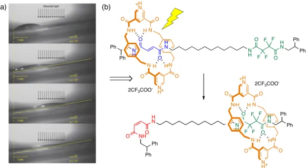
The macroscopic transport of liquid drops was demonstrated by the surface-energy switching that originated from a light-responsive molecular shuttle (rotaxane)9
(Figure 4). The rotaxane, which is physisorbed on the surface, has fumaramide and tetrafluorosuccinamide groups, and in the unperturbed state the macrocycle resides with the former by hydrogen bonding. However, UV irradiation induces the E→Z isomerization of fumaramide to maleamide, which has a low
affinity to the macrocycle. By masking the fluoroalkyl moiety, the surface energy can be changed from “polarophobic” to “polarophilic”, which results in the movement of a liquid drop (CH2I2).
Another approach was the direct bending of microcantilever beams by electrochemically switchable [3]rotaxanes directly assembled on the surface. The design elegantly resembles the natural muscle’s actin and myosin structure. The rotaxane has four stations and two macrocyclic rings, and the rings have disulfide tethers that are attached to the surface of the cantilever beams in the self-assembly. The electrochemical switching of two stations from neutral to positively charged states results in the contraction of the distance between the rings, which is translated into
(a) (b) H N N H H N Ph O O O Ph F F F F N O N H N O N O H N N O H H H H 2CF3COO -Ph NH Ph O Ph N H H N N H H N Ph Ph O O O O Ph F F F F N O N H N O N O H N N O H H H H 2CF3COO
-Figure 4. Light-driven transport (a) of a liquid drop (CH2I2) on a self-assembled monolayer (SAM) of
11-mercaptodecanoic acid on gold deposited on mica. The light-switchable rotaxane (b) was physisorbed onto the SAM. Reproduced with permission from ref 9.
Figure 5. Design of a molecular muscle based on rotaxanes and its operation (bending of cantilever beams) under redox control. Reproduced with permission from ref 10. Copyright (2005) American Chemical Society.
We are interested in those types of molecular machines that generate useful mechanical outputs (artificial muscles). We believe that the incorporation of molecular machines into a polymeric form, if aligned properly, can enable us to effectively translate the nanoscopic events into macroscopic work. In this dissertation, we propose new molecular systems, which change
Chapter 1 Introduction
installment into polymers will be described. We seek to exploit dimensional changes of the molecular machines to create systems that can be considered as molecular muscles, or artificial muscles. To accomplish our goals, we make use of electroactive polymers (EAPs), which are a class of polymeric materials that has been spotlighted as artificial muscles.11
However, our approach is in sharp contrast to conventional EAP actuators in that we would like to take advantage of molecular events, not just bulk phase behavior.
Conducting Polymer Actuators: The Conventional Mechanisms
EAP actuators exhibit dimensional changes in response to electrical stimulation. EAPs can be classified into two basic groups depending on the activation mechanism: electronic and ionic.11
The electronic EAPs change their dimensions by the attraction force due to the applied electric field. Although the electronic EAPs exhibit relatively large force and rapid response time, they generally require very high voltages (~100 MV/m) that are close to dielectric breakdowns. On the contrary, ionic EAPs only need very low driving voltages (1~5 V); they are driven by diffusion of ions. However, they require an electrolyte, which may cause problems in open-air conditions, or with long-term use.
Conducting polymers can be classified as the ionic EAP because the ions’ egress and ingress are responsible for the polymers’ deformation.12
As shown in Figure 6, a conducting polymer gains positive charge due to oxidation caused by the applied voltage. Then, counter-ions and accompanying solvent molecules are incorporated to balance the charge, which causes the expansion of volume. This process is reversible, so upon reduction the polymer returns to its original volume by expelling the counter-ions. This mechanism is often referred to as a
H N N H H N N H * * H N N H H N N H * * = = Oxidation counteranions
Figure 6. Swelling mechanism represented by polypyrrole. The polymer increases in dimension when oxidized due to the ingress of counterions (typically PF6- in organic solvents).
Polypyrrole and related systems are the most developed conducting polymers for actuators.12,13
Polypyrroles are often fabricated into the tri-layer system, in which two polypyrrole films sandwich a polymeric electrolyte layer.14
This tri-layer actuator can bend in either direction according to the voltage polarities. Polypyrrole can also be easily deposited onto conductive substrates via an electrochemical oxidative polymerization method using pyrrole monomers. This makes them useful for microfabrication.
Optimization for the polypyrrole’s performance has been continued to create materials that display one or two performance metrics that are equal or exceed that of natural muscle.15
However, the performance that allows proper comparison with natural muscle has not been realized in a single device. Therefore, continuing efforts are still needed and our group has contributed to this area with a different approach – molecular mechanisms.
Chapter 1 Introduction
Molecular Actuators: Mechanisms and Designs
In a conventional swelling mechanism, speed is intrinsically limited by the ion’s mobility. In addition, strain cannot exceed values imposed by the space that is occupied by the ions and associated solvent molecules. It should be noted that the individual polymer chains remain chemically unchanged and the reversible oxidation and reduction process in the polymer causes the ion flow. The macroscopic polymer object retains its shape and the incorporation of counterions causes the swelling-based volume change.
Throughout this thesis, we will refer to materials having geometrical changes at the molecular level to create volume changes as molecular actuators. It should be noted that unlike bulk actuators (e.g., polypyrrole), molecular actuators harness the dimensional variation of the single strands of the polymer. We envision that if we utilize the conformational changes of single polymer chains, we may obtain fast responses and large strains, which are not limited to the ion’s mobility. These single molecule actuators have potential applications to the bottom-up development of nano-devices and machines. The conformational change of the single strand would come from the monomer unit’s response to the external stimulation, namely electrochemical. We propose two types of molecular mechanisms: expanding and contracting (Figure 7).
(a) (b) [Ox] [Red] [Ox] [Red]
Figure 7. Expanding (a) and contracting (b) molecular mechanisms for actuation under redox control. Expanding model exploits the conformational changes of monomer units, while in the contracting model new chemical bond induces the contraction through a molecular hinge.
In the expanding model, the monomer moiety has a bent geometry in its neutral state. However, it becomes planar when it is oxidized, resulting in expansion. We can exploit the aromatization of non-aromatic system as the driving force. For example, we proposed thianthrene as the candidate, which is bent in its neutral state, and calculations showed about 7% increase upon oxidation (Figure 8a).16
Marsella et al. reported a polymer which was based on the bent-to-planar transformation of cyclooctatetraene moiety under redox control (Figure 8b).17
Although not experimentally confirmed, theoretical calculations predicted ~6% increase when oxidized.
In the contracting model, the key in the design is a hinged molecule that connects electroactive segments. Upon electrochemical oxidation, new chemical bonds between the oxidized species
Chapter 1 Introduction
electroactive segments (Figure 9).18
The driving force for contraction is the π-dimer formation
between the radical cations of oligothiophenes.
(a) (b) S S S S - n e -+ n e -S S S S n n Bu Bu n - 2 e -+ 2 e -2+
Figure 8. Expanding molecular actuators: a thianthrene-based polymer (a) and a cyclooctatetraene-based polymer (b). They utilize a bent-to-planar transformation caused by the aromatization from 8 to 6-π electrons.
OO OO R H S S H R S S Molecular hinge Electroactive segments OO OO R H S S H R S S - n e -+ n e -n n 2( ) !-Dimer formation
Figure 9. Contracting molecular actuator with a calix[4]arene as a molecular hinge. The new chemical bond (π-dimer) would be the driving force for the transformation.
In this dissertation, we first confirm by the model compound study that π-dimer formation can indeed be a driving force for the calix[4]arene-based molecular actuator (Chapter 2). We propose another molecular hinge, binaphthol moiety, for the contracting model. The syntheses of polymers with binaphthols and their characterization, including signatures of oligothiophene
the binaphthol polymer as a chiral amine sensor in Chapter 4. To create actuators that make use of the expanding model, we propose new conjugated seven-membered ring systems with heteroatoms (thiepin with sulfur, Chapter 5, and azepine with nitrogen, Chapter 6) and their syntheses and characterizations will be described. Inspired by the fact that sulfoxide has very low extrusion barrier in the related system, we applied the thiepin molecules to create a peroxide sensor (Chapter 5). In addition, during the investigation of phenol functional groups in conducting polymers, we found that strategic positioning of phenol groups can render a conjugation-broken meta-linked system just as conductive as a fully conjugated para-linked isomeric system, which will be described in Chapter 7.
Chapter 1 Introduction
References and Notes
(1) Borman, S. Chemical & Engineering News 2007, 85, 13–16.
(2) (a) Mavroidis, C.; Dubey, A.; Yarmush, M. L. Annu. Rev. Biomed. Eng. 2004, 6, 363–395. (b) Kinbara, K.; Aida, T. Chem. Rev. 2005, 105, 1377–1400.
(3) For reviews: (a) Jimenez-Molero, M. C.; Dietrich-Buchecker, C.; Sauvage, J. –P. Chem. Comm. 2003, 1613–1616. (b) Kottas, G. S.; Clarke, L. I.; Horinek, D.; Michl, J. Chem. Rev.
2005, 105, 1281–1376. (c) Balzani, V.; Credi, A.; Ferrer, B.; Silvi, S.; Venturi, M. Top.
Curr. Chem. 2005, 262, 1–27. (d) Braunschweig, A. B.; Northrop, B. H.; Stoddart, J. F. J. Mater. Chem. 2006, 16, 32–44. (e) Browne, W.; Feringa, B. L. Nature Nanotech. 2006, 1, 25–35. (f) Kay, E. R.; Leigh, D. A.; Zerbetto, F. Angew. Chem. Int. Ed. 2007, 46, 72–191. (4) Kelly, T. R.; De Silva, H.; Silva, R. A. Nature 1999, 401, 150–152.
(5) (a) Koumura, N.; Zijlstra, R. W.; van Delden, R. A.; Harada, N.; Feringa, B. L. Nature
1999, 401, 152–155. (b) Feringa, B. L.; van Delden, R. A.; ter Wiel, M. K. J. Pure Appl.
Chem. 2003, 75, 563–575.
(6) Tseng, H. –R.; Wu, D. M.; Fang, N. X. L.; Zhang, X.; Stoddart, J. F. ChemPhysChem
2004, 5, 111–116.
(7) Whitesides, G. M. Sci. Am. 2001, 285, 78–84.
(8) Vicario, J.; Katsonis, N.; Ramon, B. S.; Bastiaansen, C. W. M.; Broer, D. J.; Feringa, B. L. Nature 2006, 440, 163.
(9) Berná, J.; Leigh, D.; Lubomsak, M.; Mendoza, S. M.; Pérez, E. M.; Rudolf, P.; Teobaldi, G.; Zerbetto, F. Nature Mater. 2005, 4, 704–710.
(10) Liu, Y.; Flood, A. H. Bonvallet, P. A.; Vignon, S. A.; Northrop, B. H.; Tseng, H. –R.; Jeppesen, J. O.; Huang, T. J.; Brough, B.; Baller, M.; Magonov, S.; Solares, S. D.; Goddard, W. A.; Ho, C. –M.; Stoddart, J. F. J. Am. Chem. Soc. 2005, 127, 9745–9759. (11) Electroactive Polymer (EAP) Actuators as Artificial Muscles – Reality, Potential, and
Challenges; Bar-Cohen, Y., Ed.; SPIE Press: Bellingham, 2001. (12) Baughman, R. H. Synth. Met. 1996, 78, 339–353.
(13) Smela, E. Adv. Mater. 2003, 15, 481–494, and references therein.
(14) Madden, P. G. A. Ph.D. Thesis, Massachusetts Institute of Technology, 2003.
(15) (a) Hunter, I.; Lafontaine, S. In Tech. Dig. IEEE. Solid State Sensors Actuators Workshop
1992, 178–185. (b) Madden, J. D. W. Ph.D. Thesis, Massachusetts Institute of Technology,
2000.
(16) Yu, H. –h. Ph.D. Thesis, Massachusetts Institute of Technology, 2003.
(17) (a) Marsella, M. J. Acc. Chem. Res. 2002, 35, 944–951. (b) Marsella, M. J.; Reid, R. J.; Estassi, S.; Wang, L. -S. J. Am. Chem. Soc. 2002, 124, 12507–12510.
(18) (a) Anquetil, P. A.; Yu, H. -h.; Madden, J. D.; Madden, P. G.; Swager, T. M.; Hunter, I. W. Smart Structures and Materials 2002: EAPAD, Proc. Of SPIE 2002, 4695, 424–434. (b) Yu, H. -h.; Xu, B.; Swager, T. M. J. Am. Chem. Soc. 2003, 125, 1142–1143. (c) Yu, H. -h.; Swager, T. M. IEEE J. Oceanic Eng. 2004, 29, 692–695.
Chapter 2 π-Dimers OO OO S S S S OC16H33 OC16H33 OO OO S S S S S OC16H33 S C16H33O S S S S OC16H33 OC16H33 O O O O [Ox]
Chapter 2.
π-Dimer Formation as the Driving Force for
π
Calix[4]arene-based Molecular Actuator
Our group designed a calix[4]arene-based molecular actuator (Scheme 1), in which electroactive oligothiophenes are connected by the calix[4]arene scaffold.1
We proposed that each calix[4]arene moiety acts as a molecular hinge and that new noncovalent interactions between oxidized oligothiophenes (i.e., π-dimer or π-stack) can drive the dimensional changes. The π-dimer (or π-stack) is a nonconventional interaction that has drawn particular attention as a
charge transporting entity in conducting polymers.2
π-Dimer formation has been demonstrated in solution and in the solid state for oligothiophene derviatives.3,4
Scheme 1. Model of the Calix[4]arene-based Molecular Actuator.
In order to take advantage of such interactions as a driving force for molecular actuators, we considered that a segmented polymer would be more suitable than a fully conjugated one because it is able to attain the maximum interaction due to the spatial confinement of the radical cation’s wavefunctions. It should be noted, however, that higher degrees of spatial confinement would be expected to result in coulombic repulsion of like charges and hence counterion and solvent effects are expected.
The ab initio calculations by Scherlis and Marzari modeling the behavior of one actuating unit supported the concept that π-dimer formation (or π-stacking) between oxidized oligothiophenes
Chapter 2 π-Dimers
calix[4]arene hinges and the stacking interactions between oligothiophenes, they demonstrated with molecular dynamic simulations that the model molecules can change their shape in response to electrochemical oxidation.5b
The π-dimers of oxidized oligothiophenes are found unstable in gas phase but a polarizable solvent (e.g., acetonitrile) can dramatically stabilize the charged dimers with a moderate binding energy (5.2 kcal/mol for therthiophene).5a
Herein, we present a series of model studies designed to test the hypothesis that π-dimer formation can indeed be a
driving force for the calix[4]arene-based molecular actuator.
π-Dimers
Conduction in doped conjugated polymers is understood in terms of the polaron/bipolaron model7
which has been widely investigated experimentally and computationally. In simplified terms, a polaron is a radical cation delocalized to a few repeating units in a conjugated polymer, and a bipolaron is as a dication with no unpaired electrons. Due to the spinless nature of the highly doped polythiophenes as revealed by electron paramagnetic resonance (EPR), bipolarons (not polarons) were believed to be the charge carriers. However, since Hill and coworkers first reported the dimerization of oligothiophene cation radicals, the π-dimer (also a spinless entity) has emerged as an alternative model for the bonding in doped polythiophenes.2,3,8,9
This is not to say that it is incompatible with the polaron/bipolaron model. Indeed, a diamagnetic π-dimer is best considered an inter-chain bipolaron. Nevertheless, the study of this new type of chemical bonding (π-dimer or π-stack) is very important for understanding charge transport through polymer chains.
π
temperatures or at high concentration. π-Dimers have also been observed in the solid state; in
particular, our group reported π-dimer formation in polythiophene hybrids of transition-metal
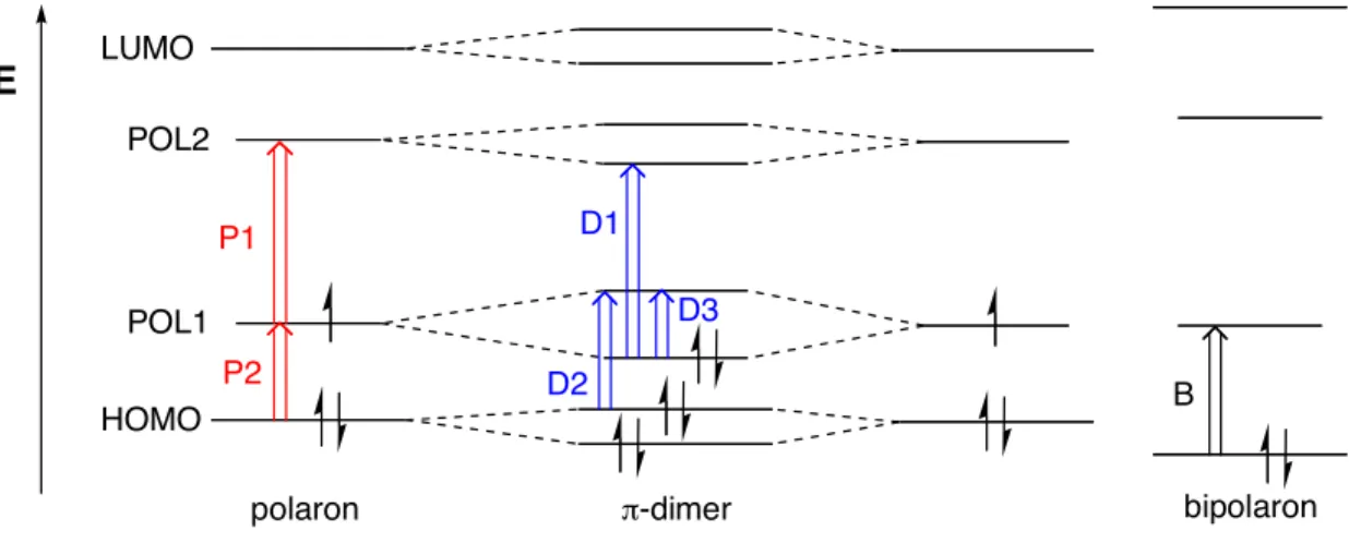
bis(salicylidenimine)s.4 E P1 P2 D2 D1 D3 B LUMO HOMO POL1 POL2
polaron !-dimer bipolaron
Figure 1. Schematic representation of electronic structures of three oxidized states. (Adapted from ref 3d) When two oligothiphenes are linked with an alkyl chain3c,d
or constrained to a cofacial arrangement,3e
dimer formation is greatly enhanced and observed even at room temperature.
π-Dimers can be easily detected by spectroscopic methods; diminished EPR intensities and shifted UV-vis-NIR absorptions are considered to be diagnostic indicators of π-dimer formation.
Electronic structures of these π-electron species have been proposed by several groups (Figure 1).3a,b,d
When one electron is removed from the π-electron system, two new energy levels emerge
due to the relaxation of the nuclear coordinates (polaron-like structure), allowing two sub-bandgap transitions (P1 and P2). In the π-dimer state, two singly occupied molecular orbitals
(SOMOs) mix together and form a new bond, resulting in the disappearance of EPR signals. In optical spectroscopy, three allowed transitions (D1–D3) are observed. In addition to the charge transfer transition (D3), D1 and D2 are comparable to polaronic transitions P1 and P2,
Chapter 2 π-Dimers
Synthesis of Model Compounds
To conduct model studies of the actuating unit, we synthesized 1–4, which contain a calix[4]arene hinge and two oligothiophene derivatives as electroactive segments (Scheme 2). Firstly, we sought to examine which, if any, of these compounds would give rise to stable radical cations when oxidized, and whether π-dimer formation would take place. Secondly, the effect of
the calix[4]arene’s conformation (cone vs. 1,3-alternate) was the subject of investigation.
Oligothiophene derivatives were connected to the upper rim of the calix[4]arene moiety. Compound 1 has free hydroxy groups on the benzene ring (lower rim) whereas compounds 2–4 contain alkoxy groups. Calix[4]arenes 1–3 existed predominantly in the cone conformation as determined by 1
H-NMR spectroscopy, whereas 4 adopted the 1,3-alternate conformation. We postulate that the calix[4]arene moiety of 1 is conformationally more rigid than that of 2, 3, or 4 due to lower rim hydrogen bonding.10
A long alkyl chain (n-C16H33) was installed to
oligothiophene moieties in order to increase their solubility.
Calix[4]arene precursors 6, 7 and 9 were prepared according to literature procedures (see Experimental Section). Compound 8 was synthesized from 7 via Stille coupling with 2-tributylstannylthiophene, followed by electrophilic iodination. The Stille cross-coupling reactions between calix[4]arenes 6–9 and tributylstannylated bithiophene derivative 12 furnished the desired compounds 1–4 in moderate yields. For the comparison, compound 5 was also prepared as a monomeric version of compound 2.
π Scheme 2.a Br OO OO Br OO OO S S I I Br OO OO Br H H O Br S S OC16H33 Bu3Sn OO OO S S S S OC16H33 C16H33O OO OO S S S S S OC16H33 S C16H33O OO OO S S S S OC16H33 C16H33O H H O S S OC16H33 1 2 3 5 6 7 8 10 12 OC16H33 Br i, ii iii, iv v 11 Br O O O O Br 9 S S S S OC16H33 C16H33O 4 O O O O
aReagents: (i) 2-Tributylstannylthiophene, Pd
2(dba)3, (t-Bu3PH)BF4, KF, DMF, 80 °C, 6 h, 93%. (ii) n-BuLi, -40 °C,
then I2, 36%. (iii) 5-Tributylstannyl-2,2’-bithiophene, Pd2(dba)3, (t-Bu3PH)BF4, KF, DMF, 80 °C, 6 h, 72%. (iv)
n-BuLi, Bu3SnCl, -40 °C to room temperature, 86%. (v) Pd2(dba)3, (t-Bu3PH)BF4, KF, THF/DMF, 70 °C, 6 h, 41%
(1), 61% (2), 49% (3), 59% (4), 83% (5)
π-Dimers between Oxidized Oligothiophene Derivatives: UV-vis
We examined several oxidizing agents to produce radical cations of 1–5 and finally chose Et3OSbCl6, a Meerwein’s salt, as a 1-electron oxidant
11
(not as an alkylating agent) because it is relatively easy to handle and more importantly, it does not exhibit a strong absorbance above 300 nm in the UV-vis spectrum. Therefore, we were able to monitor the diminution of the neutral absorption and the concurrent evolution of new absorptions without any interference (Figure 2).
Chapter 2 π-Dimers
cations, which were stable to moisture. However, the color was slowly lost (returned to their neutral state) when air was bubbled into the solution.
400 500 600 700 800 900 1000 300 400 500 600 700 800 900 1000 1100 A b s o rb a n c e ( a .u .) Wavelength (nm) 4 1 2 3 (a) (b) (c) (d) (e) 5
Figure 2. UV-vis spectral changes of 1–5 in CH2Cl2 at room temperature upon the increasing addition of the
oxidant Et3OSbCl6. Spectra of neutral absorptions of 1–5 are displayed by dashed lines.
For the oxidation of monomeric 5 in CH2Cl2 under ambient conditions (Figure 2e), the initial
π-π* absorption (382 nm) decreased and new peaks (645, 1079 nm) developed, which can be attributed to the polaronic absorptions (radical cations, Figure 1). These sub-bandgap transitions with vibronic structures are in good accord with literature precedent.3
We were not able to observe the formation of dications even after adding excess amounts of Et3OSbCl6 under the
above conditions.
Remarkably distinct behavior was observed upon oxidation of 2 (Figure 2b). Polaronic peaks, similar in shape to those of 5•+
, were dominant at the low levels of oxidation (Figure 2e). However, as more oxidant was added, blue-shifted absorptions were evident. Such blue-shifts are
π
upon oxidation of 3, the terthiophene-substituted version (Figure 2c). It is known that longer oligothiophene forms stronger dimers, probably due to the reduced coulombic repulsion.7c
The results of the present study are noteworthy in that a stable π-dimer is generated at room temperature in a solvent of low dielectric constant (CH2Cl2) from a framework as short as two
thiophenes.
Table 1. Absorption Maximaa and Half-Wave Potentialsb of 1~5
Absorption Maxima / nm Half-Wave Potentials / V
Compounds
Neutral Polaronic π-Dimeric E1/21 E1/22
1 384 665, >1100 - 0.33 0.57
2 382 655, 1084 593, 948 0.33 0.83
3 409 730, >1100 663, 1062 0.23 0.71
4 382 658, 1094 598, 955 0.33 0.82
5 382 645, 1079 - 0.42 0.76
aAbsorption were measured in CH
2Cl2 upon addition of oxidant Et3OSbCl6 at room temperature. bHalf-wave
potentials (all vs. Fc/Fc+) were measured in CH
2Cl2 with 0.1 M TBAPF6 as a supporting electrolyte under ambient
conditions.
Interestingly, when we added the oxidant Et3OSbCl6 to 1, only polaronic absorptions were
observed in the UV-vis spectra (Figure 2a). The only difference is that 1 has free hydroxyl groups at the lower rim of the calix[4]arene moiety. We attribute this reluctance to form π-dimer
formation to the calix[4]arene’s conformational rigidity resulting from lower-rim hydrogen bonding. However, it should be noted here that the π-dimer formation is coupled to the
“motional” flexibility of the calix[4]arene hinge (hydrogen-bonded vs. tetraalkylated).
There is a possibility that the hydroxyl groups of 1 were alkylated by the Meerwein’s salt. To address this, we reduced the oxidized solution of 1 by NH4OH and took the
1
H-NMR from the recovered compound. Indeed, we found the phenolic protons were no longer evident and
Chapter 2 π-Dimers
point the alkylation took place. That is, was it during the oxidation, or during the reduction step with NH4OH? If the alkylation occurred during oxidation, the absorption of 1 should have
resembled that of 2. However, the different electronic absorption spectra of oxidized 1 and 2 suggest that the oxidation of 1 with the Meerwein’s salt was not accompanied by alkylation. Moreover, oxidation of 1 with FeCl3 resulted in a similar absorption spectrum to that obtained
using Et3OSbF6 (Figure 3).
0 0.2 0.4 0.6 0.8 1 1.2 300 400 500 600 700 800 900 1000 1100 A b s o rb a n c e ( a .u .) Wavelength (nm) 1 FeCl3
Figure 3. UV-vis spectral changes of 1 in CH2Cl2 at room temperature upon the increasing addition of the oxidant
FeCl3. The dashed line represents the neutral absorption of 1.
The effect of the calix[4]arene’s conformation on the dimer formation appeared minimal. Oxidation profiles of 1,3-alternate 4 are very similar to those of cone 2 (Figure 2d vs. 2b). Literature precedent suggests that these conformations should be fixed; when the hydroxyl groups on the lower rim of the calix[4]arene are alkylated with propyl groups or larger substituents, rotation through the annulus of the macrocycle is not observed at room temperature.10
Therefore, there is no possibility of interconversion of 4 to 2, and vice versa. We can conclude that the cone and 1,3-alternate conformations of 2 and 4 are sufficiently flexible to
π
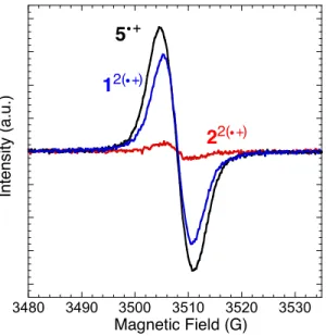
EPR and DPV
π-Dimer formation was further confirmed by EPR spectroscopy. EPR spectra were acquired for each of the CH2Cl2 solutions (0.2 mM for 1–2, 0.4 mM for 5) at room temperature. As
expected, bis(radical cation) 12(•+)
(Figure 1, blue) was EPR active, showing a rather broad and featureless signal, which is very similar to radical cation 5•+
(black). Note that 12(•+)
consists of two independent radical cations. In contrast, 22(•+)
(red) was almost EPR silent, which indicates that the two radical cations are bound to form a π-dimer.
3480 3490 3500 3510 3520 3530 Magnetic Field (G) In te n s ity ( a .u .) 5 12( ) 22( )
Figure 4. 9-GHz EPR spectra of radical cations 12(•+) (blue), 22(•+) (red), and 5•+ (black) in CH
2Cl2 at room
temperature.
The evolution of the EPR signals of oxidized 2 and 5 was monitored as the oxidant (Et3OSbCl6
in CH2Cl2) was added incrementally (Figure 5). In the case of 5, the signal increased gradually to
maximum. However, the initially developed signal for 2 decreased as more oxidant was added. This is in accord with what was observed by the UV-vis spectroscopy (Figure 2b); in 2, radical cations appeared at the initial stages of oxidation, but the π-dimer dominated at higher oxidation
Chapter 2 π-Dimers 3500 3510 3520 3500 3510 3520 2 5 In te n s ity ( a .u .) Magnetic Field (G) 0.5 equiv 1.0 equiv 1.5 equiv 2.0 equiv 1.0 equiv 0.75 equiv 0.5 equiv (a) (b)
Figure 5. 9-GHz EPR spectra of 2 (a) and 5 (b) measured in CH2Cl2 at room temperature with increasing addition
of oxidant Et3OSbCl6.
Oxidation potentials of dimeric 2 and monomeric 5 were measured in CH2Cl2 solutions with
0.1 M TBAPF6 as a supporting electrolyte under ambient conditions (Figure 6). In cyclic
voltammetry, both 2 and 5 showed two 1-electron oxidation peaks, which correspond to radical cation(s) and dication(s), respectively. However, the first oxidation of 2 took place at lower potential than that of 5 (0.33 and 0.42 V, respectively, vs. Fc/Fc+
), and the peak was broader. In contrast, the second oxidation of 2 was shifted to the higher potential. Differential pulse voltammetry (DPV, Figure 6b) reveals the differences more clearly. The first oxidation of the dimeric 2 occurred at a lower potential and the peak was broader, while the second oxidation was at higher potential when compared to the monomeric 5. These data also are consistent with the π-dimer formation because an over-potential would be required to oxidize the π-π-dimer, which is a
π -3 -2 -1 0 1 2 3 4 5 -0.2 0 0.2 0.4 0.6 0.8 1 C u rr e n t (µ A ) Potential (V vs Fc/Fc+) 2 5 -0.2 0 0.2 0.4 0.6 0.8 1 -0.2 0 0.2 0.4 0.6 0.8 1 Potential (V vs Fc/Fc+) N o rm a liz e d C u rr e n t 2 5 CH2Cl2 (a) (b)
Figure 6. Cyclic voltammograms (a) and differential pulse voltammograms (b) of 2 (blue) and 5 (red) in CH2Cl2
solution (~ 0.5 mM) on Pt button electrodes with 0.1 M TBAPF6 as a supporting electrolyte.
Conclusion
We have demonstrated that a stable π-dimer is formed upon oxidation of compounds 2, 3, and
4, the model units of the proposed molecular actuator, in the solvent of low dielectric constant
(CH2Cl2) at room temperature. Evidence from UV-vis, EPR, and DPV are all in agreement with
the π-dimer formation. In addition, we found that π-dimer formation is dependent upon the conformational flexibility of the calix[4]arene hinge. We are currently investigating polymeric materials of these compounds.
Chapter 2 π-Dimers
Experimental Section
General. NMR spectra were recorded on a Varian Mercury-300, Bruker Advance-400, or
Varian Inova-500 spectrometer. Chemical shifts were reported in ppm and referenced to residual solvent peaks (CDCl3: δ 7.27 ppm for
1
H, δ 77.23 ppm for 13
C). High-resolution mass spectra
(HR-MS) were obtained on a Bruker Daltonics APEX II 3 Tesla FT-ICR-MS. UV-vis spectra were obtained using a HP 8453 diode array spectrometer. Electrochemical measurements were carried out using an Autolab PGSTAT 10 or PGSTAT 20 potentiostat (Eco Chemie) in a three-electrode cell configuration consisting of a quasi-internal Ag wire reference three-electrode (BioAnalytical Systems) submerged in 0.01 AgNO3 / 0.1 M tetrabutylammonium
hexafluorophosphate (TBAPF6) in anhydrous CH3CN, a Pt button (1.6 mm in diameter) electrode
as the working electrode, and a Pt coil as the counter electrode. The ferrocene/ferrocenium (Fc/Fc+
) redox couple was used as an external reference. Half-wave potentials of Fc/Fc+
were observed between 210-245 mV vs Ag/Ag+
in CH2Cl2. EPR spectra were obtained using a Bruker
Model EMX Electron Paramagnetic Resonance Spectrometer operating as the X-band with 100 kHz modulation at room temperature. All air and water sensitive synthetic manipulations were performed under an argon or nitrogen atmosphere using standard Schlenk techniques.
Materials. Spectroscopic grade CH2Cl2 was purchased from Aldrich for electrochemistry.
TBAPF6 was recrystallized in ethanol prior to use. Anhydrous DMF was purchased from Aldrich
as Sure-Seal Bottles and used as received. THF was purified by passage through two alumina columns of an Innovative Technologies purification system. All other chemicals were of reagent grade and used as received. Compounds 6, 7 and 9 were prepared by literature procedures.12
π
standard Williamson ether synthesis with alkyl bromides. 5-Tributylstannyl-2,2’-bithiophene was synthesized by a known procedure.14
O O O O S S
11,23-Bis(thiophen-2-yl)-25,26,27,28-tetrapropoxycalix[4]arene (A). In a Schlenk tube
equipped with a stir bar were combined 6 (0.766 g, 1 mmol), Pd2(dba)3·CHCl3 (31 mg, 3 mol %),
and t-Bu3PH·BF4 (19 mg, 6.6 mol %). To the mixture, after purging three times with Ar, were
added 2-tributylstannylthiophene (0.827 mL, 2.5 mmol), KF (0.349 g, 6 mmol), and anhydrous DMF (3 mL). The mixture was allowed to stir at 80 °C for 6 h, at which time it was cooled to room temperature and methanol was added to precipitate product. The crude product was isolated by filtration and thoroughly washed with methanol. It was then passed through a short silica gel pad (dichloromethane as eluent). The concentrated product was further purified by recrystallization (dichloromethane/methanol), yielding 0.705 g (93%) of white solid. 1H-NMR
(400 MHz, CDCl3) δ: 7.22 (s, 4H), 7.17 (dd, 2H, J = 5.1, 1.0 Hz), 7.13 (dd, 2H, J = 3.6, 1.0 Hz), 7.00 (dd, 2H, J = 5.1, 3.6 Hz), 6.39 (s, 6H), 4.48 (d, 4H, J = 13 Hz), 4.00 (t, 4H, J = 8.0 Hz), 3.77 (t, 4H, J = 7.0 Hz), 3.21 (d, 4H, J = 13 Hz), 1.96 (m, 8H), 1.09 (t, 6H, J = 7.5 Hz), 0.96 (t, 6H, J = 7.4 Hz). 13 C-NMR (125 MHz, CDCl3) δ: 157.57, 155.81, 145.02, 136.94, 133.70, 128.26, 128.01, 127.99, 126.36, 123.82, 122.43, 122.16, 77.12, 76.89, 31.22, 23.65, 23.33, 10.89, 10.25. HR-MS (ESI): calcd for C48H52O4S2 [M+Na]
+
Chapter 2 π-Dimers O O O O S S I I 11,23-Bis(5-iodothiophen-2-yl)-25,26,27,28-tetrapropoxycalix[4]arene (8). To A (0.250 g,
0.33 mmol) dissolved in THF (10 mL) was added n-BuLi (0.413 mL, 0.66 mmol) at –40 °C. It was then removed from the cooling bath and allowed to stir for 1 h at room temperature. The mixture was cooled to –40 °C again and then quenched with iodine (0.168 mg, 0.66 mmol). After being diluted with ethyl acetate at room temperaure, the organic layer was washed with a saturated aqueous solution of Na2S2O3 and brine, dried over MgSO4, and evaporated under
reduced pressure. The crude product was purified by column chromatography (chloroform:hexane 1:3), yielding 0.120 g (36 %) of a white solid. 1
H-NMR (400 MHz, CDCl3) δ: 6.98 (d, 2H, J = 3.8 Hz), 6.78 (d, 4H, J = 6.5 Hz), 6.72 (pseudo-t, 2H, J = 6.5 Hz), 6.70 (s, 4H), 6.42 (d, 2H, J = 3.8 Hz), 4.48 (d, 4H, J = 13 Hz), 3.93 (t, 4H, J = 7.7 Hz), 3.85 (t, 4H, J = 7.4 Hz), 3.19 (d, 4H, J = 13 Hz), 1.96 (m, 8H), 1.05 (t, 6H, J = 7.4 Hz), 1.02 (t, 6H, J = 7.5 Hz). 13 C-NMR (125 MHz, CDCl3) δ: 156.85, 156.72, 150.79, 137.71, 135.54, 135.21, 128.60, 127.58,
125.54, 123.14, 122.47, 77.11, 76.92, 31.15, 23.52, 23.39, 10.63, 10.45. HR-MS (ESI): calcd for C48H50I2O4S2 [M+Na]
+
, 1031.1132; found, 1031.1133.
S
S OC16H33
π
2 mol %), and 5-tributylstannyl-2,2’-bithiophene (4.89 mL, 13 mmol). After purging three times with Ar, KF (1.74 g, 30 mmol) and anhydrous DMF (20 mL) were added. The mixture was then allowed to stir at 80 °C for 6 h, at which time it was cooled to room temperature and methanol was added. The suspension was filtered and the resulting solid washed thoroughly with methanol. It was then passed through a short pad of silica gel, eluting with chloroform. The concentrated product was further purified by recrystallization (chloroform/methanol), yielding 3.46 g (72%) of white solid. 1
H-NMR (300 MHz, CDCl3) δ: 7.52 (pseudo-d, 2H, J = 8.7 Hz),
7.21 (dd, 1H, J = 5.1, 1.2 Hz), 7.19 (dd, 1H, J = 3.6, 1.2 Hz), 7.13 (d, 1H, J = 3.7 Hz), 7.11 (d, 1H, J = 3.7 Hz), 7.03 (dd, 1H, J = 5.1, 3.6 Hz), 6.92 (pseudo-d, 2H, J = 8.7 Hz) 3.99 (t, 2H, J = 6.6 Hz), 1.81 (m, 2H), 1.54–1.20 (m, 26H), 0.89 (t, 3H, J = 6.9 Hz). HR-MS (ESI): calcd for C30H42OS2 [M+H] + , 483.2750; found, 483.2754. S S OC16H33 Bu3Sn 5-Tributylstannyl-5’-(4-Hexadecyloxyphenyl)-2,2’-bithiophene (12). To B (0.950 g, 1.9
mmol) dissolved in THF (50 mL) was added n-BuLi (1.31 mL, 2.1 mmol) at –40 °C. It was then removed from the cooling bath and allowed to stir for 1 h at room temperature. The mixture was cooled to –40 °C again and then quenched with tributylstannyl chloride (0.814 mL, 2.2 mmol). After being diluted with ethyl acetate at room temperaure, the organic layer was washed with water and brine, dried over MgSO4, and evaporated under reduced pressure. The crude product
was used without further purification. Yield: 1.41 g (86 %, assuming 90% purity) of pale yellow solid. 1
Chapter 2 π-Dimers
J = 7.3 Hz), 0.89 (t, 3H, J = 7.0 Hz). 13
C-NMR (125 MHz, CDCl3) δ: 158.97, 143.15, 142.99,
136.60, 136.33, 136.11, 127.04, 127.00, 124.75, 124.48, 122.74, 115.06, 68.33, 32.16, 29.91~29.94 (5C), 29.89, 29.83, 29.80, 29.62, 29.60, 29.47, 29.17, 27.49, 26.26, 22.93, 14.37, 13.91, 11.08. HR-MS (ESI): calcd for C42H68OS2Sn [M]
+ , 772.3704; found, 772.3742. O O O O S S S S OC16H33 OC16H33 H H
11,23-Bis[5’-(4-hexadecyloxyphenyl)-2,2’-bithiophen-5-yl]-25,27-dihydroxy-26,28-dipropoxycalix[4]arene (1). In a Schlenk tube equipped with a stir bar were combined 6 (0.081
g, 0.12 mmol), Pd2(dba)3·CHCl3 (6.2 mg, 5 mol %), t-Bu3PH·BF4 (3.8 mg, 11 mol %), 12 (0.198
g, 0.25 mmol), THF (0.8 mL), and DMF (0.4 mL). To the mixture, which was degassed by free-pump-thaw (three times), was added KF (0.049 g, 0.72 mmol) under Ar. The mixture was allowed to stir at 70 °C for 6 h, at which time it was cooled to room temperature and methanol was added. The crude product was isolated by filtration and thoroughly washed with methanol. It was then passed through a short pad of silica gel, eluding with dichloromethane. The concentrated product was further purified by recrystallization (dichloromethane/methanol, two times). Yield: 0.072 g (41%) of yellow solid. 1
H-NMR (300 MHz, CDCl3) δ: 8.52 (s, 2H), 7.52
(pseudo-d, 4H, J = 9.0 Hz), 7.32 (s, 4H), 7.27 (d, 2H, J = 2.1 Hz), 7.14–7.10 (m, 4H), 7.08 (d, 2H, J = 3.9 Hz), 7.01 (d, 4H, J = 7.5 Hz), 6.92 (pseudo-d, 4H, J = 9.0 Hz), 6.82 (t, 2H, J = 7.5
π
MHz, CDCl3) δ: 159.02, 153.73, 152.11, 143.89, 142.92, 136.23, 135.46, 133.27, 129.38,
128.77, 127.02, 126.98, 126.24, 125.67, 125.47, 124.29, 124.14, 122.79, 122.36, 115.09, 77.23, 76.97, 68.35, 32.15, 31.68, 29.92–29.89, 29.83, 29.80, 29.62, 29.59, 29.46, 26.25, 23.72, 22.92, 14.36, 11.15. HR-MS (ESI): calcd for C94H116O6S4 [M+Na]
+ , 1491.7547; found, 1491.7527. O O O O S S S S OC16H33 OC16H33
11,23-Bis[5’-(4-hexadecyloxyphenyl)-2,2’-bithiophen-5-yl]-25,26,27,28-tetrapropoxycalix[4]arene (2). Using the similar procedure for the synthesis of 1, compound 7
(0.171 g, 0.22 mmol) was treated with Pd2(dba)3·CHCl3 (11 mg, 5 mol %), t-Bu3PH·BF4 (7 mg,
11 mol %), 12 (0.386 g, 0.51 mmol), THF (1.4 mL), DMF (0.7 mL), and KF (0.077 g, 1.3 mmol). Yield: 0.207 g (61%) of yellow solid. 1
H-NMR (400 MHz, CDCl3) δ: 7.43 (pseudo-d, 4H, J = 8.8 Hz), 7.00 (d, 2H, J = 3.8 Hz), 6.96 (d, 2H, J = 3.8 Hz), 6.83 (pseudo-d, 4H, J = 8.8 Hz), 6.82–6.78 (m, 10H), 6.72 (pseudo-t, 2H, J = 7.5 Hz), 6.65 (d, 2H, J = 3.7 Hz), 4.49 (d, 4H, J = 13 Hz), 3.96 (t, 4H, J = 6.6 Hz), 3.93 (t, 4H, J = 7.6 Hz), 3.86 (t, 4H, J = 7.4 Hz), 3.20 (d, 4H, J = 13 Hz), 1.96 (m, 8H), 1.78 (m, 4H), 1.57–1.28 (m, 52H), 1.04 (t, 6H, J = 7.4 Hz), 1.02 (t, 6H, J = 7.4 Hz), 0.90 (t, 6H, J = 7.0 Hz). 13 C-NMR (125 MHz, CDCl3) δ: 158.83, 156.85, 156.55, 143.33, 142.54, 136.42, 135.53, 135.53. 135.47, 135.23, 128.62, 128.12, 127.04, 126.87, 125.48, 124.21, 123.92, 122.56, 122.46, 114.97, 77.11, 76.97, 68.30, 32.16, 31.22, 29.93–29.94, 29.91,
Chapter 2 π-Dimers O O O O S S S S S OC 16H33 S OC16H33
11,23-Bis[5’-(4-hexadecyloxyphenyl)-2,2’-bithiophen-5-yl]-25,26,27,28-tetrapropoxycalix[4]arene (3). Using the similar procedure for the synthesis of 1, compound 8
(0.075 g, 0.073 mmol) was treated with Pd2(dba)3·CHCl3 (3.8 mg, 5 mol %), t-Bu3PH·BF4 (2.3
mg, 11 mol %), 12 (0.128 g, 0.17 mmol), THF (1 mL), DMF (0.5 mL), and KF (0.025 g, 1.3 mmol). Yield: 0.062 g (61%) of orange solid. 1
H-NMR (400 MHz, CDCl3) δ: 7.41 (pseudo-d, 4H, J = 8.7 Hz), 6.96 (s, 4H), 6.95 (d, 4H, J = 8.7 Hz), 6.92 (d, 2H, J = 3.7 Hz), 6.85 (m, 4H), 6.38 (pseudo-d, 4H, J = 8.7 Hz), 6.71 (d, 2H, J = 3.7 Hz), 6.63 (s, 4H), 6.51 (d, 2H, J = 3.7 Hz), 4.49 (d, 4H, J = 13 Hz), 3.98 (t, 4H, J = 7.9 Hz), 3.96 (t, 4H, J = 6.6 Hz), 3.79 (t, 4H, J = 7.0 Hz), 3.20 (d, 4H, J = 13 Hz), 1.96 (m, 8H), 1.77 (m, 4H), 1.42–1.27 (m, 52H), 1.07 (t, 6H, J = 7.4 Hz), 0.98 (t, 6H, J = 7.4 Hz), 0.89 (t, 6H, J = 7.0 Hz). 13 C-NMR (125 MHz, CDCl3) δ: 158.94, 157.30, 156.17, 143.67, 142.93, 136.69, 135.87, 135.81, 135.73, 135.04, 134.96, 128.87, 128.11, 127.08, 126.92, 125.31, 124.32, 123.81, 123.54, 122.73, 122.48, 122.34, 115.12, 115.00, 77.21, 76.93, 68.32, 32.17, 31.22, 29.95–29.93, 29.91, 29.88, 29.85, 29.70, 29.61, 29.54, 26.31, 23.62, 23.35, 22.94, 14.38, 10.81, 10.33. HR-MS (ESI): calcd for C108H132O6S6 [M+Na]
+
, 1739.8240; found, 1739.8210.
π S S S S OC16H33 OC16H33 O O O O
11,23-Bis[5’-(4-hexadecyloxyphenyl)-2,2’-bithiophen-5-yl]-25,26,27,28-tetrapropoxycalix[4]arene (4). Using the similar procedure for the synthesis of 1, compound 9
(0.072 g, 0.094 mmol) was treated with Pd2(dba)3·CHCl3 (4.9 mg, 5 mol %), t-Bu3PH·BF4 (3.0
mg, 11 mol %), 12 (0.155 g, 0.20 mmol), THF (0.8 mL), DMF (0.4 mL), and KF (0.033 g, 0.56 mmol). Yield: 0.087 g (59%) of yellow solid. 1
H-NMR (300 MHz, CDCl3) δ: 7.42 (pseudo-d, 4H, J = 8.7 Hz), 7.25 (s, 4H), 7.04 (d, 4H, J = 7.5 Hz), 7.00 (d, 2H, J = 3.9 Hz), 6.94 (d, 2H, J = 3.9 Hz), 6.84 (s, 4H), 6.82 (pseudo-d, 4H, J = 8.7 Hz), 6.69 (t, 2H, J = 7.5 Hz), 4.00 (t, 4H, J = 6.6 Hz), 3.73 (t, 4H, J = 6.9 Hz), 3.67 (t, 4H, J = 7.2 Hz), 3.59 (s, 8H), 1.97 (m, 4H), 1.83 (m, 8H), 1.50–1.28 (m, 52H), 1.15 (t, 6H, J = 7.5 Hz), 1.05 (t, 6H, J = 7.5 Hz), 0.90 (t, 6H, J = 6.9 Hz). 13 C-NMR (125 MHz, CDCl3) δ: 158.76, 156.51, 156.43, 143.75, 142.32, 136.76, 135.23, 133.94, 133.30, 130.22, 127.50, 127.34, 127.14, 126.84, 124.30, 123.68, 122.53, 122.25, 121.69, 114.95, 74.87, 74.81, 68.30, 35.81, 32.16, 29.95–29.93, 29.90, 29.89, 29.86, 29.72, 29.61, 29.58, 26.32, 24.30, 24.15, 22.93, 14.37, 11.34, 10.97, 10.90. HR-MS (ESI): calcd for C100H128O6S4
[M+Na]+
, 1575.8486; found, 1575.8463.
O
S S
Chapter 2 π-Dimers
11,23-Bis[5’-(4-hexadecyloxyphenyl)-2,2’-bithiophen-5-yl]-25,26,27,28-tetrapropoxycalix[4]arene (5). Using the similar procedure for the synthesis of 1, compound 10
(0.016 g, 0.065 mmol) was treated with Pd2(dba)3·CHCl3 (2.0 mg, 3 mol %), t-Bu3PH·BF4 (1.2
mg, 6.6 mol %), 12 (0.083 g, 0.098 mmol), DMF (0.7 mL), and KF (0.023 g, 0.39 mmol). Yield: 0.035 g (83%) of yellow solid. 1 H-NMR (400 MHz, CDCl3) δ: 7.52 (pseudo-d, 2H, J = 8.8 Hz), 7.27 (s, 2H), 7.14 (d, 1H, J = 3.7 Hz), 7.12 (pseudo-s, 2H), 7.11 (d, 1H, J = 3.7 Hz), 6.92 (pseudo-d, 2H, J = 8.8 Hz), 3.99 (t, 2H, J = 6.6 Hz), 3.76 (t, 2H, J = 6.6 Hz), 2.33 (s, 6H), 1.85 (m, 4H), 1.56–1.27 (m, 26H), 1.10 (t, 3H, J = 7.4 Hz), 0.89 (t, 3H, J = 7.0 Hz). 13 C-NMR (125 MHz, CDCl3) δ: 159.08, 156.09, 143.22, 143.08, 136.40, 136.01, 131.76, 129.65, 127.05, 126.91, 126.26, 124.40, 124.32, 123.31, 122.81, 115.10, 74.21, 68.35, 32.15, 29.91~29.93 (5C), 29.89, 29.83, 29.80, 29.62, 29.60, 29.46, 26.25, 23.86, 22.92, 16.61, 14.36, 10.88. HR-MS (ESI): calcd for C41H56O2S2 [M]
+
π
References and Notes
(1) (a) Anquetil, P. A.; Yu, H. -h.; Madden, J. D.; Madden, P. G.; Swager, T. M.; Hunter, I. W. Smart Structures and Materials 2002: EAPAD, Proc. Of SPIE 2002, 4695, 424–434. (b) Yu, H. -h.; Xu, B.; Swager, T. M. J. Am. Chem. Soc. 2003, 125, 1142–1143. (c) Yu, H. -h.; Swager, T. M. IEEE J. Oceanic Eng. 2004, 29, 692–695.
(2) Miller, L. L.; Mann, K. R. Acc. Chem. Res. 1996, 29, 417–423.
(3) For example, see (a) Bäuerle, P.; Segelbacher, U.; Maier, A.; Mehring, M. J. Am. Chem. Soc. 1993, 115, 10217–10223. (b) Graf, D. D.; Duan, R. G.; Campbell, J. P.; Miller, L. L.; Mann, K. R. J. Am. Chem. Soc. 1997, 119, 5888–5899. (c) Satou, T.; Sakai, T.; Kaikawa, T.; Takimiya, K.; Otsubo, T.; Aso, Y. Org. Lett. 2004, 6, 997–1000. (d) Sakai, T.; Satou, T.; Kaikawa, T.; Takimiya, K.; Otsubo, T.; Aso, Y. J. Am. Chem. Soc. 2005, 127, 8082– 8089. (e) Knoblock, K. M.; Silvestri, C. J.; Collard, D. M. J. Am. Chem. Soc. 2006, 128, 13680–13681. (f) Yamazaki, D.; Nishinaga, T.; Tanino, N.; Komatsu, K. J. Am. Chem. Soc.
2006, 128, 14470–14471.
(4) Kingsborough, R. P.; Swager, T. M. J. Am. Chem. Soc. 1999, 121, 8825–8834.
(5) (a) Scherlis, D.; Marzari, N. J. Phys. Chem. B 2004, 108, 17791–17795. (b) Scherlis, D.; Marzari, N. J. Am. Chem. Soc. 2005, 127, 3207–3212.
(6) A different mechanism involving a repulsive electrostatic interaction, rather than by an attractive π-π stacking, was suggested: see Casanovas, J.; Zanuy, D.; Alemán, C. Angew.
Chem. Int. Ed. 2006, 45, 1103–1105.
(7) Bredas, L. L.; Street, G. B. Acc. Chem. Res. 1985, 18, 309–315.
Chapter 2 π-Dimers
Mater. 1992, 4, 1106–1113. (c) Bäuerle, P.; Segelbacher, U.; Gaudl, K. –U.; Huttenlocher, D.; Mehring, M. Angew. Chem. Int. Ed. 1993, 32, 76–78.
(9) As a spinless entity, chain dimer (two polarons on a single chain) and σ-dimer were also proposed. For the chain dimer, see: van Haare, J. A. E. H.; Havinga, E. E.; van Dongen, J. L. J.; Janssen, R. A. J.; Cornil, J., Brédas, J. -L. Chem. Eur. J. 1998, 4, 1509–1522. For the σ-dimer, see: Smie, A.; Heinze, J. Angew. Chem. Int. Ed. 1997, 36, 363–367.
(10) Shinkai, S. Tetrahedron 1993, 49, 8933–8968.
(11) Rathore, R.; Kumar, A. S.; Lindeman, S. V.; Kochi, J. K. J. Org. Chem. 1998, 63, 5847– 5856.
(12) (a) Stastny, V.; Lhoták, P,; Stibor, I.; König, B. Tetrahedron 2006, 62, 5748–5755. (b) Casnati, A.; Fochi, M.; Minari, P.; Pochini, A.; Reggiani, M. Gazz. Chim. Ital. 1996, 126, 99–106. (c) Linnane, P.; James, T. D.; Shinkai, S. J. Chem. Soc., Chem. Commun. 1995, 1997–1998.
(13) Larsen, M.; Krebs, F. C.; Harrit, N.; Jorgensen, M. J. Chem. Soc., Perkin Trans. 2, 1999, 1749–1757.
Spectrum 1.1H-NMR spectrum of A (400 MHz, CDCl 3). Spectrum 2.13C-NMR spectrum of A (125 MHz, CDCl 3). O O O O S S
Chapter 2 Appendix NMR Spectrum 3.1H-NMR spectrum of 8 (400 MHz, CDCl 3). Spectrum 4.13C-NMR spectrum of 8 (125 MHz, CDCl). O O O O S S I I
Spectrum 5.1H-NMR spectrum of B (300 MHz, CDCl 3).
S S
Chapter 2 Appendix NMR Spectrum 6.1H-NMR spectrum of 12 (400 MHz, CDCl 3). Spectrum 7.13C-NMR spectrum of 12 (125 MHz, CDCl 3). S S OC16H33 Bu3Sn
Spectrum 8.1H-NMR spectrum of 1 (300 MHz, CDCl 3). Spectrum 9.13C-NMR spectrum of 1 (125 MHz, CDCl 3). O O O O S S S S OC16H33 OC16H33 H H
Chapter 2 Appendix NMR Spectrum 10.1H-NMR spectrum of 2 (400 MHz, CDCl 3). Spectrum 11.13C-NMR spectrum of 2 (125 MHz, CDCl 3). O O O O S S S S OC16H33 OC16H33
Spectrum 12.1H-NMR spectrum of 3 (400 MHz, CDCl 3). Spectrum 13.13C-NMR spectrum of 3 (125 MHz, CDCl 3). O O O O S S S S S OC 16H33 S OC16H33
Chapter 2 Appendix NMR Spectrum 14.1H-NMR spectrum of 4 (400 MHz, CDCl 3). Spectrum 15.13C-NMR spectrum of 4 (125 MHz, CDCl 3). S S S S OC16H33 OC16H33 O O O O
Spectrum 16.1H-NMR spectrum of 5 (400 MHz, CDCl 3). Spectrum 17.13C-NMR spectrum of 5 (125 MHz, CDCl 3). O S S OC16H33
Chapter 3 Binaphthyl Actuator
Chapter 3.
Binaphthyl – A Molecular Hinge
We have proposed “molecular actuators” that change their dimensions via a novel molecular mechanism in Chapter 1.1
In contrast to conventional conducting polymer actuators, which operate through absorption and release of counter-ions and solvents under electrochemical oxidation and reduction, our molecular actuator designs utilize the conformational changes of the individual polymer chain at the molecular level. Following this concept, we proposed two mechanisms for molecular actuators; the expansion and the contraction. In the expanding mechanism, the initially bent moieties are forced to be flat under redox control. The driving force is the aromatization, gained by oxidizing a non-aromatic system. Cyclooctatetraene2
and thianthrene3
have been suggested as possible candidates to produce this behavior.
To display a contracting mechanism, we have developed a calix[4]arene-based conducting polymer.1b
In this system, the calix[4]arene scaffold functions as a molecular hinge, through which electroactive segments are brought together and apart to form a reversible (intermolecular) bond. We utilize π-dimer formation as the driving force for the actuation, which we
unequivocally proved in model compound studies (see Chapter 2).
In parallel with the calix[4]arene-based system, a new building block containing binaphthyl units was developed as another potential hinge candidate. The binaphthyl has a hinge comprised of a 1,1’ C-C bond between two naphthyl units. As described in Figure 1a, our design involves the electroactive segments (oligothiophenes, for example) that are connected through a binaphthyl hinge. As we oxidize the electroactive segments, radical cations are generated and the new chemical bond called π-dimer can potentially drive the dimensional changes. Figure 1b
Chapter 3 Binaphthyl Actuator
We have long pursued development of segmented electroactive polymers that lack extended conjugation. We believe that if we confine the wavefunctions into a finite region, we can expect a better interaction due to the large coefficients. However, the stability of charged species is generally improved when the species have more delocalized structures. It is also important to consider that the generated charges will display coulombic repulsion, which can offset any effects gained by the wavefunction confinement.
HO HO Electroactive segments Molecular Hinge (a) (b)
Figure 1. (a) Design of binaphthyl containing molecular actuators. (b) The computer-generated model of the
binaphthyl polymer’s conformational change under redox control.
In this chapter, we describe the synthesis of this new class of materials and their electrochemical properties. We also developed a new binaphthyl monomer with oligothiophenes of different connectivity to promote a better interaction.
Synthesis of Binaphthyl Polymers: The First Generation
As a result of the richness in chemistry of binaphthyls,4
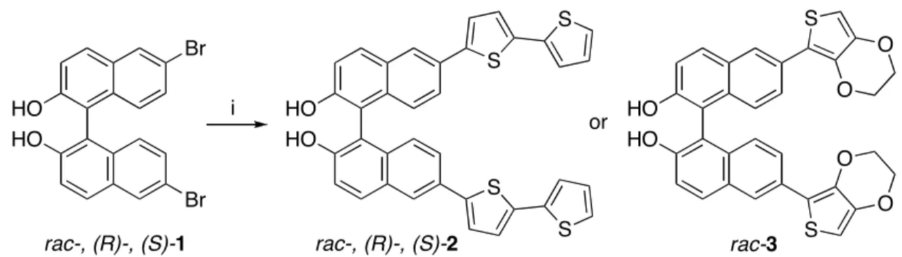
dibromobinaphthol 1 is commercially available both in racemic and enantiomeric forms (R and S). Thus, we were able to synthesize the electropolymerizable monomers 2 and 3 in a single step with a Stille coupling reaction in moderate yields (Scheme 1). Compound 2 is stable for storage in the solid state, but we observed slow decomposition under air in case of compound 3, perhaps due to oxidation.
Scheme 1.a HO HO Br Br HO HO S S S S HO HO S S O O O O i or
rac-, (R)-, (S)-1 rac-, (R)-, (S)-2 rac-3
aReagents: (i) Pd
2(dba)3·CHCl3, t-Bu3P, 5-tributylstannyl-2,2’-bithiophene, KF, NMP, 70 °C, 24 h, 61~66%.
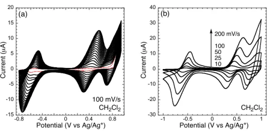
Figure 2 shows the electropolymerization of monomer 2 (racemic) to create electrode-surface confined polymer films and the scan-rate dependence of the electroactive polymers in CH2Cl2.
The electrodeposition was performed under swept potential conditions with 0.1 M TBAPF6 as a
supporting electrolyte in air. The shift of the onset potential of the second scan (Figure 2a), when compared to the first scan, is indicative of generating a more extended conjugated system (i.e., thiophene-thiophene coupling).5
As more films were deposited, the peak potentials slightly shifted and the increase in current with each scan slightly diminished. These effects are common with sluggish diffusion of ions into and out of the film, and the overall resistive loss through the thickness of the film results in a reduced potential (IR drop) at the film surface. We observed two
Chapter 3 Binaphthyl Actuator
oxidation and reduction couples, which is consistent with the generation of polaron (radical cation) and bipolaron (dication) types of species.6
-30 -20 -10 0 10 20 30 40 -0.6 -0.4 -0.2 0 0.2 0.4 0.6 0.8 1 -60 -40 -20 0 20 40 60 80 -0.2 0 0.2 0.4 0.6 0.8 1
Potential (V vs Ag/Ag+) Potential (V vs Ag/Ag+)
C u rr e n t (µ A ) C u rr e n t (µ A ) (a) (b) CH2Cl2 100 mV/s CH2Cl2 200 mV/s 100 50 25 10
Figure 2. (a) Electropolymerization of rac-2 (~1.5 mM) on a Pt button electrode. The dotted line represents the
first scan. (b) CVs of a poly(rac-2) film at different scan rates in a monomer free solution. All measurements were carried out in CH2Cl2 with 0.1 M TBAPF6 as a supporting electrolyte.
-60 -40 -20 0 20 40 60 80 0 0.2 0.4 0.6 0.8 1 0 20 40 60 80 0 0.2 0.4 0.6 0.8 1 Potential (V vs Ag/Ag+) C u rr e n t (µ A ) C o n d u ct iv it y (S /c m ) CH2Cl2 10 mV/s 10 mV/s 40 mV offset
Figure 3. The CV (dotted line) and in situ conductivity measurement (solid line) of a film of poy(rac-2) on 5-µm
interdigitated Pt microelectrode in CH2Cl2 with 0.1 M TBAPF6 as a supporting electrolyte.


![Figure 9. Contracting molecular actuator with a calix[4]arene as a molecular hinge](https://thumb-eu.123doks.com/thumbv2/123doknet/14681590.559382/19.918.153.767.531.768/figure-contracting-molecular-actuator-calix-arene-molecular-hinge.webp)