HAL Id: hal-02323748
https://hal.archives-ouvertes.fr/hal-02323748
Preprint submitted on 21 Oct 2019HAL is a multi-disciplinary open access
archive for the deposit and dissemination of sci-entific research documents, whether they are pub-lished or not. The documents may come from teaching and research institutions in France or abroad, or from public or private research centers.
L’archive ouverte pluridisciplinaire HAL, est destinée au dépôt et à la diffusion de documents scientifiques de niveau recherche, publiés ou non, émanant des établissements d’enseignement et de recherche français ou étrangers, des laboratoires publics ou privés.
Gonzalo Quiroga Artigas, Pascal Lapébie, Lucas Leclère, Philip Bauknecht,
Julie Uveira, Sandra Chevalier, Gáspár Jékely, Tsuyoshi Momose, Evelyn
Houliston
To cite this version:
Gonzalo Quiroga Artigas, Pascal Lapébie, Lucas Leclère, Philip Bauknecht, Julie Uveira, et al.. A G-protein-coupled receptor mediates neuropeptide-induced oocyte maturation in the jellyfish Clytia. 2019. �hal-02323748�
1
A G-protein-coupled receptor mediates neuropeptide-induced
oocyte maturation in the jellyfish Clytia
Gonzalo Quiroga Artigas1#, Pascal Lapébie1, Lucas Leclère1, Philip Bauknecht 2, Julie Uveira1, Sandra Chevalier1, Gáspár Jékely2,3, Tsuyoshi Momose1 and Evelyn Houliston1*
1. Sorbonne University, CNRS, Villefranche-sur-mer Developmental Biology Laboratory (LBDV), 06230 Villefranche-sur-mer, France
2. Max Planck Institute for Developmental Biology, Spemannstraße 35, 72076 Tübingen, Germany. 3. Living Systems Institute, University of Exeter, Stocker Road, EX4 4QD, Exeter, UK
# current address: GQA: The Whitney Laboratory for Marine Bioscience, University of Florida, St. Augustine, FL, USA
*Corresponding author: E. Houliston houliston@obs-vlfr.fr
Short title: The Clytia oocyte maturation hormone receptor
2
Abstract
The reproductive hormones that trigger oocyte meiotic maturation and release from the ovary vary
greatly between animal species. Identification of receptors for these Maturation Inducing
Hormones (MIHs), and understanding how they initiate the largely conserved maturation process,
remain important challenges. In hydrozoan cnidarians including the jellyfish Clytia hemisphaerica,
MIH comprises neuropeptides released from somatic cells of the gonad. We identified the receptor
(MIHR) for these MIH neuropeptides in Clytia using cell culture-based "deorphanization" of
candidate oocyte-expressed GPCRs. MIHR mutant jellyfish generated using CRISPR-Cas9 had
severe defects in gamete development or in spawning both in males and females. Female gonads,
or oocytes isolated from MIHR mutants, failed to respond to synthetic MIH. Treatment with the
cAMP analogue 5’Br-cAMP to mimic cAMP rise at maturation onset rescued meiotic maturation
and spawning. Injection of inhibitory antibodies to GαS into wild type oocytes phenocopied the
MIHR mutants. These results provide the molecular links between MIH stimulation and meiosis initiation in hydrozoan oocytes. Molecular phylogeny grouped Clytia MIHR with a subset of
bilaterian neuropeptide receptors including Neuropeptide Y, Gonadotropin Inhibitory Hormone,
pyroglutamylated RFamide and Luqin, all upstream regulators of sexual reproduction. This
identification and functional characterisation of a cnidarian peptide GPCR advances our
understanding of oocyte maturation initiation and sheds light on the evolution of
neuropeptide-hormone systems.
Non-standard Abbreviations: MIH - Maturation Inducing Hormone; GVBD - Germinal Vesicle
3
Introduction
Oocyte meiotic maturation is an essential process for animal sexual reproduction. It transforms the
tetraploid, fully-grown, ovarian oocyte into a haploid female gamete [1]. The core biochemical and
cellular pathways operating within the oocyte during maturation are highly conserved between animals
across the phylogenetic spectrum. Activation of the CyclinB-Cdk1 kinase complex assures meiotic
progression from prophase I arrest into M phase, while parallel Mos-MAP kinase activation steers
polar body formation as well as cytostatic arrest once maturation is complete [2,3]. In contrast the
upstream physiological processes vary widely, as does the molecular nature of the maturation inducing
hormones (MIHs) that act on the oocyte to trigger maturation [1,4]. This reflects clade-specific
acquisition of endocrine tissues such as ovarian follicles, the corpus cardiacum/corpus allatum in
insects, or the pituitary in vertebrates [5,6]. These tissues act downstream of other neuroendocrine sites
such as the vertebrate hypothalamus to integrate environmental, behavioural and physiological
information in order to achieve optimal conditions for gamete development and release [7]. The
complex evolutionary history of hormonal reproductive regulation has made it challenging to unravel
the crucial regulatory events operating within the oocyte at the onset of maturation.
G-protein coupled receptors (GPCRs), the largest superfamily of integral transmembrane receptors [8],
are good candidates to serve as MIH receptors. These 7-transmembrane domain proteins activate a
variety of cytoplasmic signalling pathways via Gα and/or Gβγ subunits that become released from
receptor-associated heterotrimeric Gαβγ protein upon ligand binding [9–11]. Members of the four
main Gα subunits classes, Gαs, Gαi Gαq and Gα12/13, associate variously with members of the vast
GPCR family. In vertebrates, constitutively active GPCRs are coupled to Gαs, which stimulates
adenylate cyclase to maintain high cytoplasmic cAMP concentrations in ovarian oocytes [12–14].
These high cAMP levels help hold the oocyte in an immature state, with the cell cycle arrested in
4
vary between species and are not fully understood (see Discussion). In marked contrast, oocytes of
many invertebrate species show a rise in cytoplasmic cAMP concentration upon MIH stimulation that
is required for meiotic maturation [6]. In these species, GPCRs working through Gαs are thus good
candidates to trigger oocyte maturation, rather than to inhibit it as in vertebrates. The identification of
such receptors could help to understand the origin of this diversity by providing an evolutionary
perspective.
Here we identify the MIH receptor (MIHR) in the hydrozoan jellyfish Clytia hemisphaerica as a
GPCR likely working through Gαs, and determine its in vivo function. In Clytia and other hydrozoan
species, MIH consists of PRPamide and related tetrapeptides that are released upon light stimulation
from opsin-expressing gonad ectoderm cells, and act at the plasma membrane [15,16]. Neuropeptides
such as these are of major importance in cnidarian biology, acting both as “neuroendocrine” mediators
of physiological transitions such as metamorphosis, as well as in fast neuromuscular transmission
regulating swimming and feeding mediated by ligand-gated ion channels [reviewed by 17,18]. We
uncovered an oocyte-expressed Class A GPCR in Clytia (MIHR) activated by MIH peptides, which is
to our knowledge the first characterized cnidarian neuropeptide GPCR [19,20]. Antibody inhibition
experiments further provided evidence that Gαs links MIHR activation to cAMP production to trigger
maturation [21,22]. CRISPR-Cas9 mediated mutation of the MIHR gene revealed an essential in vivo
function of MIHR in initiating oocyte maturation. Phylogenetic analysis of the MIHR sequence
identified an evolutionary link to a subset of bilaterian neuropeptide-hormone GPCR families,
including several upstream regulators of reproduction. These results allow us to propose a new
scenario for the evolution of hormonal signalling pathways regulating oocyte maturation.
5
Results
Selection of candidate MIH GPCRs
Amidated neuropeptides like MIH commonly signal through GPCRs, although other receptor types can
also be used [23]. As the first step to select candidate MIH receptors, we compiled a comprehensive
catalogue of Clytia GPCRs from a Clytia reference transcriptome covering all life-cycle stages. Our
bioinformatics pipelines first retrieved all sequences predicted to code for 7 transmembrane domain
(7TM domain) proteins and bearing GPCR-related Pfam tags. An initial list of 761 sequences was then
assigned by Pfam to the three main GPCR classes: A (rhodopsin-like), B (secretin-like) and C
(metabotropic glutamate-like)[8,24] or to an “other” category (which included for instance sweet-taste
receptors and cAMP-like receptors). The final dataset of 536 class-sorted putative Clytia GPCRs
obtained after removal of duplicates (File S1) may be a slight overestimate due to some incorrectly
identified or incomplete sequences. We focussed on the 377 class-A GPCRs, since most neuropeptide
GPCRs belong to this class [8, 25].
An important criterion for the selection of MIH-receptor candidates from the class-A GPCR list was
enrichment in oocytes (Fig.1A). We mapped Illumina HiSeq mRNA reads previously obtained from
Clytia gonad tissues [16] and life cycle stage [26] against all putative GPCR sequences. Profile clustering revealed three groups of sequences with oocyte-enriched expression (Fig. S1). We further
narrowed down the number of potential MIHRs from these expression groups to 96 sequences, taking
into account also Pfam indicators and sequence similarity with a set of bilaterian GPCRs [25; see
Figure 6 for clustering of a subset of sequences closest to the Clytia MIHR]. Finally we compiled a
short-list of 16 candidates for functional testing, using high expression level in oocytes as the final
6
Identification of the Clytia MIH receptor by GPCR deorphanization assay
We used a cell culture-based GPCR deorphanization assay to identify the MIH receptor from our
candidate shortlist. cDNAs for each candidate were transfected into CHO-K1 cells along with an
aequorin-GFP luminescence reporter that measures Ca2+ mobilization downstream of a promiscuous
Gα protein [27,28]. We first screened the 16 candidate Clytia GPCRs against a mixture of 33 synthetic
amidated peptides (including MIHs) predicted to be generated from previously identified putative
Clytia neuropeptide precursors [15] and one additional one identified from transcriptome data (File S4). Given our imperfect knowledge of pro-peptide processing in cnidarians, some of the synthetic peptides
may not correspond to endogenous peptides [29,30]. Only one of the 16 GPCRs was activated by this
peptide mixture. This receptor responded well to all 4 synthetic Clytia MIH tetrapeptides, as well as to
related Cladonema MIH peptides that can also activate Clytia oocyte maturation, but not to other Clytia
peptide mixtures or poorly-active MIH penta-/tripeptides. We termed this Clytia GPCR the Maturation
Inducing Hormone Receptor (MIHR). The activity of individual peptides at 1 µM to stimulate the
MIHR closely matched their in vivo ability to induce oocyte maturation [15; Fig.1B].
The Clytia MIH amidated peptides are produced from two precursor genes Che-pp4 (generating
multiple copies of WPRAa, WPRYa and WPRPa) and Che-pp1 (multiple copies of WPRPa and
RPRGa) [15; File S4]. Their predicted structural similarity suggests they bind the same site on the
receptor with different affinities. Dose-response curves for each of the 4 Clytia MIH neuropeptides,
generated from three independent experiments, showed half-maximal effective concentration (EC50)
values in the high-nanomolar or low-micromolar range for all 4 MIHs, with RPRYamide showing the
highest activity and RPRGamide the lowest (Fig. 1C). To our knowledge, Clytia MIH and MIHR are
7
Slow growth, gametogenesis disruption and spawning failure in MIHR mutant jellyfish
To determine the function of MIHR in vivo we generated Clytia MIHR knockout (KO) polyp colonies
using CRISPR/Cas9 mutagenesis (see Methods). This gene-editing technique is very effective in
Clytia, allowing extensive bi-allelic mutation of target genes already in F0 polyp colonies [16, 63]. Three guide RNAs designed to target the 3rd transmembrane domain of the MIHR protein were tested
by genotyping at the planula stage, and the most effective one used to generate polyp colonies. After
genotyping around the target site, we selected and propagated for phenotypic analysis six colonies
showing no detectable wild type sequence and carrying mainly frame-shift mutations (Table 1). All six
mutant polyp colonies expanded slowly compared to wild type colonies. They displayed variable
morphologies, with two of them rambling pronouncedly away from the glass substrate (Fig. 2A; Table
1), but all produced active gonozooids that budded baby medusae. Growth of these mutant baby
medusae was slower for the six mutants than for wild types, but they were able to reach the full adult
size of about 1cm in diameter. The adult jellyfish all swam less vigorously than wild types. The slow
growth of mutant polyps and medusae was not due to any obvious feeding problems: both forms could
capture Artemia nauplii without difficulty.
Of the six MIHR mutant colonies, three produced male jellyfish and three females. In addition to the
shared defects in growth and swimming behaviour, they showed one of two distinct phenotypes
affecting gonad development. For one female and one male MIHR mutant (n5-8 and n5-24
respectively), the gonads of the adult jellyfish developed poorly, failing to accumulate gametes as
occurs during wild type adult growth (Fig. 2B). Examination of the gonads by confocal microscopy
revealed the presence of small oocytes for n5-8 and a thin spermatogenic zone for n5-24. This suggests
that mutation of the MIHR gene can compromise gametogenesis in male and female jellyfish. One
possible explanation is that gamete development can stall completely when overall growth of the
jellyfish is poor. In marked contrast, jellyfish from the four other MIHR mutant polyp colonies
8
spermatozoids (n5-6, n5-10; Fig. 2D). In the female medusae, the gonads were grossly inflated by
accumulation of fully-grown immature oocytes, due to failure in light-induced oocyte maturation and
subsequent release of the unfertilised egg (see below). In males, the gonads were markedly deformed
and irregular. Following dark-light transitions that induced spawning in wild type males, some local
sperm release was observed from rupture sites of the gonad epithelium, but the sperm remained
concentrated at the gonad surface and failed to disperse.
Overall this initial phenotype analysis of six independent MIHR mutant F0 colonies indicated that
MIHR has non-essential functions in medusa growth and in regulating medusa swimming. These could
relate to expression of MIH and MIHR at other sites in the jellyfish, notably the manubrium
(mouth/stomach structure) and tentacles (see below). In addition, two distinct gonad defects were
observed: failure of gamete development or over-accumulation of mature gametes. These had no
obvious relationship to particular genotypes of the corresponding F0 polyp colonies (Table 1) and so
may be the consequence of MIHR mutation in different genetic or epigenetic backgrounds. In any case,
the accumulation of immature, fully grown oocytes in medusae from two independent MIHR mutants
strongly supported a key role for this GPCR in oocyte maturation, as characterized further below.
Clytia MIHR is expressed in oocytes and also in tentacle cells
The defects observed in behaviour and growth as well as in gametogenesis in MIHR mutant jellyfish
suggested that this receptor may have roles both in the gonads and other sites. In situ hybridization
detection of MIHR mRNA in adult jellyfish (medusae) and in isolated gonads (Fig. 3A, B) supported
this idea. Strong expression was expressed in developing oocytes and in male gametes, consistent with
the ability of MIH to induce male spawning [15]. Expression was also detected in clusters of small
somatic cells located within each tentacle bulb. Individual cells from these clusters extended in a line
9
involved in regulating tentacle contraction. The MIH receptor in these tentacle cells could potentially
bind MIH neuropeptides produced by neural/neuroendocrine cells of the endodermal gastrovascular
system, detectable using an antibody recognising the PRPamide peptides produced from both Che-pp1
and Che-pp4 [anti-PRPa; 15; Fig. 3C]. These two precursors are expressed respectively exclusively in
the manubrium and tentacle, as well as being co-expressed in scattered cells of the gonad [15]. As well
as the cells in the gonad ectoderm that mediate spawning, the anti-PRPa antibody decorates cells in the
tentacle especially at its junction with the circular gastrovascular canal, and in the endoderm of the
manubrium feeding/digestive organ [15,16] (Fig. 3C). Comparison of in situ hybridization patterns for
MIHR and MIH showed that the ligand and receptor-expressing cells in the tentacles have distinct distributions: the single file of MIHR cells lies on the rounded oral side [see 31], whereas the MIH cells
form two flanking lines (Fig. 3B). The position and morphology of these cells suggest that they
correspond to two sub-populations of neural cells positioned between the tentacle endoderm and
ectoderm in hydrozoans [31,32]. No MIHR-expressing cells were detected in the manubrium, only
non-specific staining of the manubrium floor. Our inability to detect MIHR expression close to these
manubrium PP1-expressing cells may be due to the limitations of the in situ hybridization technique,
with low expression in the manubrium and/or other tissues not sufficient to provide a clear signal.
Another possibility is that a second receptor is expressed in the manubrium and more sensitive to
RPRGa/WPRPa peptides. Alternatively, the MIHR-expressing cells at the base of the tentacles might
respond to endocrine MIH tetrapeptides circulating in the gastrovascular system, for example secreted
in response to nutritional status. Regulation by these cells of tentacle contractions, and directly or
indirectly of swimming behaviour, could account for the sluggish movement and poor growth of MIHR
10
Clytia MIHR is the essential oocyte receptor for oocyte maturation
We used jellyfish from the two MIHR female mutant colonies showing the swollen-gonad phenotype
(n5-13 and n5-23) to characterize the role of this GPCR in oocyte maturation. For n5-23 the
predominant genotypes were a 4 base deletion which introduces a premature STOP codon, and a 55
base insertion, but a small proportion of wild type cells was also detected. Colony n5-13 contained a
majority 4 base deletion but also a high proportion of a 3 base deletion. We performed a series of
oocyte maturation and spawning assays, comparing the responses of wild type and MIHR KO isolated
gonads and oocytes to different stimuli (Figure 4). Isolated wild type gonads underwent oocyte
maturation and spawning in response to light stimulation or treatment with the synthetic MIH peptide
WPRPamide (100nM) as previously shown [15], but MIHR mutant gonads did not respond (Fig. 4A,
B). In contrast, treatment with 4mM 5Br-cAMP, a cell-permeable analogue of cAMP which induces
hydrozoan oocyte maturation by mimicking the early cytoplasmic cAMP rise [3,21], rescued the
phenotype of MIHR mutant gonads, efficiently triggering oocyte maturation and spawning. 5-Br-cAMP
treatments but not MIH peptides also promoted maturation of isolated fully grown MIHR mutant
oocytes (Fig. 4C). Br-cAMP-matured MIHR mutant oocytes could be fertilized to develop into planula
larvae, although had lower development and metamorphosis rates than wild type oocytes.
This demonstration of a cAMP-reversible maturation initiation failure in mutant female jellyfish
confirmed that the Clytia MIHR has an essential in vivo function as the oocyte receptor for MIH.
MIH-induced oocyte maturation blocked by an inhibitory Gαs antibody
GPCR activation can lead to a cytoplasmic cAMP concentration rise in the responding cell as a result of
adenylate cyclase stimulation via S type Gα subunits of heterotrimeric G proteins. We tested the role of
Gαs in Clytia oocyte maturation by injecting isolated oocytes with a specific inhibitory antibody
11
oocytes were tested for maturation competence by incubation at the end of the experiment in
5’-Br-cAMP. Any that failed to mature when treated with cAMP were discounted in the analyses. Oocytes
injected with anti-Gαs responded less efficiently than oocytes injected with PBS or a control anti-GST
antibody when treated with synthetic MIH at low and then high doses (10nM then 100nM WPRPamide;
Kinetics observed in one of three equivalent experiments shown in Fig. 5A). These results strongly
suggest that MIHR acts mainly through Gαs.
Based on these findings we can now propose a model for maturation initiation in Clytia oocytes
through MIH, MIHR, Gαs and cAMP (Fig. 5B). A light cue triggers MIH release from
neuroendocrine-type cells in the gonad ectoderm via an essential opsin protein [16]. The MIH peptides act on MIHR at
the oocyte surface to promote Gαs activation, stimulating adenylate cyclase and thus promoting an
increase in cytoplasmic cAMP concentration, essential for the transition into meiotic M phase.
Clytia MIHR is related to a bilaterian superfamily of neuropeptide hormone receptors
Much progress has been made in understanding the relationships between GPCR-neuropeptide families
between protostomes and deuterostomes [20,25,28,35,36], and more recently with those in acoels and
cnidarians [37]. We performed sequence-similarity based clustering to explore the relationship of the
Clytia MIHR sequence with known GPCR families in Bilateria using established datasets from human and the annelid Platynereis [25,28 Fig. 6A]. The Clytia MIHR fell within the majority group of
cnidarian GPCRs that cluster with a superfamily of peptide hormone receptors including human
neuropeptide Y, neuropeptide FF, Tachykinin, Orexin/allastropin, Elevenin, and EFLGa/Thyrotropin
releasing hormone, as well as Luqin from Platynereis (Fig. 6B). Adding to this analysis sequences
recovered from the anthozoan cnidarian Nematostella highlighted the extensive independent expansion
of GPCR-A sequence since cnidarian and bilaterian neuropeptide systems split [37]. This analysis
12
oocyte maturation in vertebrates, including the constitutively active GPR3, gonadotropin releasing
hormone (GnRH) receptor or Luteinizing/Follicle-stimulating hormone (LH/FSH) receptor (see
Introduction and Figure 7). A GPCR from Nematostella annotated as a GnRH receptor [38] belongs as
sister to distinct ‘superfamily’ including the bilaterian GnRH, vasotocin, CCAP corazonin and achatin
receptor families.
Maximum Likelihood phylogenetic analyses of the large GPCR cluster containing MIHR confirmed
phylogenetic support for a GPCR ‘superfamily’ associating two distinct cnidarian groups (A and B in
Fig. 6C, D) with a set of bilaterian neuropeptide hormone receptors [37]. Cnidarian GPCR group A
contained Clytia MIHR, six other Clytia GPCRs and a large number of Nematostella receptors. It
showed weakly-supported association with a set of bilaterian peptide hormone receptor families
including those for neuropeptide Y/neuropeptide F, GnIH/neuropeptide FF, RYa/Luqin, Tachykinin
and pyroglutamylated RFamide peptide (QRFP) [25,28,37]. Group B included two Clytia GPCRs and
associated with further bilaterian neurohormonal receptor families including Elevenin, EFLGa/TRH
and Orexin/Allatotropin receptors (Fig.6C,D). The low support values for many deep branch
relationships within this superfamily makes it difficult to determine definitive evolutionary
relationships between the neuropeptide hormone receptors and cnidarian group A and B GPCRs. We
can nevertheless conclude that Clytia MIHR along with eight other Clytia GPCRs and a very large
group of Nematostella GPCRs have a common ancestor with a subset of bilaterian peptide hormone
receptor families that includes many involved in regulating sexual reproduction and feeding (see
13
Discussion
GPCR deorphanization followed by targeted gene mutation allowed us to identify Clytia MIHR as the
neuropeptide receptor responsible for initiating oocyte meiotic maturation. MIHR female mutant
medusae accumulated fully-grown oocytes that failed to mature unless treated with cAMP analogues
to bypass the receptor. We further provided evidence that a Gαs subunit links MIHR activation to the
increase in cytoplasmic cAMP concentration in the oocyte that initiates maturation. MIHR belongs to a
superfamily of neuroendocrine GPCRs involved in regulating reproduction but also nutrition, as well
as diverse other physiological processes. Consistent with a wider role for Clytia MIH-MIHR, MIHR
expression was detected in jellyfish tentacle cells that may respond to MIH neuropeptides produced in
the gastrovascular system, while MIHR mutants showed sluggish behaviour and slow growth, with
gamete development also stalled in jellyfish from some mutants. These findings thus open doors to an
improved understanding of the vital animal process of oocyte maturation and to its evolution. More
widely they shed light on the evolution of the neurohormonal regulation of sexual reproduction in the
animal kingdom.
An oocyte receptor for maturation initiation
Our work shows that Clytia MIHR is entirely responsible for the oocyte response to MIH, allowing us
to fill in the main molecular actors leading to oocyte meiotic maturation (Fig. 5B): MIH is released
from specialized gonad ectoderm cells in response to a dark-light transition at dawn via the essential
photoresponsive GPCR Opsin9 [16]. Binding of MIH to MIHR at the oocyte surface is likely swiftly
followed by a rise in cytoplasmic cAMP levels in the oocyte to activate cAMP-dependent Protein
Kinase (PKA), as has been shown in other hydrozoan species [22]. Our antibody inhibition
experiments strongly implicate Gαs-stimulated adenylate cyclase activity in driving this cAMP rise.
PKA leads to activation of the Cdk1-CyclinB and Mos-MAP kinase systems, that have highly
14
secretion and meiotic maturation will require the identification of PKA substrates from Clytia oocytes
that interact with regulators of Mos translation and/or Cdk1-CyclinB autoactivation.
cAMP rises initiate oocyte maturation not only in hydrozoans but also in a diverse range of
protostome and deuterostome species [6], so it is tempting to speculate that the straightforward
pathway linking MIH secretion to meiotic maturation in Clytia may have retained its main features
from a distant cnidarian-bilaterian ancestor. A useful step towards testing this idea would be to search
for MIHR superfamily GPCRs in oocytes of nemertean, ascidian, bivalves or ophiuroid species that
depend on cAMP signalling for maturation. If confirmed as the ancestral state, the Clytia MIH-MIHR
system could provide a paradigm for the molecular dissection of a common, cAMP-stimulated,
mechanism for oocyte meiotic resumption.
Evolution of reproductive regulation
The role of cAMP in initiating oocyte maturation is not universal. In some species such as starfish it has
no significant role [41], while in vertebrates, high cAMP levels in the ovarian oocyte maintain the
prophase arrest. Clues to understanding how these marked differences arose during evolution may be
found by considering the GPCRs involved in regulating gamete maturation and release across species.
In vertebrates, GPCRs are involved in triggering oocyte maturation at several levels along the H-P-G
axis (Figure 7). In ovarian oocytes, constitutively active GPCRs of the GPR3/6/12 family help maintain
prophase arrest via Gαs-cAMP [12–14]. Luteinizing Hormone receptors in the surrounding follicle cells
provide upstream signalling also via Gαs-cAMP. The follicle cells signal is transmitted to the oocyte in
mouse via a cGMP decrease through gap-junctions [42], or in fish and amphibians via steroid hormone
cocktails that act on one or more types of membrane receptor, potentially including GPCR progestin
receptors linked to Gαi that contribute to lowering cAMP levels [39,40,43,44]. Upstream of the
vertebrate follicle, LH production from the pituitary is under control of the master reproductive
15
pairs influence production of GnRH by the hypothalamus, as well as the gonadotropins LH and FSH by
the pituitary. Molecular phylogeny showed that Clytia MIHR is not closely to receptors of the core
H-P-G GPCRs, but rather forms a superfamily with receptors of these “upstream” neuropeptide hormones,
notably GnIH, QRFP, Neuropeptide Y and NkB. Production of GnRH and gonadotropins along the
H-P-G axis are inhibited by GnIH and stimulated by QRFPs [45,46], while the Tachykinin family
hormone NkB acts in a group of hypothalamus neurons together with Kisspeptin and Dynorphin A in
the generation of cyclic GnRH pulses [47]. The ancestral cnidarian-bilaterian GPCR for MIHR and the
receptors to all these vertebrate hormones may thus already had a role in regulating gamete production
and/or release. In this case, vertebrate GPCRs in the hypothalamus and the pituitary, rather than those
in gonad follicle cells or gametes, would have retained the trace of this ancestor. Likewise, protostome
members of the MIHR GPCR group regulate sexual reproduction indirectly from sites far from the
gonad. The planarian NeuropeptideY NPY-8 regulates gametogenesis through its GPCR expressed in
CNS neuroendocrine hormone cells [48], while nematode Luqin peptides produced by pharyngeal
neurons regulate egg laying via serotonergic RIH interneurons [49].
An ancient regulatory system linking reproduction and nutrition?
It is striking that many bilaterian neuropeptide GPCRs closely related to Clytia MIHR are known for
roles in regulating feeding and nutritional balance. In mammals these include GPR83/PEN [50] as well
as QRFP receptors and Neuropeptide Y receptors, which provide important links between metabolic
state and reproductive regulation [51-53], in conjunction with GnIH signalling [54]. Related hormone
receptor families from protostomes are also known for regulating feeding, including those for
Drosophila Leucokinin [55] and Luqins in arthropods (RYamides) and nematodes [49,56]. It is thus tempting to propose that the ancestral GPCR of the Clytia MIHR and these bilaterian hormone receptor
families was involved in integrating sexual reproduction with nutritional status. It should be noted,
however, that bilaterian neuropeptide GPCRs in this superfamily also function in many other
16
sexual reproduction with feeding, notably including Kisspeptin, involve GPCRs only distantly related
to MIHR [35,37,57]. To unravel further the evolutionary history of hormonal regulation of gamete
production in relation to nutrition it will be of great interest to examine in detail the function of MIHR
in Clytia relating to the sluggish movement and poor growth of MIHR mutants, but also to determine
17
Methods
Animals
Sexually mature jellyfish generated from laboratory maintained Clytia hemisphaerica polyp colonies
(“Z strains”) [26] were fed regularly with Artemia nauplii and cultured under light-dark cycles to allow
daily spawning. Red Sea Salt brand artificial seawater (ASW) was used for all culture and experiments.
Selection of candidate Clytia MIH Receptors
From a comprehensive Clytia reference transcriptome (86,606 contigs) derived from mixed larva, polyp
and jellyfish samples,we generated a list of predicted protein sequences from complete and incomplete
ORFs using a homemade script [58]. This dataset was screened using TMHMM 2.0c to produce a list
of Clytia complete protein sequences coding for 7 transmembrane domain proteins (7TMD) including
putative incomplete sequences containing 2 to 7 transmembrane domains. Scanning with Interproscan
5.22 was used to generate a list of 761 potential Clytia GPCRs with Pfam tags related to 7TMD
receptors were retained, sorted by class. CD-HIT [59] was run with 95% identity to eliminate sequence
duplicates, obtaining a final dataset of 536 Clytia GPCRs (sequences in File S1). Illlumina HiSeq 50nt
reads from mRNA isolated from manually dissected gonad ectoderm, endoderm, growing oocytes and
fully-grown oocytes [16] and from other Clytia life cycle stages including polyp and planula larvae [26]
were mapped against all candidate GPCR sequences using Bowtie2 [60]. The counts for each contig
were normalized per total of reads of each sample and contig length to allow expression comparisons
between genes and samples.
Presumptive GPCR sequences were separated in groups using a hierarchical pipeline based on the
correlation of their expressions (gplots package in R). Z-scores were obtained after standardization of
the counts in the different sequenced samples for each GPCR candidate and plotted in a heat map using
R (Fig. S1). For clustering, a previous collection of GPCRs[25] was complemented with several
18
was performed using CLANS2 [61] with a BLOSUM62 matrix and a p-value cutoff of 1.e-30. Based
on this information, Pfam signatures, reciprocal BLASTs and high expression level in oocytes, a subset
of 16 top candidates was manually selected (sequences and accession numbers in FileS2).
Candidate receptor cloning
The selected Clytia GPCRs were cloned from gonad-extracted cDNA into pcDNA3.1(+) (Thermo
Fisher Scientific, Waltham, MA, USA) using the Gibson Assembly® Cloning Kit (New England
Biolabs) [61]. pcDNA3.1(+) vector was linearized with BamHI and NotI restriction enzymes. Primers
were designed using the Gibson Cloning option in Geneious v8. Forward primers consisted of the
overhang left after BamHI vector linearization followed by the Kozak consensus sequence
(CGCCACC), a start codon (ATG), and a sequence corresponding to the target sequence. Reverse
primers consisted of the overhang left after NotI vector linearization followed by a STOP codon, and a
reverse complementary sequence to the target sequence. The primers for all cloned GPCRs are listed in
File S3. Polymerase chain reaction was performed using Phusion polymerase (New England Biolabs).
Cloned GPCRs were sequenced using primers for T7: TAATACGACTCACTATAGGG and BGHrev:
TAGAAGGCACAGTCGAGG.
Receptor deorphanization
Cell culture GPCR ligand response assays were performed as described [28]. Briefly, CHO-K1 cells
were cultured in Ham’s F12 Nut Mix medium (Thermo Fisher Scientific) with 10% foetal bovine
serum. Cells were seeded in 96-well plates at approximately 10.000 cells/well and transfected the
following day with pcDNA3.1(+) plasmids encoding each of the 16 candidate Clytia GPCRs (File S2),
the promiscuous Gα-16 protein [63], and a reporter construct GFP-apoaequorin [64] (60 ng each) using
the transfection reagent TurboFect (Thermo Fisher Scientific). After 2 days of expression, the medium
was removed and replaced with Hank’s Balanced Salt Solution (HBSS) supplemented with 1.8 mM
19
37 °C for 2 hours, cells were tested by adding synthetic peptides (GenScript) in HBSS supplemented
with 1.8 mM Ca2+ and 10 mM glucose. A list of all synthetic peptides used is provided in File S4.
Luminescence was recorded for 45 s in a plate reader (BioTek Synergy Mx or Synergy H4; BioTek,
Winooski, VT, USA). Data were integrated over the 45-s measurement period and recorded as technical
triplicates in each case. Data were normalized using the response of Platynereis FLamide receptor to 1
µM AKYFL-NH2 [28]. Dose-response curves were obtained using concentrations between 0.01 nM
and 100 µM for each peptide. Data for dose-response curves were recorded in triplicate for each
concentration and the experiment was repeated independently 3 times. Dose-response curves were
fitted with a four-parameter curve using Prism 6 (GraphPad, La Jolla, CA, USA) and were normalized
to the calculated upper plateau values (100% activation).
Generation of CRISPR-Cas9 mutant Clytia polyp colonies
Following our established protocol [65] MIHR small guide RNA (sgRNA) was assembled by
hybridising crRNA and tracrRNA synthesized at IDT (Integrated DNA Technologies), obtaining a final
concentration of 50 µM. sgRNA was kept at -80ºC until use. The crRNA MIHR sequence is shown in
File S5. We avoided off-target matches by scanning the Clytia genome assembly at
http://crispor.tefor.net. Purified Cas9 protein in Cas9 buffer (10 mM Hepes, 150 mM KCl) provided by
J-P Concordet (MNHN Paris) was diluted to 10 µM. sgRNA was added to Cas9 protein in excess
(~2:1) prior to injection and incubated for 10 minutes at room temperature. The final Cas9
concentration was adjusted to 4 µM and for sgRNA to 10 µM. The mixture was centrifuged at 14,000
rpm for 10 minutes at room temperature before injection (2-3% of egg volume) into unfertilized eggs
within 1 hour after spawning, prior to fertilization.
Injected embryos were cultured for 3 days in Millipore-filtered sea water (MFSW) at 18-20°C.
Metamorphosis of planula larvae into polyps was induced about 72 hours after fertilization by placing
20
metamorphosis peptide (GNPPGLW-amide), followed by overnight incubation. Slides with fixed
primary polyps were transferred to small aquariums kept at 24°C, a temperature which favours the
establishment of female colonies [66]. Primary polyps and young polyp colonies were fed twice a day
with smashed Artemia nauplii until they were grown enough to be fed with swimming nauplii.
Following colony vegetative expansion, a single well-growing colony on each slide was maintained as
a founder. After several weeks of growth, polyp colonies were genotyped to assess mutation efficiency
and mosaicism, and medusae were collected from the most strongly mutant colony (MIHR KO) for
further experimentation.
Genotyping
Genomic DNA from Clytia polyps was purified using DNeasy blood/tissue extraction kit (Qiagen). The
MIHR target site was amplified by PCR using Phusion DNA polymerase. Primers used for genotyping are listed in File S5. PCR products were sequenced and mutation efficiency was assessed using TIDE
analyses [64]. In cases where Agarose gel analysis of the PCR product revealed large insertions or
deletions, this was cloned into pGEM easy vector and clones randomly selected for individual
sequencing.
Gonad spawning assays
Sexually mature MIHR mutant medusae from colonies n5-13 and n5-23 and wild type medusae were
cultured on the same day-night cycle. Individual gonads were dissected in the evening after afternoon
spawning. To test the light response, one group of each was transferred to 100 µl MFSW in wells of
96-well plastic plates, covered overnight and re-exposed to white light the following day. To test responses
to MIH and cAMP, other groups of dissected gonads were cultured overnight in constant light, then
transferred to 96-well plastic plates for two hours. MIH was added to wells as an equal volume of 200
nM WPRPamide [GenScript; 15] stock in MFSW was added to give a final concentration of 100 nM.
21
Sigma Aldrich) in distilled water was added to give a final concentration of 4 mM. Gonads were
washed after 5 minutes incubation in 5-Br-cAMP. Oocyte maturation was scored after 30 minutes as
Germinal Vesicle breakdown (GVBD), i.e. visible dissolution of the oocyte nuclear membrane upon
entry into M phase, which in all cases was followed by spawning about 1 hour later. Gonads that
showed premature maturation or spawning due to manipulation stress were excluded from analysis.
Oocyte maturation assays
Fully grown oocytes were isolated manually from dissected gonads of wild type and MIHR KO mutant
jellyfish prepared as above. Maturation assays were performed in wells of 96-well plates or small
plastic petri dishes lined with 2% agarose in MFSW. Spontaneous maturation occurs at a low frequency
in control oocytes. To test MIH-induced maturation, 10 µl WPRPamide from 1 µM solution in MFSW
were added to a final concentration of 100 nM. In some experiments, 5-Br-cAMP from a 20 mM stock
in H2O was used at a final concentration of 4 mM. GVBD was scored at 30 minutes, every 5 minutes,
depending on experiments.
Antibody injection into isolated oocytes was performed using Nanoject or Narisige compressed air
microinjection systems [68, 69]. Solutions were centrifuged at 14000 rpm at 4°C for 5 minutes before
use, and approximately 2% oocyte volume injected. An inhibitory anti-GS antibody [33] was
concentrated to 8 mg/ml in PBS by three passages through a ULTRAFREE spin column (Millipore-
UFV5BQK25). A purified anti-GST antibody (Sigma 67781) at 8.3 mg/mL was used as a control.
GPCR molecular phylogeny
Clustering analysis was performed using CLANS2 [59] with a BLOSUM62 matrix and a p-value cutoff
of 1e-40 or 1e-50. Sequences were retrieved from NCBI (Homo and Nematostella) and from [28] for
the deorphanized Platynereis GPCRs. Nematostella GPCRs were identified using HMMER [70].
Identifiers for the sequences used are given in Files S6 and S7. Multiple alignments were generated
22
from the alignment, provided in File S8. Phylogenetic analyses were performed using RaxML v8.2.9
[72] and the model PROTGAMMAGTR with Bootstrap support calculated from 500 replicates. The
resulting tree file was visualized with FigTree (http://tree.bio.ed.ac.uk/software/figtree/).
In situ hybridization
A urea-based protocol for in situ hybridization was used as previously [15,16,32].
Immunofluorescence
For co-staining of neuropeptides and tyrosinated tubulin, dissected Clytia gonads, whole medusae were
fixed overnight at 18ºC in HEM buffer (0.1 M HEPES pH 6.9, 50 mM EGTA, 10 mM MgSO4)
containing 3.7% formaldehyde, then washed five times in PBS containing 0.1% Tween20 (PBS-T).
Treatment on ice with 50% methanol/PBS-T then 100% methanol plus storage in methanol at -20ºC
improved visualization of microtubules in neural cells. Samples were rehydrated, washed several times
in PBS-0.02% Triton X-100, then one time in PBS-0.2% Triton X-100 for 20 minutes, and again
several times in PBS-0.02% Triton X-100. After overnight incubation at 4ºC in PBS with 3% BSA they
were incubated in a rabbit anti-PRPa antibody [15] and a rat monoclonal anti-Tyr tubulin (YL1/2,
Thermo Fisher Scientific) in PBS/BSA at room temperature for 2 h. After washes, the specimens were
incubated with secondary antibodies (Rhodamine goat anti-rabbit and Cy5 donkey anti-rat-IgG;
Jackson ImmunoResearch, West Grove, PA) overnight in PBS at 4ºC, and nuclei stained using Hoechst
dye 33258. Images were acquired using a Leica SP8 confocal microscope and maximum intensity
projections of z-stacks prepared using ImageJ software.
Statistics
23
Acknowledgements
We thank our LBDV colleagues Philippe Dru for Bioinformatics support, Maeva Goulais for assistance
with immunofluorescence and Céline Hebras for antibody injections. Thanks also to J-P Concordet
(MNHN Paris) for generously providing Cas9 protein, Noriyo Takeda (Hiroshima University) for the
anti-PRPamide antibody and Laurinda Jaffe (University of Connecticut Health Centre) for the anti-GαS
antibody. Funding was provided by the Marie Curie ITN NEPTUNE and French ANR grant
OOCAMP–ANR-13-BSV2-0008, as well as core CNRS and Sorbonne University funding to the
LBDV. For animal care and microscopy we thank of the Service Aquariologie (especially Alexandre
Jan) and the Villefranche-sur-mer Imaging platform of the Institut de la Mer de Villefranche (IMEV)
24
References
1. Voronina E, Wessel GM. The regulation of oocyte maturation. Curr Top Dev Biol. 2003;58: 53– 110.
2. Verlhac M-H, Terret M-E. Oocyte Maturation and Development. F1000Res. 2016;5. doi:10.12688/f1000research.7892.1
3. Amiel A, Leclère L, Robert L, Chevalier S, Houliston E. Conserved functions for Mos in eumetazoan oocyte maturation revealed by studies in a cnidarian. Curr Biol. 2009;19: 305–311. 4. Von Stetina JR, Orr-Weaver TL. Developmental control of oocyte maturation and egg activation
in metazoan models. Cold Spring Harb Perspect Biol. 2011;3: a005553.
5. Hartenstein V. The neuroendocrine system of invertebrates: a developmental and evolutionary perspective. J Endocrinol. 2006;190: 555–570.
6. Deguchi R, Takeda N, Stricker SA. Comparative biology of cAMP-induced germinal vesicle breakdown in marine invertebrate oocytes. Mol Reprod Dev. 2011;78: 708–725.
7. Le Tissier P, Campos P, Lafont C, Romanò N, Hodson DJ, Mollard P. An updated view of hypothalamic-vascular-pituitary unit function and plasticity. Nat Rev Endocrinol. 2017;13: 257– 267.
8. Kristiansen K. Molecular mechanisms of ligand binding, signaling, and regulation within the superfamily of G-protein-coupled receptors: molecular modeling and mutagenesis approaches to receptor structure and function. Pharmacol Ther. 2004;103: 21–80.
9. Neves SR, Ram PT, Iyengar R. G protein pathways. Science. 2002;296: 1636–1639.
10. Oldham WM, Hamm HE. Heterotrimeric G protein activation by G-protein-coupled receptors. Nat Rev Mol Cell Biol. 2008;9: 60–71.
11. Chakravorty D, Assmann SM. G protein subunit phosphorylation as a regulatory mechanism in heterotrimeric G protein signaling in mammals, yeast, and plants. Biochem J. 2018;475: 3331– 3357.
12. Kalinowski RR, Berlot CH, Jones TLZ, Ross LF, Jaffe LA, Mehlmann LM. Maintenance of meiotic prophase arrest in vertebrate oocytes by a Gs protein-mediated pathway. Dev Biol. 2004;267: 1–13.
13. Freudzon L, Norris RP, Hand AR, Tanaka S, Saeki Y, Jones TLZ, et al. Regulation of meiotic prophase arrest in mouse oocytes by GPR3, a constitutive activator of the Gs G protein. J Cell Biol. 2005;171: 255–265.
14. Nader N, Dib M, Daalis A, Kulkarni RP, Machaca K. Role for endocytosis of a constitutively active GPCR (GPR185) in releasing vertebrate oocyte meiotic arrest. Dev Biol. 2014;395: 355– 366.
15. Takeda N, Kon Y, Quiroga Artigas G, Lapébie P, Barreau C, Koizumi O, et al. Identification of jellyfish neuropeptides that act directly as oocyte maturation-inducing hormones. Development.
25
2018;145. doi:10.1242/dev.156786
16. Quiroga Artigas G, Lapébie P, Leclère L, Takeda N, Deguchi R, Jékely G, et al. A gonad-expressed opsin mediates light-induced spawning in the jellyfish. Elife. 2018;7.
doi:10.7554/eLife.29555
17. Takahashi T, Takeda N. Insight into the Molecular and Functional Diversity of Cnidarian Neuropeptides [Internet]. International Journal of Molecular Sciences. 2015. pp. 2610–2625. doi:10.3390/ijms16022610
18. Bosch TCG, Klimovich A, Domazet-Lošo T, Gründer S, Holstein TW, Jékely G, et al. Back to the Basics: Cnidarians Start to Fire. Trends Neurosci. 2017;40: 92–105.
19. Grimmelikhuijzen CJP, Hauser F. Mini-review: the evolution of neuropeptide signaling. Regul Pept. 2012;177 Suppl: S6–9.
20. Elphick MR, Mirabeau O, Larhammar D. Evolution of neuropeptide signalling systems. J Exp Biol. 2018;221. doi:10.1242/jeb.151092
21. Freeman G, Ridgway EB. The role of cAMP in oocyte maturation and the role of the germinal vesicle contents in mediating maturation and subsequent developmental events in hydrozoans. Rouxs Arch Dev Biol. 1988;197: 197–211.
22. Takeda N, Kyozuka K, Deguchi R. Increase in intracellular cAMP is a prerequisite signal for initiation of physiological oocyte meiotic maturation in the hydrozoan Cytaeis uchidae. Dev Biol. 2006;298: 248–258.
23. Jékely G, Melzer S, Beets I, Kadow ICG, Koene J, Haddad S, et al. The long and the short of it - a perspective on peptidergic regulation of circuits and behaviour. J Exp Biol. 2018;221.
doi:10.1242/jeb.166710
24. Audet M, Bouvier M. Restructuring G-protein- coupled receptor activation. Cell. 2012;151: 14– 23.
25. Jékely G. Global view of the evolution and diversity of metazoan neuropeptide signaling. Proc Natl Acad Sci U S A. 2013;110: 8702–8707.
26. Leclère L, Horin C, Chevalier S, Lapébie P, Dru P, Peron S, et al. The genome of the jellyfish Clytia hemisphaerica and the evolution of the cnidarian life-cycle. Nat Ecol Evol. 2019;3: 801– 810.
27. Tunaru S, Lättig J, Kero J, Krause G, Offermanns S. Characterization of Determinants of Ligand Binding to the Nicotinic Acid Receptor GPR109A (HM74A/PUMA-G) [Internet]. Molecular Pharmacology. 2005. pp. 1271–1280. doi:10.1124/mol.105.015750
28. Bauknecht P, Jékely G. Large-Scale Combinatorial Deorphanization of Platynereis Neuropeptide GPCRs. Cell Rep. 2015;12: 684–693.
29. Nielsen SKD, Koch TL, Hauser F, Garm A, Grimmelikhuijzen CJP. De novo transcriptome assembly of the cubomedusa Tripedalia cystophora, including the analysis of a set of genes involved in peptidergic neurotransmission. BMC Genomics. 2019;20: 175.
26
30. Hayakawa E, Watanabe H, Menschaert G, Holstein TW, Baggerman G, Schoofs L. A combined strategy of neuropeptide prediction and tandem mass spectrometry identifies evolutionarily conserved ancient neuropeptides in the sea anemone Nematostella vectensis. PLoS One. 2019;14: e0215185.
31. Condamine T, Jager M, Leclère L, Blugeon C, Lemoine S, Copley RR, et al. Molecular characterisation of a cellular conveyor belt in Clytia medusae. Dev Biol. 2019;
doi:10.1016/j.ydbio.2019.09.001
32. Sinigaglia C, Thiel D, Hejnol A, Houliston E, Leclère L. A safer, urea-based in situ hybridization method improves detection of gene expression in diverse animal species. Dev Biol. 2018;434: 15– 23.
33. Gallo CJ, Hand AR, Jones TL, Jaffe LA. Stimulation of Xenopus oocyte maturation by inhibition of the G-protein alpha S subunit, a component of the plasma membrane and yolk platelet
membranes. J Cell Biol. 1995;130: 275–284.
34. Mehlmann LM, Jones TLZ, Jaffe LA. Meiotic arrest in the mouse follicle maintained by a Gs protein in the oocyte. Science. 2002;297: 1343–1345.
35. Mirabeau O, Joly J-S. Molecular evolution of peptidergic signaling systems in bilaterians. Proc Natl Acad Sci U S A. 2013;110: E2028–37.
36. Zandawala M, Moghul I, Yañez Guerra LA, Delroisse J, Abylkassimova N, Hugall AF, et al. Discovery of novel representatives of bilaterian neuropeptide families and reconstruction of neuropeptide precursor evolution in ophiuroid echinoderms. Open Biol. 2017;7.
doi:10.1098/rsob.170129
37. Thiel D, Franz-Wachtel M, Aguilera F, Hejnol A. Xenacoelomorph Neuropeptidomes Reveal a Major Expansion of Neuropeptide Systems during Early Bilaterian Evolution [Internet]. Molecular Biology and Evolution. 2018. pp. 2528–2543. doi:10.1093/molbev/msy160
38. Anctil M. Chemical transmission in the sea anemone Nematostella vectensis: A genomic perspective. Comp Biochem Physiol Part D Genomics Proteomics. 2009;4: 268–289. 39. Haccard O, Jessus C. Oocyte Maturation, Mos and Cyclins—A Matter of Synthesis: Two
Functionally Redundant Ways to Induce Meiotic Maturation [Internet]. Cell Cycle. 2006. pp. 1152–1159. doi:10.4161/cc.5.11.2800
40. Nagahama Y, Yamashita M. Regulation of oocyte maturation in fish. Dev Growth Differ. 2008;50 Suppl 1: S195–219.
41. Kishimoto T. MPF-based meiotic cell cycle control: Half a century of lessons from starfish oocytes. Proceedings of the Japan Academy, Series B. 2018. pp. 180–203.
doi:10.2183/pjab.94.013
42. Jaffe LA, Egbert JR. Regulation of Mammalian Oocyte Meiosis by Intercellular Communication Within the Ovarian Follicle [Internet]. Annual Review of Physiology. 2017. pp. 237–260.
doi:10.1146/annurev-physiol-022516-034102
43. Zhu Y, Rice CD, Pang Y, Pace M, Thomas P. Cloning, expression, and characterization of a membrane progestin receptor and evidence it is an intermediary in meiotic maturation of fish
27
oocytes. Proc Natl Acad Sci U S A. 2003;100: 2231–2236.
44. Ben-Yehoshua LJ, Lewellyn AL, Thomas P, Maller JL. The Role of Xenopus Membrane
Progesterone Receptor β in Mediating the Effect of Progesterone on Oocyte Maturation [Internet]. Molecular Endocrinology. 2007. pp. 664–673. doi:10.1210/me.2006-0256
45. Patel SR, Murphy KG, Thompson EL, Patterson M, Curtis AE, Ghatei MA, et al.
Pyroglutamylated RFamide Peptide 43 Stimulates the Hypothalamic-Pituitary-Gonadal Axis via Gonadotropin-Releasing Hormone in Rats. Endocrinology. 2008. pp. 4747–4754.
doi:10.1210/en.2007-1562
46. Tsutsui K, Bentley GE, Kriegsfeld LJ, Osugi T, Seong JY, Vaudry H. Discovery and Evolutionary History of GnIH and Kisspeptin: New Key Neuropeptides Controlling Reproduction [Internet]. Journal of Neuroendocrinology. 2010. p. no–no. doi:10.1111/j.1365-2826.2010.02018.x
47. Hu G, Lin C, He M, Wong AOL. Neurokinin B and reproductive functions: “KNDy neuron” model in mammals and the emerging story in fish. Gen Comp Endocrinol. 2014;208: 94–108. 48. Saberi A, Jamal A, Beets I, Schoofs L, Newmark PA. GPCRs Direct Germline Development and
Somatic Gonad Function in Planarians. PLoS Biol. 2016;14: e1002457.
49. Luqin-like RYamide peptides regulate food-evoked responses in C. elegans. eLife 2017;6: e28877. doi:10.7554/eLife.28877
50. Gomes I, Bobeck EN, Margolis EB, Gupta A, Sierra S, Fakira AK, et al. Identification of GPR83 as the receptor for the neuroendocrine peptide PEN. Sci Signal. 2016;9: ra43.
51. Chartrel N, Picot M, El Medhi M, Arabo A, Berrahmoune H, Alexandre D, et al. The
Neuropeptide 26RFa (QRFP) and Its Role in the Regulation of Energy Homeostasis: A Mini-Review. Front Neurosci. 2016;10: 549.
52. Navarro VM, Fernández-Fernández R, Nogueiras R, Vigo E, Tovar S, Chartrel N, et al. Novel role of 26RFa, a hypothalamic RFamide orexigenic peptide, as putative regulator of the gonadotropic axis. J Physiol. 2006;573: 237–249.
53. Wójcik-Gładysz A, Polkowska J. Neuropeptide Y--a neuromodulatory link between nutrition and reproduction at the central nervous system level. Reprod Biol. 2006;6 Suppl 2: 21–28.
54. Tsutsui K, Ubuka T. GnIH Control of Feeding and Reproductive Behaviors. Front Endocrinol . 2016;7: 170.
55. Al-Anzi B, Armand E, Nagamei P, Olszewski M, Sapin V, Waters C, et al. The leucokinin pathway and its neurons regulate meal size in Drosophila. Curr Biol. 2010;20: 969–978. 56. Suppressive effects of dRYamides on feeding behavior of the blowfly, Phormia regina.
Zoological Lett. 2015;1: 35. doi:10.1186/s40851-015-0034-z
57. Shahjahan M, Kitahashi T, Parhar IS. Central pathways integrating metabolism and reproduction in teleosts. Front Endocrinol . 2014;5: 36.
58. Lapébie P, Ruggiero A, Barreau C, Chevalier S, Chang P, Dru P, et al. Differential responses to Wnt and PCP disruption predict expression and developmental function of conserved and novel
28
genes in a cnidarian. PLoS Genet. 2014;10: e1004590.
59. Fu L, Niu B, Zhu Z, Wu S, Li W. CD-HIT: accelerated for clustering the next-generation sequencing data. Bioinformatics. 2012;28: 3150–3152.
60. Langmead B, Salzberg SL. Fast gapped-read alignment with Bowtie 2. Nat Methods. 2012;9: 357– 359.
61. Frickey T, Lupas A. CLANS: a Java application for visualizing protein families based on pairwise similarity. Bioinformatics. 2004;20: 3702–3704.
62. Gibson DG, Young L, Chuang R-Y, Craig Venter J, Hutchison CA, Smith HO. Enzymatic
assembly of DNA molecules up to several hundred kilobases [Internet]. Nature Methods. 2009. pp. 343–345. doi:10.1038/nmeth.1318
63. Offermanns S, Simon MI. G alpha 15 and G alpha 16 couple a wide variety of receptors to phospholipase C. J Biol Chem. 1995;270: 15175–15180.
64. Baubet V, Le Mouellic H, Campbell AK, Lucas-Meunier E, Fossier P, Brúlet P. Chimeric green fluorescent protein-aequorin as bioluminescent Ca2+ reporters at the single-cell level. Proc Natl Acad Sci U S A. 2000;97: 7260–7265.
65. Momose T, De Cian A, Shiba K, Inaba K, Giovannangeli C, Concordet J-P. High doses of CRISPR/Cas9 ribonucleoprotein efficiently induce gene knockout with low mosaicism in the hydrozoan Clytia hemisphaerica through microhomology-mediated deletion. Sci Rep. 2018;8: 11734.
66. Carré D, Carré C. Origin of germ cells, sex determination, and sex inversion in medusae of the genus Clytia (Hydrozoa, leptomedusae): the influence of temperature. J Exp Zool. 2000;287: 233– 242.
67. Easy quantitative assessment of genome editing by sequence trace decomposition. Nucleic Acids Research 2014;42: e168. doi:10.1093/nar/gku936
68. Momose T, Houliston E. Two oppositely localised frizzled RNAs as axis determinants in a cnidarian embryo. PLoS Biol. 2007;5: e70.
69. Yasuo H, McDougall A. Practical Guide for Ascidian Microinjection: Phallusia mammillata. Adv Exp Med Biol. 2018;1029: 15–24.
70. Potter SC, Luciani A, Eddy SR, Park Y, Lopez R, Finn RD. HMMER web server: 2018 update. Nucleic Acids Res. 2018;46: W200–W204.
71. Edgar RC. MUSCLE: a multiple sequence alignment method with reduced time and space complexity. BMC Bioinformatics. 2004;5: 113.
72. Stamatakis A. RAxML-VI-HPC: maximum likelihood-based phylogenetic analyses with thousands of taxa and mixed models [Internet]. Bioinformatics. 2006. pp. 2688–2690. doi:10.1093/bioinformatics/btl446
73. Lutz LB, Kim B, Jahani D, Hammes SR. G protein beta gamma subunits inhibit nongenomic progesterone-induced signaling and maturation in Xenopus laevis oocytes. Evidence for a release
29
of inhibition mechanism for cell cycle progression. J Biol Chem. 2000;275: 41512–41520.
74. Nader N, Courjaret R, Dib M, Kulkarni RP, Machaca K. Release from Xenopus oocyte prophase I meiotic arrest is independent of a decrease in cAMP levels or PKA activity. Development.
30
Figure Legends
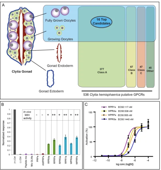
Figure 1. Identification of the Clytia MIH receptor (MIHR)
A) Diagram of a Clytia gonad showing the different tissues used for RNA-seq and GPCR expression comparisons.
536 putative GPCRs identified in Clytia mixed-stages transcriptome were assigned to the three main GPCR classes (A, B, C) or “other” based on Pfam signatures. 16 top candidate MIH GPCRs were selected based on oocyte enrichment, relatedness to known bilaterian class-A neuropeptide GPCRs and Pfam information. B) Luminescence response of CHO-K1 cells expressing the putative Clytia MIH GPCR treated with neuropeptide mixes lacking MIH activity (purple bars), MIH tetrapeptides identified from Clytia hemisphaerica (blue bars) or Cladonema radiatum (green bars), or related penta- and tripeptides previously shown to be ineffective in triggering oocyte maturation in
vivo (red bars) [15]. Empty pcDNA3.1 vector was used as negative control, and Platynereis FLamide and its receptor
as a positive control (grey bars). Peptide concentrations all 1µM. Absolute units of luminescence were normalized using the positive control; data are shown as mean ± standard error of the mean (n= 3). MIH tetrapeptides were selectively able to activate the Clytia GPCR, the responses closely matching the in vivo MIH activity of each peptide tested on Clytia oocytes as indicated [summarised results from 15]. C) Dose–response curves of Clytia MIHR challenged with four variant Clytia MIH tetrapeptides. One of 3 independent experiments with equivalent results is shown. Luminescence values were normalized relative to the maximum of fitted dose–response curves, and are shown as mean ± standard error of the mean (n= 3). Half maximal effective concentration (EC50) values were calculated as means of 3 independent experiments.
31
Figure 2. Phenotypes of Clytia MIHR mutants
Light and confocal microscope images of MIHR mutant F0 polyp colonies and jellyfish. A) Morphology of a wild type (WT) colony (Z11, top) and a MIHR mutant colony (n5-13, bottom). All MIHR mutant colonies contained gastrozooid and gonozooid polyps (pink and yellow arrows respectively), however the connecting stolons in some mutant colonies (see Table 1) were convoluted and frequently detached from the glass substrate, while stolons of wild type colonies were straight and adhered tightly. B) Fully grown mutant jellyfish n5-8 and n5-24 (bottom row) compared to wild type (top row) showed poorly developed gonads (white arrows) and sluggish behaviour but are otherwise indistinguishable. Centre (females) and right (males) panels show confocal microscope images through the gonads of adult medusae; Nuclei are stained with Hoechst 33342 (cyan) and F-actin with Phalloidin (Red). In the n5-8 female gonad, small oocytes (arrows) can be detected between the endodermal (en) and ectodermal (ec) layers, but no large growing oocytes (oo) are present compared to the wild type female gonad. In the n5-24 male gonad, the spermatogenic zone (asterisk) between endoderm and ectoderm is much thinner than in the wild type male gonad.
C) Comparison of mutant n5-23 and n5-13 (bottom row) female medusae gonads (white arrows) swollen by an
accumulation of large oocytes to wild type (Z11) female medusae (top row). Right panels show gonads dissected from 3-week old n5-23 medusae 10 hours after a light cue that induced spawning in the wild type but not the mutant gonad. D) Fully grown mutant n5-10 and n5-6 male medusa (bottom row) compared to wild type (Z13) male medusae (top row) have deformed gonads (white arrows). Right panels show isolated gonads, illustrating the thickened and irregular spermatogenic layer (arrowheads) in the mutant. Scale bars 1mm for light microscope jellyfish and polyp images; 200µm for isolated gonad images; 50µm for confocal images.
32
Figure 3. Sites of MIHR expression in the Clytia medusa
A) In situ hybridization detection of MIHR mRNA in a Clytia female medusa. Strong purple MIHR signal (orange
arrows) was detected in oocytes within the gonads, as well as in scattered cells in tentacles. B) Comparison of the distribution of the MIH receptor and ligand expressing cells in different medusa structures as labelled, detected by
in situ hybridization using probes to MIHR (top row) and to the MIH peptide precursors PP1 and/or PP4 as
indicated (bottom row). Orange arrows point oocytes, developing spermatozoa and tentacle MIHR cells, and white arrows indicate MIH cells. The focal plane in the male gonad image is through the centre to illustrate the position of the MIH cells in the ectodermal layer. Weak staining at the base of the manubrium (black arrow) in A and B is frequently observed with probes for many genes and is probably due to a specific trapping of the colour reagent. Scale bars: 100 µm. C) Confocal images of the three main sites of MIH-expressing cells (white arrows) in medusae, visualized using anti-PRPamide antibody (MIH: white), anti-tyrosinated tubulin (magenta) and Hoechst staining of nuclei (blue). Summed Z stacks are shown in all cases except for the gonad tubulin and DNA staining, where a single plane was selected through the centre of the gonad. All scale bars 100µm.
33
Figure 4. Oocyte maturation failure in MIHR mutant medusae
Oocyte maturation assays performed using isolated gonads (A, B) or isolated oocytes (C) from MIHR mutant jellyfish compared to wild type (WT). In A and C, bar heights represent mean percentages of 3 independent experiments and error bars show standard deviations. Total gonad or oocyte numbers for each treatment are indicated in grey. A) Spawning response of isolated gonads from WT and n5-23 female medusae. Three treatments were compared as indicated above each panel: Light: light stimulation after incubation in the dark; MIH: treatment of light-maintained gonads with 100nM WPRPamide; cAMP: treatment of light-maintained gonads with 4mM 5-Br-cAMP. No oocyte maturation or spawning were observed in MIHR KO gonads upon light stimulation or MIH treatment, while 5-Br-cAMP treatment provoked oocyte maturation and spawning. The Fisher exact test showed significant differences (F= 0) between wild type and mutant responses to light and MIH, but not for the cAMP treatment (F=1). B) Light microscope images illustrating gonads from an equivalent experiment performed with n5-13 female MIHR mutant medusae 120 minutes after the indicated treatments. Scale bar 500µm. C) Response of fully grown oocytes isolated from WT and n5-23 MIHR mutant gonads to MIH and 5-Br-cAMP treatments as in A . Both treatments triggered maturation of WT oocytes, visible after 20-30 minutes as germinal vesicle breakdown (GVBD), but only 5-Br-cAMP induced maturation of MIHR KO oocytes. Control experiments using the 5-Br-cAMP solvent (distilled water) showed a low level of spontaneous maturation in both cases. Fisher exact test did not show significant differences between WT and mutant oocytes in the control (F= 0.101) or cAMP treated (F= 0.216) groups, but did so in the MIH assays (F= 0).
34
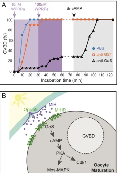
Figure 5. MIH-induced oocyte maturation blocked by an inhibitory Gαs antibody
Involvement of Gαs in Clytia oocyte maturation. A) Results of an antibody inhibition experiment. Maturation response (scored as % GVBD over time) of isolated oocytes injected with antibodies or buffer, then challenged first with a low dose of MIH (10nM WPRPamide), then with a higher dose (100nM WPRPamide) and finally with 5’-Br-cAMP to verify maturation competence, as indicated by the coloured arrows and background shading. Oocytes injected with PBS (blue circles) or a control anti-GST antibody (orange squares) responded efficiently to MIH, whereas very few oocytes injected with anti-Gαs (black triangles) underwent GVBD after treatment with the low dose of MIH and only 29% following the high dose. The number of oocytes per group in this experiment was 30, 28 and 30 respectively. All of them had undergone GVBD by the end of the experiment. Times from the start of the first incubation are shown on the x axis. Equivalent results were obtained in two other experiments.
B) Scheme illustrating the proposed cascade initiating Clytia oocyte maturation initiation. Following light
stimulation after a dark period, Opsin9 mediates release of MIH neuropeptides from specialised cells (purple) of the gonad ectoderm (cyan). Activation of the MIHR (green) at the oocyte surface releases GαS to promote an increase in cytoplasmic cAMP, activating PKA. Unknown PKA substrates likely trigger in parallel Cdk1 activation and thus GVBD, and Mos1 synthesis to initiate the MAPK cascade.
35
Figure 6. Relationship of Clytia MIHR to bilaterian neuropeptide hormone GPCRs
(A) Sequence-similarity-based clustering using Clans2 of all identified class-A GPCRs from Clytia, human (olfactory receptors excluded) and Platynereis deorphanized GPCRs [28] BLASTP p-value < 1e-40. (B) Cluster map of the largest cluster (circled in red in A) keeping only sequences that show at least 2 connections with the central cluster. BLASTP p-value < 1e-40. (C) More stringent cluster map (p-value < 1e-50) of the same
sequences as in (A) plus all Nematostella GPCR-A sequences. Only clusters containing at least 5 sequences from at least 2 species were kept. All connections with p-value < 1e-40 are shown. (D) Maximum likelihood analysis of the sequences contained inside the dashed area shown in (C) using RaxML (PROTGAMMAGTR) with 500 Bootstrap replicates (BR). Tree file in Fig. S2. AstA, Galanin, AstC and Kisspeptin receptors were included as outgroup. AKH, adipokinetic hormone; AstC, allatostatin-C; AstA, allatostatin-A; GnRH, gonadotropin releasing hormone; LH/FSH, Luteinizing/Follicle-stimulating hormone receptor; GPR3, G protein-coupled receptor 3;
36
MCH, melanin-concentrating hormone; NPY, neuropeptide Y; NPFF, neuropeptide FF; PRLH, prolactin releasing hormone; P2Y, purinoceptor; QRFP, pyroglutamylated RFamide peptide; TRH, thyrotropin releasing hormone. Colour code: Homo sapiens: blue, Platynereis dumerilli: green, Nematostella vectensis: black, Clytia: orange. Red star: Clytia MIHR.
Figure 7. Schematic comparison of GPCR regulation of Clytia and vertebrate oocyte maturation
Simplified view of the tissues, hormones and receptors involved in regulating oocyte maturation in Clytia, and in fish/amphibians and mammals. For simplicity we have not included protostome or echinoderm models. The principle peptide hormones of the reproductive hypothalamus-pituitary-gonadal axis (GnRH and LH/FSH) are in pink, and those for which the receptors group phylogenetically with Clytia MIHR in “Group A” (Fig. 6) in purple. Peptide hormones: Clytia MIH; Neuropeptide Y (NPY); Gonadotropin Inhibitory Hormone (GnIH); Gonadotropin Releasing Hormone (GnRH); Luteinizing Hormone (LH); glutamine RF amide peptide (QRFP); Neurokinin B (NkB); C-type natriuretic peptide (CNP). All their receptors, except the guanylyl cyclase
natriuretic peptide receptor 2 (NPR2) activated by CNP, are GPCRs (green). Constitutively active (CA) GPCRs in vertebrate oocytes maintain cytoplasmic cAMP levels high prior to maturation. Several types of oocytes receptor (orange) may respond to steroid hormones (Pg) in different species of amphibians and fish, but the relative importance of multiple downstream signalling pathways remains to be clarified [1,43,73,74]. See text for discussion.