Distant Chaperones and N-Glycan Signals: New Mechanisms of Secretory Pathway Proteostasis
by
Madeline Y. Wong B.S., Yale University (2013)
Submitted to the Department of Chemistry
in Partial Fulfillment of the Requirements for the Degree of Doctor of Philosophy in Chemistry
at the
MASSACHUSETTS INSTITUTE OF TECHNOLOGY September 2018
@ 2018 Massachusetts Institute of Technology. All rights reserved.
Signature redacted
Signature of Author_______________Department of Che stry August 27, 18
Signature redacted
Certified by Accepted by MASSACHUSETTS INST MASSACHUSETS INT OF TECHNOLOGYNOV 1 4 2018
Matthew D. Shoulders Whitehead Career Development Associate Professor of Chemistry Thesis SupervisorSignature redacted
TUTE -<oDert vv. I-ield
Haslam and Dewey Professor of Chemistry Chair, Departmental Committee on Graduate Students
This doctoral thesis has been examined by a
committee of the Department of Chemistry as follows:
Signature redacted
(7Whitehead
Elizabeth M. Nolan Associate Professor of Chemistry Thesis Committee Chair
Signature redacted
Matthew D. Shoulders Career Development Associate Professor of Chemistry Thesis Supervisor
Signature redacted
Jeremiah A. Johnson Associate Professor of Chemistry
Distant Chaperones and N-Glycan Signals: New Mechanisms of Secretory Pathway Proteostasis
by
Madeline Y. Wong
Submitted to the Department of Chemistry on August 27, 2018 in Partial Fulfillment of the Requirements for the Degree of Doctor of Philosophy in
Chemistry Abstract
Approximately one-third of all cellular proteins traverse the secretory pathway. After translation and folding in the endoplasmic reticulum (ER), proteins are transported through the Golgi to their final locations inside or outside of cells. At each step, proteins are helped by chaperones, which both shepherd proteins towards their native structures and serve as gatekeepers for export. Many proteins in the secretory pathway are also modified by installation of polysaccharides on specific asparagine residues. These N-glycans are installed in the ER as uniform precursors, but are trimmed and built up by Golgi glycan maturation enzymes into a striking array of epitopes. N-glycans act as a second mechanism to stabilize protein structure and prevent the release of misfolded proteins. Outside the cell, N-glycans on cell surfaces and secreted, soluble proteins allow cells to interact with each other, with their environment, and with distal tissues.
During development, cells encounter physiological ER stress incurred by high levels of sustained protein production. Unresolved protein misfolding, on the other hand, results in pathological ER stress and tissue dysfunction. Prior work has used small model substrates to show that cells utilize secretory pathway chaperones and tune N-glycosylation to respond to ER stress. This thesis examines how cells use similar strategies to accommodate challenging cargoes such as collagen-1. In the human body, collagen-I constitutes the primary protein component of bone, skin, and other organs; collagen-I misfolding results in pathological ER stress and connective tissue diseases. We therefore set out 1) to identify cellular components required for collagen-I secretion that could be targeted to address disease and 2) to assess the effects of ER stress on both cellular N-glycan structures and individual glycoproteins.
Here, we employ a high-throughput assay for collagen-I secretion and find that the cytosolic isoform of Hsp90 is required for collagen-I export. We also show that intracellular stress signaling alters the structures of cell surface and secreted N-glycans. Finally, we demonstrate that the collagen-I N-glycan buffers collagen-I folding against destabilizing mutations and ER stress. Our results identify potential therapeutic leads for collagen misfolding diseases and point to new mechanisms for maintaining secretory pathway proteostasis.
Thesis Supervisor: Matthew D. Shoulders Title: Whitehead CD Associate Professor
Acknowledgements
This thesis would not have been possible without the generosity and assistance of many people. My thanks go to: the chemical waste and radiation safety personnel at MIT, for keeping the lab space clean and safe; my collaborators in MIT core facilities and around the world, who freely offered their expertise, helped with experiments, and welcomed me into their labs; my thesis committee members, for their thoughtful comments on this work; my lab mates, past and present - for sharing time, reagents, and protocols, and for the antics as well; Matt, for being a better advisor than I could have asked for; and family and friends, who gave invaluable support, advice, and fellowship on this road.
Were the whole realm of nature mine, That were an offering far too small;
Love so amazing, so divine, Demands my soul, my life, my all.
CONTENTS
Abstract 3
Acknowledgements 5
PART ONE
Chapter I 15
A Proteostasis Approach to Treating the Collagenopathies
Chapter II 29
Development of a High-Throughput Assay for Collagen-I Secretion
Summary 29
Introduction 30
Results 30
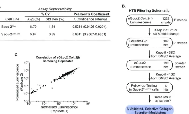
Luminescent Assay Design and Validation 30
High-Throughput Screening for Collagen-I Secretion Modulators 34
The Hsp90 Inhibitor 17-AAG Reduces the Secretion of Endogenous Collagen-I 37
17-AAG Reduces Collagen-I Secretion via a Post-Translational Effect 38
17-AAG Selectively Reduces Collagen-I, but Not General Protein, Secretion 39
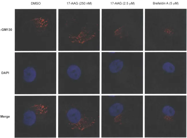
17-AAG Treatment Does Not Result in Intracellular Collagen-I Deposits 42
The Broad-Spectrum Anti-Parasitic Nitazoxanide Also Reduces
Collagen-I Secretion 44
Discussion 44
Materials and Methods 46
Cell Lines and Reagents 46
Vector and Stable Cell Line Construction 47
High-Throughput Screening and Dose-Response Curves 47
Co-Immunoprecipitation Experiments 47
Precipitation of Collagen-I from Conditioned Media 48
Disulfide-Dependent Assembly Assays 48
Pepsin Digestion 48
Fluorescence-Assisted Cell Sorting 49
Quantitative PCR (qPCR) 49
Pulse Labeling 49
Adenoviral Production 49
Transthyretin and Fibulin-3 Secretion 49
Metabolic Labeling of Secreted Proteins 49
Confocal Microscopy 50 Electron Microscopy 50 Statistical Analyses 50 Funding 51 References 52 Chapter 1I1 57
A Role for Cytosolic Hsp90P in Collagen-I Secretion
Summary 57
Introduction 58
Results 59
Structurally Diverse Hsp90 Inhibitors Reduce the Secretion of
Endogenous Collagen-I 59
Reduced Collagen-I Secretion Does Not Require the Heat Shock Response 60
Grp94 Knockdown Does Not Inhibit Collagen-I Secretion 61
Isoform-Selective Inhibitors Suggest that Cytosolic Hsp903 is Specifically
Required for Collagen-I Secretion 64
Genetic Knockdown of Hsp90a and Hsp903 65
Genetic Knockdown of USP19 and the Co-Chaperone NudCL 68
Discussion 69
Materials and Methods 73
Cell Lines and Reagents 73
Western Blotting Analysis 73
Lentivirus Production 73
Quantitative PCR (qPCR) 73
shRNA Knockdown 74
Funding 74
PART TWO
Chapter IV 81
Adapting Secretory Proteostasis and Function through the Unfolded Protein Response
Summary 81
Introduction 82
The UPR in Health and Disease 82
Development, Professional Secretory Cells, and Immunity 83
Emerging Functions of the UPR 85
Dysregulated ER Proteostasis and Disease 85
Concept Summary 86
Targeting the UPR to Modulate ER Proteostasis 86
Stress-Dependent Methods to Modulate the UPR 86
Stress-Independent Methods to Modulate the UPR 87
Activating the UPR to Address Diseases Linked to Dysregulated ER Proteostasis 88
Concept Summary 91
Beyond the UPR 91
Targeting A TP-Dependent Chaperone Systems in the ER 91
Targeting ERAD 93
Concept Summary 94
Conclusions 94
References 96
Chapter V 105
XBP1s Activation Can Globally Remodel N-Glycan Structure Distribution Patterns
Summary 105
Introduction 106
Results 107
Experimental Platform and Workflow to Scrutinize Effects of XBP1s
on the N-Glycome 107
XBP1s Remodels the HEKXBPls Cell Membrane N-Glycome 112 XBPls Remodels HeLaxBP"s Membrane N-Glycoproteomes in a
Cell Type-Dependent Manner 113
XBP1s Does Not Significantly Alter the Proteomic Composition of the HEKXBPls
Secretome 120
Selective XBP1s Induction Remodels the Glycogene Transcriptome 120
Discussion 123
Materials and Methods 125
Cells and Reagents 125
RNA Extraction, Real-Time qPCR, RNA-Seq and Membrane/Secretome
Preparation for Glycomic Analyses 125
Lectin Microarray Glycomic Analyses 125
MALDI-TOF MS and TOF/TOF MS/MS Glycomic Analyses 126
GC-MS Glycan Linkage Analyses 126
Lectin Flow Cytometry and Metabolic Assays 126
Proteomics Analysis 127
Data Deposition 127
Funding 127
References 128
Chapter VI 135
Identification of a Functional Role for the Conserved N-Glycan on the Collagen-I C-Terminal Propeptide
Summary 135
Introduction 136
Results 137
The Collagen-I N-Glycan is Highly Conserved Across Species 137
The N-Glycan is Required for Secretion of Misfolding Collagen-I Variants 139
Chemically Phenocopying Genetic Removal of the N-Glycan Sequon 140
The N-Glycan Protects Cola1 (I) Against Stress-Induced Aggregation 141
Discussion 141
Materials and Methods 143
Cell Lines and Reagents 143
Vector Construction 143
Adenoviral Production 144
Adenoviral Transduction 144
Castanospermine Treatment 144
Funding 145
References 146
Chapter VIl 149
Perspectives on the Field
References 153
CHAPTER I
A Proteostasis Approach to Treating the Collagenopathies Summary
The collagen-misfolding diseases, or collagenopathies, encompass mutations in more than 31 genes and show clinical phenotypes in most connective tissues. Single mutations that affect the folding of collagen chains can prevent secretion of sufficient protein levels for tissue function; in other cases, mutant chains accumulate in the endoplasmic reticulum (ER), resulting in sustained ER stress that leads to cellular dysfunction or death. The most severe disease phenotypes often stem from a third class of mutants, whose deposition in the extracellular matrix (ECM) disrupts packing of wild-type collagen fibrils or other ECM components. In addition to these disease mechanisms, loss-of-function variants of chaperones required for collagen folding, or mutations in other ECM proteins that share the same secretion pathway, also contribute to chondro- and skeletal dysplasias. Cells employ a transcriptional program called the Unfolded Protein Response (UPR) that is responsible for restoring homeostasis in the ER upon cellular or organismal insult, and targeting each of the three arms of the UPR has shown promise for diverse protein misfolding diseases. Increasing evidence suggests that UPR-focused therapeutic strategies may be able to mitigate the consequences of collagen misfolding and greatly improve patient outlook. Beyond the UPR, targeting individual components of the cellular proteostasis network also holds therapeutic potential. In this chapter, we discuss application of these strategies to the unique proteostatic challenges of collagen misfolding diseases, and review potential directions for building upon recent advances in this area.
Dysregulated Proteostasis and the Collagenopathies
Collagen is a strange, surprising protein. It completely lacks enzymatic activity, and yet regulates processes such as receptor signaling, cell migration, and tissue repair.' The structure of mature type I collagen (collagen-) is a simple right-handed triple helix formed from strands of repeating Gly-X-Y amino acid motifs, but relies on specific sequence properties and post-translational modifications to pre-organize and stabilize the triple helix.2'
3 Among the twenty-eight other types of collagen, the triple helix is subjected to an impressive array of permutations, from the interrupted helices of the FACIT (Fibril Associated Collagens with Interrupted Triple helices) family to the modular, organizing appendages on the termini of network-forming collagens (reviewed in [1] and [4]). Outside the cell, the collagens assemble with themselves, with each other, and with other ECM components to form and maintain diverse tissues.
Unsurprisingly, delays or difficulties in any of these processes-translation, modification, triple helix assembly, secretion, or fibril formation-cause disease. The affected tissues and phenotypes are as varied as the collagens themselves, but many involve factors such as insufficient or excess protein deposition, cellular stress, inflammation, and apoptosis.5 Because the molecular basis for disease is not always clear, with some collagen genotypes giving rise to
6
multiple distinct phenotypes, one approach to treating the collagenopathies would be to target the network of cellular proteins responsible for collagen biosynthesis and turnover. Focusing on regulators of collagen protein homeostasis (proteostasis) is not unprecedented: over twenty-five years ago, small molecule inhibitors of the collagen prolyl-4-hydroxylase (P4H) enzyme were presented as potential therapeutic compounds for cases of excess collagen deposition, and compounds that selectively inhibit or activate P4H in the absence of general toxicity are now available.f~9 Moreover, collagen does not contain clear binding sites or chemical handles; thus, targeting the chaperones and modifying enzymes that regulate its production could expand druggable space while simultaneously addressing multiple aspects of proteostatic imbalance
(see Section 3.3 of [10]).'
The Unfolded Protein Response
Collagen is synthesized and assembled in the ER, which safeguards organellar proteostasis by means of the Unfolded Protein Response (UPR). Similar to the cytosolic heat shock response, the UPR tunes protein folding and degradation machinery to restore ER proteostasis upon accumulation of misfolded proteins. Activation of the three transmembrane sensors IRE1, ATF6, and PERK produces transcription factors XBP1s, ATF6(1-373), and ATF4, respectively, that together increase levels of chaperones, enhance protein degradation, and stall new protein synthesis to allow the ER to recover. If ER stress continues, however, PERK-induced ATF4 activity will increase levels of a protein termed CHOP in a signaling cascade that leads to apoptosis.
The advent of chemical tools for selective, stress-independent activation of individual arms of the UPR" has clarified the overlapping functions of each branch, as well as revealed physiological roles for individual UPR transcription factors.12 XBP1s activity, for instance, has
been shown to regulate processes such as memory formation, innate immunity, and cell non-autonomous signaling.-13 5 ATF6 has been less extensively studied, but has been linked to
regulation of ER size and mesodermal differentiation.16' 17 Importantly, selective activation of individual arms of the UPR also mitigates protein misfolding in both gain-of-function and loss-of-function disease models. In particular, ATF6 activation has been shown to reduce secretion and aggregation of amyloid-forming mutants of transthyretin, 18 '9 as well as increase clearance of
(toxic) protein aggregates, either through ER-associated degradation by the proteasome or autophagy.2 0 XBP1s activation is also able to reduce secretion of amyloidogenic proteins.19
Intriguingly, proteomics-based approaches to identify components of the collagen-I proteostasis network found that a number of novel interactors are transcriptional targets of XBP1s and/or
collagen- biosynthesis, but also may have positive effects in the collagenopathies; we discuss these possibilities in more detail below.
IREI-XBPls
IRE1 is the oldest stress sensor of the UPR, and fulfills dual roles in response to ER stress. Accumulation of misfolded proteins titrates the ER chaperone BiP away from the luminal portion of IRE1, causing oligomerization and auto-phosphorylation by the IRE1 kinase domain 2
The IRE1 RNase domain subsequently splices XBP1 mRNA to generate active XBP1s (spliced) mRNA, which is translated to give the XBP1s transcription factor. In addition to generating active XBP1s, IRE1 also degrades selective subsets of gene transcripts in a process known as regulated IRE1-dependent decay. XBP1s was previously shown to enable antibody production during B-cell differentiation by expanding the ER and increasing secretory pathway capacity.2 4
Given that many fibrillar collagens are too large to fit into standard COP-Il vesicles, and that collagen-producing tissues are responsible for secreting large amounts of ECM proteins in addition to collagen, it is plausible that XBP1s facilitates a similar change during cartilage or bone development. Indeed, in addition to regulating the expression of genes involved in I biosynthesis, the IRE1/XBP1s axis has numerous physiological roles in collagen-producing tissues. IRE1 and XBP1s inhibit ER stress-induced apoptosis in differentiating cartilage.26 XBP1s also controls the expression of the transcription factor Osterix, required for osteoblast mineralization, in addition to chondrocyte proliferation and hypertrophic growth plate shortening in cartilage maturation.27
28
The effects of IRE1-XBPls signaling, though, are multi-faceted, complicating efforts to evaluate XBP1s as a therapeutic target for the collagenopathies. A transgenic mouse model in which a 13-base pair deletion mutant of Coll0a(l) accumulates in hypertrophic chondrocytes shows XBP1s splicing and increased BiP expression, suggestive of a protective UPR that allows chondrocytes to survive ER stress. Chondrocyte differentiation in these mice, however, is disrupted, leading to delayed endochondral bone formation and chondrodysplasia in a process that may involve XBP1s-mediated increases in miRNA-214 .29 3 1 Stress-induced IRE1 activity has been proposed to exacerbate fibrosis through degradation of miR-150 and production of XBP1s.3 2 Moreover, expression of the Schmid metaphyseal chondrodysplasia (MCDS) mutant
Coll0a(l) N617K in XBP1s-depleted cartilage revealed that UPR activation suppresses chondrocyte differentiation through CHOP, not XBP1s.33 Thus, while XBP1s activity can protect
cells from apoptosis following collagen retention and ER stress, hyper-activation of XBP1s can also disrupt normal development, or in other cases may not be related to pathology at all.3
Still, targeted methods to control IRE1 and/or XBP1s activity independent of ER stress may prove useful in addressing the collagenopathies. Clearance of misfolded collagen by autophagy restored bone growth in osteoblasts expressing the osteogenesis imperfecta (01) variant Cola2(1) G610C, and autophagy has been proposed as an innate protective mechanism for dealing with collagen aggregates that accompany expression of many disease variants.3 3
-Small molecule autophagy activators such as carbamazepine (CBZ) have yielded promising results in several instances, 3 94
-but may require high doses that could produce off-target effects. A more selective approach might be XBP1s-mediated induction of autophagy, which could reduce disease burden by removing aggregated collagen deposits and alleviating pathological ER stress. Doxycycline-controlled expression of XBP1s has been used to profile UPR transcriptional targets and achieve sustained, stress-independent XBP1s activity , small molecule activators of IRE1 have also been developed that are well-suited for short (-8 h)
45
periods of IRE1 activity. Application of similar strategies to collagen disease models may identify a therapeutic window for IRE1 and/or XBP1s activation that can resolve collagen misfolding in the absence of long-term effects on differentiation (Figure 1.1).
N-NH
HN lk /
> <
N!< HN~~ 0Na SCH 3 IPA E R IRE1 PIP21 ;e l
CBZ XBP1s O NH Nucleus Beclin-1 AutophagyFigure 1.1: Methods to regulate IRE1/XBPls activity for addressing the collagenopathies.
The small molecule IPA activates IRE1 RNase activity independent of its kinase domain, leading to splicing of XBP1 mRNA and production of the active transcription factor XBP1s. Among XBP1s transcriptional targets is Beclin-1, a positive regulator of autophagy that could help clear misfolded collagen aggregates. Carbamazepine (CBZ) also activates autophagy, but through IRE1 and XBPIs-independent signaling pathways. INS = inositol; IP3 = inositol
1,4,5-trisphosphate; PIP2 = Phosphatidylinositol 4,5-bisphosphate. Phosphoinositol signaling
A TF6 and A TF6-like transcription factors
Like XBP1s, ATF6 has physiological roles in the development of collagen-producing tissues. Overexpression of active or dominant-negative ATF6 by adenoviral transduction linked ATF6 expression to matrix mineralization downstream of bone morphogenic protein 2 (BMP2) and Runx2 signaling.46 Activation of ATF6 also conferred protection against experimental inflammatory arthritis by C/EBPP and mTOR-mediated inhibition of NF-KB signaling.47 ATF6
regulates a partially overlapping set of genes with IRE1, but is activated by a distinct mechanism, in which the transcription factor is transported to the Golgi and processed by site 1 (S1) and site 2 (S2) proteases to give an active N-terminal fragment (ATF6f).
Engineered cell lines, in which ATF6 is fused to a metastable, small molecule-responsive domain, have been used to examine effects of selective, stress-independent ATF6 activation.'' 18 An attractive alternate strategy is direct, small-molecule activation of ATF6 by compounds identified in a recent high-throughput screen.48 However, further studies have
revealed that the mechanism of action for one of these compounds involves covalent modification of ER chaperones PDIA3, 4, and 5, as well as ERP29.49 The extent of modification
was incomplete (-29%), but several of these chaperones have potential roles in collagen-I biosynthesis,2' and so thorough characterization of ATF6-activating compounds will be needed to avoid counterproductive effects.
Beyond ATF6, a family of ATF6-like transcription factors has been identified with strong connections to collagen physiology. Despite low sequence homology, the osteoblast-specific OASIS and chondrocyte-specific BBF2H7 share key features of ATF6 structure, including a transcriptional activation domain, transmembrane domain, and motif for site 1 (S1) protease processing.50 BBF2H7 increases expression of Sec23a, a gene required for collagen secretion, to promote chondrogenesis and protect chondrocytes from UPR-induced apoptosis; BBF2H7 knockout mice accordingly show chondrodysplasia along with a distended ER.27 In
osteoblasts, OASIS increases expression of Colal(I) to facilitate bone formation.52 OASIS and
BBF2H7 are regulated differently than ATF6, through stabilization of protein levels as opposed to disulfide reduction-mediated monomerization, and may not respond to the identified ATF6 activators.53 Still, clarification of OASIS and UPRE binding motifs could allow for similar
reporter-based screening approaches to discover compound leads for modulating these ATF6-like transcription factors in ways that could prove beneficial in the collagenopathies (Figure 1.2). PERK
As the UPR branch responsible for initiating apoptotic signaling cascades, PERK is often treated more as part of the problem than the solution in protein misfolding diseases. ER stress-induced PERK signaling, however, is required for osteogenesiS54 and is activated in chondrocytes to facilitate collagen secretion.55
But while PERK/ATF4 signaling protects chondrocytes from apoptosis, it also promotes chondrodysplasia by reverting chondrocyte differentiation.56 Notably, the small molecule ISRIB,57 which blocks PERK signaling by rendering
cells insensitive to elF2a phosphorylation, restored differentiation and lessened skeletal deformities in a mouse model of MCDS.56 A similar approach using salubrinal showed positive
effects in an osteoarthritis model.58
However, the related compound guanbenz had no therapeutic effect, and the classification of these two compounds as elF2a phosphorylation inhibitors has been challenged.59~1 Obtaining a clearer understanding of the mode of action of PERK signaling inhibitors will thus be critical for understanding PERK pathophysiology in the context of collagen misfolding (Figure 1.3).
> N N 1NN N ,N I KSC-34 HN _'N CI H PDIU Collagen-I 4-ER SIP si i ATF6f
A
NucleusA" BBF2H7AFigure 1.2: Methods to regulate A TF6 activity for addressing the collagenopathies. The small
molecule 147 activates ATF6 by inhibiting the prolyl disulfide isomerases (PDIs) that promote
ATF6 oligomerization (ATF6o); inhibition of PDIA3, PDIA4, and/or PDIA5 promotes formation
of monomeric ATF6 (ATF6m), which is free to traffic to the Golgi. Processing by site 1 (S1P)
and site 2 (S2P) proteases yields the active transcription factor ATF6f. Among ATF6 transcriptional targets is BGLAP (Osteocalcin), a positive regulator of osteoblast differentiation. The a-site specific PDIA1 inhibitor KSC-34 does not target PDIA3 or PDIA4, but could potentially prevent PDIA1-mediated hydroxylation of collagen-I in the absence of UPR activation. BBF2H7 and OASIS are two members of a family of ATF6-like transcription factors whose target genes are important for chondrogenesis and osteoblast differentiation, respectively. Methods for regulating the activity of these transcription factors may therefore prove useful for chondro- or skeletal dysplasias.
0 -N H OH .4 ER BB I Golgi Processing OASISA 11 A7T 6t Ab ATF6m% Am OASIS;k
PERK 0~ S NH NH CI N CI CI Salubrinal?
K A
peIF ISRIB Guanabenz? CI H N'N N NH2 NHI
Translation CI 0 H - o ,,AN B "a o H ISRIB ER CHOP 2a ATF4 Chondrocyte DifferentiationFigure 1.3: Methods to regulate PERK activity for addressing the collagenopathies. The
small molecule ISRIB desensitizes cells to downstream signaling of phosphorylated elF2a. Salubrinal and guanabenz have also been proposed to block effects of elF2a phosphorylation, but their mechanism of action is not yet confirmed. PERK/ATF4 signaling can increase levels of the protein CHOP, which initiates apoptotic signaling cascades, but PERK/ATF4 signaling has also been implicated in chondrocyte differentiation. More work is therefore needed to determine therapeutic opportunities for modulating PERK activity in the
context of the collagenopathies.
Targeting downstream proteostasis components
The class of compounds that has been most widely used to date in addressing the collagenopathies is chemical chaperones. 4-Phenylbutyric acid (PBA) reduced cell death in 01 patient fibroblasts by promoting autophagic clearance of mutant Colal (1) and Cola2(l) chains and has been used to reduce ER stress in Col2a(IV) mutant lines.2
63 PBA also reduced ER stress and autophagy, but increased Col5a(IV) expression in fibroblasts from patients with Alport Syndrome (AS).64 Another chemical chaperone, trimethylamine N-oxide (TMAO), had a
larger effect on collagen-I triple helix thermostability when incubated with purified wild type,
R789C Col2a(l), or R992C Col2a(l) molecules than when added to cell culture,5 suggesting
that such compounds may act through direct binding to the target molecule rather than by modulating the cellular proteostasis network.6 This explanation is consistent with the ability of
PBA to increase Col5a(IV) transcripts in both AS patient lines, but improve secretion of only one mutant. In a separate study, PBA had minimal effects on retention of aggregated cartilage oligomeric matrix protein (COMP), underscoring the need to consider misfolding,
disease-related proteins in the context of the proteostasis network.6 7
More promising than chemical chaperones, though still untested, are therapeutic strategies that target individual components of the collagen proteostasis network. The existence of at least nine genes linked to recessive forms of 01 demonstrates that individual collagen chaperones can (and do) influence collagen production.68 Key advantages of this approach-in
contrast to the chemical chaperones-include disease-specific protein targets, improved mechanistic understanding, and access to existing medicinal chemistry knowledge. Extensive work on prolyl disulfide isomerase (PDI) inhibitors, for instance, enabled the recent development of a compound, KSC-34, that selectively binds the a-site catalytic domain.9 Importantly,
compound treatment has minimal effects on the UPR, indicating that PDIA1 can be targeted independent of induced ER stress. Similar improvements to the selectivity of inhibitors for BiP and other collagen-I chaperones will thus provide new opportunities for testing a proteostasis-targeted therapeutic approach in collagen disease models.
Conclusions
The UPR is integrally linked with development and disease in collagen-producing tissues. Connections between IRE1, ATF6, PERK, and multiple collagenopathies provide compelling evidence that regulating the different arms of the UPR could address diseases with varied causes and phenotypes. Due to the dual roles of UPR activation in development and disease, therapeutic approaches that target individual components of the proteostasis network will likely improve selectivity. However, many collagen-misfolding diseases result from inactivating mutations in collagen chaperones, and small molecule activators for these proteins are rare. Still, we note that significant advances in pharmacologic methods to tune UPR activity have come from the development of new screening platforms. Continued studies of disease-relevant signaling pathways can thus inform the design of screens for new compound leads. In the following chapters, we apply a similar approach to collagen-I proteostasis. We report the design and application of a high-throughput assay to identify selective small molecule modulators of collagen-I secretion (Chapter II). We then describe the results of mechanistic studies on a validated screening hit, 17-allylaminogeldanamycin (17-AAG), and the insights we obtain regarding its cellular target (Chapter 1I). Our results demonstrate that elements of the proteostasis network not traditionally considered to be collagen biosynthetic enzymes may also be important players in the collagenopathies. In addition, our findings emphasize the importance of the continued study of collagen biosynthesis and the physiology of collagen-producing tissues for discovery of new therapeutic targets.
References
[1] Ricard-Blum, S. (2011) The collagen family, Cold Spring Harbor Perspect. Biol. 3, a004978. [2] Shoulders, M. D., and Raines, R. T. (2009) Collagen structure and stability, Annu. Rev.
Biochem. 78, 929-958.
[3] Ishikawa, Y., and Bachinger, H. P. (2013) A molecular ensemble in the rER for procollagen maturation, Biochim. Biophys. Acta 1833, 2479-2491.
[4] Brinckmann, J. (2005) Collagens at a Glance, In Collagen: Primer in Structure, Processing and Assembly (Brinckmann, J., Notbohm, H., and MUller, P. K., Eds.), pp 1-6, Springer Berlin Heidelberg, Berlin, Heidelberg.
[5] Jobling, R., D'Souza, R., Baker, N., Lara-Corrales, I., Mendoza-Londono, R., Dupuis, L., Savarirayan, R., Ala-Kokko, L., and Kannu, P. (2014) The collagenopathies: review of clinical phenotypes and molecular correlations, Curr. Rheumatol. Rep. 16, 394. [6] Makareeva, E., Mertz, E. L., Kuznetsova, N. V., Sutter, M. B., DeRidder, A. M., Cabral, W.
A., Barnes, A. M., McBride, D. J., Marini, J. C., and Leikin, S. (2008) Structural heterogeneity of type I collagen triple helix and its role in osteogenesis imperfecta, J. Biol. Chem. 283, 4787-4798.
[7] Pihlajaniemi, T., Myllyla, R., and Kivirikko, K. I. (1991) Prolyl 4-hydroxylase and its role in collagen synthesis, J. Hepatol. 13 Suppl. 3, S2-7.
[8] Vasta, J. D., Andersen, K. A., Deck, K. M., Nizzi, C. P., Eisenstein, R. S., and Raines, R. T. (2016) Selective Inhibition of Collagen Prolyl 4-Hydroxylase in Human Cells, ACS Chem. Biol. 11, 193-199.
[9] Vasta, J. D., and Raines, R. T. (2016) Human Collagen Prolyl 4-Hydroxylase Is Activated by Ligands for Its Iron Center, Biochemistry 55, 3224-3233.
[10] Wong, M. Y., DiChiara, A. S., Suen, P. H., Chen, K., Doan, N.-D., and Shoulders, M. D. (2018) Adapting Secretory Proteostasis and Function Through the Unfolded Protein Response, In Coordinating Organismal Physiology Through the Unfolded Protein Response (Wiseman, R. L., and Haynes, C. M., Eds.), pp 1-25, Springer International Publishing, Cham.
[11] Shoulders, M. D., Ryno, L. M., Genereux, J. C., Moresco, J. J., Tu, P. G., Wu, C., Yates, J. R., 3rd, Su, A. I., Kelly, J. W., and Wiseman, R. L. (2013) Stress-independent activation of XBP1 s and/or ATF6 reveals three functionally diverse ER proteostasis environments,
Cell Rep. 3, 1279-1292.
[12] Plate, L., and Wiseman, R. L. (2017) Regulating Secretory Proteostasis through the Unfolded Protein Response: From Function to Therapy, Trends Cell Biol. 27, 722-737. [13] Martinon, F., Chen, X., Lee, A. H., and Glimcher, L. H. (2010) TLR activation of the
transcription factor XBP1 regulates innate immune responses in macrophages, Nat. Immunol. 11, 411-418.
[14] Martinez, G., Vidal, Rene L., Mardones, P., Serrano, Felipe G., Ardiles, Alvaro 0., Wirth, C., Valdes, P., Thielen, P., Schneider, Bernard L., Kerr, B., Valdes, Jose L., Palacios, Adrian G., Inestrosa, Nibaldo C., Glimcher, Laurie H., and Hetz, C. (2016) Regulation of Memory Formation by the Transcription Factor XBP1, Cell Rep. 14, 1382-1394.
[15] Taylor, R. C., and Dillin, A. (2013) XBP-1 is a cell-nonautonomous regulator of stress resistance and longevity, Cell 153, 1435-1447.
[16] Maiuolo, J., Bulotta, S., Verderio, C., Benfante, R., and Borgese, N. (2011) Selective activation of the transcription factor ATF6 mediates endoplasmic reticulum proliferation triggered by a membrane protein, Proc. Natl. Acad. Sci. U.S.A. 108, 7832-7837.
[17] Kroeger, H., Grimsey, N., Paxman, R., Chiang, W.-C., Plate, L., Jones, Y., Shaw, P. X., Trejo, J., Tsang, S. H., Powers, E., Kelly, J. W., Wiseman, R. L., and Lin, J. H. (2018) The unfolded protein response regulator ATF6 promotes mesodermal differentiation, Sci. Signaling 11, eaan5785.
[18] Chen, J. J., Genereux, J. C., Qu, S., Hulleman, J. D., Shoulders, M. D., and Wiseman, R. L. (2014) ATF6 Activation Reduces the Secretion and Extracellular Aggregation of
Destabilized Variants of an Amyloidogenic Protein, Chem. Biol. 21, 1564-1574.
[19] Cooley, C. B., Ryno, L. M., Plate, L., Morgan, G. J., Hulleman, J. D., Kelly, J. W., and
Wiseman, R. L. (2014) Unfolded protein response activation reduces secretion and extracellular aggregation of amyloidogenic immunoglobulin light chain, Proc. Nat/. Acad.
Sci. U.S.A. 111, 13046-13051.
[20] Smith, S. E., Granell, S., Salcedo-Sicilia, L., Baldini, G., Egea, G., Teckman, J. H., and Baldini, G. (2011) Activating transcription factor 6 limits intracellular accumulation of mutant alpha(1)-antitrypsin Z and mitochondrial damage in hepatoma cells, J. Biol.
Chem. 286, 41563-41577.
[21] DiChiara, A. S., Taylor, R. J., Wong, M. Y., Doan, N. D., Rosario, A. M., and Shoulders, M.
D. (2016) Mapping and Exploring the Collagen-I Proteostasis Network, ACS Chem. Biol.
11, 1408-1421.
[22] Walter, P., and Ron, D. (2011) The unfolded protein response: from stress pathway to homeostatic regulation, Science 334, 1081-1086.
[23] Hollien, J., and Weissman, J. S. (2006) Decay of Endoplasmic Reticulum-Localized mRNAs
During the Unfolded Protein Response, Science 313, 104-107.
[24] Shaffer, A. L., Shapiro-Shelef, M., Iwakoshi, N. N., Lee, A.-H., Qian, S.-B., Zhao, H., Yu, X., Yang, L., Tan, B. K., Rosenwald, A., Hurt, E. M., Petroulakis, E., Sonenberg, N.,
Yewdell, J. W., Calame, K., Glimcher, L. H., and Staudt, L. M. (2004) XBP1,
Downstream of Blimp-1, Expands the Secretory Apparatus and Other Organelles, and Increases Protein Synthesis in Plasma Cell Differentiation, Immunity 21, 81-93.
[25] Malhotra, V., and Erlmann, P. (2015) The pathway of collagen secretion, Annu. Rev. Cell
Dev. Biol. 31, 109-124.
[26] Han, X., Zhou, J., Zhang, P., Song, F., Jiang, R., Li, M., Xia, F., and Guo, F. J. (2013)
IRE1alpha dissociates with BiP and inhibits ER stress-mediated apoptosis in cartilage development, Cell Signal. 25, 2136-2146.
[27] Hughes, A., Oxford, A. E., Tawara, K., Jorcyk, C. L., and Oxford, J. T. (2017) Endoplasmic
Reticulum Stress and Unfolded Protein Response in Cartilage Pathophysiology; Contributing Factors to Apoptosis and Osteoarthritis, Int. J. Mol. Sci. 18, e665.
[28] Cameron, T. L., Gresshoff, I. L., Bell, K. M., Pirog, K. A., Sampurno, L., Hartley, C. L.,
Sanford, E. M., Wilson, R., Ermann, J., Boot-Handford, R. P., Glimcher, L. H., Briggs, M.
D., and Bateman, J. F. (2015) Cartilage-specific ablation of XBP1 signaling in mouse
results in a chondrodysplasia characterized by reduced chondrocyte proliferation and delayed cartilage maturation and mineralization, Osteoarthritis Cartilage 23, 661-670.
[29] Tsang, K. Y., Chan, D., Cheslett, D., Chan, W. C. W., So, C. L., Melhado, I. G., Chan, T. W.
Y., Kwan, K. M., Hunziker, E. B., Yamada, Y., Bateman, J. F., Cheung, K. M. C., and Cheah, K. S. E. (2007) Surviving Endoplasmic Reticulum Stress Is Coupled to Altered Chondrocyte Differentiation and Function, PLoS Biol. 5, e44.
[30] Roberto, V. P., Gavaia, P., Nunes, M. J., Rodrigues, E., Cancela, M. L., and Tiago, D. M. (2018) Evidences for a New Role of miR-214 in Chondrogenesis, Sci. Rep. 8, 3704. [31] Dong, L., Jiang, C. C., Thorne, R. F., Croft, A., Yang, F., Liu, H., de Bock, C. E., Hersey, P.,
and Zhang, X. D. (2011) Ets-1 mediates upregulation of McI-1 downstream of XBP-1 in human melanoma cells upon ER stress, Oncogene 30, 3716-3726.
[32] Heindryckx, F., Binet, F., Ponticos, M., Rombouts, K., Lau, J., Kreuger, J., and Gerwins, P. (2016) Endoplasmic reticulum stress enhances fibrosis through IRE1alpha-mediated
degradation of miR-1 50 and XBP-1 splicing, EMBO Mol. Med. 8, 729-744.
[33] Cameron, T. L., Bell, K. M., Gresshoff, I. L., Sampurno, L., Mullan, L., Ermann, J., Glimcher,
Pathways Suppress C/EBP-beta Mediated Chondrocyte Differentiation in ER-Stress Related Skeletal Disease, PLoS Genet. 11, e1005505.
[34] Ishikawa, T., Kashima, M., Nagano, A. J., Ishikawa-Fujiwara, T., Kamei, Y., Todo, T., and Mori, K. (2017) Unfolded protein response transducer IRE1-mediated signaling
independent of XBP1 mRNA splicing is not required for growth and development of medaka fish, eLife 6, e26845.
[35] Mirigian, L. S., Makareeva, E., Mertz, E. L., Omari, S., Roberts-Pilgrim, A. M., Oestreich, A. K., Phillips, C. L., and Leikin, S. (2016) Osteoblast Malfunction Caused by Cell Stress Response to Procollagen Misfolding in a2(l)-G61 OC Mouse Model of Osteogenesis Imperfecta, J. Bone Miner. Res. 31, 1608-1616.
[36] Ishida, Y., Yamamoto, A., Kitamura, A., Lamande, S. R., Yoshimori, T., Bateman, J. F., Kubota, H., and Nagata, K. (2009) Autophagic elimination of misfolded procollagen aggregates in the endoplasmic reticulum as a means of cell protection, Mol. Biol. Cell 20, 2744-2754.
[37] Makareeva, E., Sun, G., Mirigian, L. S., Mertz, E. L., Vera, J. C., Espinoza, N. A., Yang, K., Chen, D., Klein, T. E., Byers, P. H., and Leikin, S. (2018) Substitutions for arginine at position 780 in triple helical domain of the alphal (1) chain alter folding of the type I procollagen molecule and cause osteogenesis imperfecta, PLoS One 13, e0200264. [38] Lisse, T. S., Thiele, F., Fuchs, H., Hans, W., Przemeck, G. K., Abe, K., Rathkolb, B.,
Quintanilla-Martinez, L., Hoelzlwimmer, G., Helfrich, M., Wolf, E., Ralston, S. H., and Hrabe de Angelis, M. (2008) ER stress-mediated apoptosis in a new mouse model of osteogenesis imperfecta, PLoS Genet. 4, e7.
[39] Mullan, L. A., Mularczyk, E. J., Kung, L. H., Forouhan, M., Wragg, J. M. A., Goodacre, R., Bateman, J. F., Swanton, E., Briggs, M. D., and Boot-Handford, R. P. (2017) Increased intracellular proteolysis reduces disease severity in an ER stress-associated dwarfism, J. Clin. Invest. 127, 3861-3865.
[40] Forouhan, M., Sonntag, S., and Boot-Handford, R. P. (2018) Carbamazepine reduces disease severity in a mouse model of metaphyseal chondrodysplasia type Schmid caused by a premature stop codon (Y632X) in the CollOal gene, Hum. Mol. Genet. ddy253.
[41] Vidal, R. L., Matus, S., Bargsted, L., and Hetz, C. (2014) Targeting autophagy in neurodegenerative diseases, Trends Pharmacol. Sci. 35, 583-591.
[42] Margariti, A., Li, H., Chen, T., Martin, D., Vizcay-Barrena, G., Alam, S., Karamariti, E., Xiao, Q., Zampetaki, A., Zhang, Z., Wang, W., Jiang, Z., Gao, C., Ma, B., Chen, Y. G.,
Cockerill, G., Hu, Y., Xu, Q., and Zeng, L. (2013) XBP1 mRNA splicing triggers an autophagic response in endothelial cells through BECLIN-1 transcriptional activation, J. Biol. Chem. 288, 859-872.
[43] Lee, A. H., Iwakoshi, N. N., and Glimcher, L. H. (2003) XBP-1 regulates a subset of
endoplasmic reticulum resident chaperone genes in the unfolded protein response, Mol. Ce// Biol. 23, 7448-7459.
[44] Dewal, M. B., DiChiara, A. S., Antonopoulos, A., Taylor, R. J., Harmon, C. J., Haslam, S. M., Dell, A., and Shoulders, M. D. (2015) XBPls Links the Unfolded Protein Response to the Molecular Architecture of Mature N-Glycans, Chem. Biol. 22, 1301-1312.
[45] Mendez, A. S., Alfaro, J., Morales-Soto, M. A., Dar, A. C., McCullagh, E., Gotthardt, K., Li, H., Acosta-Alvear, D., Sidrauski, C., Korennykh, A. V., Bernales, S., Shokat, K. M., and Walter, P. (2015) Endoplasmic reticulum stress-independent activation of unfolded protein response kinases by a small molecule ATP-mimic, eLife 4, e05434.
[46] Jang, W. G., Kim, E. J., Kim, D. K., Ryoo, H. M., Lee, K. B., Kim, S. H., Choi, H. S., and Koh, J. T. (2012) BMP2 protein regulates osteocalcin expression via Runx2-mediated Atf6 gene transcription, J. Biol. Chem. 287, 905-915.
[47] Nakajima, S., Hiramatsu, N., Hayakawa, K., Saito, Y., Kato, H., Huang, T., Yao, J., Paton,
A. W., Paton, J. C., and Kitamura, M. (2011) Selective Abrogation of BiP/GRP78 Blunts
Activation of NF-KB through the ATF6 Branch of the UPR: Involvement of C/EBPP and mTOR-Dependent Dephosphorylation of Akt, Mol. Cell Biol. 31, 1710-1718.
[48] Plate, L., Cooley, C. B., Chen, J. J., Paxman, R. J., Gallagher, C. M., Madoux, F., Genereux, J. C., Dobbs, W., Garza, D., Spicer, T. P., Scampavia, L., Brown, S. J.,
Rosen, H., Powers, E. T., Walter, P., Hodder, P., Wiseman, R. L., and Kelly, J. W.
(2016) Small molecule proteostasis regulators that reprogram the ER to reduce
extracellular protein aggregation, eLife 5, e15550.
[49] Paxman, R., Plate, L., Blackwood, E. A., Glembotski, C., Powers, E. T., Wiseman, R. L., and Kelly, J. W. (2018) Pharmacologic ATF6 activating compounds are metabolically activated to selectively modify endoplasmic reticulum proteins, eLife 7, e37168.
[50] Kondo, S., Saito, A., Asada, R., Kanemoto, S., and Imaizumi, K. (2011) Physiological
unfolded protein response regulated by OASIS family members, transmembrane bZIP transcription factors, IUBMB Life 63, 233-239.
[51] Saito, A., Hino, S.-i., Murakami, T., Kanemoto, S., Kondo, S., Saitoh, M., Nishimura, R.,
Yoneda, T., Furuichi, T., Ikegawa, S., Ikawa, M., Okabe, M., and Imaizumi, K. (2009) Regulation of endoplasmic reticulum stress response by a BBF2H7-mediated Sec23a pathway is essential for chondrogenesis, Nat. Cell Biol. 11, 1197-1204.
[52] Murakami, T., Saito, A., Hino, S., Kondo, S., Kanemoto, S., Chihara, K., Sekiya, H.,
Tsumagari, K., Ochiai, K., Yoshinaga, K., Saitoh, M., Nishimura, R., Yoneda, T., Kou, I., Furuichi, T., Ikegawa, S., Ikawa, M., Okabe, M., Wanaka, A., and Imaizumi, K. (2009) Signalling mediated by the endoplasmic reticulum stress transducer OASIS is involved in
bone formation, Nat. Cell Biol. 11, 1205-1211.
[53] Kondo, S., Hino, S. I., Saito, A., Kanemoto, S., Kawasaki, N., Asada, R., Izumi, S.,
Iwamoto, H., Oki, M., Miyagi, H., Kaneko, M., Nomura, Y., Urano, F., and Imaizumi, K. (2012) Activation of OASIS family, ER stress transducers, is dependent on its
stabilization, Cell Death Differ. 19, 1939-1949.
[54] Saito, A., Ochiai, K., Kondo, S., Tsumagari, K., Murakami, T., Cavener, D. R., and Imaizumi, K. (2011) Endoplasmic reticulum stress response mediated by the PERK-eIF2(alpha)-ATF4 pathway is involved in osteoblast differentiation induced by BMP2, J.
Biol. Chem. 286, 4809-4818.
[55] Hisanaga, S., Miyake, M., Taniuchi, S., Oyadomari, M., Morimoto, M., Sato, R., Hirose, J., Mizuta, H., and Oyadomari, S. (2018) PERK-mediated translational control is required
for collagen secretion in chondrocytes, Sci. Rep. 8, 773.
[56] Wang, C., Tan, Z., Niu, B., Tsang, K. Y., Tai, A., Chan, W. C. W., Lo, R. L. K., Leung, K. K.
H., Dung, N. W. F., Itoh, N., Zhang, M. Q., Chan, D., and Cheah, K. S. E. (2018) Inhibiting the integrated stress response pathway prevents aberrant chondrocyte differentiation thereby alleviating chondrodysplasia, eLife 7, e37673.
[57] Sidrauski, C., Acosta-Alvear, D., Khoutorsky, A., Vedantham, P., Hearn, B. R., Li, H.,
Gamache, K., Gallagher, C. M., Ang, K. K., Wilson, C., Okreglak, V., Ashkenazi, A., Hann, B., Nader, K., Arkin, M. R., Renslo, A. R., Sonenberg, N., and Walter, P. (2013) Pharmacological brake-release of mRNA translation enhances cognitive memory, eLife
2, e00498.
[58] Hamamura, K., Nishimura, A., lino, T., Takigawa, S., Sudo, A., and Yokota, H. (2015)
Chondroprotective effects of Salubrinal in a mouse model of osteoarthritis, Bone Joint
Res. 4, 84-92.
[59] Crespillo-Casado, A., Chambers, J. E., Fischer, P. M., Marciniak, S. J., and Ron, D. (2017)
PPP1R15A-mediated dephosphorylation of elF2alpha is unaffected by Sephin1 or Guanabenz, eLife 6, e26109.
[60] Das, I., Krzyzosiak, A., Schneider, K., Wrabetz, L., D'Antonio, M., Barry, N., Sigurdardottir, A., and Bertolotti, A. (2015) Preventing proteostasis diseases by selective inhibition of a phosphatase regulatory subunit, Science 348, 239-242.
[61] Tsaytler, P., Harding, H. P., Ron, D., and Bertolotti, A. (2011) Selective Inhibition of a Regulatory Subunit of Protein Phosphatase 1 Restores Proteostasis, Science 332, 91-94.
[62] Besio, R., lula, G., Garibaldi, N., Cipolla, L., Sabbioneda, S., Biggiogera, M., Marini, J. C., Rossi, A., and Forlino, A. (2018) 4-PBA ameliorates cellular homeostasis in fibroblasts from osteogenesis imperfecta patients by enhancing autophagy and stimulating protein secretion, Biochim. Biophys. Acta 1864, 1642-1652.
[63] Murray, L. S., Lu, Y., Taggart, A., Van Regemorter, N., Vilain, C., Abramowicz, M., Kadler, K. E., and Van Agtmael, T. (2014) Chemical chaperone treatment reduces intracellular accumulation of mutant collagen IV and ameliorates the cellular phenotype of a COL4A2 mutation that causes haemorrhagic stroke, Hum. Mol. Genet. 23, 283-292.
[64] Wang, D., Mohammad, M., Wang, Y., Tan, R., Murray, L. S., Ricardo, S., Dagher, H., van Agtmael, T., and Savige, J. (2017) The Chemical Chaperone, PBA, Reduces ER Stress and Autophagy and Increases Collagen IV alpha5 Expression in Cultured Fibroblasts From Men With X-Linked Alport Syndrome and Missense Mutations, Kidney Int. Rep. 2, 739-748.
[65] Gawron, K., Jensen, D. A., Steplewski, A., and Fertala, A. (2010) Reducing the effects of intracellular accumulation of thermolabile collagen I mutants by increasing their thermostability in cell culture conditions, Biochem. Biophys. Res. Commun. 396, 213-218.
[66] Omachi, K., Kamura, M., Teramoto, K., Kojima, H., Yokota, T., Kaseda, S., Kuwazuru, J., Fukuda, R., Koyama, K., Matsuyama, S., Motomura, K., Shuto, T., Suico, M. A., and Kai, H. (2018) A Split-Luciferase-Based Trimer Formation Assay as a High-throughput
Screening Platform for Therapeutics in Alport Syndrome, Cell Chem. Biol. 25, 634-643. [67] Posey, K. L., Coustry, F., Veerisetty, A. C., Liu, P., Alcorn, J. L., and Hecht, J. T. (2014)
Chondrocyte-specific pathology during skeletal growth and therapeutics in a murine model of pseudoachondroplasia, J. Bone Miner. Res. 29, 1258-1268.
[68] Marini, J. C., and Blissett, A. R. (2013) New Genes in Bone Development: What's New in Osteogenesis Imperfecta, J. Clin. Endocrinol. Metab. 98, 3095-3103.
[69] Cole, K. S., Grandjean, J. M. D., Chen, K., Witt, C. H., O'Day, J., Shoulders, M. D., Wiseman, R. L., and Weerapana, E. (2018) Characterization of an A-Site Selective Protein Disulfide Isomerase Al Inhibitor, Biochemistry 57, 2035-2043.
CHAPTER II
Development of a High-Throughput Assay for Collagen-I Secretion Summary
Collagen overproduction is a feature of fibrosis and cancer, while insufficient deposition of functional collagen molecules and/or the secretion of malformed collagen are common in genetic disorders like osteogenesis imperfecta. Collagen secretion is an appealing therapeutic target in these and other diseases, as secretion directly connects intracellular biosynthesis to collagen deposition and biological function in the extracellular matrix. However, small molecule and biological methods to tune collagen secretion are severely lacking. Their discovery could prove useful not only in the treatment of disease, but also in providing tools for better elucidating mechanisms of collagen biosynthesis. We developed a cell-based, high-throughput luminescent assay of collagen type I secretion and used it to screen for small molecules that selectively enhance or inhibit that process. Among several validated hits, the Hsp9O inhibitor 17-allylaminogeldanamycin (17-AAG) robustly decreases the secretion of collagen- by our model cell line and by human primary cells. In these systems, 17-AAG reduces collagen-I secretion post-translationally and is not a global inhibitor of protein secretion. Beyond 17-AAG, we also identify the broad-spectrum antiparasitic compound nitazoxanide (NTZ) as a post-translational inhibitor of collagen-I secretion. Our results highlight the potential of a cell-based high-throughput screen for selective modulators of collagen secretion.
Contributions
Data from this chapter (Figures 2.1-10) were previously published in Wong et al. Biochemistry
2018, 57, 2814-2827, from which this chapter is adapted in part. Reprinted (adapted) with
permission. Copyright 2018 American Chemical Society. We thank Dr. Ngoc-Duc Doan for providing the Saos-2TREX cells used to generate the screening cell-lines and for assistance with confocal microscopy; Dr. Andrew S. DiChiara for experimental advice and for generating Figure
2.5E; Louis J. Papa, Ill for generating the fibulin-3 and transthyretin adenoviral constructs; Dr.
Nicki Watson for assistance with processing and imaging electron microscopy samples; and Dr. Jaime H. Cheah and Christian K. Soule for assistance with assay validation and high-throughput screening. We also thank Professor John Hulleman at the University of Texas Southwestern Medical Center in Dallas, TX for experimental advice and feedback on assay design.
Introduction
By both function and sheer mass percentage, collagen constitutes the major component
of animal tissue.' Twenty-eight distinct types of collagen play important roles in architecturally diverse extracellular matrices, ranging from skin and bone to cartilage and basement membranes. In addition to providing the structural framework for these tissues, the collagens have dynamic functions in numerous biological processes.3'
, For example, collagens engage
integrins on cell surfaces, influence wound-healing responses and inflammation, and play critical roles in cell differentiation, organ development, and tissue maintenance.
Collagen biosynthesis is a complex process, encompassing extensive post-translational modifications, folding and assembly, propeptide cleavage, secretion, and extracellular fibril formation.5 It is not surprising, then, that dysregulated collagen homeostasis is closely related to numerous pathologies.6 Fibrosis is characterized by collagen overproduction and often leads to organ damage or failure.7 Many cancers also feature high levels of collagen secretion and matrix remodeling, which promote metastasis.8
-10 Conversely, insufficient deposition of properly
structured collagen and/or excessive accumulation of misfolded collagen molecules give rise to an array of phenotypically diverse pathologies." These genetic disorders, ranging from osteogenesis imperfecta to Ehlers-Danlos syndrome, are typically caused by mutations in collagen genes or chaperones.1 2
Despite substantial work characterizing fibrosis13 and the collagenopathies," 14 many of
the underlying disease mechanisms remain poorly defined, especially with respect to why and how collagen homeostasis fails. Biological and small molecule tools that selectively target the processes involved in the biosynthesis of collagen type 1, the most abundant collagen type, 1 could yield new mechanistic insights, in addition to providing leads for therapeutic strategies. However, relatively few small molecules that can selectively alter either collagen-I folding or secretion have been identified to date.16-18 Furthermore, high-throughput methods to facilitate
the discovery and design of new compounds are limited, in large part because assaying the folding and secretion of a protein that not only lacks enzymatic activity but also requires a complex cellular folding environment5 is inherently challenging.
To address this need, we developed a high throughput cell-based, luminescent assay for modulators of collagen-I secretion. We screened 1228 known bioactive and FDA-approved compounds, validating a number of hit molecules that selectively affect collagen-I secretion but not the secretion of a control protein. Of particular interest, we find that the Hsp90 inhibitor 17-allylaminogeldanamycin (17-AAG) selectively reduces the secretion of our collagen-I reporter. We confirmed the ability of 17-AAG to reduce the secretion of endogenous collagen-I from human primary cells independent of effects on collagen-I transcripts or synthesis. Beyond
17-AAG, we also identify the broad-spectrum antiparasitic compound nitazoxanide (NTZ) as
another potential inhibitor of collagen-I secretion. Follow-up studies using these and other screening hits are thus expected to provide additional insights into mechanisms of collagen-I biosynthesis and secretion, as well as new potential opportunities for therapeutic intervention.
Results
Luminescent Assay Design and Validation. Because collagen-I lacks enzymatic activity, current
methods for assaying collagen-I levels in moderate- to high-throughput fashion primarily rely on the binding of hydrophobic dyes to deposited collagen fibers.9 , 20 Other assays are conducted in
vitro with purified collagen-I and recombinant collagen binding proteins' 21 or measure collagen
transcription,22 rather than the many post-transcriptional processes key to collagen biosynthesis.
We reasoned that appending a luciferase enzyme to collagen-I would allow for direct monitoring of fusion protein secretion, with increased sensitivity from the enzymatic signal amplification. With this approach, the screen would be suitable for discovering both activators and inhibitors and would not require prior knowledge of collagen folding, quality control, and secretion pathways. For our reporter molecule, we selected the engineered, or "enhanced",
variant of Gaussia luciferase (eGLuc2). eGLuc2 is natively secreted, small, bright, ATP-independent, and highly stable, and has been used successfully for high-throughput screening.2 -27 As native collagen-I is a 2:1 Colal(1):Cola2() heterotrimer,28 we attached
eGLuc2 to Cola2(l) to control the stoichiometry of fusion protein incorporation. We positioned eGLuc2 at the extreme N-terminus to avoid interfering with triple-helix nucleation by collagen's C-terminal propeptide (Figure 2.1A). We also included the eGLuc2 signal sequence to direct eGLuc2.Colca2(l) to the secretory pathway. Finally, we engineered the fusion protein gene to be under control of a tetracycline repressor-regulated, doxycycline (Dox)-inducible promoter both to simplify the long-term propagation of stable cell lines and to provide temporal control of eGLuc2.Cola2() expression.
We next engineered Saos-2 osteosarcoma cells, which are osteoblast-like and produce endogenous collagen-I,3 0
31 to stably and inducibly express eGLuc2.Cola2(l). We had previously shown that Saos-2 cells stably expressing the tetracycline repressor (termed Saos-2-TREx cells) can be used to control the heterologous expression of antibody epitope-tagged collagen-I variants.3 2 Building on that work, we transduced Saos-2-TREx cells with an eGLuc2.Coa2(l)-encoding lentivirus, selected for stable gene incorporation, validated protein expression by immunoblotting (Figure 2.2A), and isolated a genetically homogeneous single-colony line for
assay development. eGLuc2.Cola2() secretion can be assayed in the resulting Saos-2GLuc.Co1
cells by inducing transcription with Dox and then measuring the extent of secretion via a luminescence assay on the conditioned media (Figure 2.1A). We verified that the intensity of the extracellular luminescent signal is increased in response to sodium ascorbate (Figure 2.2B), which induces collagen-I translation and also facilitates collagen-I secretion by recycling the prolyl-4-hydroxylase metallocofactor.' " " The level of eGLuc2.Cola2() secretion is also strongly reduced by treatment with the ER-to-Golgi transport inhibitor Brefeldin A (Figure 2.2C).35 Thus, the secretion of our eGLuc2.Cola2() fusion protein is dependent on mechanisms known to be involved in collagen export. In tandem, we also prepared a Saos-2GLuC cell line, which inducibly expresses unfused eGLuc2 under the tetracycline repressor, as a control for our
high-throughput screen.
Although we and others3 2'
36 have generated N-terminal fusions to collagen-I and shown
that such proteins are well-behaved, we sought to directly evaluate whether the presence of eGLuc2 disrupts collagen-I folding or assembly. We first determined whether eGLuc2.Cola2() associates with Colal (1) intracellularly. Immunoprecipitating eGLuc2.CoIca2() with an a-GLuc
antibody from Saos-2GLuc.Col cell lysate revealed that both eGLuc2.Cola2(l) and Colal() are enriched in the eluted fraction (Figure 2.1B), indicating formation of a stable heteromeric complex, as expected, and suggesting that eGLuc2.Colca2(l) can successfully assemble with Cola (1).
We next sought to determine if secreted eGLuc2.Cola2(l) also formed stable heteromers with Colal(1). As disulfide-bond formation in the C-terminal propeptide is needed to initiate collagen-I triple-helix formation,5 we precipitated collagen-I from conditioned media of Saos-2 GLuc.Col cells and assayed for disulfide-dependent assembly by SDS-PAGE (Figure 2.1C). We
observed that secreted eGLuc2.Cola2(l) associates with Colal(I), as detected by the co-staining of a-GLuc and Colal(l) antibodies under nonreducing conditions. While we observe monomeric (-190 kDa) and dimeric (-245 kDa) species for both Colal (I) and eGLuc2.Cola2() under non-reducing conditions, likely owing to disulfide shuffling during SDS-PAGE separation, the banding pattern is consistent with previous observations by us and others for native collagen-I.37 38 Importantly, in the presence of the reducing agent 1,4-dithiothreitol (DTT), both
eGLuc2.CoIa2(l) and Colal(l) collapse to the monomeric molecular weight, as expected for dissociation of disulfide-linked trimers.