Publisher’s version / Version de l'éditeur:
The Journal of Physical Chemistry A, 106, 1, pp. 170-175, 2002-01-10
READ THESE TERMS AND CONDITIONS CAREFULLY BEFORE USING THIS WEBSITE. https://nrc-publications.canada.ca/eng/copyright
Vous avez des questions? Nous pouvons vous aider. Pour communiquer directement avec un auteur, consultez la première page de la revue dans laquelle son article a été publié afin de trouver ses coordonnées. Si vous n’arrivez pas à les repérer, communiquez avec nous à PublicationsArchive-ArchivesPublications@nrc-cnrc.gc.ca.
Questions? Contact the NRC Publications Archive team at
PublicationsArchive-ArchivesPublications@nrc-cnrc.gc.ca. If you wish to email the authors directly, please see the first page of the publication for their contact information.
NRC Publications Archive
Archives des publications du CNRC
This publication could be one of several versions: author’s original, accepted manuscript or the publisher’s version. / La version de cette publication peut être l’une des suivantes : la version prépublication de l’auteur, la version acceptée du manuscrit ou la version de l’éditeur.
For the publisher’s version, please access the DOI link below./ Pour consulter la version de l’éditeur, utilisez le lien DOI ci-dessous.
https://doi.org/10.1021/jp047148f
Access and use of this website and the material on it are subject to the Terms and Conditions set forth at
Density Functional Theory Analysis of Nickel Octaethylporphyrin
Ruffling
Stoll, Lindy K.; Zgierski, Marek Z.; Kozlowski, Pawel M.
https://publications-cnrc.canada.ca/fra/droits
L’accès à ce site Web et l’utilisation de son contenu sont assujettis aux conditions présentées dans le site LISEZ CES CONDITIONS ATTENTIVEMENT AVANT D’UTILISER CE SITE WEB.
NRC Publications Record / Notice d'Archives des publications de CNRC:
https://nrc-publications.canada.ca/eng/view/object/?id=61fe7128-8efa-4f51-847f-6a0f4fb214ed https://publications-cnrc.canada.ca/fra/voir/objet/?id=61fe7128-8efa-4f51-847f-6a0f4fb214edDensity Functional Theory Analysis of Nickel Octaethylporphyrin Ruffling
Lindy K. Stoll,†Marek Z. Zgierski,‡ and Pawel M. Kozlowski*,†Department of Chemistry, UniVersity of LouisVille, LouisVille, Kentucky 40292, and Steacie Institute for Molecular Science, National Research Council of Canada, Ottawa, Ontario, Canada K1A OR6
ReceiVed: June 25, 2001; In Final Form: October 25, 2001
The results of density functional theory (DFT) energy calculations and geometry optimizations for selected planar and ruffled conformers of nickel octaethylporphyrin (NiOEP) are reported. Calculated geometric parameters show remarkably good agreement with experimental X-ray crystallography data. The tendency for ruffling of the porphyrin macrocycle to allow for a shorter Ni-N bond is accurately predicted by DFT calculations. Energy values indicate that ruffling of the macrocycle lowers the energy of the different conformers by about 0.2 kcal/mol.
Introduction
Metalloporphyrins have been extensively studied because they have important and diverse roles in biological systems including heme proteins,1cytochromes,2and photosynthetic active
cen-ters,3 and because they display unique photophysical and
spectroscopic properties.4Among different metalloporphyrins,
nickel(II) porphyrins are considered to be exemplary systems for spectroscopic studies because they form stable nonfluores-cent four-coordinate complexes without the complication of axial ligation.5 For many nickel(II) complexes, nearly complete
vibrational assignment has been established on the basis of multiple isotope studies with infrared and multiple wavelength resonance Raman [RR] spectroscopy.6-8 In particular, nickel
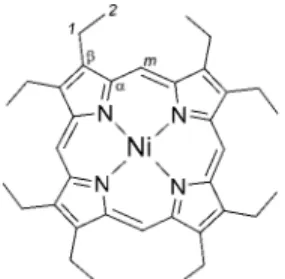
octaethylporphyrin, NiOEP (Figure 1), has been considered to be a good model for the heme prosthetic group because like all physiological porphyrins, the porphyrin macrocycle retains its 4-fold symmetry and has carbon substituents at the eight pyrrole positions. The eight ethyl substituents give rise to a high level of symmetry without being too sterically bulky. NiOEP has played a key role as a model system for spectroscopic characterization of heme proteins by RR spectroscopy.9In this
context, NiOEP has been used by many researchers as a primary model for the development of the empirical force field. In earlier studies Kitagawa and co-workers used NiOEP with several isotopomers to establish the vibrational assignment of certain in-plane vibrations of the porphyrin skeleton and to identify various over- and combination tones.6 Spiro and co-workers
significantly extended and improved the quality of the empirical force field, resulting in development of a consistent porphyrin force field.7The resulting force field was used to assign all
in-plane vibrations7a,7bwith an attempt to characterize out-of-plane
vibrations as well.7c
NiOEP has interesting conformational properties because different orientations of the eight ethyl groups can generate a number of different conformers. Indeed, X-ray crystallography data has been published for three conformers of NiOEP. Meyer reported X-ray data for a tetragonal form of NiOEP,10Cullen
and Meyer followed this with data for a triclinic form (later referred to as the triclinic A form),11 and Shelnutt and
co-workers reported X-ray data for a second triclinic form, called triclinic B.12It is well-known that nickel(II) porphyrins have a
tendency to deviate from planarity.13 This deviation from
planarity is attributed to the combination of the Ni-N bond being too short to fit the central cavity of a planar pophyrin and the presence of substituents on the periphery of the macrocycle.14The bond shortening is accommodated by ruffling
of the porphyrin ring, an effect which has been observed crystallographically and spectroscopically.12,14,15
Although a large amount of experimental data has been reported for NiOEP, there has not been an attempt to character-ize this system quantum mechanically. In this paper we report the results of density functional theory (DFT) energy calculations and geometry optimizations for selected planar and ruffled conformers of NiOEP. The ultimate goal of this project is to develop a high quality vibrational force field and establish definite vibrational assignment similar to those presented recently for nickel porphine (NiP)16and nickel
tetraphenylpor-phyrin (NiTPP).17Contrary to these previously studied nickel
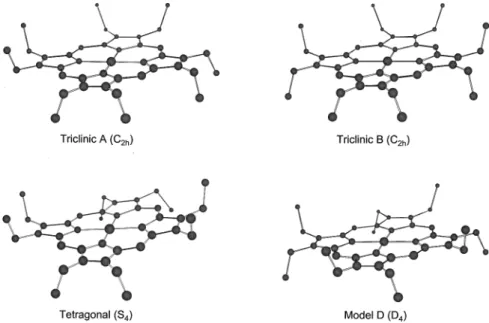
porphyrins, the full structural and vibrational analysis of NiOEP presents a more complex problem than for other nickel systems because of the various possible ethyl group orientations. From a combinatorial point of view there are over 200 possible conformers of NiOEP with respect to ethyl orientation, of which about 25 are unique, nonredundant conformers. In this study we concentrate on the energies and geometries of four conform-ers (Figure 2). These conformconform-ers were chosen because X-ray crystallography data has been reported for three of them,10-12
and the fourth has been used as a model in other NiOEP
* Author to whom correspondence should be addressed. E-mail pawel@louisville.edu.
†University of Louisville.
‡Steacie Institute for Molecular Science, National Research Council of
Canada.
Figure 1. Structure of nickel octaethylporphyrin and its carbon atom labeling scheme.
170 J. Phys. Chem. A 2002, 106,170-175
10.1021/jp012416k CCC: $22.00 © 2002 American Chemical Society Published on Web 12/08/2001
studies.7bThe detailed vibrational analysis will be presented in
future publication.18
Computational Methods
Calculations reported in this paper were carried out using gradient-corrected density functional theory (DFT) with the Becke-Lee-Young-Parr composite exchange correlation func-tional (B3LYP) as implemented in the Gaussian19 suite of
programs for electronic structure calculations. The B3-LYP level of theory with 6-31G(d) [for H, C, and N atoms] and Ahlrichs VTZ (for Ni)20 basis sets, successfully used in previous
calculations on metalloporphyrins,16,17,21-23 was employed in
the present study. Results and Discussion
A. Energies. Table 1 summarizes the results of DFT optimized energy values for NiOEP conformers. These energies, relative to the energy calculated for the tetragonal conformer (S4symmetry), are plotted in Figure 3. Initially, geometries were
optimized for four NiOEP conformers that correspond to tetragonal with S4symmetry, triclinic A and B forms with C2h
symmetry, and model D with D4symmetry24(Figure 2). The
Figure 2. Structures of NiOEP conformers showing the orientation of the ethyl substituent groups.
Figure 3. DFT optimized energies of NiOEP conformers relative to the tetragonal (S4) conformer. TABLE 1: Comparison of DFT Optimized Energy Values for NiOEP Conformers
energy relative to S4 planar vs ruffled
conformer symmetry energy (a.u.) (10-4a.u.) (kcal/mol) (kcal/mol)
tetragonal ruffled S4 -3125.819185 0.00 0.00 triclinic A planar C2h -3125.818939 2.46 0.15 0.21 ruffled CS -3125.819278 -0.93 -0.06 triclinic B planar C2h -3125.818748 4.36 0.27 0.24 ruffled C2 -3125.819133 0.52 0.03 model D planar D4 -3125.818606 5.78 0.36 0.21 ruffled D2 -3125.818945 2.40 0.15
lowest energy was found for the tetragonal S4conformer, though
the energy difference between other conformers, i.e., planar triclinic A and B as well as planar model D, was found to be small in the range of 0.15-0.36 kcal/mol (Table 1, Figure 3). For each conformer under consideration, frequency calcula-tions were performed and analyzed. All harmonic frequencies for the optimized geometry for the tetragonal S4conformer were
real, indicating that the optimized structure corresponds to a stable energy minimum (Figure 4). In the three other cases, one imaginary frequency was found which was associated with the planarity of the porphyrin core imposed by symmetry
con-straints. Interestingly, eigenvector analysis revealed that in all three cases the imaginary frequency was associated with ruffling of the porphyrin core. Consequently, each of these three optimized structures was distorted by lowering the symmetry of the initial structure along the eigenvector representing the imaginary frequency, and the resulting distorted structures were re-optimized and frequencies were recalculated. The final analysis of conformers show that for the planar triclinic A conformer with C2hsymmetry, the minimum energy corresponds
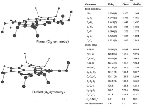
to a ruffled structure with Cssymmetry (Figure 5). Similarly,
the minimum corresponding to the triclinic conformer B (C2h
Figure 4. Comparison of calculated and experimentally determined structures of NiOEP, tetragonal conformer.aMeyer, E. F. Acta Crystallogr. 1972, B28, 2162.bCullen, D. L.; Meyer, E. F. J. Am. Chem. Soc. 1974, 96, 2095.cThe torsional angle, in degrees, of the opposite pyrrole ring planes with respect to the N-Ni-N axis which connects them.dJentzen, W.; Turowska-Tyrk, I.; Scheidt, W. R.; Shelnutt, J. A. Inorg. Chem. 1996,
35, 3559.eThe root-mean-square out-of-plane displacement, in 10-2Å, following the definition in Jentzen et al. Inorg. Chem. 1996, 35, 3559.
Figure 5. Comparison of calculated and experimentally determined structures of NiOEP, triclinic A conformer.aCullen, D. L.; Meyer, E. F. J. Am.
Chem. Soc. 1974, 96, 2095-2102.bThe torsional angle, in degrees, of the opposite pyrrole ring planes with respect to the N-Ni-N axis which connects them.cJentzen, W.; Turowska-Tyrk, I.; Scheidt, W. R.; Shelnutt, J. A. Inorg. Chem. 1996, 35, 3559.dThe root-mean-square out-of-plane displacement, in 10-2Å, following the definition in Jentzen et al. Inorg. Chem. 1996, 35, 3559.
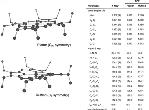
symmetry) has ruffled C2symmetry (Figure 6). The distortion
of model D (D4symmetry) leads to a structure of D2symmetry
with a ruffled porphyrin core (Figure 7). The ruffling of the porphyrin macrocycle accounts for about a 0.2 kcal/mol lowering of energy in each of the conformers. The ruffled forms of the triclinic conformers along with the tetragonal conformer, which is also ruffled, have energy values within a range of 0.09 kcal/mol (Figure 3) and are essentially degenerate. Upon correction for zero-point vibrational energy, these three con-formers are all within 0.3 kcal/mol of each other.
In spectroscopic studies on the conformational properties of NiOEP in solution, Jentzen and co-workers15ddecomposed the
sublines of ν19and ν10on the polarized resonance Raman spectra
for NiOEP and determined that the nonplanar conformer is favored energetically in CH2Cl2 and CS2 solutions. Their
experimentally obtained value of 0.7 kcal/mol is slightly higher but is of the same order of magnitude as that of the 0.2 kcal/ mol calculated value.
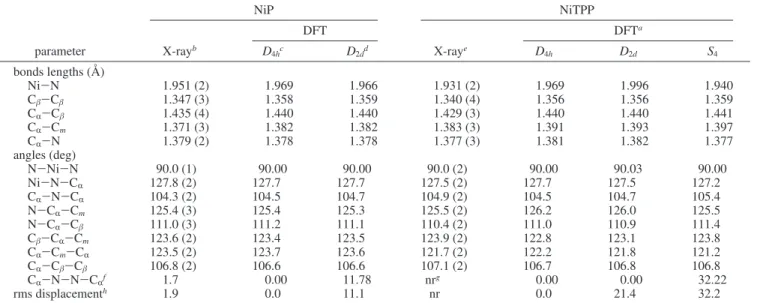
B. Optimized Geometries. Values of the optimized geo-metrical parameters for each conformer together with available X-ray data are summarized in Figures 4-7. Table 2 gives values for two other nickel porphyrins: nickel porphine (NiP) and nickel tetraphenylporphyrin (NiTPP).
Figure 6. Comparison of calculated and experimentally determined structures of NiOEP, triclinic B conformer.aBrennan, T. D.; Scheidt, W. R.; Shelnutt, J. A. J. Am. Chem. Soc. 1988, 110, 3919.b The torsional angle, in degrees, of the opposite pyrrole ring planes with respect to the N-Ni-N axis which connects them.cJentzen, W.; Turowska-Tyrk, I.; Scheidt, W. R.; Shelnutt, J. A. Inorg. Chem. 1996, 35, 3559.dThe root-mean-square out of plane displacement, in 10-2Å, following the definition in Jentzen et al. Inorg. Chem. 1996, 35, 3559.
Figure 7. Comparison of calculated and experimentally determined structures of NiOEP, model D conformer.aThe torsional angle, in degrees, of the opposite pyrrole ring planes with respect to the N-Ni-N axis which connects them.bThe root-mean-square out-of-plane displacement, in 10-2
Å, following the definition in Jentzen et al. Inorg. Chem. 1996, 35, 3559.
The DFT calculations for the planar and ruffled conformers of triclinic A, triclinic B, and model D show the expected decrease in the Ni-N bond length with the ruffling of the porphyrin macrocycle. Overall, the DFT calculations for bond lengths are in good agreement with reported experimental values. The experimental Cβ-Cβ, CR-N, and C1-C2bond lengths are
somewhat shorter than the corresponding calculated bond lengths for the tetragonal, triclinic A, and triclinic B conformers, and this can be attributed to crystal packing forces that are not present in the theoretical models.
For NiOEP, NiP, and NiTPP, the Cβ-Cβbond length appears to be insensitive to ruffling. Additionally, the calculated Cβ -Cβ bond distances are larger for the NiOEP conformers than
for the NiP and NiTPP conformers, which can be attributed to the fact that of the three molecules, only NiOEP has carbon substituents at the Cβposition. Likewise, NiTPP has longer CR
-Cmbond distances than the other two molecules, and only NiTPP
has carbon substituents at the Cmposition.
The calculated bond angles also show good agreement with the data for the three conformers characterized experimentally. In general, the calculated CR-N-N-CRdihedral angles
com-pare favorably with the reported experimental values. Calcula-tions of the ruffled conformers give larger values for the CR
-N-N-CR dihedral angles and root-mean-square out-of-plane
displacements,25another measure of planarity of the porphyrin
macrocycle, as compared to their planar counterparts, as expected. For the triclinic A and B conformers, the CR-
N-N-CRdihedral angles found by X-ray are smaller than those
calculated, and this may also be attributed to crystal packing forces. The CR-N-N-CR dihedral angle in the tetragonal
conformer, however, does not follow this trend. This dihedral angle had a significantly smaller calculated value compared to experiment. This may be the result of comparing gas-phase calculations with solid-state X-ray data or of possible error in the calculated values, experimental values, or both. Closer analysis of the porphyrin skeleton bond lengths show that the experimental Ni-N bond length of the tetragonal conformer is significantly shorter, i.e., 1.929(3) Å, in comparison to the triclinic A and B conformers, which have Ni-N bond lengths of 1.958(2) and 1.952 (4) Å, respectively. The optimized
theoretical Ni-N bond lengths for the three conformers are consistent with values of 1.970, 1.961, and 1.963 Å. This similarity among the optimized conformers might indicate that the experimental Ni-N bond length was not determined accurately for the tetragonal conformer.
Conclusion
The values obtained through geometry optimization of NiOEP using density functional theory show remarkably good agree-ment with experiagree-mental X-ray results. As such, DFT appears to be a good choice for performing calculations on the complex structures of metalloporphyrins. DFT calculations show that three NiOEP conformers, which crystallize in alternative pol-ymorphs, have essentially degenerate energy values. All inves-tigated conformers show significant ruffling distortion. The origin of the ruffling distortion is attributed to the fact that the nickel ion is too small for the natural porphyrin cavity size, which has a 2.00 Å radius.13With a distance shorter than 2.00
Å, the Ni-N bonds draw the pyrrole rings toward the center through a ruffling distortion of the macrocycle. Since the planar geometry maximizes the π overlap, the equilibrium structure would find a balance between the energy gain from shorter bonds and the energy penalty to the π system from ruffling. On the basis of present DFT calculations for NiOEP, the energy corresponding to this ruffling distortion can be accurately estimated to be equal to 0.2 kcal/mol.
References and Notes
(1) (a) Heme and Heme Proteins; Chance, B., Estabrock, R. W., Yontenani, T., Eds.; Academic Press: New York, 1966. (b) Antonini, E.; Brunori, M. Hemoglobin and Myoglobin in Their Reactions with Ligands; North-Holland Publishing Co.: Amsterdam, 1971. (c) Hemoglobins; Everse, J., Vandegriff, K. D., Winslow, R. M., Eds.; Academic Press: San Diego, 1994. (c) Hemoglobin and Oxygen Binding; Ho, C., Ed.; Elsevier Biomedi-cal: New York 1982.
(2) (a) Lemburg, R.; Barrett, J. Cytochromes; Academic Press: London, New York, 1973. (b) Moore, G. R.; Pettigrew, G. W. Cytochromes c:
EVolutionary, Structural, and Physicochemical Aspects; Springer-Verlag:
Berlin, New York, 1990. (c) Cytochrome P450 Protocols; Phillips, I. R., Shephard, E., Eds.; Humana Press: Totowa, NJ, 1998. (d) Cytochrome
P450; Johnson, E. F., Waterman, M. R., Eds.; Academic Press: San Diego, 1996.
TABLE 2: Calculated and Experimentally Determined Geometries of NiP and NiTPP
NiP NiTPP DFT DFTa parameter X-rayb D 4hc D2dd X-raye D4h D2d S4 bonds lengths (Å) Ni-N 1.951 (2) 1.969 1.966 1.931 (2) 1.969 1.996 1.940 Cβ-Cβ 1.347 (3) 1.358 1.359 1.340 (4) 1.356 1.356 1.359 CR-Cβ 1.435 (4) 1.440 1.440 1.429 (3) 1.440 1.440 1.441 CR-Cm 1.371 (3) 1.382 1.382 1.383 (3) 1.391 1.393 1.397 CR-N 1.379 (2) 1.378 1.378 1.377 (3) 1.381 1.382 1.377 angles (deg) N-Ni-N 90.0 (1) 90.00 90.00 90.0 (2) 90.00 90.03 90.00 Ni-N-CR 127.8 (2) 127.7 127.7 127.5 (2) 127.7 127.5 127.2 CR-N-CR 104.3 (2) 104.5 104.7 104.9 (2) 104.5 104.7 105.4 N-CR-Cm 125.4 (3) 125.4 125.3 125.5 (2) 126.2 126.0 125.5 N-CR-Cβ 111.0 (3) 111.2 111.1 110.4 (2) 111.0 110.9 111.4 Cβ-CR-Cm 123.6 (2) 123.4 123.5 123.9 (2) 122.8 123.1 123.8 CR-Cm-CR 123.5 (2) 123.7 123.6 121.7 (2) 122.2 121.8 121.2 CR-Cβ-Cβ 106.8 (2) 106.6 106.6 107.1 (2) 106.7 106.8 106.8 CR-N-N-CR f 1.7 0.00 11.78 nrg 0.00 0.00 32.22 rms displacementh 1.9 0.0 11.1 nr 0.0 21.4 32.2
aRush, T. S.; Kozlowski, P. M.; Piffat, C. A.; Kumble, R.; Zgierski, M. Z.; Spiro, T. G. J. Phys. Chem. B 2000, 104, 5020.bJentzen, W.;
Turowska-Tyrk, I.; Scheidt, W. R.; Shelnutt, J. A. Inorg. Chem. 1996, 35, 3559.cThis work.dKozlowski, P. M.; Rush, T. S.; Jarzecki, A. A.;
Zgierski, M. Z.; Chase, B.; Piffat, C.; Ye, B-H.; Li, X-Y.; Pulay, P.; Spiro, T. G. J. Phys. Chem. A 1999, 103, 1357.eMaclean et al. Aust. J. Chem.
A 1996, 49, 1273.fThe torsional angle, in degrees, of the opposite pyrrole ring planes with respect to the N-Ni-N axis which connects them.gNot
reported.hThe root-mean-square out-of-plane displacement, in 10-2Å, following the definition in Jentzen et al. Inorg. Chem. 1996, 35, 3559.
(3) The Photosynthetic Reaction Center; (a) Deisenhofer, J., Norris, J. R., Eds.; Academic Press: San Diego, 1993. (b) The Photosynthetic
Bacterial Reaction Center II: Structure, Spectroscopy, and Dynamics; Breton, J., Vermeglio, A., Eds.; Plenum Press: New York, 1992. (c) The
Reaction Center of Photosynthetic Bacteria: Structure and Dynamics;
Michel-Beyerle, E., Ed.; Springer: Berlin, New York, 1996.
(4) (a) Spiro, T. G.; Strekas, T. C. Proc. Natl. Acad. Sci. U.S.A. 1972,
69,2622. (b) Strekas, T. C.; Spiro, T. G. Biochim. Biophys. Acta 1972,
278, 188.
(5) Scheidt, W. R.; Lee, Y. J. Struct. Bonding 1987, 64, 1. (6) (a) Abe, M.; Kitagawa, T.; Kyoguku, Y. J. Chem. Phys. 1978, 69, 4526. (b) Kitagawa, T.; Abe, M.; Ogoshi, H. J. Chem. Phys. 1978, 69, 4516.
(7) (a) Li, X. Y.; Czernuszewicz, R. S.; Kincaid, J. R.; Su, Y. O.; Spiro, T. G. J. Am. Chem. Soc. 1990, 94, 31. (b) Li, X. Y.; Czernuszewicz, R. S.; Kincaid, J. R.; Stein, P.; Spiro, T. G. J. Am. Chem. Soc. 1990, 94, 47. (c) Li, X. Y.; Czernuszewicz, R. S.; Kincaid, J. R.; Spiro, T. G. J. Am. Chem.
Soc. 1989, 111, 7012.
(8) Hu, S.; Mukherjee, A.; Piffat, C.; Mak, R. S. W.; Li, X. Y.; Spiro, T. G. Biospectroscopy 1995, 1, 395.
(9) (a) Kitagawa, T.; Ozaki, Y. Struct. Bonding 1987, 64, 71. (b) Spiro, T. G.; Li, X. Y. In Biological Applications of Raman Spectroscopy; Spiro, T. G., Ed.; Wiley: New York, 1988; Vol. 3, p 1. (c) Spiro, T. G.; Czernuszewicz, R. S.; Li, X. Y. Coord. Chem. ReV. 1990, 100, 541. (d) Procyk, A. D.; Bocian, D. F. Annu. ReV. Phys. Chem. 1992, 43, 465.
(10) Meyer, E. F. Acta Crystallogr. 1972, B28, 2162.
(11) Cullen, D. L.; Meyer, E. F. J. Am. Chem. Soc. 1974, 96, 2095. (12) Brennan, T. D.; Scheidt, W. R.; Shelnutt, J. A. J. Am. Chem. Soc.
1988, 110, 3919.
(13) Hoard, J. L. Science 1971, 174, 1295.
(14) Jentzen, W.; Turowska-Tyrk, I.; Scheidt, W. R.; Shelnutt, J. A.
Inorg. Chem. 1996, 35, 3559.
(15) (a) Alden, R. G.; Crawford, B. A.; Doolen, R.; Ondrias, M. R.; Shelnutt, J. A. J. Am. Chem. Soc. 1989, 111, 2070. (b) Alden, R. G.; Ondrias, M. R.; Shelnutt, J. A. J. Am. Chem. Soc. 1990, 112, 691. (c) Shelnutt, J. A.; Medforth, C. J.; Berber, M. D.; Barkigia, K. M.; Smith, K. M. J. Am.
Chem. Soc. 1991, 113, 4077. (d) Jentzen, W.; Unger, E.; Karvounis, G.;
Shelnutt, J. A.; Dreybrodt, W.; Schweitzer-Stenner, R. J. Phys. Chem. 1996,
100, 14184-14191.
(16) Kozlowski, P. M.; Rush, T. S.; Jarzecki, A. A.; Zgierski, M. Z.; Chase, B.; Piffat, C.; Ye, B.-H.; Li, X.-Y.; Pulay, P.; Spiro, T. G. J. Phys.
Chem. A 1999, 103, 1357.
(17) Rush, T. S.; Kozlowski, P. M.; Piffat, C. A.; Kumble, R.; Zgierski, M. Z.; Spiro, T. G. J. Phys. Chem. B 2000, 104, 5020.
(18) Stoll, L. K.; Zgierski, M. Z.; Kozlowski, P. M. Unpublished results. (19) Frisch, M. J.; Trucks, G. W.; Schlegel, H. B.; Scuseria, G. E.; Robb, M. A.; Cheeseman, J. R.; Zakrzewski, V. G.; Montgomery, J. A.; Stratmann, R. E.; Burant, J. C.; Dapprich, S.; Millam, J. M.; Daniels, A. D.; Kudin, K. N.; Strain, M. C.; Farkas, O.; Tomasi, J.; Barone, V.; Cossi, M.; Cammi, R.; Mennucci, B.; Pomelli, C.; Adamo, C.; Clifford, S.; Ochterski, J.; Petersson, G. A.; Ayala, P. Y.; Cui, Q.; Morokuma, K.; Malick, D. K.; Rabuck, A. D.; Raghavachari, K.; Foresman, J. B.; Cioslowski, J.; Ortiz, J. V.; Stefanov, B. B.; Liu, G.; Liashenko, A.; Piskorz, P.; Komaromi, I.; Gomperts, R.; Martin, R. L.; Fox, D. J.; Keith, T.; Al-Laham, M. A.; Peng, C. Y.; Nanayakkara, A.; Gonzalez, C.; Challacombe, M.; Gill, P. M. W.; Johnson, B.; Chen, W.; Wong, M. W.; Andres, J. L.; Gonzalez, C.; Head-Gordon, M.; Replogle, E. S.; Pople, J. A. Gaussian 98, revision A.3; Gaussian, Inc.: Pittsburgh, PA, 1998.
(20) Scha¨fer, A.; Horn, H.; Ahlrichs, R. J. Phys. Chem. 1992, 97, 2571. (21) Kozlowski, P. M.; Spiro, T. G.; Berces, A.; Zgierski, M. Z. J. Phys.
Chem. B 1998, 102, 2603.
(22) Spiro, T. G.; Kozlowski, P. M. J. Am. Chem. Soc. 1998, 120, 4524. (23) Spiro, T. G.; Kozlowski, P. M.; Zgierski, M. Z. J. Raman Spectrosc.
1998, 29, 869.
(24) Because this work focuses on the comparison of theoretical values with experimental X-ray data, the crystallographic descriptions of the different conformers were used to identify the different conformers. Hence the conformer with S4symmetry is referred to as the tetragonal conformer,
though the crystal structure was not involved in the DFT calculations. Likewise, the two conformers with C2h(planar) symmetry are referred to
as triclinic A and triclinic B as appropriate.
(25) Root-mean-square out-of-plane displacements were calculated using the method described in ref 14.