HAL Id: inserm-03223397
https://www.hal.inserm.fr/inserm-03223397
Submitted on 11 May 2021HAL is a multi-disciplinary open access
archive for the deposit and dissemination of sci-entific research documents, whether they are pub-lished or not. The documents may come from teaching and research institutions in France or abroad, or from public or private research centers.
L’archive ouverte pluridisciplinaire HAL, est destinée au dépôt et à la diffusion de documents scientifiques de niveau recherche, publiés ou non, émanant des établissements d’enseignement et de recherche français ou étrangers, des laboratoires publics ou privés.
Spastic co-contraction, rather that spasticity, is
associated with impaired active function in adults with
acquired brain injury: A pilot study
Alexandre Chalard, David Amarantini, Joseph Tisseyre, Philippe Marque,
Jessica Tallet, D. Gasq
To cite this version:
Alexandre Chalard, David Amarantini, Joseph Tisseyre, Philippe Marque, Jessica Tallet, et al.. Spas-tic co-contraction, rather that spasSpas-ticity, is associated with impaired active function in adults with acquired brain injury: A pilot study. Journal of Rehabilitation Medicine, Foundation for Rehabilita-tion InformaRehabilita-tion, 2019, 51 (4), pp.307-311. �10.2340/16501977-2528�. �inserm-03223397�
JRM
JRM
J
our nal ofR
ehabilitationM
edicineJRM
J
our nal ofR
ehabilitationM
edicineSHORT COMMUNICATION
SPASTIC CO-CONTRACTION, RATHER THAN SPASTICITY, IS ASSOCIATED WITH
IMPAIRED ACTIVE FUNCTION IN ADULTS WITH ACQUIRED BRAIN INJURY: A
PILOT STUDY
Alexandre CHALARD, PT, MS1,2, David AMARANTINI, PhD1, Joseph TISSEYRE, MS1, Philippe MARQUE, MD, PhD1,3,
Jessica TALLET, PhD1 and David GASQ, MD, PhD1,4
From the 1ToNIC, Toulouse NeuroImaging Center, Université de Toulouse, Inserm, UPS, 2Ipsen Innovation, Les Ulis, 3Department of Physical Medicine and Rehabilitation and 4Department of Functional Physiological Explorations, University Hospital of Toulouse, Hôpital de Rangueil, Toulouse, France
LAY ABSTRACT
Spasticity and spastic co-contraction are expressions of muscle overactivity that occur in spastic paresis syn-drome after a brain injury. The objective of the pre-sent pilot study was to improve our understanding of the respective adverse consequences of spasticity and spastic co-contraction on motor disability. In contrary to spasticity, spastic co-contraction is strongly associa-ted with motor impairment in subjects with brain injury. Therapies should be directed toward reducing spastic co-contraction in order to improve motor function.
Objective: To elucidate the adverse consequences of spasticity and spastic co-contraction of elbow flex-ors on motor impairment and upper limb functional limitation.
Design: A pilot case-controlled prospective observa-tional study.
Subjects: Ten brain-injured adults, and 10 healthy controls.
Methods: The co-contraction index was computed from electromyographic recordings of elbow flexors during sub-maximal (25% Maximal Voluntary Cont-raction) isometric elbow extension. Spasticity was assessed with the Tardieu scale, upper limb limita-tion using a goniometer during active elbow exten-sion, motor selectivity with the Fugl-Meyer Assess-ment for the upper limb, and motor function with the Action Research Arm Test.
Results: Greater co-contraction occurred in patients with brain injury compared with controls. In cont-rast to spasticity, strong associations were found between the co-contraction index, the limitation of active elbow extension, the Fugl-Meyer Assessment, and the Action Research Arm Test.
Conclusion: This pilot study suggests that spastic co-contraction rather than spasticity is an important factor in altered upper limb motricity in subjects with brain injury, leading to abnormal restricting arm movement patterns in subjects with more seve-re motor impairment. Practical applications diseve-rectly concern the pre- and post-therapeutic evaluation of treatments aimed at improving motor skills in sub-jects with brain injury.
Key words: brain injury; hemiplegia; muscle hypertonia; up-per extremity.
Accepted Jan 29, 2019; Epub ahead of print Feb 15, 2019 J Rehabil Med 2019; 51: 307–311
Correspondence address: David Gasq, Toulouse NeuroImaging Cen-ter, CHU Purpan, Pavillon Baudot, place du Dr Baylac 31024 Toulouse, France. E-mail: david.gasq@inserm.fr
M
uscle overactivity, including spasticity andspas-tic co-contraction in parspas-ticular, describes invo-luntary motor unit recruitment, which occurs in spastic
paresis syndrome after a brain injury, such as stroke or traumatic brain injury (1). Spasticity is defined as an increase in velocity-dependent stretch reflexes, and is clinically manifested by excessive responses to mus-cle stretch (2). Spasticity is used as a convenient way to assess muscle overactivity during passive and fast muscular stretch. Spastic co-contraction, as assessed by electromyography using the muscle co-contraction index (3), refers to increased antagonist muscle recru-itment triggered by the volitional command of agonist muscles in the absence of a phasic stretch (1). It has been well established that spasticity and spastic co-contraction have different underlying physiological mechanisms (1), but their consequences on motor function remain to be confirmed and elucidated. It has been suggested that spastic co-contraction may contri-bute to limitations in active movement (4). However, to date, the impact of this disabling form of muscle overactivity on motor function in brain-injured adults has been only sparsely and indirectly studied (5). In addition, most treatments aimed at improving upper limb function, such as rehabilitation or botulinum toxin, focus on spasticity as the primary outcome in clinical practice (6).
The aim of the present pilot study was therefore to elucidate the adverse consequences of spasticity and spastic co-contraction of elbow flexors on upper limb motor impairment and disability. The results of this study may have direct application in improving the evaluation and implementation of treatments aimed at improving motor function in subjects with brain injury.
JRM
JRM
J
our nal ofR
ehabilitationM
edicineJRM
J
our nal ofR
ehabilitationM
edicine 308 A. Chalard et al. METHODS ParticipantsThis pilot case-controlled prospective observational study included 10 adults with brain injury (HEMI) and 10 control participants (CONTROL) (see Table I for participants’ demo-graphics). The inclusion criteria were: brain injury for at least 6 months caused by an acquired cerebral lesion (single stroke or traumatic brain injury); strength of paretic triceps brachii (rated at least at 3/5 on the Held-Deseilligny Scale, corresponding to extension of the forearm against slight resistance); and no anti-spastic treatment during the 3 previous months. Exclusion criteria were: severe cognitive disorders with limited compre-hension of basic instructions; neurodegenerative conditions other than the acquired brain injury; elbow contracture (loss of passive elbow extension or flexion); and upper limb pain during movement. Ethics approval was obtained from the local institutional review board at Paul Sabatier University Hospital (No. 07-0716, Toulouse, France) and written informed consent was obtained from all participants. The study was conducted in accordance with the amended Declaration of Helsinki and conforms to all STROBE guidelines, reporting the required information accordingly.
Materials
Net torque around the elbow joint was recorded at 1 kHz using a Con-Trex MJ calibrated dynamometer (CMV AG, Dubendorf, Switzerland).
The surface electromyographic signal (EMG) was recorded at 1 kHz using Ag–AgCl bipolar electrodes in bipolar confi-guration with an inter-electrode distance of 20 mm,, using an MP150 system (Biopac Systems Inc., Goleta, CA, USA). The reference electrode was placed on the left ulnar head. Biceps brachii and brachioradialis were selected as the elbow flexors acting as antagonist muscles during elbow extension.
Recordings were made on the non-dominant side in the CON-TROL group and on the paretic side in HEMI group.
Torque and EMG data were synchronized automatically using a rectangular triggering pulse signal and analysed offline.
Procedure
The experimental procedure comprised 2 steps.
First step. To perform the following clinical assessment (7):
spasticity of elbow flexors, limitation to active elbow extension,
motor selectivity and motor function were assessed respec-tively, using the Tardieu scale, a goniometer during repetitive and dynamic voluntary elbow extensions at a preferred rate, the upper limb motor section of the Fugl-Meyer Assessment scale, and the Action Research Arm Test (Table I). Maximal net elbow extension torque value was taken as a functional marker of triceps brachii paresis, the Fugl-Meyer score as a motor selectivity assessment, and the Action Research Arm Test as a motor function assessment.
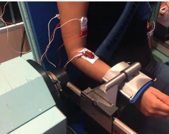
Second step. Participants were seated on the dynamometer chair
with their upper body strapped firmly, the upper arm positioned along the trunk, and the elbow flexed at 90° (Fig. 1). The parti-cipants exerted 3 isometric maximal voluntary contractions of the elbow in flexion and in extension for a duration of 5 s, with 1 min rest between each contraction and 3 min rest between flexion and extension contractions. After collection of maximal voluntary contraction data, participants performed 2 sets of 5-s elbow isometric extension sub-maximal contractions while receiving visual feedback on their actual torque in relation to a target torque. Each set included 6 contractions at 25% Maxi-mal Voluntary Contraction (MVC), corresponding to a level of force required for daily activities (8). The time between each contraction was 30 s; each set was separated by a 3-min rest period to minimize fatigue.
Table I. Participants’ demographics
Participants Sex Age, year Pathology Side and location of cerebral injury Disease course, month
Fugl-Meyer Assessment score
(upper limb/66) Action Research Arm Test (/57) Control Group
(n = 10)
6 Males
4 Females 30 (15)* – – – – –
Brain Injured Group
1 Female 47 Ischaemic stroke Left, cortical & subcortical 48 55 39 2 Male 66 Haemorrhagic stroke Left, thalamic 14 51 36 3 Male 59 Ischaemic stroke Left, latero-bulbar 27 59 55 4 Male 52 Ischaemic stroke Left, subcortical 43 48 18 5 Male 64 Ischaemic stroke Left, cortical & subcortical 39 60 57 6 Male 71 Ischaemic stroke Right, cortical & subcortical 60 44 23 7 Male 33 Ischaemic stroke Right, cortical & subcortical 146 54 51 8 Male 41 Traumatic brain injury Left, cortical & subcortical 132 46 29 9 Male 57 Traumatic brain injury Right, cortical & subcortical 360 18 0 10 Male 63 Haemorrhagic stroke Left, subcortical 60 32 0 *Mean (standard deviation (SD)), indicates a significant difference in age between HEMI and CONTROL groups (p < 0.05).
Fig. 1. Illustration of the arm and forearm positions used to perform
torque and electromyographic recordings during isometric elbow extension on the calibrated dynamometer.
JRM
JRM
J
our nal ofR
ehabilitationM
edicineJRM
J
our nal ofR
ehabilitationM
edicinep < 0.01), with a mean difference (SD) of 7.6±12.9%.
Lower net elbow extension torque during maximal vol-untary contraction was found in the HEMI compared
with the CONTROL group (w19 = –2.34, p<0.05), with
a mean difference (SD) of 17.6 ± 17 Nm. Results of the Spearman’s correlations are shown in Table III.
DISCUSSION
This pilot study aimed to elucidate the consequences of spasticity and spastic co-contraction of elbow flexors on limitation of active elbow extension in adults with brain injury. A strong association was found between the co-contraction index and (i) the limitation of ac-tive elbow extension, (ii) the Fugl-Meyer Assessment score, and (iii) the score on Action Research Arm Test. Conversely, no significant correlation was found bet-ween spasticity and any of the variables cited above.
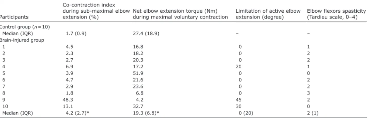
These results are thus the first to show that spastic co-contraction primarily contributes to a deficit in active elbow extension in adults with brain injury, which occurs even in the absence of spasticity (see, for example, Table II, HEMI participants 5 and 10). These findings confirm the absence of an association between spasticity and spastic co-contraction, supporting the idea that they refer to different forms of overactivity with different underlying physiological mechanisms
Data processing
Net torque was low-pass filtered at 100 Hz with a 6th-order zero-lag Butterworth filter. EMG data were 10–400-Hz band-pass filtered (4th-order zero-lag Butterworth filter), full-wave rectified, and smoothed at 9 Hz to obtain the linear envelopes. The co-contraction index was determined as the ratio (expres-sed in percentage) between the root mean square value of the elbow flexors EMG envelopes during the sub-maximal elbow extensions and the root mean square of the same muscle during the highest maximal voluntary elbow flexion contraction (9).
Statistical analysis
Non-parametric analysis using Wilcoxon rank-sum test was per-formed to compare the co-contraction index and the maximal net elbow extension torque between HEMI and CONTROL groups.
Non-parametric Spearman’s correlations (rs) were performed
to investigate the relationship between co-contraction index, elbow-flexor spasticity, maximal net elbow extension torque with: (i) limitation to active elbow extension, (ii) elbow-flexor spasticity, (iii) Fugl-Meyer Assessment score for the upper limb, and (iiii) Action Research Arm Test. It is notable that that there was a significant difference in age between the HEMI and CONTROL groups (Table I). However, preliminary analysis of the data showed a lack of any correlation with age (p > 0.05), enabling the results to be interpreted independently of age.
RESULTS
A higher co-contraction index occurred in the HEMI
group compared with the CONTROL group (w19 = 3.4,
Table II. Co-contraction index, net elbow extension torque during maximal voluntary contraction and clinical characteristics
Participants
Co-contraction index during sub-maximal elbow
extension (%) Net elbow extension torque (Nm) during maximal voluntary contraction Limitation of active elbow extension (degree) Elbow flexors spasticity (Tardieu scale, 0–4) Control group (n = 10) Median (IQR) 1.7 (0.9) 27.4 (18.9) – – Brain-injured group 1 4.5 16.8 0 1 2 2.3 18.2 0 2 3 2.7 20.3 0 2 4 6.9 17.2 20 1 5 3.9 51.9 0 0 6 4.7 21.6 0 2 7 2.9 23.6 0 2 8 1.8 6.8 0 3 9 48.3 4.2 45 2 10 13.1 32.7 30 0 Median (IQR) 4.2 (2.7)* 19.3 (6.8)* 0 (20) 2 (1) *Indicates a significant difference between HEMI and CONTROL groups (p < 0.05).
IQR: interquartile range.
Table III. Spearman correlations (95% confidence interval) for HEMI participants between co-contraction index, net elbow extension
torque during maximal voluntary contraction and clinical variables
Limitation of active elbow
extension Elbow flexors spasticity Fugl-Meyer Assessment score (upper limb) Action Research Arm Test Co-contraction index during the sub-maximal
elbow extension 0.88** [0.59, 0.97] 0.01 [–0.59, 0.60] –0.86** [–0.96, –0.53] –0.66* [–0.90, –0.09] Elbow flexors spasticity –0.23 [–0.72, 0.42] – 0.10 [–0.53, 0.66] 0.06 [–0.63, 0.56] Net elbow extension torque during maximal
voluntary contraction –0.26 [–0.74, 0.40] –0.49 [–0.92, –0.08] 0.44 [–0.21, 0.82] 0.41 [–0.19, 0.80] *Indicates a significant correlation at p < 0.05. **Indicates a significant correlation at p < 0.01.
JRM
JRM
J
our nal ofR
ehabilitationM
edicineJRM
J
our nal ofR
ehabilitationM
edicine 310 A. Chalard et al.(1). Most importantly, in agreement with a suggestion made in a previous report (10), these results unequivo-cally establish that spasticity and spastic co-contraction have different functional repercussions with regards to impaired motor function in adults with brain injury.
It is well known that selective muscle activation is necessary for skilled and coordinated upper limb movements, which implies concomitant activation of agonists and relaxation of antagonists. Lesions that damage the corticospinal pathways, such as stroke or traumatic brain injury, cause long-lasting impairment of the ability to produce selective patterns of EMG activity (11). However, it has been shown that, during rehabilitation, the decrease in co-contraction index is correlated with the improvement in Fugl-Meyer score among post-stroke subjects (12). In agreement with a previous study (4), our finding of a strong association between the co-contraction index and Fugl-Meyer Assessment score thus highlights the importance of impaired motor selectivity as a mechanism that con-tributes to greater spastic co-contraction.
The adverse consequence of the presence of spastic co-contraction on upper limb motor impairment is further supported by the significant association bet-ween the co-contraction index and the score on Action Research Arm Test, taken to reflect upper limb functio-nal limitation. Furthermore, by afunctio-nalysing EMG-based assessment of spastic co-contraction in patients with a wide range of motor impairment, our results highlight that the more severe the motor impairment, the greater the co-contraction index.
These results also failed to show an association bet-ween the net elbow extension torque during maximal voluntary contraction, taken as a functional marker of triceps brachii paresis, and either the limitation of active elbow extension, the score of Fugl-Meyer As-sessment or of the Action Research Arm Test. These findings indicate that motor weakness is not a primary factor limiting active elbow extension, and lead to the conclusion that non-selective motricity and function impairment are not directly linked to motor weakness.
Taken together, the above results demonstrate the detrimental impact of spastic co-contraction on upper limb motor function, leading to abnormal restricting arm movement patterns, especially in subjects with more severe motor impairment.
Despite strong evidence of improvement in spasti-city induced by botulinum toxin treatment, few studies have shown effectiveness in improving active function and active movement (13, 14). A limitation that may explain this lack of efficacy in active function is the use of spasticity, as assessed during passive stretching, as a marker of muscle overactivity during movement (15). In contrast, the change in spastic co-contraction
after injection of botulinum toxin has been poorly studied. From a clinical perspective, these results are in line with the Subcommittee of the American Aca-demy of Neurology (13), which recommend the use and development of a method and outcome regarding motor function and active movement. We support the requirement to report active range of motion as an out-come of muscle overactivity treatments (7), and the use of EMG-based quantification of spastic co-contraction as a relevant tool for providing effective interventions related to altered recruitment of antagonist muscles and limitation of active movement.
Study limitations
Although a limitation of this pilot study is the small sample size, significant findings were made relative to the link between spastic co-contraction and clinical scores. Any generalization of these results, however, should be viewed with caution, especially because hemiparetic subjects had different aetiologies of brain injury (i.e. stroke and traumatic brain injury) and that little is known about the influence of the type of brain injury on spastic co-contraction.
This pilot study assessed the co-contraction index during submaximal isometric contraction, while spastic co-contraction is sensitive to stretch (1). Thus, future research should investigate co-contraction during ac-tive elbow extension.
Conclusion and clinical implications
The results of this pilot study suggest that spastic co-contraction alters upper limb function in subjects with hemiparetic brain injury. These findings may partially explain the lack of data concerning the efficacy of treatments, such as botulinum toxin to improve up-per limb function (6, 13, 14). Although measurement of spasticity is usually performed to assess muscle overactivity, these results highlight the importance of considering spastic co-contraction to assess active motor function, and support further studies on changes in spastic co-contraction after injection of botulinum toxin, in connection with the improvement in active function. Practical applications arising from this work are to improve the assessment of factors that restrict movement and implementation of treatments aimed at improving motor function in subjects with brain injury.
ACKNOWLEDGEMENT
Alexandre Chalard is an employee of Ipsen Innovation within the framework of a Conventions Industrielles de Formation par la REcherche (CIFRE) PhD fellowship. Others authors in this study declare that they have no conflict of interest in relation to this study.
JRM
JRM
J
our nal ofR
ehabilitationM
edicineJRM
J
our nal ofR
ehabilitationM
edicine REFERENCES1. Gracies J-M. Pathophysiology of spastic paresis. II: emergence of muscle overactivity. Muscle Nerve 2005; 31: 552–571.
2. Lance JW. The control of muscle tone, reflexes, and mo-vement: Robert Wartenberg Lecture. Neurology 1980; 30: 1303–1313.
3. Kellis E, Arabatzi F, Papadopoulos C. Muscle co-activation around the knee in drop jumping using the co-contraction index. J Electromyogr Kinesiol 2003; 13: 229–238. 4. Chae J, Yang G, Park BK, Labatia I. Muscle weakness and
cocontraction in upper limb hemiparesis: relationship to motor impairment and physical disability. Neurorehabil Neural Repair 2002; 16: 241–248.
5. Silva CC, Silva A, Sousa A, Pinheiro AR, Bourlinova C, Silva A, et al. Co-activation of upper limb muscles during reaching in post-stroke subjects: an analysis of the cont-ralesional and ipsilesional limbs. J Electromyogr Kinesiol 2014; 24: 731–738.
6. Wissel J, Ward AB, Erztgaard P, Bensmail D, Hecht MJ, Lejeune TM, et al. European consensus table on the use of botulinum toxin type A in adult spasticity. J Rehabil Med 2009; 41: 13–25.
7. Yelnik AP, Simon O, Parratte B, Gracies JM. How to clinically assess and treat muscle overactivity in spastic paresis. J Rehabil Med 2010; 42: 801–807.
8. Marshall MM, Armstrong TJ. Observational assessment of forceful exertion and the perceived force demands of daily activities. J Occup Rehabil 2004; 14: 281–294.
9. Vinti M, Couillandre A, Hausselle J, Bayle N, Primerano A, Merlo A, et al. Influence of effort intensity and gastroc-nemius stretch on co-contraction and torque production in the healthy and paretic ankle. Clin Neurophysiol 2013; 124: 528–535.
10. Sarcher A, Raison M, Ballaz L, Lemay M, Leboeuf F, Trudel K, et al. Impact of muscle activation on ranges of motion during active elbow movement in children with spastic hemiplegic cerebral palsy. Clin Biomech 2015; 30: 86–94. 11. Schieber MH, Lang CE, Reilly KT, McNulty P, Sirigu A.
Selective activation of human finger muscles after stroke or amputation. Adv Exp Med Biol 2009; 629: 559–575. 12. Hu X, Tong KY, Song R, Tsang VS, Leung PO, Li L. Variation
of muscle coactivation patterns in chronic stroke during robot-assisted elbow training. Arch Phys Med Rehabil 2007; 88: 1022–1029.
13. Simpson DM, Gracies J-M, Graham K, Hallett M, Miyasaki J, Naumann M, et al. Assessment: botulinum neurotoxin for the treatment of spasticity (an evidence-based review). Neurology 2009; 73: 736–747.
14. Foley N, Pereira S, Salter K, Fernandez MM, Speechley M, Sequeira K, et al. Treatment with botulinum toxin impro-ves upper-extremity function post stroke: a systematic review and meta-analysis. Arch Phys Med Rehabil 2013; 94: 977–989.
15. Sheean G, Lannin NA, Turner-Stokes L, Rawicki B, Snow BJ, Cerebral Palsy Institute. Botulinum toxin assessment, intervention and after-care for upper limb hypertonicity in adults: international consensus statement. Eur J Neurol 2010; 17: 74–93.