Nanostructuring polyetheretherketone for medical implants
Jasmin Althaus 1 – 4 , Celestino Padeste 2 , Joachim K ö ser 3 ,
Uwe Pieles 3 , Kirsten Peters 4 and Bert M ü ller 1, *
1 Biomaterials Science Center (BMC) , University of Basel, c/o University Hospital Basel, 4031 Basel , Switzerland
2 Laboratory for Micro- and Nanotechnology , Paul Scherrer Institute, 5232 Villigen , Switzerland
3 Institute of Chemistry and Bioanalytics , University of Northwestern Switzerland, School of Life Sciences, 4132 Muttenz , Switzerland
4 Department of Cell Biology , University of Rostock, 18057 Rostock , Germany
Abstract
Surface roughness is a vital factor for medical implants since the cells of the surrounding tissue interact with the underly-ing substrate on the micro- and nanometer scales. In order to improve the surface morphology of implants, appropriate large-area micro- and nanostructuring techniques have to be identifi ed being applicable to irregularly shaped structures. We demonstrate that plasma treatments of polyetheretherke-tone (PEEK) thin fi lms produce nanostructured surfaces in a reproducible manner. They are easily tailored by varying plasma intensity using oxygen and ammonia as process gases. It was observed that roughness and nanostructure density lin-early depend on plasma intensity. Oxygen plasma turned out to exhibit a stronger effect compared to ammonia plasma at the same processing conditions. For cell interaction studies, the mean size of the nanostructures was intentionally varied between 10 nm and 100 nm. In vitro experiments revealed that human mesenchymal stem cells (hMSC) adhere inho-mogenously on untreated PEEK fi lms, but the plasma treat-ment with oxygen or ammonia allows the hMSC to adhere and proliferate. Fluorescence microscopy of the cells on the PEEK fi lms turned out to be diffi cult because of the strong auto-fl uorescence of the PEEK substrate. Stains including the whole cell vital stain Calcein-AM allowed cell morphol-ogy studies on plasma-treated PEEK fi lms. In the case of the analysis of cell compartments such as the actin cytoskeleton, confocal laser scanning microscopy (CLSM) was success-fully applied.
Keywords: atomic force microscopy; auto-fl uorescence; confocal laser scanning microscopy; human mesenchymal stem cells; nanostructures; oxygen and ammonia plasma treatments; polyetheretherketone (PEEK); Rat-2 fi broblasts.
Introduction
The surface chemistry, the surface roughness, and the stiff-ness of an implant play a crucial role in the biocompatibi-lity of medical implants (1 – 3) because adherent cells interact with the accessible micro- and nanometer-size features (4 – 6) . Titanium bone implants are usually sandblasted and etched to reach the necessary micro- and nanometer-scale roughness for appropriate osseointegration. For an increasing number of muscolo-skeletal applications polymer materials have been used, which exhibit the advantage of being radiolucent which is benefi cial following the soft tissue formation around the implant. Their limited mechanical properties, however, restrict the materials selection. Therefore, the high-performance polymer polyetheretherketone (PEEK), which exhibits the desired mechanical properties, received the FDA-approval for trauma, orthopedic and spinal implants in 1982, and expe-riences an increasing use for medical implants and beyond. Besides reasonable mechanical properties it also exhibits outstanding chemical resistance, which makes it suitable as a biocompa tible material and is already used in applications such as spinal disc cages and housings of pacemakers (7) . For bone implants, PEEK surfaces have to be activated to allow for proper cell attachment. Plasma treatment belongs to the promising methods because of the ease of the process and the reproducible control of the fi nal surface chemistry. We hypo-thesize that the plasma treatment can be exploited to generate and tailor nanostructures for improved osseointegration. Our hypothesis is also based on the recent observation of Dalby et al. (8) , that patterns of nanostructures on polymeric substrates cause osteogenic differentiation of mesenchymal stem cells. They have found that, in contrast to highly ordered nanostruc-tures, the randomly arranged nanostructures induce osteogenic differentiation. Therefore, we assume that the plasma etching, which leads to a random distribution of nanostructures on the PEEK surfaces, could be well suited for bone implants.
To study the cell-biomaterial interactions including cell morphology, fl uorescence microscopy and confocal laser scanning microscopy (CLSM) are widely used. In the case of PEEK substrates, the application of fl uorescence microscopy is critical because of the strong auto-fl uorescence of the poly-meric material. Hunter et al. (9) , investigated the attachment and proliferation of osteoblasts and fi broblasts on biomateri-als for orthopedic use, and explicitly excluded PEEK from the
*Corresponding author: Dr. Bert Müller, Thomas Straumann Professor für Materialwissenschaft in der Medizin, Biomaterials Science Center (BMC), Universität Basel, c/o Universitätsspital, 4031 Basel, Schweiz
Phone: +41 61 265 9660
E-mail: [email protected], www.bmc.unibas.ch Previously published online January 19, 2012
immuno-fl uorescence study due to the prominent auto-fl uore-scence of the material. To overcome the problem, Briem et al. (10) replaced the immuno-fl uorescence stains with Giemsa stains when investigating the response of primary fi broblasts and osteoblasts to plasma-treated PEEK. In order to address the problem of the auto-fl uorescence, we have carefully ana-lyzed the auto-fl uorescence of commercially available PEEK fi lms (APTIV ™ Series from VICTREX, Hofheim, Germany) to explore the possible origin of the strong background fl uorescence. One cause could arise from fl uorescent addi-tives. Therefore, PEEK has been chemically synthesized without any additive according to Risse et al. (11) . Finally, we demonstrate that utilizing CLSM allows for the investiga-tion of fl uorescence-stained cells on micro meter-thin PEEK sheets with minimized background signal.
Materials and methods PEEK sheet pretreatment
Hot embossing with a HEX03 press (JENOPTIK Mikrotechnik GmbH, Germany) at a temperature of 160 ° C and a pressure of 100 kN served to fl atten commercially available amorphous APTIV ™ PEEK fi lms (Series 2000, Victrex Europa GmbH, Hofheim, Germany) with nominal thicknesses of 6 μ m, 12 μ m, 25 μ m and 50 μ m between two polished 4-inch silicon wafers.
Plasma treatment
Oxygen/argon or ammonia plasma treatments (Piccolo system, Plasma Electronic, Neuenburg, Germany) activated the embossed PEEK sheets. The embossing-marked fi lms were placed at the bot-tom of the plasma chamber. Subsequently, the chamber was evacuat-ed, fl ushed for a period of 5 min with a 2:1 mixture of oxygen/argon (200/100 sccm, 99.5/99.2 % , Messer, Lenzburg, Switzerland) or am-monia (200 sccm, 99.98 % , Messer, Lenzburg, Switzerland) and then equilibrated for further 5 min with oxygen/argon (20/10 sscm) or ammonia (30 sccm). The plasma treatments using a power of 10 W, 25 W, 50 W, 75 W, 100 W, 125 W, 150 W, 175 W, or 200 W always lasted 5 min.
Atomic force microscopy measurements (AFM)
AFM measurements enabled us to determine the surface rough-ness and island density of plasma-activated PEEK sheets. Measurements were performed in TappingMode ® in air under dry conditions on a Dimension IIIa instrument (Veeco, Mannheim, Germany) using silicon cantilevers with a Si 3 N 4 coating and a
tip radius of 20 nm, a spring constant of 40 N/m and a resonance frequency of 325 kHz (NSC15/A1BS, Mikromasch, CA, USA; manufacturers specifi cations). The scan area was set to 2 × 2 μ m 2 .
Data processing and roughness evaluation was done using the Nanoscope software. For each specimen the island density was determined from three individual characteristic square areas, each with approximately 100 islands.
Transmission and fl uorescence scans
PEEK was synthesized according to Risse et al. (11) . A tert -butyl substituted PEEK was prepared by nucleophilic substitution of tert
-butylhydroquinone and 4,4 ′ -difl uorobenzophenone (Sigma-Aldrich). The tert -butyl substituent was cleaved via reversed Friedel-Crafts alkylation using trifl uoromethansulfonic acid. The specially synthe-sized PEEK and corresponding starting materials in powder form were fi lled into 96-well plates until the bottom was covered.
The PEEK fi lms were stamped out to fi t into the 96-well plate format. The transmission measurements carried out in a 96-well quartz glass plate (Hellma, Müllheim, Germany) were recorded for wavelengths ranging from 240 nm to 1000 nm. The fl uorescence ex-periments in black 96-well plates were performed with a TECAN micro-plate reader infi nite 200, equipped with a UV Xenon fl ash lamp (TECAN trading AG, Switzerland) using bottom reading with a detector gain of 60. The excitation wavelengths were varied be-tween 350 nm and 800 nm in steps of 10 nm. The corresponding emission was acquired 30 nm above excitation wavelengths to 850 nm in 5 nm steps.
Cell culture
Rat-2 fi broblasts were cultured in DMEM medium under standard conditions (5 % CO 2 , 37 ° C). The fi broblasts were seeded over night
on 70 % ethanol sterilized class cover slips and PEEK sheets. Human mesenchymal stem cells (hMSC) were isolated from liposuction-derived adipose tissue as described previously by Peters et al. (12) and seeded (96-well plate, 10 4 cells/cm 2 , triplicates) on the
differently plasma-treated PEEK substrates for a period of 14 days. Cell cultivation was done under standard conditions.
Fluorescence staining
Cells were washed with phosphate buffered saline (PBS) and fi xed with 4 % paraformaldehyde in PBS for 10 min at room temperature. The cells were then permeabilized with 0.2 % Triton X-100, washed, and incubated with a 1:40 dilution of mouse anti-human vincu-lin (Sigma-Aldrich, Buchs, Switzerland) and a 1:2000 dilution of TRITC-conjugated phalloidin (Sigma-Aldrich, Buchs, Switzerland) in PBS for 1 h at room temperature. Subsequently, the cells were labeled using a 1:1000 dilution of goat anti-mouse Alexa 488 sec-ondary antibody (Invitrogen) in PBS during 30 min. The cells were visualized on a BX-51 fl uorescence microscope equipped with a fl uorescence unit, and a Fluo-View 1000 confocal laser scanning mi-croscope, both from Olympus (Hamburg, Germany).
hMSC were vital-stained with Hoechst 33324 (1:2000, Sigma-Aldrich, Taufkirchen, Germany) and Calcein-AM (Biomol GmbH, Hamburg, Germany; 1:200, ATT Bioquest) in DMEM medium during 15 min at a temperature of 37 ° C. The staining medium was exchanged with fresh solution before imaging.
Scanning electron microscopy (SEM)
The plasma-treated PEEK fi lms were coated with Au/Pd during 30 s using a current of 20 mA and applied a vacuum of 6 Pa (sputter coater Polaron, Thermo VG Scientifi c, Germany). Substrates were investi-gated with the fi eld emission scanning electron microscope Supra 40 VP (Carl Zeiss, Jena, Germany) with an applied acceleration voltage of 10 kV using the InLens detector.
Results
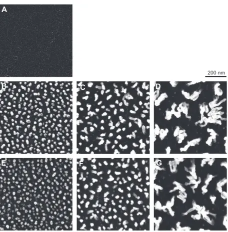
Plasma treatment is a common process to chemically acti-vate polymer surfaces (10, 13 – 16) , which is a prerequisite to
100 50 0 500 nm A B C D F G H E I Height level, nm
Figure 1 AFM images of plasma-treated PEEK fi lms.
The 25 μ m-thick fi lms were plasma-treated for 5 min. (A) untreated, (B-E) ammonia-plasma-treated, plasma power from left to right: 10 W, 50 W 100 W and 200 W. (F-I) oxygen-plasma-treated, plasma power from left to right: 10 W, 50 W 100 W and 200 W.
achieve proper cell attachment. The process gases, oxygen and ammonia, modify the PEEK surface and lead to nano-structures as represented by the AFM images in Figure 1 .
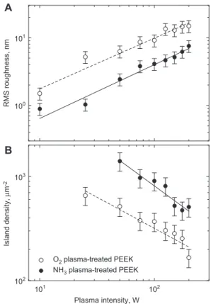
The plasma treatment with a duration of 5 min and the variation of the power between 10 and 200 W results in nanostructures of increasing size and PEEK surfaces with increasing roughness. This fi nding clearly indicates that plasma activation does not only change the chemical nature of the PEEK sheet but also induces signifi cant changes in the surface morphology. These changes are also refl ected in water contact angle measurements (data not shown). From the AFM measurements shown as an example in Figure 1, quantitative data were derived. The root-mean-square (RMS) roughness exhibits an almost perfectly linear behavior as a function of plasma power (see Figure 2 A). The RMS rough-ness measurements of the oxygen plasma-treated surfaces show twice as large values to those of the ammonia treat-ments. Nevertheless, the island densities for the ammonia plasma treatments determined via area measurements of about 100 islands (island counting on certain areas), turned out to show values twice as high as the ones for the oxygen treatments (cp. Figure 2B).
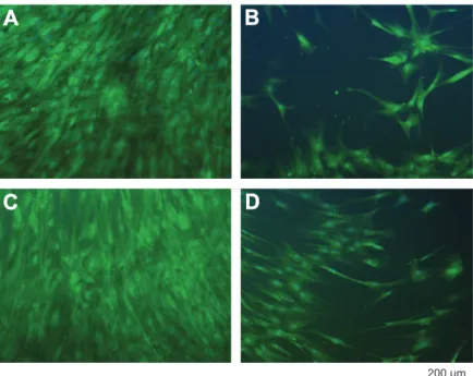
The adipose tissue-derived hMSC, cultured on the differently treated PEEK substrates and the tissue culture polystyrene (TCPS) control for a period of 14 days, show a morphology characteristic for the plasma activation. While the hMSC on the PEEK sheet treated with a power of 10 W are similar to the ones on the control substrate, i.e., in a healthy state, the hMSC on the untreated PEEK have not properly adhered and spread out (see Figure 3 ). Also on harshly oxygen plasma-treated PEEK, the cells did not adhere and spread.
In order to study the cells on the PEEK fi lms, fl uorescence stains were applied with the aim to visualize the
integrin-RMS roughness, nm 101 103 100 101 102 102 Plasma intensity, W Island density, μ m -2 O2 plasma-treated PEEK NH3 plasma-treated PEEK A B
Figure 2 RMS roughness and island density of plasma-treated PEEK fi lms.
The 25 μ m-thick fi lms were plasma-treated for 5 min.
mediated focal adhesions. Integrins are heterodimeric cell membrane spanning receptors and connect the extracellu-lar matrix (ECM) with the actin cytoskeleton of the cell.
200 μm
A
B
C
D
Figure 3 hMSC on oxygen-plasma-treated PEEK fi lms.
hMSC from adipose tissue were cultured for 14 days under standard conditions. The 25 μ m-thick fi lms were plasma-treated for 5 min. (A) TCPS control, (B) untreated, (C) oxygen plasma power 10 W, (D) oxygen plasma power 200 W.
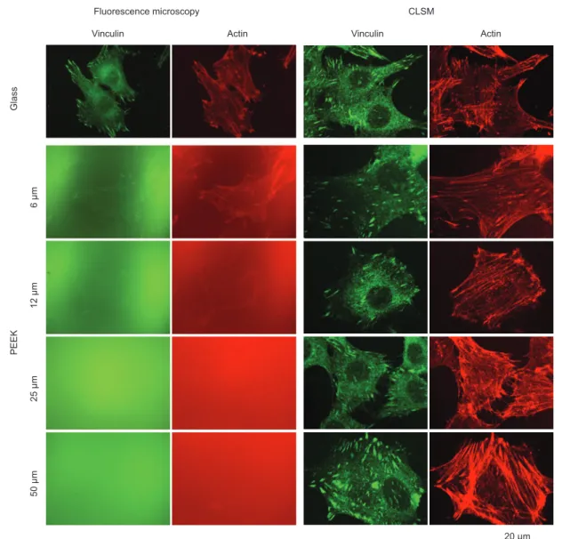
Here, the intracellular part of the integrin receptor binds to a protein complex including vinculin, which itself directly binds to actin. Therefore, co-localization of actin and vin-culin is regarded as proper verifi cation for the presence of focal adhesions (17) . Rat-2 fi broblasts were seeded on APTIV ™ PEEK sheets of different thicknesses and stained for vinculin and actin, giving rise to green- and red-colored features, respectively. Figure 4 shows the related images obtained with conventional epifl uorescence microscopy and CLSM.
The images displayed in the fi rst row of Figure 4 show the controls (rat-2 fi broblasts on glass slides). Here, the back-ground is low and the green vinculin stain (FITC-labeled) clearly points to the focal adhesions of the cells. The red-colored actin cytoskeleton (TRITC-labeled) spans over the entire individual cells. The APTIV ™ PEEK sheets exhibit a strong background fl uorescence that seriously compli-cates the feature extraction using conventional fl uorescence microscopy. This background fl uorescence linearly increased with the fi lm thickness. Hence, three-dimensional structures made out of PEEK, e.g., medical implants, can hardly be included into such fl uorescence studies that strongly depend on the focal plane. In the model system studied, replacing the conventional fl uorescence microscopy by CLSM, the background could signifi cantly be reduced (see Figure 4). The characteristic pattern of the fl uorescence stained cyto-skeletal elements was obvious and similar to the control. The fl exible 6 μ m-thin PEEK fi lms, however, exhibit an inhomo-geneous background associated with their non-wavy surface. Therefore, one can reasonably assume that the investiga-tion of cells on non-planar PEEK implant surfaces becomes complicated.
In order to characterize the optical properties of the PEEK fi lms, the transmission spectra have been recorded (see Figure 5 ) for the four selected thicknesses.
With the exception of the Fabry-P é rot fringes that are very obvious for the 6 μ m-thin sheets, the maximum transmission corresponds to about 80 % . Below 370 nm, the PEEK sheets are opaque.
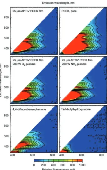
The background of the fl uorescence images implies char-acteristic auto-fl uorescence of PEEK. To obtain a detailed view about the auto-fl uorescence of the PEEK sheets, spec-troscopic fl uorescence measurements for wavelengths rang-ing from 350 to 850 nm have been recorded and are presented in the diagrams of Figure 6 .
To exclude that the fl uorescence originates from any polymer additive, the PEEK polymer was synthesized according to the description of Risse et al. (11) and compared with the commercially available APTIV ™ PEEK fi lms from VICTREX. As shown in Figure 6, the PEEK fi lms and the pure PEEK exhibit very similar emission spectra. Consequently, the auto-fl uorescence is an inherent property of the PEEK polymer.
Strong emission resulted upon excitation at wave-lengths between 370 and 550 nm with a broader maxi-mum for excitation wavelengths between 370 and 450 nm. Oxygen and ammonia plasma treatments did not change the emission profi les. The materials for the PEEK synthesis, 4,4 ′ -difl uorobenzophenone and tert-butylhydroquinone, were analyzed concerning fl uorescence emission as well. While 4,4 ′ -difl uorobenzophenone showed fl uorescence behavior similar to PEEK, tert-butylhydroquinone did not emit light in the excitation wavelength range between 400 and 800 nm.
Discussion and conclusions
The observed nanostructuring of PEEK by oxygen and ammonia plasma treatments is likely due to etching. To exclude a thermal effect, as the temperature within the plasma chamber increases during the processing by several Kelvin, we have performed an additional experiment comparing the nanostructures of a 20-min treatment with a four times 5 min processing. After each 5-min treatment, we have opened the chamber to harvest a part of the PEEK sheet for scanning electron microscopy imaging. Figure 7 displays these images, which do not show any signifi cant difference between step-wise and the one-step processes.
Therefore, one can conclude that thermal effects are neg-ligible. We speculate that the local etching rate depends on the molecule orientation within the semi-crystalline polymer. When etching is described as a negative growth, similar to the facet-depending growth rates in single-crystalline solids, the orientations with fast etching rates gradually disappear, which
Actin Actin Vinculin Vinculin Fluorescence microscopy CLSM Glass 50 μm 6 μm 12 μm 25 μm 20 μm PEEK
Figure 4 Rat-2 fi broblasts seeded on glass and differently thick APTIV ™ PEEK fi lms.
Actin stain: phalloidin-TRITC, vinculin stain: monoclonal anti-vinculin and goat-anti-mouse A-488.
300 500 700 900 0.0 0.2 0.4 0.6 0.8 1.0 Wavelength, nm Relative transmission 50 μm 25 μm 12 μm 6 μm
Figure 5 Transmission spectra of PEEK fi lms. (Film thickness indicated.)
Figure 6 Fluorescence spectra from APTIV ™ PEEK fi lms, synthe-sized PEEK and starting materials.
Specimens were excited with wavelengths ranging from 350 nm to 800 nm in 10 nm-steps. Emission spectra up to a wavelength of 850 nm were recorded 30 nm above excitation wavelength (5 nm steps). The 25 μ m-thick fi lms were plasma-treated for 5 min.
Surface nanostructures have a vital infl uence on protein adsorption and cellular response, making them a key param-eter in characterizing the interactions between biological systems and man-made biomaterials (4, 19 – 21) . In contrast to metallic and ceramic biomaterials, however, nanostructur-ing polymer materials is still poorly understood, although this group of biomaterials has numerous advantages and the plasma-induced roughness increase of polymers has been known for more than a decade (22) . First, the plasma ment can be realized on large areas, as required for the treat-ment of medical implants. Second, it can be combined with other structuring technologies such as soft lithography, rep-lica molding or polymer de-mixing (23) . Third, the plasma treatment generates random nanostructures, which seem to be favored by hMSC for osteogenic differentiation over highly ordered structures as recently described by Dalby et al. (8) . Fourth, plasma treatment is a fast, dry, and reproducible pro-cess and easy to apply on large scales and therefore suitable for mass production. As a consequence, this method enables the production of large substrate areas necessary for in vitro cell culture studies with primary cells like hMSC to reach sta-tistically signifi cant statements. However, one drawback of the plasma-induced activation (chemical modifi cation) is the creation of instable surfaces, which change their properties including the water contact angle over time, due to decay of the reactive molecular species remaining.
A further drawback of optimizing PEEK for medical implants is the optical imaging of cells on PEEK surfaces due to auto-fl uorescence (9, 10) . The detailed investigation of the fl uorescence of commercially available PEEK fi lms (VICTREX) and pure PEEK revealed that the broadband auto-fl uorescence is an inherent material property and not caused by any additive. As the aromatic ring structures of the PEEK monomers consist of highly delocalized electrons, they are expected to be excitable to a greater extent than tightly bound electrons. According to the results of the fl uorescence analysis, fl uorescent dyes that emit in the UV and infrared spectral regions should be less prone to background fl uores-cence interference. Therefore, Alexa Fluor 350 and Alexa Fluor 633 are promising candidates for fl uorescence micros-copy on PEEK substrates, since the corresponding fi lters are less common but available for most fl uorescent microscopes. Due to the relatively long wavelength of 633 nm, a loss in resolution has to be taken into account. Notably, the cell live stain Calcein-AM, which has its emission maximum at 488 nm, can be applied to visualize cells on PEEK fi lms. The cell stain is strong compared to a cell compartment stain and a cell has a height of several micrometers, shifting the focal plane away from the auto-fl uorescent PEEK surface.
PEEK belongs to the promising biomaterials for a variety of medical implants. To reach cell attachment and the desired cell response, the surface has to be activated. Plasma treat-ment is a suitable method to activate the PEEK surface, asso-ciated with the generation of nanostructures. Their density and size can be tailored by the choice of the process gas, the plasma power, and the duration of the plasma treatment. In summary our results guide a path towards improving the bio-compatibility of PEEK implants.
is linked to the formation of larger and larger nanostructures. Oxygen plasma exhibits a higher etching rate than ammonia plasma. This phenomenon can be explained, because the two plasma-activated reaction gases exhibit a different level of chemical reactivity causing the difference in the etching rate. Consequently, the nanostructures on the PEEK sheets can be tailored using the plasma power, the duration of processing, and the choice of the process gas.
AFM measurements directly provide data on the substrate roughness. In many experiments one observes, however, that the roughness of nanostructured surfaces depends on the scanning speed and the choice of the scanning range (18) . Counting the nanometer-size features is rather independent on the spatial resolution of the imaging method and there-fore a reliable parameter to compare specimens from differ-ent batches (19) . Hence, we recommend selecting the process parameters via the nanostructure density rather than using RMS values derived from AFM data.
Acknowledgments
We gratefully acknowledge Nadja Chiapparelli for the AFM mea-surements and Marcus Waser for the synthesis of PEEK. We thank Helmut Schift, Konrad Vogelsang and Mirco Altana for supporting us with knowledge about hot embossing and VICTREX for supplying us with APTIV ™ PEEK fi lms. Stefanie Adam kindly supported the cell culture work. Financial support was provided by grants from the Swiss Nanoscience Institute (project 6.2), the Rectors Conference of the Swiss Universities (CRUS), the European Union and the Federal State of Mecklenburg-Vorpommern, Germany (ESF/IV-WM-B34-0006/08).
References
1. Ponche A, Bigerelle M, Anselme K. Relative infl uence of surface topography and surface chemistry on cell response to bone implant materials. Part 1: physico-chemical effects. Proc Inst Mech Eng H 2010;224:1471 – 86.
2. Anselme K, Ponche A, Bigerelle M. Relative infl uence of sur-face topography and sursur-face chemistry on cell response to bone implant materials. Part 2: biological aspects. Proc Inst Mech Eng H 2010;224:1487 – 507.
3. Engler AJ, Sen S, Sweeney HL, Discher DE. Matrix Elasticity Directs Stem Cell Lineage Specifi cation. Cell 2006;126:677 – 89. 4. Riedel M, M ü ller B, Wintermantel E. Protein adsorption and
monocyte activation on germanium nanopyramids. Biomaterials 2001;22:2307 – 16.
5. Variola F, Brunski JB, Orsini G, Tambasco de Oliveira P, Wazen R, Nanci A. Nanoscale surface modifi cations of medically rele-vant metals: state-of-the art and perspectives. Nanoscale 2011;3: 335 – 53.
6. Dohan Ehrenfest D, Coelho P, Kang B, Sul Y, Albrektsson T. Classifi cation of osseointegrated implant surfaces: materials, chemistry and topography. Trends Biotechnol 2010;28:198 – 206. 7. Kurtz SM, Devine JN. PEEK biomaterials in trauma, orthopedic,
and spinal implants. Biomaterials 2007;28:4845 – 69.
8. Dalby MJ, Gadegaard N, Tare R, Andar A, Riehle MO, Herzyk P, et al. The control of human mesenchymal cell differentiation using nanoscale symmetry and disorder. Nat Mater 2007;6:997 – 1003. 9. Hunter A, Archer CW, Walker PS, Blunn GW. Attachment and proliferation of osteoblasts and fi broblasts on biomaterials for orthopaedic use. Biomaterials 1995;16:287 – 95.
10. Briem D, Strametz S, Schr ö der K, Meenen NM, Lehmann W, Linhart W, et al. Response of primary fi broblasts and osteoblasts to plasma treated polyetheretherketone (PEEK) surfaces. J Mater Sci-Mater M 2005;16:671 – 7.
11. Risse W, Sogha DY. Synthesis of soluble high molecular weight poly(aryl1 ether ketones) containing bulky substitutents. Macromolecules 1990;23:4029 – 33.
12. Peters K, Salamon A, Van Vlierberghe S, Richly J, Kreutzer M, Neumann H-G, et al. A new approach for adipose tissue regen-eration based on human mesenchymal stem cells in contact to hydrogels-an in vitro study. Adv Eng Mater 2007;11:B155 – 61. 13. Ha SW, Hauert R, Ernst KH, Wintermantel E. Surface
analy-sis of chemically-etched and plasma-treated
polyetherether-200 nm
A
B C D
E F G
Figure 7 SEM images of 25 μ m-thin PEEK fi lms after 50 W oxygen plasma treatment with varying exposure time. ( A) untreated, (B) 5 min, (C) 10 min, (D) 20 min, (E) 5 min, (F) 2 × 5 min (G) 4 × 5 min.
ketone (PEEK) for biomedical applications. Surf Coat Tech 1997;96:293 – 9.
14. Ha SW, Kirch M, Birchler F, Eckert KL, Mayer J, Wintermantel E, et al. Surface activation of polyetheretherketone (PEEK) and formation of calcium phosphate coatings by precipitation. J Mater Sci-Mater M 1997;8:683 – 90.
15. Cortese B, Morgan H. Controlling the wettability of hierarchically structured thermoplastics. Langmuir 2011; dx.doi.org/10.1021/ la203741b.
16. Tsougeni K, Vourdas N, Tserepi A, Gogolides E, Cardinaud C. Mechanisms of oxygen plasma nanotexturing of organic polymer surfaces: from stable super hydrophilic to super hydrophobic surfaces. Langmuir 2009;6:11748 – 59.
17. Geiger B. A 130K protein from chicken gizzard: its localization at the termini of microfi lament bundles in cultured chicken cells. Cell 1979;18:193 – 205.
18. M ü ller B, Riedel M, Michel R, Paul SMD, Hofer R, Heger D, et al. Impact of nanometer-scale roughness on contact-angle hys-teresis and globulin adsorption. J Vac Sci Technol B 2001;19: 1715 – 20.
19. M ü ller B. Natural Formation of nanostructures: from fundamen-tals in metal heteroepitaxy to applications in optics and biomate-rials science. Surf Rev Lett 2001;8:169 – 228.
20. Lord MS, Foss M, Besenbacher F. Infl uence of nanoscale surface topography on protein adsorption and cellular response. Nano Today 2010;5:66 – 78.
21. Latour R. Biomaterials: Protein-surface Interactions. Encyclo-pedia of Biomaterials and Bioengineering. 2008: pp. 27 – 284. 22. Chan CM, Ko TM, Hiraoka H. Polymer surface modifi cation by
plasmas and photons. Surf Sci Rep 1996;24:1 – 54.
23. Engel E, Michiardi A. Nanotechnology in regenerative medicine: the materials side. Trends Biotechnol 2008;26:39 – 47.
Jasmin Althaus received her diploma in chemistry from the University of Applied Sciences Northwestern Switzerland in 2004. After working at the Friedrich Miescher Institute in Basel as a research assistant in the group of Jan Hofsteenge, she moved on to the Biocenter in Basel where she earned her MSc in molecular biology in 2008. She is currently working towards her PhD degree in biomedical engineering on the effect of plasma-treated PEEK fi lms on human mesenchymal stem cell differentiation at the University of Basel in close collaboration with Paul Scherrer Institute and University of Applied Sciences Northwestern Switzerland as well as the medical faculty of the University of Rostock.
Celestino Padeste studied chemistry at the University of Z ü rich from where he received a PhD degree on inorganic solid state-gas phase reactions in 1989. After a Post-doc at the University of New South Wales in Sydney/Australia in the fi eld of surface analysis of catalyst systems he was employed at the Micro- and Nanotechnology Laboratory at the Paul Scherrer Institute in Villigen/Switzerland. His work is focused on the design of functional surfaces by com-bination of nanostructuring methods including synchrotron radiation-based lithography with chemical surface modifi ca-tion, polymer grafting and protein immobilization.
Joachim K ö ser received a diploma in biology from the University of Heidelberg. Following his PhD thesis at the German Cancer Research Center he moved to the Biocenter in Basel to work in the group of Ueli Aebi on the nuclear pore complex. Subsequently, he joined the spin-off company Concentris GmbH where he was respon-sible for the application development for cantilever sensors and now is working in the group of Uwe Pieles at the University for Applied Sciences Northwestern Switzerland in the fi eld of medical nano-bio-technology.
Uwe Pieles studied chemistry at the universities in Bielefeld and G ö ttingen (1981–1985) and obtained his diploma (1985) and the PhD degree (1988) at the Max-Planck Institute for experimental medicine under the supervision of Friedrich Cramer. Between 1989 and 1991 he worked as a postdoc-toral researcher at the European Molecular Biology Laboratory. From 1991 to 1996 he worked as a laboratory head in the Central Research Laboratories of Ciba/Novartis in Switzerland (Basel). He became a group leader in the pharmaceutical research of Altana AG Germany (Konstanz, 1997 – 2000). Since 2000 he is a full professor and head of nanotechnology at the University of Applied Sciences Northwestern Switzerland in Muttenz. In 2007 he was rewarded with the Collano Prize. His main research activities include surface science, surface chemistry in materials sci-ence, chemical and biochemical sensors, and nanoparticles in catalysis and biochemical applications.
Kirsten Peters studied biology and did her diploma thesis (1993 – 1994) at the Westf ä lische Wilhelms-University in M ü nster, Germany, where she also received her PhD under the supervision of J ü rgen Rauterberg (1994 – 1998). Between 1999 and 2006 she worked as a senior post-doctoral researcher at the Institute of Pathology at the Johannes Gutenberg-University Mainz (C. James Kirkpatrick). Since 2006 she is the head of the Junior Research Group (Department of Cell Biology, University of Rostock, Germany). In 2008 she received her habilitation in molecular medicine from the Johannes Gutenberg-University in Mainz.
Bert M ü ller received a diploma in mechanical engineering (1982), followed by an MSc from the Dresden University of Technology and the PhD from the University of Hannover, Germany in 1989 and 1994, respectively. From 1994 to 2001, he worked as a researcher at the Paderborn University, Germany, EPF Lausanne, ETH Zurich. He became a faculty mem-ber of the Physics Department at ETH Zurich in April 2001. After his election as Thomas Straumann-Chair for Materials Science in Medicine at the University of Basel, Switzerland and his appointment at the Surgery Department of the University Hospital Basel in September 2006, he founded the Biomaterials Science Center. He also teaches physics and materials science at the ETH Zurich and the Universities of Basel and Bern.