European Journal of Cardio-thoracic Surgery 13 (1998) 200 – 202
How to do it
Protective subpleural blanketing of intrathoracic esogastric anastomosis
after esophagectomy
A. Spiliopoulos, J.H. Robert *, N. Murith, G. Chassot
Unit of thoracic surgery, Clinic of cardio6ascular and thoracic surgery, Uni6ersity Hospital, CH-1211, Gene6a14, Switzerland
Received 27 May 1997; received in revised form 13 October 1997; accepted 19 November 1997
Abstract
Esophagectomy followed by intrathoracic anastomosis is threatened by leakage which may prove all the more serious that mediastinal contamination is extensive. In the technique presented, the esogastric anastomosis is slipped under the upper mediastinal pleura which is kept intact, after the azygos vein has been ligated and divided. This pleural ‘blanket’ may act as an efficient barrier against potential digestive spillage into the mediastinum. © 1998 Elsevier Science B.V. All rights reserved.
Keywords: Intrathoracic; Esogastric; Anastomosis
Surgery for intrathoracic esophageal carcinoma is seldom rewarded by lasting survivals [1]. Moreover, survival can be dramatically shortened by early postop-erative complications [2], such as anastomotic leakage, which occurs in 5 – 10% of cases. This appears particu-larly dangerous [3] when it originates from an anasto-mosis lying unprotected in the mediastinum, as is usually the case [4] (cervical esogastric anastomoses seem to reduce the death toll of this complication [5] but are not as often resorted to).
This report describes a useful and easy-to-do adjunct to the standard Ivor – Lewis procedure which may re-duce the consequences of potential anastomotic leaks: the intrathoracic esogastric anastomosis is placed in the posterior mediastinum and is blanketed under the up-per mediastinal pleura, which is kept intact, and acts as an efficient barrier against potential digestive spillage.
1. Description of the procedure
The abdominal and most of the right chest steps are those of a classic Ivor – Lewis procedure [4]. Both di-aphragmatic crura are divided, most of the greater omentum is excised, and a gastroplasty is performed with a GIA-55 stapler (Auto Suture, Ho¨ri, Switzer-land), together with a pylorotomy (occasionally an extramucosal pyloroplasty).
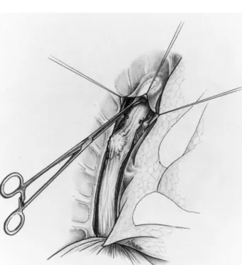
A right posterolateral thoracotomy is performed in the fifth intercostal space. The azygos vein is doubly ligated and excised, and the mediastinal pleura above it gently detached from the underlying proximal esopha-gus using a blunt-tipped clamp (Fig. 1). This creates a ‘pleural tent’ under which the esogastric anastomosis will be placed (Fig. 2).
After the tumor and the esophagus have been ex-cised, the gastric tube to which the lesser curvature is still attached is then drawn into the thorax and an end-to-side anastomosis performed with a PCEEA 28 mm stapler (Auto Suture, Ho¨ri, Switzerland). The end of the gastric tube is cut at least 2 cm above the anastomosis with a TA-60 stapler (Ethicon, Spreiten-* Corresponding author. Tel.: + 41 22 3727875; fax: + 41 22
3727880.
1010-7940/98/$19.00 © 1998 Elsevier Science B.V. All rights reserved.
A. Spiliopoulos et al./European Journal of Cardio-thoracic Surgery13 (1998) 200 – 202 201
bach, Switzerland). The anastomosis is then slipped under the pleural tent and the gastric tube fixed to the adjacent pleura with a dozen separated stitches (Fig. 2). The posterior and inferior chest drain is placed in contact with the gastroplasty and is left in place until the postoperative radio-opaque swallow has shown the anastomosis to be tight.
2. Results
This technique has been used in 43 consecutive pa-tients with esophageal cancer over the last five years. Meglumine diatrozoate (Gastrografin®, Schering Suisse,
Schlieren, Switzerland) swallow performed within ten days of surgery has revealed no anastomotic leakage, with the possible exception of a patient who suffered an extended and eventually lethal necrosis of his gastro-plasty.
Two other patients died, the first of cardiac arrhyth-mia, the second of ARDS. Morbidity affected 17 pa-tients, mostly due to pulmonary complications. Finally, three patients required successful endoscopic dilation of anastomotic stenoses which appeared within the first three months.
For comparison purposes, we retrieved data pertain-ing to 46 consecutive Ivor – Lewis operations which were performed in our department for squamous cell carcinoma of the esophagus between 1977 and 1988, i.e. prior to the use of the pleural tent. Nine patients suffered anastomotic leaks (six of them fatal), and eight patients in all died postoperatively (17%).
Fig. 2. Lateral view after completion of the esogastric anastomosis. Inset: anterior view; arrows = mediastinal pleura.
3. Comments
Esogastric anastomoses are threatened by leakage. Apart from omentum which is almost invariably re-sorted to in one way or another (to hook up the gastric tube to the mediastinum [3,6], for instance, or to help bury the gastric staple line), few if any anatomical structures of the mediastinum lend themselves to anas-tomotic reinforcement. Performing anastomoses at the cervical level is frequently advocated in view of the lower morbidity and mortality caused by anastomotic leaks. The artifice described herein, however, should not discourage surgeons from performing esophagec-tomies with intrathoracic anastomoses, provided of course the slightly more extensive excision afforded by cervical re-establishment [7] is not warranted for other reasons. In spite of the obvious absence of randomiza-tion in both series presented, use of the pleural tent described herein does seem to diminish the incidence of anastomotic leakage (nine in 46 in the first period versus only one in 43 in the second).
Finally, end-to-side esogastric anastomoses may help prevent reflux, a known complication of esophageal substitutions with stomach [8]. Although not docu-mented by postoperative pH metries, this anti-reflux effect may be optimized by the pleural tent described herein, which keeps the anastomosis flat and maintains its valve-like configuration (Fig. 2, inset).
Acknowledgements
Many thanks to Joan and Marcel Robert and to Joan Robert-Yap for reviewing the manuscript. Fig. 1. Preparation of the pleural tent. The azygos vein has been
A. Spiliopoulos et al./European Journal of Cardio-thoracic Surgery13 (1998) 200 – 202
202
References
[1] Turnbull ADM, Ginsberg RJ. Options in the surgical treatment of esophageal carcinoma. Chest Surg Clin North Am 1994;4:315 – 29.
[2] Infante M, Valente M, Andreani S, Catanese C, Dal Fante M, Pizetti P, Giudice G, Basilico M, Spinelli P, Ravasi G. Conserva-tive management of esophageal leaks by transluminal endoscopic drainage of the mediastinum or pleural space. Surgery 1996;119:46 – 50.
[3] Wilson SE, Stone R, Scully M, Ozeran L, Benfield JR. Modern management of anastomotic leak after esophagogastrectomy.
Am J Surg 1982;144:95 – 101.
[4] Bains MS. Ivor Lewis esophagectomy. Chest Surg Clin North Am 1995;5:515 – 26.
[5] Se´gol P, Salame E, Bonvalot S, Maurel J, Gignoux M. Tech-nique actuelle des plasties gastriques en chirurgie æsophagienne. Ann Chir Paris 1996;50:13 – 20.
[6] Fekete F, Breil P, Ronsse H, Tossen JC, Langonnet F. EEA stapler and omental graft in esophagogastrectomy. Ann Surg 1981;193:825 – 30.
[7] Chasseray VM, Kiroff GK, Buard JL, Launois B. Cervical or thoracic anastomosis for esophagectomy for carcinoma. Surg Gynecol Obstet 1989;169:55 – 62.
[8] Postlethwait RW. Colonic interposition for esophageal substitu-tion. Surg Gynecol Obstet 1983;156:377 – 83.