locytic sarcoma to all-trans-retinoic acid plus interferon alfa-2a [letter]. J Natl Cancer Inst 1996;88:1494-5.
(4) Lazzarino M, Corso A, Regazzi MB, Iacona I, Bernasconi C. Modulation of all-trans ret-inoid acid pharmacokinetics in acute promy-elocytic leukaemia by prolonged interferon-a therapy. Br J Haematol 1995;90:928-30. (5) Koller E, Krieger O, Kasparu H, Lutz D.
Restoration of all-trans retinoic acid sensitiv-ity by interferon in acute promyelocytic leu-kaemia [letter]. Lancet 1991;338:1154-5. (6) Bugge CJ, Rodriguez LC, Vane FM.
Deter-mination of isotretinoin or etretinate and their major metabolites in human blood by re-versed phase high performance liquid chro-matography. J Pharm Biomed Anal 1985;3: 269-77.
(7) Smith DC, Jacob HE, Lotze MT, Branch RA, Adedoyin A, Stiff D, et al. A phase I trial of interferon-a2a (IFN-a) and all-trans retinoic acid (ATRA): a pharmacokinetic assessment. Proc ASCO 1994;13:134.
(8) Kalvakolanu DV, Sen GC. Differentiation-dependent activation of interferon-stimulated gene factors and transcription factor NF-kappa B in mouse embryonal carcinoma cells. Proc Natl Acad Sci U S A 1993;90: 3167-71.
(9) Kalvakolanu DV, Kolla V, Linder DJ, Weihau X, Freund R, Borden EC. Retinoic acid en-hances IFN-regulated genes via the modula-tion of STAT1 protein in a breast tumor cell line. J Interferon Cytokine Res 1995;15 (sup-pl):S217.
(10) Marth C, Doxenbichler G, Widschwendter M. Combination of retinoic acid and interfer-ons: mechanism of synergistic interaction. J Interferon Cytokine Res 1995;15 (suppl): S216.
(11) Lippman SM, Kavanagh JJ, Paredes-Espinoza M, Delgadillo-Madrueno F, Paredes-Casillas P, Hong WK, et al. 13-cis-retinoic acid plus interferon a-2a: highly active systemic therapy for squamous cell carcinoma of the cervix. J Natl Cancer Inst 1992;84:241-5. (12) Athanasiadis I, Kies MS, Miller M,
Gan-zenko N, Joob A, Marymont M, et al. Phase II study of the all-trans-retinoic acid and
a-interferon in patients with advanced
non-small cell lung cancer. Clin Cancer Res 1995; 1:973-9.
Notes
Affiliation of authors: Section of Hematology/
Oncology, University of Chicago Medical Center, IL.
Correspondence to: Gini F. Fleming, M.D.,
Section of Hematology/Oncology, University of Chicago Medical Center, 5841 S. Maryland Ave., MC 2115, Chicago, IL 60637-1470.
The study on which these patients were treated was supported in part by Public Health Service grant GCRC M01RR00055 from the National Center for Research Resources, National Institutes of Health, Department of Health and Human Ser-vices.
A Rare Case of
Prednimustine-Induced
Myoclonus
Prednimustine is frequently used in the treatment of chronic lymphocytic leukemia, non-Hodgkin’s lymphoma, Hodgkin’s disease, and ovarian and breast cancers. However, care should be taken to recognize early neurologic side effects that may have disabling conse-quences.
We report here a rare neurologic side effect associated with administration of prednimustine. A 79-year-old woman presented with weight loss, night sweats, asthenia, bilateral inguinal lymphadenopathy, and splenomegaly. She had had a 19-year history of low-grade follicular lymphoma, which had been treated 8 years previously by vari-ous chemotherapy regimens, including prednimustine.
A biopsy of an inguinal adenopathy disclosed Hodgkin’s disease, histologi-cally different from the previously known lymphoma. She was treated with an oral combination of lomustine at a dose of 80 mg/m2on day 1 and etopo-side at a dose of 100 mg/m2and pred-nimustine at a dose of 60 mg/m2, both given on days 1 to 5 (corresponding to a total dose of chlorambucil of 6 mg/kg), and she was premedicated with allopu-rinol and ondansetron. On day 3, she noticed abnormal jerks of her hands, which increased during the subsequent days and finally involved the four limbs. On day 6, unable to stand, she fell and broke her right arm. Neurologic assess-ment at the emergency room disclosed severe myoclonia of the four limbs. Her mental state and conscious state were normal, and laboratory tests showed no electrolyte abnormalities. Clonazepam was introduced, and the myoclonia gradually decreased and disappeared on day 8. A medical history revealed that the patient had experienced the same hand jerks 8 years before while taking prednimustine. We replaced prednimus-tine with prednisone in subsequent treat-ment cycles, and the myoclonus did not reappear.
Prednimustine is an ester of prednis-olone and chlorambucil. Elimination of chlorambucil and its metabolite
phenyl-acetic mustard is prolonged after oral administration of prednimustine com-pared with chlorambucil alone. Neuro-toxicity was dose limiting in a phase I trial of high-dose chlorambucil (1), and chlorambucil-related myoclonus has been described (2) in a 71-year-old woman treated for a lymphocytic lym-phoma with a 5-day regimen of chlor-ambucil and deltacortisone. In this patient, the jerks appeared on day 3, cul-minated on day 7, and disappeared gradually after another week.
Three cases of prednimustine-in-duced myoclonus have also been re-ported (3) in women previously treated with cisplatin for an ovarian cancer. The myoclonia developed on day 4 during a 5-day regimen of prednimustine (120 mg/m2 per day) and disappeared after cessation of the chemotherapy and ad-ministration of diazepam. Hypomagne-semia found in these three patients was considered a possible contributing fac-tor.
The mechanism of chlorambucil-induced myoclonus is not known. My-oclonus arises from the central nervous system in a complex interaction between brain stem, cortex, and cerebellum. It is found in several neurologic diseases, in-cluding mitochondrial myopathies with central nervous system dysfunction, in which deficiencies of components of the respiratory chain are observed (4). Similarly, the neurotoxicity of an alkyl-ating agent (5) is due to the interference with oxidative phosphorylation in the mitochondria by their metabolites.
CHRISTIANMONNERAT MARCGANDER SERGE LEYVRAZ
References
(1) Blumenreich MS, Woodcock TM, Sherrill EJ, Richman SP, Gentile PS, Epremian BE, et al. A phase I trial of chlorambucil administered in short pulses in patients with advanced malig-nancies. Cancer Invest 1988;6:371-5. (2) LaDelfa I, Bayer N, Myers R, Hoffstein V.
Chlorambucil-induced myoclonic seizures in an adult [letter]. J Clin Oncol 1985;3:1691-2. (3) Martin M, Diaz-Rubio E, Casado A, Valverde JJ, Garcia Urra D, Lopez-Martin JA, et al. Prednimustine-induced myoclonus—a report of three cases. Acta Oncol 1994;33:81-2. (4) Hopkins LC, Rosing HS. Myoclonus and
mi-tochondrial myopathy. Adv Neurol 1986;43: 105-17.
(5) Kupfer A, Aeschlimann C, Wermuth B, Cerny
T. Prophylaxis and reversal of ifosfamide en-cephalopathy with methylene-blue. Lancet 1994;343:763-4.
Notes
Affiliation of authors: Centre Pluridisciplinaire
d’Oncologie, University Hospital Lausanne, Swit-zerland.
Correspondence to: Christian Monnerat, M.D.,
Centre Pluridisciplinaire d’Oncologie, CHUV BH 10, Rue du Bugnon 46, CH-1011 Lausanne, Swit-zerland.
Circulating Prostate-Specific
Antigen/CD14-Double-Positive
Cells: a Biomarker Indicating
Low Risk for Hematogeneous
Metastasis of Prostate Cancer
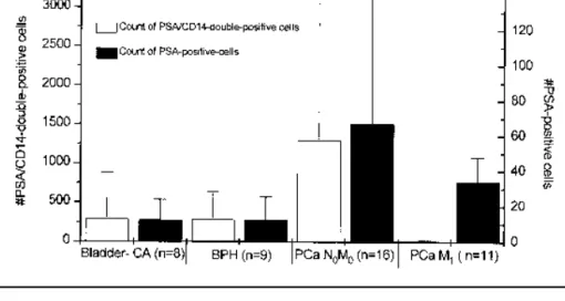
The presence of cells in the blood stream of prostate cancer patients that stain positively for both prostate-specific antigen (PSA) and for the monocyte marker CD14 seems to indi-cate a low risk of metastasis formation. This cellular biomarker might be help-ful in the assessment of the prognosis for prostate cancer patients with or-gan-restricted disease. It could be used to define a subgroup of patients with a low risk of life-threatening bone me-tastasis, although these patients show evidence of circulating prostate cancer cells by flow cytometry (1) or PSA-PCR2 (2).
We describe here 16 patients with N0M0 stage prostate cancer and 11 pa-tients with prostate cancer with bone metastasis (stage M1) as well as two control groups (eight patients with blad-der cancer and nine patients with be-nign prostatic hyperplasia). So that we could determine the number of PSA/ CD14-double-stained cells in the pe-ripheral blood of the patients by flow cytometry, 10 mL of EDTA-treated blood was drawn from the patients by venipuncture, and the erythrocytes were depleted by density-gradient centrifuga-tion. The cells were stained simulta-neously with anti-PSA–fluorescein isothiocyanate (FITC) (Coulter-Im-munotech, Hamburg, Federal Republic of Germany) and an antibody labeled with CD14–phycoerythrin (PE) (Di-anova, Hamburg). Controls for
nonspe-cific binding of the antibody, negative controls with blood from healthy wo-men, and positive controls with LNCaP cells were performed. The PSA-positive and PSA/CD14-double-positive cells were counted by a count gate discrimi-nation procedure adjusted to the exclu-sion of nonspecific, stained cells in the FL1 (FITC)/FL2 (PE) plot of the nega-tive control; two million peripheral white blood cells were analyzed for each sample after erythrocyte depletion.
The data in Fig. 1 suggest that the exclusive detection of molecular signals, either by immunologic means or by polymerase chain reaction, representing PSA-positive cells in the circulation, is not sufficient to estimate the risk of me-tastasis in prostate cancer.
We are entering a new period of re-search on circulating cancer cells. In 1991, Smith et al. (3) introduced the polymerase chain reaction for tissue-specific cell detection. Modern tools in immunology and in molecular biology now provide a means to unveil the meaning of circulating cancer cells (4). BURKHARD BRANDT CORDGRIWATZ OLAF BRINKMANN KURTHS. ZA¨NKER
References
(1) Hamdy FC, Lawry J, Anderson JB, Parsons MA, Rees RC, Williams JL. Circulating pros-tate specific antigen-positive cells correlate with metastatic prostate cancer. Br J Urol 1992;69:392-6.
(2) Olsson CA, de Vries GM, Raffo AJ, Benson MC, O’Toole K, Cao Y, et al. Preoperative reverse transcriptase polymerase chain reac-tion for prostate specific antigen predicts treat-ment failure following radical prostatectomy. J Urol 1996;155:1557-62.
(3) Smith B, Selby P, Southgate J, Pittman K, Bradley C, Blair GE. Detection of melanoma cells in peripheral blood by means of reverse transcriptase and polymerase chain reaction. Lancet 1991;338:1227-9.
(4) Brandt B, Junker R, Heidl S, Griwatz C, Sem-jonow A, Brinkmann O, et al. Isolation of prostate-derived single cells and cell clusters from human peripheral blood. Cancer Res. In press.
Notes
Affiliations of authors: B. Brandt (Institut fu¨r
Klinische Chemie und Laboratoriumsmedizin), O. Brinkmann (Klinik und Poliklinik fu¨r Urologie), Westf. Wilhelms-Universita¨t, Mu¨nster, Federal Republic of Germany; C. Griwatz, K. S. Za¨nker, Institut fu¨r Immunologie, Universita¨t Witten/ Herdecke, Witten, Federal Republic of Germany.
Correspondence to: Burkhard Brandt, Ph.D,
In-stitut fu¨r Klinische Chemie und Laboratoriums-medizin, Westf. Wilhelms-Universita¨t, 48149 Mu¨nster, Federal Republic of Germany. Fig. 1. In patients with N0M0 or M1 stage prostate cancer (PCa), prostate-specific antigen (PSA)-positive cells (right Y axis) were increased compared with those in the control patients. Only in patients with stage M1 disease were the PSA/CD14-double-positively stained cells (left Y axis) significantly decreased compared with those in the control patients (P<.05, paired Wilcoxon test). A statistically significant increase in PSA/CD14-double-positive cells was measured in patients with M1 stage prostate cancer compared with patients with N0M0 prostate cancer (P<.01). BPH4 benign prostatic hyperplasia. Error bars represent standard deviations.
CORRESPONDENCE Journal of the National Cancer Institute, Vol. 89, No. 2, January 15, 1997 174