© The Author 2014. Published by Oxford University Press on behalf of The Gerontological Society of America. 809 doi:10.1093/gerona/glu121
Original Article Advance Access publication August 14, 2014
This is an Open Access article distributed under the terms of the Creative Commons Attribution Non-Commercial License (http://creativecommons.org/licenses/ by-nc/4.0/), which permits non-commercial re-use, distribution, and reproduction in any medium, provided the original work is properly cited. For commercial re-use, please contact journals.permissions@oup.com
Original Article
Circulating Proteomic Signatures of
Chronological Age
Cristina Menni,
1,*
Steven J. Kiddle,
2,3,*
Massimo Mangino,
1Ana Viñuela,
1Maria Psatha,
1Claire Steves,
1Martina Sattlecker,
2,4Alfonso Buil,
5Stephen Newhouse,
2,4Sally Nelson,
6Stephen Williams,
6Nicola Voyle,
2,3Hilkka Soininen,
7Iwona Kloszewska,
8Patrizia Mecocci,
9Magda Tsolaki,
10Bruno Vellas,
11Simon Lovestone,
12Tim D. Spector,
1Richard Dobson,
2,4,$and Ana M. Valdes
1,13,$1Department of Twin Research and Genetic Epidemiology, King’s College London. 2Institute of Psychiatry, King’s College London. 3Medical Research Council Social, Genetic and Developmental Psychiatry Centre, King’s College London. 4National Institute for Health Research Biomedical Research Centre for Mental Health and Biomedical Research Unit for Dementia at South London and Maudsley NHS Foundation, London. 5Department of Genetic Medicine and Development, University of Geneva Medical School, Switzerland. 6SomaLogic, Boulder, Colorado. 7Department of Neurology, University of Eastern Finland and Kuopio University Hospital, Finland. 8 Medical University of Lodz, Poland. 9Institute of Gerontology and Geriatrics, University of Perugia, Italy. 10Department of Neurology III, Aristotle University, Thessaloniki, Greece. 11Institut national de la sante et de la recherche medicale University of Toulouse, France. 12Department of Psychiatry, University of Oxford. 13Academic Rheumatology, University of Nottingham.
Address correspondence to Cristina Menni, PhD, Department of Twin Research, King’s College London, St Thomas’ Hospital Campus, 4th Floor South Wing Block D, Westminster Bridge Road, London SE1 7EH, UK. Email: cristina.menni@kcl.ac.uk
*These authors contributed equally to this work.
$These authors contributed equally to this work.
Abstract
To elucidate the proteomic features of aging in plasma, the subproteome targeted by the SOMAscan assay was profiled in blood samples from 202 females from the TwinsUK cohort. Findings were replicated in 677 independent individuals from the AddNeuroMed, Alzheimer’s Research UK, and Dementia Case Registry cohorts. Results were further validated using RNAseq data from whole blood in TwinsUK and the most significant proteins were tested for association with aging-related phenotypes after adjustment for age. Eleven proteins were associated with chronological age and were replicated at protein level in an independent population. These were further investigated at gene expression level in 384 females from the TwinsUK cohort. The two most strongly associated proteins were chordin-like protein 1 (meta-analysis β [SE] = 0.013 [0.001], p = 3.66 × 10−46) and pleiotrophin (0.012 [0.005], p = 3.88 × 10−41). Chordin-like protein 1 was also significantly correlated with birthweight (0.06 [0.02], p = 0.005) and with the individual Framingham 10-years cardiovascular risk scores in TwinsUK (0.71 [0.18], p = 9.9 × 10⁻⁵). Pleiotrophin is a secreted growth factor with a plethora of functions in multiple tissues and known to be a marker for cardiovascular risk and osteoporosis. Our study highlights the importance of proteomics to identify some molecular mechanisms involved in human health and aging.
Key Words: Aging—Proteomics—Early development—Blood biomarkers—Nucleotide—Aptamers. Received May 5, 2014; Accepted June 24, 2014.
Aging is a complex multifactorial process, which represents the accu-mulation of molecular, cellular, and organ damage over time leading to loss of function and increased vulnerability to disease and death (1). A number of genetic and nongenetic factors, including lifestyle and environmental factors, contribute to human aging. The aging pro-cess manifests probably due to a progressive accumulation of diverse adverse molecular changes resulting from a combination of environ-mental, epigenetic, posttranslational, microbial, and lifestyle factors that increase the risk of death (2). It has been postulated that health outcomes during aging can be determined during early development (3). Observational and experimental evidence increasingly supports a relation between growth and development during fetal and infant life and health in later years, termed the developmental origins of health and disease (3). For example, an individual’s health status in middle and old age, and the risk of premature death due to cardiovascular or metabolic disease, is dependent on perinatal and early life traits such as birthweight (3). Identifying age biomarkers can help to pre-dict and monitor age-related physiological decline and disease, and, importantly, it can also provide molecular insights into the aging pro-cess and into early developmental propro-cesses that influence aging (4–7). Metabolomic and glycomics analysis of blood samples have success-fully been used to identify key molecular mechanisms underlying human health and aging (7,8). Additional molecular signatures of health and aging can be found using high-throughput proteomics. However, due to the high cost, this has been relatively understudied. Recently, three studies on aging using high-throughput proteomics identified proteins whose plasma levels (9,10) and cerebrospinal fluid (CSF) levels (11) substantially change with increasing age. However, these studies either did not apply a correction for multiple testing or did not validate their findings in independent cohorts. Proteomics profiling in the CSF study (11) was obtained using SOMAscan (12), a Slow Off-rate Modified Aptamer (SOMAmer)-based capture array. SOMAscan involves the use of SOMAmers (single-stranded DNA aptamers) to assay proteins in multiplex using DNA microarrays. As such, SOMAscan quantifies the level of the subproteome of proteins targeted by SOMAmers. A total of 1,129 of these SOMAmers are currently available, and a third has been assessed for specificity using pull-down and mass spectrometry.
The SOMAscan approach has previously been used by us and others to study plasma proteins related to Alzheimer and related phenotypes (13–15).
In this study, we use the SOMAscan approach to assess the extent to which proteins are correlated with chronological age in a cohort of female twins with independent replication. We further investigate gene expression levels for those proteins that correlate with age using RNAseq data from whole blood in twins. Finally, we examine the association of specific proteins with factors relating to biologic aging such as birthweight and cardiovascular risk.
Methods
Study Populations
Study subjects were twins enrolled in the TwinsUK (TUK) registry, a national register of adult twins. Twins were recruited as volunteers by successive media campaigns without selecting for particular dis-eases or traits (12). In this study, we analyzed data from 206 female twins who have SOMAscan protein data available.
Replication Cohort—AddNeuroMed, Alzheimer’s Research UK, and Dementia Case Registry
The replication cohort was drawn from a combination of AddNeuroMed (ANM) and the Alzheimer’s Research UK/Maudsley
BRC Dementia Case (ARUK/DCR) Registry at King’s Health Partners Alzheimer’s Disease research cohorts (referred to collectively as ANM + ARUK + DCR) at King’s College London with SOMAscan data available as previously described (15). A total of 677 individu-als (both males and females) were included in the analysis.
All samples and information were collected with written and signed informed consent. All studies were approved by the local research ethics committees.
Protein Measures and Quality Control
Proteins were measured using a SOMAmer-based capture array called “SOMAscan” (SomaLogic Inc, Boulder, CO). SOMAmers are single-stranded DNA aptamers, which include some bases that have been chemically modified to mimic protein side chains while retain-ing the ability to base pair with standard DNA bases (allowretain-ing their levels to be quantified by PCR or microarrays). A total of 1,129 of these SOMAmers are currently available, and they can be used to analyze a biologic sample in such a way that the amount of each type of SOMAmer remaining in the sample is proportional to the amount of the corresponding protein targets in the original sample. Currently, a third of the SOMAmers have been assessed for speci-ficity using pull-down and mass spectrometry. For more details on the approach, see Gold and colleagues (12), and the SomaLogic Inc Web site (http://www.somalogic.com/Products-Services/SOMAscan/
FAQs.aspx). TUK proteomic data were collected using SOMAscan
v3, measuring 1,129 proteins, whereas ANM + ARUK + DCR pro-teomic data were collected using SOMAscan v2, measuring 1,001 proteins. All except 21 of the proteins measured by SOMAscan v2 are also measured by SOMAscan v3.
TUK proteomics data are publicly available upon request on the department website (
http://www.twinsuk.ac.uk/data-access/access-management/). We aim to make the ANM + ARUK + DCR data
available to the public as soon as possible. In the short term, the authors can be contacted and collaborations established under an agreement shared with SomaLogic.
Quality control was performed at the sample and SOMAmer level, and involved the use of control SOMAmers on the microarray and calibration samples. At the sample level, hybridization controls on the microarray were used to monitor sample-by-sample variability in hybrid-ization, while the median signal over all SOMAmers was used to moni-tor overall technical variability. The resulting hybridization scale facmoni-tor and median scale factor were used to normalize data across samples.
RNAseq
Whole blood samples from 384 female twins were prepared for sequencing and processed as previously described (16). Briefly, sam-ples were prepared for sequencing with the Illumina TruSeq sample preparation kit (Illumina, San Diego, CA) according to manufac-turer’s instructions and were sequenced on a HiSeq2000 machine. Then, the 49-bp sequenced paired-end reads were mapped to the GRCh37 reference genome.
Statistical Analysis
Statistical analysis of the protein data was carried out using Stata version 11/12.
Step 1. Data quality control
We log10 transformed the protein data as the protein concentrations were not normally distributed. Additionally, protein values ± 6 SDs were excluded as outliers.
Step 2. Identify proteins associated with chronological age In the TUK data, we looked for proteins associated with chronologi-cal age by running random intercept linear regression adjusting for family relatedness:
E X
( )
ij=
β
0+
β
iage
ij+
ζ
j+
ε
ij,
(1)where Xij is the protein residual of twin j from pair i; ζj is the family-specific error component, which represents the omitted family char-acteristics or unobserved heterogeneity. We corrected for multiple comparisons using Bonferroni correction thus giving a significant threshold (p = .05/(1,129 proteins) = 4.43 × 10⁻⁵).
Step 3. Replication in the ANM + ARUK + DCR sample of significant proteins
We replicated our significant findings in the ANM + ARUK + DCR data running linear regressions adjusting for sex and recruitment centre. We combined discovery and validation results using an inverse variance Han and Eskin random effect meta-analysis (17).
Step 4. Influence of expression on age-related proteins (TUK) Rank normalized reads per exon were used to assess the age effect on exon expression. A linear mixed model was fitted to examine age effect on gene expression in R (http://www.r-project.org/) with the lmer function in the lme4 package (18). Confounding factors in the models included as fixed effects were primer insert size, GC content mean, and batch. Random effect confounding factors included primer index, date, family relation-ship, and zygosity. The p values to assess significance for age effect were calculated from the Chi-square distribution with 1 degree of freedom using likelihood ratio as the test statistic, while comparing a null model (expression ~ fixed covariates + random covariates) versus a full model with age (expression ~ age + fixed covariates + random covariates).
Step 5. Heritability of age-related proteins in TUK
We estimated heritability using structural equation modeling to separate the observed phenotypic variance into three latent sources of variation: additive genetic variance (A), shared/common environmental variance (C), and nonshared/unique environmental variance (E) (19). Heritability is defined as the proportion of the phenotypic variation attributable to genetic factors and is given by the equation, h2 = (A)/(A+ C + E).
Step 6. Identify a proteomics panel of independent proteins associated with chronological age (TUK)
Although the hypothesis underlying our work is that protein levels change with age, in order to investigate which proteins are correlated independently of each other with age we carried out a backward lin-ear regression in the TUK data, with age as the dependent variable. This was performed on a total of 11 proteins, identified in Step 2 and replicated at protein level (Step 3) or with consistent association with age at gene expression level (Step 4). We used p = .01 as a cutoff for the removal of variables from the model.
Step 7. Assess whether the protein panel associated with chronological age is also associated with known markers of early development and cardiovascular risk (TUK)
To determine whether the identified proteins are also markers of early development, we examined the association of the age-associ-ated proteins and birthweight in twins, by running random intercept linear regression adjusting for age and family relatedness.
Finally, we explored the association of selected proteins with the Framingham 10-years cardiovascular risk (20) in twins.
Results
The demographic characteristics of the study populations are pre-sented in Table 1 and Supplementary Table S1. Age was found to correlate in TUK with 13 proteins after accounting for multiple test-ing (p < 4.4 × 10⁻⁵) and family relatedness (Table 2). Of these
pro-teins, 10 were also found to associate with age in the independent ANM + ARUK + DCR cohort after adjusting for sex and recruitment centre (Table 2). Stratifying ANM + ARUK + DCR by gender and/or diagnosis did not change the results (data not shown).
We further tested in 384 females (age range 39–83 years) from TUK the association of whole blood gene expression of the genes encoding all 13 proteins and found three of them (CST3, FSTL3, and HAVCR2) to be nominally significant (Supplementary Table S2). This gives a total of 11 proteins replicated at protein level or with consistent association with age at gene expression level (CHRDL1, CCDC80, PTN, ROR1, CST3, FSTL3, HAVCR2, IGFBP6, MMP12, TIMP1, and THBS4).
Of the 11 proteins, the circulating levels of four of them—namely PTN, ROR1, TIMP1, and CST3—had already shown to associate with age in CSF levels (all but PTN in the same direction as in this study) (11). A hypergeometric test revealed that this overlap is sig-nificant at the .05 level (overlapping population size = 831, number of CSF levels protein markers associated with age = 81, number of replicated blood marker associated with age = 11, overlapping mark-ers = 4, p = .018).
Taking advantage of the twin nature of our data, we ran a her-itability analysis (19) for each of the 11 replicated proteins. The calculated heritabilities ranged from 0% to 63% (Table 2) with cir-culating levels of 9 of the 11 replicated proteins having a genetic contribution of 31% or less.
In the TUK data, a backward linear regression including the 11 replicated proteins revealed 4 proteins—PTN, CHRDL1, MMP12, and IGFP6—to be independently associated with age. It has been extensively shown that low birth weight is associated with increased rates of coronary heart disease, stroke, type 2 diabetes adiposity, the metabolic syndrome, and osteoporosis in adult life (3). We there-fore investigated whether any of the four proteins are associated with birthweight, a marker of early development, and found that birthweight is significantly associated with CHRDL1 (β = 0.06, SE = 0.02, p = .005) after adjusting for age. Circulating levels of CHDRL1 are also significantly associated with the Framingham cardiovascular disease 10-years risk score calculated for TUK subjects after adjust-ing for age (−0.71 [−1.06; −0.36], p = 9.9 × 10⁻⁵), thus suggesting that
there is a relationship between early development and age-related
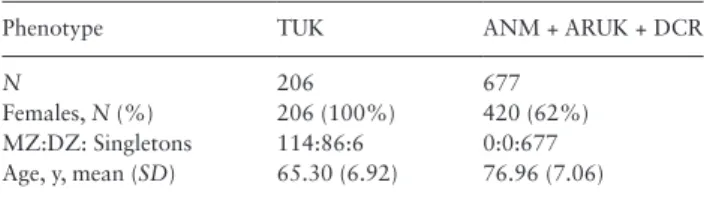
Table 1. Demographic Characteristic of the Discovery and
Replica-tion PopulaReplica-tions
Phenotype TUK ANM + ARUK + DCR
N 206 677
Females, N (%) 206 (100%) 420 (62%) MZ:DZ: Singletons 114:86:6 0:0:677 Age, y, mean (SD) 65.30 (6.92) 76.96 (7.06)
Note: ANM = AddNeuroMed; ARUK = Alzheimer’s Research UK; DCR = Maudsley BRC Dementia Case; TUK = TwinsUK; MZ = monozygotic; DZ = dizygotic.
health deterioration. Moreover, circulating levels of CHRDL1 are significantly associated with many biologic aging markers (diastolic blood pressure, dehydroepiandrosterone sulfate, force expiratory volume in one second, force vital capacity, high-density lipoprotein cholesterol, insulin resistance, and especially waist hip ratio and tri-glycerides (Table 3) after adjusting for age. Thus, although CHRDL1 levels increase with age, higher levels are actually correlated with lower cardiovascular risk and with higher weight at birth (which generally predicts better health outcomes in later age). This rela-tionship was further confirmed when we computed the difference in Framingham cardiovascular disease risk between pairs of twins and the difference in CHDRL1 levels. We find a strong significant negative correlation between the two: the difference between twins in a pair in CHDRL1 levels is negatively correlated with the differ-ence in Framingham cardiovascular disease risk (β = −0.002, SE = 0.001, p = .003).
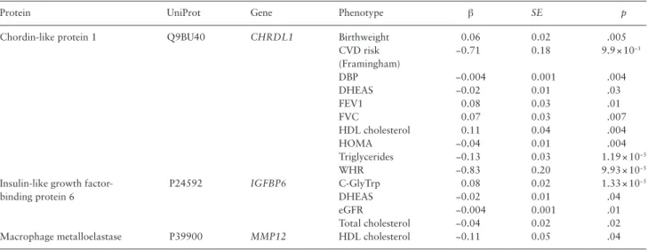
None of the remaining three proteins associate with birthweight. However, MMP12 nominally associates with high-density lipopro-tein cholesterol, while IGF6 correlates with dehydroepiandrosterone sulfate, estimated glomerular filtration rate, total cholesterol, and C-glycosyl tryptophan, a previously identified metabolite marker of aging (8) (Table 3).
For each TUK study participant, we then generated a linear com-bination of the key four proteins using the regression coefficients and created a protein-derived age variable. Plotting this variable in our samples shows a clear relationship with age (Figure 1a) and high-lights that the greatest differences in protein-derived age arise later on in life (ie, over the age of 68). For each ANM + ARUK + DCR subject, we calculated the same protein-derived age variable (model parameters determined from model fitting to TUK data), this is plot-ted in Figure 1b, showing again a clear relationship with age.
Discussion
This is the first study to use the high-throughput SOMAscan pro-teomics approach to study age-related changes in protein plasma levels. We identified 11 proteins whose levels significantly increase with increasing age and that replicated in an independent cohort at protein level or had consistent association with age at gene expres-sion level, highlighting the robustness of our results. Many of these proteins have been previously associated with age in a CSF proteins study (11). Most of these proteins have a very low heritability, con-sistent with published reports that indicate that human aging and longevity have only a modest genetic contribution and a strong Table 2. List of Proteins Significantly Associated With Age in the Discovery Cohort and Replication Cohort
Protein UniProt Gene h2 [95% CI] in
TUK
TUK ANM + ARUK + DCR RE Han and Eskin Meta-analysis
β (SE) p β (SE) p β (SE) p
Chordin-like protein 1 Q9BU40 CHRDL1 0.1 [0, 0.63] 0.014 (0.002) 2.3 × 10⁻¹² 0.012 (0.001) 1.9 × 10⁻³³ 0.013 (0.001) 3.66 × 10−46 Coiled-coil domain- containing protein 80 Q76M96 CCDC80 0.31 [0, 0.71] 0.015 (0.002) 1.7 × 10⁻⁹ 0.007 (0.001) 5.1 × 10⁻²⁹ 0.011 (0.004) 2.55 × 10−43 Pleiotrophin P21246 PTN 0.52 [0, 0.67] 0.018 (0.002) 3.6 × 10⁻¹⁵ 0.007 (0.001) 1.2 × 10⁻²⁴ 0.012 (0.005) 3.88 × 10−41 Follistatin-related protein 3* O95633 FSTL3 0.22 [0, 0.71] 0.011 (0.002) 3.6 × 10⁻⁵ 0.007 (0.0006) 3.1 × 10⁻³² 0.008 (0.002) 8.23 × 10−38 Metalloproteinase inhibitor 1 P01033 TIMP1 0 [0, 0.54] 0.009 (0.002) 1.5 × 10⁻⁸ 0.004 (0.0004) 4.4 × 10⁻²³ 0.006 (0.003) 6.74 × 10⁻³⁰ Macrophage metal-loelastase P39900 MMP12 0.31 [0, 0.77] 0.027 (0.005) 1.6 × 10⁻⁷ 0.004 (0.0004) 2.8 × 10⁻¹⁸ 0.015 (0.011) 8.92 × 10⁻²⁸ Cystatin-C* P01034 CST3 0.63 [0.12, 0.74] 0.009 (0.002) 3.2 × 10⁻⁵ 0.004 (0.0004) 5.6 × 10⁻²⁶ 0.006 (0.002) 2.68 × 10⁻²⁷ Insulin-like growth factor-binding protein 6 P24592 IGFBP6 0.26 [0, 0.63] 0.01 (0.002) 1.2 × 10⁻⁶ 0.004 (0.001) 9.9 × 10⁻¹³ 0.007 (0.003) 3.10 × 10⁻¹⁶ Tyrosine-protein ki-nase transmembrane receptor ROR1 Q01973 ROR1 0.18 [0, 0.36] 0.01 (0.002) 1.4 × 10⁻⁶ 0.004 (0.001) 1.9 × 10⁻¹¹ 0.007 (0.003) 3.86 × 10⁻¹⁶ Thrombospondin-4 P35443 THBS4 0.24 [0, 0.55] 0.008 (0.002) 4.2 × 10⁻⁵ 0.003 (0.001) 2.5 × 10⁻⁴ 0.005 (0.003) 3.47 × 10⁻⁸ Hepatitis A virus cellular receptor 2* Q8TDQ0 HAVCR2 0.24 [0, 0.61] 0.011 (0.003) 3.1 × 10⁻⁵ NA NA NA NA A disintegrin and metalloproteinase with thrombospon-din motifs 5 Q9UNA0 ADAMTS5 0.60 [0.30, 0.73]0.014 (0.003) 1.7 × 10⁻⁶ 0.0004 (0.0003) 0.21 — —
Tumor necrosis factor receptor superfamily member 27
Q9HAV5 EDA2R 0.17 [0, 0.61] 0.013 (0.002) 2.6 × 10⁻⁷ −0.0005
(0.001)
0.25 — —
Notes: ANM = AddNeuroMed; ARUK = Alzheimer’s Research UK; DCR = Maudsley BRC Dementia Case; h2 = heritability; NA = not applicable;
TUK = Twin-sUK.
environmental one (21). We also report a panel of four replicating proteins (PTN, CHRDL1, MMP12, and IGFP6) that are indepen-dently associated with chronological age.
CHRDL1
The protein most associated with chronological age is CHRDL1, a bone morphogenetic protein-4 antagonist, which is involved in bone morphogenic protein signaling and regulation and signaling by GPCR (22). Plasma levels of CHRDL1 are strongly correlated (r = 0.60) with levels of CCDC80, the protein with the second highest p value (Table 2). CHRDL1 also plays a role in regulating retinal angiogenesis through modulation of bone morphogenic protein 4 actions in endothelial cells and during anterior segment eye devel-opment (23). In our study, CHRDL1 is of particular interest as it is not only associated with chronological age but also with birth-weight, a factor affecting healthy aging and its levels have a very low heritability, with 90% of CHRDL1 levels being environmentally determined. The epidemiological observations that low birthweight
is associated with increased rates of coronary heart disease, stroke, type 2 diabetes adiposity, the metabolic syndrome, and osteoporosis in adult life (24) have been extensively replicated. Here, we find that higher birthweight is significantly associated with higher circulating levels of CHDRL1 and that higher circulating levels of CHDRL1 are in turn associated with a lower cardiovascular disease risk. Thus, a positive linear correlation with regards to chronological age does not necessarily imply that the protein is functionally linked to any bio-logic aging phenotype. Indeed, we find that CHRDL1 increases with age, but decreases for most of the markers of increased physiological deterioration (Table 3).
PTN
PTN is a secreted growth factor composed of 168 amino acid resi-dues. The name pleiotrophin is widely accepted nowadays and reflects the plethora of functions it exerts in different tissues and cell lines. Two SNPs (rs322236 and rs3959914) within the first intron of the gene encoding PTN have been associated with volumetric bone mass Table 3. Association of Proteins Independently Associated With Age in TwinsUK and Age-Related Phenotypes After Adjusting for
Chrono-logical Age (p < .05)
Protein UniProt Gene Phenotype β SE p
Chordin-like protein 1 Q9BU40 CHRDL1 Birthweight 0.06 0.02 .005 CVD risk (Framingham) −0.71 0.18 9.9 × 10⁻⁵ DBP −0.004 0.001 .004 DHEAS −0.02 0.01 .03 FEV1 0.08 0.03 .01 FVC 0.07 0.03 .007 HDL cholesterol 0.11 0.04 .004 HOMA −0.04 0.01 .004 Triglycerides −0.13 0.03 1.19 × 10⁻⁵ WHR −0.83 0.20 9.93 × 10⁻⁵
Insulin-like growth factor- binding protein 6
P24592 IGFBP6 C-GlyTrp 0.08 0.02 1.33 × 10⁻⁵
DHEAS −0.02 0.01 .04
eGFR −0.004 0.001 .01
Total cholesterol −0.04 0.02 .02 Macrophage metalloelastase P39900 MMP12 HDL cholesterol −0.11 0.05 .04
Note: CVD risk = cardiovascular disease risk, DBP = diastolic blood pressure; DHEAS = dehydroepiandrosterone sulfate; eGFR = estimated glomerular filtration rate; FEV1 = forced expiratory volume in 1 second; FVC = force vital capacity; HDL = high-density lipoprotein; HOMA = homeostatic model assessment (insulin resistance); WHR = waist hip ratio.
55 60 65 70 75 80 21 52 20 22 52 30 23 52 40 245 age
combined protein measure
60 70 80 90 10 01 05 11 01 15 120 12 51 30 age
combined protein measure
(a) Discovery sample (TUK)
(b) Replication cohorts (ANM+ARUK+DCR)
Figure 1. Protein measure and age. (a) The protein measure was calculated from each TwinsUK (TUK) participant using the coefficients from the backward
regression on age of the 11 proteins in Table 2. (b) These coefficients and blood proteins were also used to calculate a protein measure of aging in the combined ANM + ARUK + DCR cohort.
density (25). Polymorphisms in the PTN gene promoter have been also mentioned to affect bone density and might be implicated in osteoporosis (26). PTN acts via various receptors (reviewed in (27)), like the receptor protein tyrosine phosphatase β/ζ ανβ3integrin, nucleolin, N-syndecan, and anaplastic lymphoma kinase.
Mice deficient in both PTN and midkine have high mortality rates within a month of their birth (28) and exhibit female infer-tility (28). PTN-overexpressing mice exhibit abnormalities in bone formation (29–31) PTN expression has been reported in vascular-ized human atherosclerotic plaques (32) and has been also suggested as a diagnostic marker and a therapeutic target for heart failure (reviewed in (33)). In our study, we do not find PTN associated with markers of biologic aging, but this may be due to the reduced sample size of the study sample.
IGFBP-6
Insulin-like growth factor-binding protein-6 (IGFBP-6) is a secreted glycoprotein that reduces the bioavailability of IGFs in particular IGF-II (34). It has both IGF-dependent and -independent effects on cell growth. IGFBP-6 can be internalized and translocated to the nucleus and interacts with the vitamin D receptor (35) and thyroid hormone receptor α1 (36). Although we find that IGFBP-6 levels increase with age, in vitro results show that downregulation of IGFBP-6 led to premature entry into cellular senescence and that IGFBP-6 over expression increases cellular life span (37). The pleio-tropic effects of IGFBP-6 effects are confirmed in the clinical data from the TUK cohort: on the one hand, its levels are negatively cor-related with dehydroepiandrosterone sulfate and estimated glomeru-lar filtration rate both of which are known to decline with age. On the other hand, circulating levels of IGFBP-6 are also correlated with lower total cholesterol, which normally increases with age and is a marker of higher cardiovascular risk.
MMP-12
Matrix metalloproteinase-12 (MMP-12) has been strongly impli-cated in the aging process in several tissues, such as lung, skin, and brain. For example, tropoelastin and fibrillin degradation in skin by MMP-12 has been implicated in the accumulation of elastotic material in photoaged skin (38). During aging, the brain displays an increased proinflammatory status, which is associated with the pathogenesis of aging-related diseases such as Alzheimer’s and Parkinson diseases. MMPs facilitate the migration of inflamma-tory cells in tissues and modulate their inflammainflamma-tory activity. In particular, upregulated cerebral MMP-12 during aging enhances aging-associated neuroinflammation by facilitating recruitment of bone marrow-derived microglia into the brain (39). Emphysema-like change in mice due to increasing age has been shown to be facilitated by increased MMP-12 expression that accelerates elastin degrada-tion in mice lungs (40). Moreover, MMP-12 was listed as one of the 10 novel proteins for cardiovascular risk stratification (hazard ratio = 3.45 between the highest and lowest quintiles for all subsequent cardiovascular events) in over 900 individuals with a median 6-years follow-up period from the Heart and Soul study (41).
It has been widely reported that, in humans and other organ-isms, the correlations between gene expression and protein levels are not very high (~40%). Current data demonstrate a substantial role for regulatory processes occurring after messenger RNA (mRNA) is made—that is, posttranscriptional, translational, and protein degradation regulation—in controlling steady-state protein abun-dances (42). For the four proteins that we report here as consistently
and independently associated with age—namely CHRDL1, PTN, IGFBP6, and MMP12—we find that the relationship with age is the same both for mRNA and protein levels, although it is not significant with mRNA. There are many processes between transcription and translation, which result in a weak correlation between protein levels and mRNA levels and protein stability is a big factor. The fact, how-ever, that the relationship with chronological age remains suggests that the increased levels of these proteins with increasing age is not a result of an accumulation of the protein or a lack of degradation, but can be, at least to a certain extent, be attributed to gene expression and thus to a direct regulation of their levels.
We note some limitations to this study. Our discovery sample is small and consisted of middle-aged women only. We only studied the subproteome quantifiable using SOMAscan, it is therefore likely that there are many age-related proteins that were not measured by this study. There is no overlap in the blood proteins identified to be significantly associated with aging in this and the two previous dis-covery studies (9,10). This may be due to the difference in proteomic approaches or the lack of multiple testing corrections used in the previ-ous studies. In comparison, a study that looked at CSF proteins using the same proteomic approach and multiple testing corrections had a significantly overlapping set of markers of aging (11). The ability of these proteins to cross the blood–brain barrier is unknown, so it is not known whether this signal crosses between CSF and blood, or whether the same aging process affects their levels independently. PTN is associ-ated with age in a different direction in this study in comparison than in the CSF study (11), this may be due to cohort heterogeneity or tissue-specific differences. Of the 11 proteins associated with age, only three show changes in expression in the corresponding coding genes. The first most likely reason for the lack of correlation between expression and protein levels are the different methods resolution and samples sizes. The sensibilities of the different techniques to identify changes with ages were different but also the ages ranges of both sets are dif-ferent. Moreover, it has been shown that the levels of transcription are not strong predictors for protein levels due to transcriptional noise and posttranscriptional modifications as well as the higher stability of proteins compare with mRNAs (42). Finally, specificity experiments by pull-down mass spectrometry have been performed on a third of the 1,129 proteins on the menu, so some of the proteins we analyzed may have extra noise due to nonspecific binding. However, this is the case for any affinity-based protein assay, including more standard approaches, for example, antibody based. Also, SOMAscan has been shown to be a very robust technology in previous studies (43).
In conclusion, our study highlights the potential of proteomic approaches for identifying molecular pathways related to aging. At the same time, these data also illustrate some of the challenges in aging research and in particular with regards to the interpretation of omics data with regards to the role of specific molecules with regards to aging. CHRDL1 is the most strongly associated with age, with levels increasing in older individuals. However, higher levels are also correlated with lower predicted cardiovascular risk and higher birth-weight (a determinant of healthier aging). Therefore, these results need to be interpreted with caution and suggest that longitudinal studies are needed to understand the role of some of these molecules in the aging process.
Supplementary Material
Supplementary material can be found at: http://biomedgerontology. oxfordjournals.org/
Funding
SOMAscanTM and SOMAmerTM are trademarks of SomaLogic, Inc. TwinsUK: This work was supported by: the EU Framework Programme 7 small-scale focused research collaborative project EurHEALTHAging 277849; TwinsUK was funded by the Wellcome Trust; European Community’s Seventh Framework Programme (FP7/2007-2013). The study also receives support from the National Institute for Health Research (NIHR) Clinical Research Facility at Guy’s and St Thomas’ NHS Foundation Trust and NIHR Biomedical Research Centre based at Guy's and St Thomas' NHS Foundation Trust and King's College London. Tim Spector is an NIHR senior Investigator and is holder of an ERC Advanced Principal Investigator award. SNP Genotyping was performed by The Wellcome Trust Sanger Institute and National Eye Institute via National Institutes of Health/Center for Inherited Disease Research. A.V. and A.B. were supported by the EU FP7 grant EuroBATS (pro-ject no. 279749). ANM+ARUK+DCR: This work was supported by InnoMed, (Innovative Medicines in Europe), an Integrated Project funded by the European Union of the Sixth Framework program priority (FP6-2004-LIFESCIHEALTH-5); Alzheimer’s Research UK (formerly Alzheimer’s Research Trust); The John and Lucille van Geest Foundation and the NIHR Biomedical Research Centre for Mental Health and Biomedical Research Unit for Dementia at the South London and Maudsley NHS Foundation Trust and Kings College London and a joint infrastructure grant from Guy’s and St Thomas’ Charity and the Maudsley Charity; Kuopio University Hospital (H.S.) and funding from UEFBRAIN (H.S.). This study was part funded by NIHR Mental Health Biomedical Research Centre. We would like to acknowledge Abhishek Dixit for his help with data management and access. S.J.K. is supported by an MRC Career Development Award in Biostatistics (MR/L011859/1).
This paper presents independent research part funded by NIHR. The views expressed are those of the author(s) and not necessarily those of the NHS, the NIHR, or the Department of Health. Finally, we wish to express our appreciation to all study participants of the TwinsUK, ANM, ARUK, and DCR.
Conflict of Interest
S.W. is an employee and stockholder in SomaLogic. He is on the board of Venaxis Inc, a publicly held diagnostics company.
References
1. Fontana L, Partridge L, Longo VD. Extending healthy life span–from yeast to humans. Science. 2010;328:321–326. doi:10.1126/science.1172539 2. Valdes AM, Glass D, Spector TD. Omics technologies and the study of
human ageing. Nat Rev Genet. 2013;14:601–607. doi:10.1038/nrg3553 3. Gluckman PD, Hanson MA, Cooper C, Thornburg KL. Effect of in utero
and early-life conditions on adult health and disease. N Engl J Med. 2008;359:61–73. doi:10.1056/NEJMra0708473
4. Franceschi C, Campisi J. Chronic inflammation (inflammaging) and its potential contribution to age-associated diseases. J Gerontol A Biol Sci Med Sci. 2014;69:S4–S9. doi:10.1093/gerona/glu057
5. Richardson AG, Schadt EE. The role of macromolecular damage in aging and age-related disease. J Gerontol A Biol Sci Med Sci. 2014;69:S28–S32. doi:10.1093/gerona/glu056
6. Morimoto RI, Cuervo AM. Proteostasis and the aging proteome in health and disease. J Gerontol A Biol Sci Med Sci. 2014;69:S33–S38. doi:10.1093/ gerona/glu049
7. Krištic J, Vuckovic F, Menni C, et al. Glycans are a novel biomarker of chronological and biological ages. J Gerontol A Biol Sci Med Sci. 2014;69:779–789. doi:10.1093/gerona/glt190
8. Menni C, Kastenmüller G, Petersen AK, et al. Metabolomic markers reveal novel pathways of ageing and early development in human populations. Int J Epidemiol. 2013;42:1111–1119. doi:10.1093/ije/dyt094
9. Ignjatovic V, Lai C, Summerhayes R, et al. Age-related differences in plasma proteins: how plasma proteins change from neonates to adults. PLoS ONE. 2011;6:e17213. doi:10.1371/journal.pone.0017213 10. Lu J, Huang Y, Wang Y, et al. Profiling plasma peptides for the
identifica-tion of potential ageing biomarkers in Chinese Han adults. PLoS ONE. 2012;7:e39726. doi:10.1371/journal.pone.0039726
11. Baird GS, Nelson SK, Keeney TR, et al. Age-dependent changes in the cer-ebrospinal fluid proteome by slow off-rate modified aptamer array. Am J Pathol. 2012;180:446–456. doi:10.1016/j.ajpath.2011.10.024
12. Gold L, Ayers D, Bertino J, et al. Aptamer-based multiplexed prot-eomic technology for biomarker discovery. PLoS ONE. 2010;5:e15004. doi:10.1371/journal.pone.0015004
13. Lourdusamy A, Newhouse S, Lunnon K, et al. Identification of cis-regu-latory variation influencing protein abundance levels in human plasma. Hum Mol Genet. 2012;21:3719–3726. doi:10.1093/hmg/dds186 14. Sattlecker M, Kiddle SJ, Newhouse S, et al. Alzheimer’s disease biomarker
discovery using SOMAscan multiplexed protein technology. Alzheimers Dement. 2014. doi:10.1016/j.jalz.2013.09.016
15. Kiddle SJ, Sattlecker M, Proitsi P, et al. Candidate blood proteome mark-ers of Alzheimer’s disease onset and progression: a systematic review and replication study. J Alzheimers Dis. 2014;38:515–531. doi:10.3233/JAD-130380
16. Brown AA, Buil A, Viñuela A, et al. Genetic interactions affecting human gene expression identified by variance association mapping. Elife. 2014;3:e01381. doi:10.7554/eLife.01381
17. Han B, Eskin E. Random-effects model aimed at discovering associations in meta-analysis of genome-wide association studies. Am J Hum Genet. 2011;88:586–598. doi:10.1016/j.ajhg.2011.04.014
18. Bates D, Maechler M, Bolker B. lme4: linear mixed-effects models using S4 classes. R package version 0.999375–41. 2011.
19. Neale M, Cardon L. Methodology for Genetic Studies of Twins and Fami-lies. Dordrecht: Kluwer Academic Publishers; 1992.
20. D’Agostino RB Sr, Vasan RS, Pencina MJ, et al. General cardiovascular risk profile for use in primary care: the Framingham Heart Study. Circula-tion. 2008;117:743–753. doi:10.1161/CIRCULATIONAHA.107.699579 21. Brooks-Wilson AR. Genetics of healthy aging and longevity. Hum Genet.
2013;132:1323–1338. doi:10.1007/s00439-013-1342-z
22. Larman BW, Karolak MJ, Adams DC, Oxburgh L. Chordin-like 1 and twisted gastrulation 1 regulate BMP signaling following kidney injury. J Am Soc Nephrol. 2009;20:1020–1031. doi:10.1681/ASN.2008070768 23. Kane R, Godson C, O’Brien C. Chordin-like 1, a bone morphogenetic
pro-tein-4 antagonist, is upregulated by hypoxia in human retinal pericytes and plays a role in regulating angiogenesis. Mol Vis. 2008;14:1138–1148. 24. Ptitsyn AA, Weil MM, Thamm DH. Systems biology approach to identifi-cation of biomarkers for metastatic progression in cancer. BMC Bioinfor-matics. 2008;9:S8. doi:10.1186/1471-2105-9-S9-S8
25. Zmuda JM, Yerges-Armstrong LM, Moffett SP, et al. Genetic analysis of vertebral trabecular bone density and cross-sectional area in older men. Osteoporos Int. 2011;22:1079–1090. doi:10.1007/s00198-010-1296-0 26. Mencej-Bedrac S, Preželj J, Komadina R, Vindišar F, Marc J. -1227C>T
polymorphism in the pleiotrophin gene promoter influences bone mineral density in postmenopausal women. Mol Genet Metab. 2011;103:76–80. doi:10.1016/j.ymgme.2011.01.017
27. Papadimitriou E, Mikelis C, Lampropoulou E, et al. Roles of pleiotrophin in tumor growth and angiogenesis. Eur Cytokine Netw. 2009;20:180– 190. doi:10.1684/ecn.2009.0172
28. Muramatsu H, Zou P, Kurosawa N, et al. Female infertility in mice deficient in midkine and pleiotrophin, which form a distinct family of growth factors. Genes Cells. 2006;11:1405–1417. doi:10.1111/j.1365-2443.2006.01028.x
29. Li G, Bunn JR, Mushipe MT, He Q, Chen X. Effects of pleiotrophin (PTN) over-expression on mouse long bone development, fracture healing and bone repair. Calcif Tissue Int. 2005;76:299–306. doi:10.1007/s00223-004-0145-6
30. Masuda H, Tsujimura A, Yoshioka M, et al. Bone mass loss due to estrogen deficiency is compensated in transgenic mice overexpressing human osteoblast stimulating factor-1. Biochem Biophys Res Commun. 1997;238:528–533. doi:10.1006/bbrc.1997.7188
31. Hashimoto-Gotoh T, Ohnishi H, Tsujimura A, et al. Bone mass increase specific to the female in a line of transgenic mice overexpressing human osteoblast stimulating factor-1. J Bone Miner Metab. 2004;22:278–282. doi:10.1007/s00774-003-0485-8
32. Li F, Tian F, Wang L, Williamson IK, Sharifi BG, Shah PK. Pleiotrophin (PTN) is expressed in vascularized human atherosclerotic plaques: IFN-{gamma}/JAK/STAT1 signaling is critical for the expression of PTN in macrophages. FASEB J. 2010;24:810–822. doi:10.1096/fj.09-140780 33. Consortium TCADCDG. A genome-wide association study in Europeans
and South Asians identifies five new loci for coronary artery disease. Nat Genet. 2011;43:339–344. doi:10.1038/ng.782
34. Bach LA, Fu P, Yang Z. Insulin-like growth factor-binding protein-6 and cancer. Clin Sci. 2013;124:215–229. doi:10.1042/CS20120343
35. Cui J, Ma C, Qiu J, et al. A novel interaction between insulin-like growth factor binding protein-6 and the vitamin D receptor inhibits the role of vitamin D3 in osteoblast differentiation. Mol Cell Endocrinol. 2011;338:84–92. doi:10.1016/j.mce.2011.03.011
36. Qiu J, Ma XL, Wang X, Chen H, Huang BR. Insulin-like growth factor binding protein-6 interacts with the thyroid hormone receptor a1 and
modulates the thyroid hormone-response in osteoblastic differentiation. Mol Cell Biochem. 2012;361:197–208. doi:10.1007/s11010-011-1104-y 37. Micutkova L, Diener T, Li C, et al. Insulin-like growth factor binding
pro-tein-6 delays replicative senescence of human fibroblasts. Mech Ageing Dev. 2011;132:468–479. doi:10.1016/j.mad.2011.07.005
38. Chen Z, Seo JY, Kim YK, et al. Heat modulation of tropoelastin, fibrillin-1, and matrix metalloproteinase-12 in human skin in vivo. J Invest Derma-tol. 2005;124:70–78. doi:10.1111/j.0022-202X.2004.23550.x
39. Liu Y, Zhang M, Hao W, et al. Matrix metalloproteinase-12 contributes to neuroinflammation in the aged brain. Neurobiol Aging. 2013;34:1231– 1239. doi:10.1016/j.neurobiolaging.2012.10.015
40. Hu H, Zhao Y, Xiao Y, Zhang R, Song H. Disruption of plasmi-nogen activator inhibitor-1 gene enhances spontaneous enlarge-ment of mouse airspace with increasing age. Tohoku J Exp Med. 2010;222:291–296.
41. Ganz P, Stewart A, Farzaneh-Far R,et al. Stratification of cardiovascular risk using an unbiased proteomic approach. Circulation 2012;126:A14794. 42. Vogel C, Marcotte EM. Insights into the regulation of protein
abun-dance from proteomic and transcriptomic analyses. Nat Rev Genet. 2012;13:227–232. doi:10.1038/nrg3185
43. Loffredo FS, Steinhauser ML, Jay SM, et al. Growth differentiation factor 11 is a circulating factor that reverses age-related cardiac hypertrophy. Cell. 2013;153:828–839. doi:10.1016/j.cell.2013.04.015