HAL Id: tel-01717579
https://tel.archives-ouvertes.fr/tel-01717579
Submitted on 26 Feb 2018HAL is a multi-disciplinary open access archive for the deposit and dissemination of sci-entific research documents, whether they are pub-lished or not. The documents may come from teaching and research institutions in France or abroad, or from public or private research centers.
L’archive ouverte pluridisciplinaire HAL, est destinée au dépôt et à la diffusion de documents scientifiques de niveau recherche, publiés ou non, émanant des établissements d’enseignement et de recherche français ou étrangers, des laboratoires publics ou privés.
Investigation of RNA kissing complexes by native
electrospray mass spectrometry : magnesium binding
and ion mobility
Clemence Rabin
To cite this version:
Clemence Rabin. Investigation of RNA kissing complexes by native electrospray mass spectrometry : magnesium binding and ion mobility. Agricultural sciences. Université de Bordeaux, 2017. English. �NNT : 2017BORD0892�. �tel-01717579�
THÈSE PRÉSENTÉE
POUR OBTENIR LE GRADE DE
DOCTEUR DE
L’UNIVERSITÉ DE BORDEAUX
ÉCOLE DOCTORALE SCIENCES DE LA VIE ET DE LA SANTÉ
SPÉCIALITÉ INTERFACE CHIMIE-BIOLOGIE
Par Clémence RABIN
Ingénieur diplômée de l’ENSTBB
Investigation of RNA kissing complexes by native electrospray
mass spectrometry:
magnesium binding and ion mobility
Sous la direction de : Dr. Valérie GABELICA
Soutenue le 19 décembre 2017
Membres du jury :
Dr. LEIZE-WAGNER, Emmanuelle Directeur de recherche, CNRS, LSMIS Présidente Dr. ENNIFAR, Eric Directeur de recherche, CNRS, IBMC, Strasbourg Rapporteur Pr. FABRIS, Daniele Professeur à l’Université d’Albany, RNA institute Rapporteur Dr. DI PRIMO, Carmelo Chargé de recherche, INSERM, IECB Examinateur
Après avoir écrit ce manuscrit et présenté ces travaux, il est temps de clôturer avec l’écriture de mes remerciements. Une thèse, ce n’est pas que soi-même face à son sujet, c’est une multitude de moments de joie, de doutes, de tristesse et de rires partagés avec de nombreuses personnes.
En premier lieu, je remercie Emmanuelle Leize-Wagner, Eric Ennifar, Daniele Fabris et Carmelo Di Primo pour avoir accepté d’évaluer mon travail. Je souhaiterais aussi remercier Jean-Louis, directeur de l’IECB, de l’unité U1212 et aussi chef de l’équipe Mergny, pour son accueil dans l’établissement. Je souhaite ensuite remercier Valérie, notre « Big Boss ». Merci de m’avoir fait confiance durant ces 3 dernières années. Tu as vite compris que la chimie, ce n’était pas mon fort, pourtant tu m’as encouragée et suivie tout au long de ces années. Merci pour tes conseils, ta rigueur et ton exigence qui, même si parfois c’était dur, m’ont fait évoluer et avancer. J’ai pu découvrir grâce à toi le monde de la recherche en spectrométrie de masse et je ne le regrette absolument pas ! Merci.
Je tiens aussi à remercier les membres de la plateforme de spectrométrie de masse de l’IECB : Fred et Loïc. Vous faites partie intégrante de notre équipe. Fred, merci pour ton aide en instrumentation et surtout pour l’IMS, parfois capricieuse. Loïc, merci pour nos discussions, ton aide et ta patience (il en faut souvent beaucoup !).
Faire de la recherche c’est bien, mais en équipe c’est mieux ! Et quelle équipe… ! Ces 3 années m’ont permis de rencontrer de multiples personnalités, venant de tout horizon (Italie, Russie, Espagne, Inde, Canada, Japon, etc.) et que je ne risque pas d’oublier. Tout d’abord, je vais remercier les nouveaux arrivants : Anirban, Steven, Eric et Jorge. We did not spent so much time together, but I wish you the best for the future. I hope you will enjoy working at the IECB as I liked it! Ensuite, je souhaite remercier Adrien, Max, et Joséphine, collègues du « bureau au bout du couloir ». Adrien, comme je te l’ai déjà dit, je te remercie pour ton aide en début de thèse. Merci d’avoir été patient et de m’avoir transmis ton savoir ! Max… je vais te dire seulement une chose et tu comprendras « 😉 ». Joséphine, merci pour tout, autant professionnel que personnel. Même si tu as un petit côté dictateur, j’ai adoré travailler avec toi ! Aussi, tu as une petite famille au top, prenez bien soin de vous !
Ensuite, nous avons Nina. Nina, tu es une très belle rencontre ! Cette année a été pour toi aussi une grande année et je suis très heureuse d’avoir pu partager avec toi tous ces moments. Je me souviendrai toujours de cette partie de bowling endiablée pour ton EVJF ! Comme on dit chez toi : SPACIBA (si j’ai bien retenu un mot, se sera celui là ^^). Passons maintenant à mes collègues de bureau. Sandrine, ton départ n’a pas été une grande joie pour nous mais je suis contente que tu ais trouvé un poste qui te plaît. Merci pour ces bons moments et surtout pour les derniers mots que tu m’as écrits, ça m’a beaucoup touchée. Tu es la « force tranquille » incarnée, surtout ne change pas ! Ensuite, je tiens particulièrement à remercier Stefano. Tu es quelqu’un d’exceptionnel, toujours calme et posé.
J’aimerais tellement être comme toi pour ça ^^. Tu es devenu un ami très cher. Je sais que ce n’est pas toujours facile d’allier professionnel et personnel mais tout va très bien se passer, tu verras. Et puis n’oublie pas : « Quando il gatto non c’è, i topi ballano » !
J’ai aussi eu la chance de pouvoir encadrer une stagiaire au top : Louise. Merci pour ton travail qui m’a beaucoup avancé. Merci pour ton humour et ta gentillesse . J’en profite aussi pour remercier Marine, autre stagiaire, avec qui on a beaucoup ri et que j’apprécie particulièrement.
J’ai pu également faire la rencontre d’autres thésardes, devenues amies maintenant. Nassima (Chouchou), avec toi j’ai ri, j’ai pleuré, j’ai râlé, on s’est consolées mutuellement, et on n’a rien lâché ! Léonie, ma belle rencontre des JJC, avec toi aussi j’ai ri et surtout beaucoup râlé ! Ne lâche pas, si tu en es arrivée là c’est que tu as toutes les capacités pour ! Merci d’être vous les filles.
Je voudrais remercier aussi deux autres personnes de l’IECB. Elles restent souvent dans l’ombre alors qu’elles mériteraient d’être plus mises en lumière. Aurore, je pense que sans toi, l’équipe Mergny ne fonctionnerait pas aussi bien. Tu es un atout alors ne l’oublie pas. Mariline, notre hôtesse d’accueil. L’IECB serait bien triste sans toi. Merci pour ce que tu as fait pour moi ces derniers temps. Surtout ne changez pas !
Je voudrais aussi en profiter pour remercier tous les autres avec qui j’ai pu discuter, rire, et échanger durant ces 3 années : Laura, Ly, Clément, Solenne, Mona, Gilmar, Samir, Carmelo., et bien d’autres encore.
Je remercie aussi mes amis, notamment ceux qui sont là malgré la distance qui nous sépare : Pitch, Emy, Arnaud, Marie & Charlotte, ma petite Marie, Hélène.
Enfin mes derniers mots seront pour ma famille et amis proches. Je commencerai par deux personnes que j’ai l’impression de connaitre depuis des années alors que ce n’est pas le cas. Mandy, tu es quelqu’un de formidable. Merci pour tout ce que tu fais pour nous. Ton amitié nous est précieuse et pour rien au monde je n’aimerais l’échanger. Tu sais trouver les bons mots que ce soit pour nous pousser ou nous rassurer. Ensuite, il y a Julien. Mon petit Ju, j’ai pu te découvrir tout au long de cette dernière année (et demi), et en toi j’ai trouvé un véritable ami. Merci d’être toi tout simplement. Ces derniers mois, tu m’as montrée que je pourrai toujours compter sur toi (et inversement). Ton amitié est inestimable. Merci… Ensuite, Emeline, dite Luna, tu es ma meilleure amie. Même si nous sommes loin l’une de l’autre, tu es toujours là pour moi. Tu as toujours cru en moi et je t’en remercie du fond du cœur. Je suis très fière d’être la marraine de votre petit roi ! Je tiens ensuite à remercier ma famille. Ma belle-famille qui est formidable et à laquelle je tiens beaucoup. Merci de votre soutien. Merci à Mathias pour sa gentillesse et son aide photographique. Merci à mon grand-père pour tout ce qu’il
m’apporte et d’être là tout simplement. Elise, ma sœur, comme tu me l’as si bien dit, tu as assisté aux turpitudes de mon parcours et à toutes mes angoisses. Ça n’a pas toujours été facile, mais tu vois, j’y suis arrivée ! Merci d’être là Sis. Je tiens particulièrement à remercier du plus profond de mon cœur mes parents. Vous me soutenez depuis 27 ans. C’est grâce à vous que j’en suis là aujourd’hui. Vous ne m’avez jamais lâchée et je sais que vous ne le ferez jamais. Je vous aime.
Mes (presque) derniers mots seront pour l’homme de ma vie, Morgan. Cela fait bientôt 5 ans que tu partages ma vie, 5 ans de bonheur avec toi. Merci de me soutenir tous les jours. Je serai, moi aussi, toujours là pour toi. Je t’aime.
Ma dernière pensée sera pour une personne qui nous a malheureusement quittés il y a quelques années mais que je n’oublierai jamais. J’espère qu’elle est fière de moi. Tu me manques…
“If you can dream it, you can do it.“ “All our dreams can come true if we have the courage to pursue them.” - Walt Disney -
1
Table of Contents
ABSTRACT ... 5
RESUME - APERÇU EN FRANÇAIS ... 7
ABBREVIATIONS... 13
1. INTRODUCTION ... 15
I. Nucleic Acids ... 15
I.1. Bases and primary structure ... 15
I.2. Secondary and tertiary structures ... 17
II. An RNA story... 20
II.1. The biological roles of RNA ... 20
II.2. RNA structures: from RNA motifs to more complex structures ... 25
II.3. RNA structure and Cations ... 32
II.4. Why and How to study RNA-cations structures and equilibria? ... 36
III. Mass Spectrometry as a tool to study RNA ... 41
III.1. Native mass spectrometry ... 41
III.2. Ion mobility spectrometry (IMS) ... 44
III.3. Tandem mass spectrometry ... 47
IV. Scope of the thesis ... 49
IV.1. TAR-R06 kissing complex ... 49
IV.2. RNAIi-RNAIIi kissing complex ... 52
References ... 54
2. MATERIALS & METHODS ... 67
V. Sample preparation ... 67
V.1. Kissing complexes ... 67
2
V.3. Sequences used ... 68
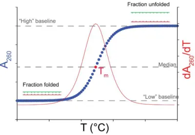
VI. Characterization in solution by thermal denaturation ... 71
VII. Mass Spectrometry experiments ... 72
VII.1. Sample preparation for mass spectrometry analysis ... 72
VII.2. Mass spectrometers used ... 72
VII.3. MS data analysis ... 78
VIII. Ion Mobility Spectrometry: how to obtain CCS values? ... 84
VIII.1. Determination of the CCS of the ATD peak center ... 84
VIII.2. CCS distribution reconstruction ... 85
IX. How to analyze MS/MS data ... 87
References ... 88
3. RESULTS AND DISCUSSION ... 89
Chapter 1. Characterization of the interaction between Mg2+ and kissing complexes by Native mass spectrometry ... 90
X. Native MS from Magnesium solutions: optimization of the method ... 90
X.1. Introduction ... 90
X.2. Results ... 91
X.3. Discussion ... 98
XI. Determination of specific cations number ... 100
XI.1. Introduction ... 100
XI.2. Results ... 100
XI.3. Discussion ... 111
XII. The extent of kissing complex stabilization by Mg2+ is correlated to the cation affinity, due to a displacement of equilibria ... 113
XII.1. Introduction ... 113
3
XII.3. Conclusion and perspectives ... 127
XIII. Mg2+ localization ... 128
XIII.1. Introduction ... 128
XIII.2. Results ... 128
XIII.3. Discussion ... 140
Chapter 2. Mass spectrometry to screen kissing complex affinities ... 145
XIV. Introduction ... 145
XV. Kissing complexes used ... 145
XVI. Results ... 146
XVI.1. Mass spectrometry experiments ... 146
XVI.2. UV melting experiments... 148
XVI.3. Surface Plasmon Resonance Experiments ... 149
XVII. Discussion and conclusion ... 152
References ... 153
4. RESULTS AND DISCUSSION ... 157
Chapter 3. DNA and RNA duplexes structures in the gas phase ... 158
XVIII. Introduction ... 158
XIX. Results and discussion ... 159
XIX.1. ESI-MS spectra ... 159
XIX.2. IM-MS results ... 163
XX. Conclusion and perspectives ... 176
XX.1. Perspectives: the effect of the charge state on DTCCS He ... 176
Chapter 4. Kissing complexes structure in the gas phase ... 179
XXI. Introduction ... 179
XXII. Results and discussion ... 179
4
XXII.2. Comparison RNA kissing complexes with RNA duplexes ... 189
XXIII. Conclusion and perspectives ... 190
References ... 191
4. GENERAL DISCUSSION AND CONCLUSIONS ... 195
XXIV. Discussion about mass spectrometry results (PART I) ... 196
XXIV.1. Mass spectrometry compatible with Magnesium salts ... 196
XXIV.2. Analysis of Mg2+ binding to RNA by native mass spectrometry ... 196
XXIV.3. Mass spectrometry as a screening tool ... 199
XXV. Discussion about the ion mobility-mass spectrometry results (PART II) ... 200
XXV.1. Initial compaction in the gas phase ... 200
XXV.2. Collision-induced compaction ... 201
5. APPENDICES ... 203
5
ABSTRACT
Besides being the molecular intermediate between DNA and proteins, RNA can have many other functions such as gene regulation (riboswitches), gene expression (mRNA and tRNA) or catalysis (ribozymes). RNA function is linked to its structure and its folding dynamics. Cations such as magnesium bind to RNA and are in some instances essential for proper folding and for stability. The need of structural and thermodynamic details about Mg2+ interactions is then of upmost importance in the study of the
structure-function relationships. The first part of our work consists in characterizing the binding equilibria between magnesium and RNA model motifs, called kissing complexes, using native mass spectrometry (MS). MS makes it possible to distinguish individual binding stoichiometries, and the present work consisted in developing a method to quantify each species, taking into account the contribution of nonspecific adducts. We also explored how tandem mass spectrometry (MS/MS) could further help localizing magnesium ions. Further, we explored the structures of RNA complexes in the gas phase using ion mobility mass spectrometry (IM-MS), with the aim to detect shape changes upon cation or ligand binding. But in contrast with anticipations, we found that DNA and RNA duplexes as well as RNA kissing complexes undergo a significant compaction at charge states naturally produced by native ESI-MS, which may hide the effect of cations. Our work showcases how mass spectrometry can bring novel information on RNA-cation binding stoichiometries and affinities, but also discusses some limitations of a gas-phase method to probe solution structures.
7
RESUME - APERÇU EN FRANÇAIS
Introduction
En plus d’être l’intermédiaire entre l’ADN et les protéines, l’ARN est impliqué dans plusieurs processus biologiques : régulation et expression des gènes (riboswitches, ARNm et ARNt) ou encore catalyse (ribozymes). La fonction de chaque ARN est liée à sa structure et à sa dynamique de repliement. L’ARN peut adopter différents motifs structuraux secondaires tels que le simple brin, les duplexes ou encore les hairpins, mais aussi des motifs tertiaires comme les pseudoknots ou encore les kissing complexes. Des études ont montré l’importance des cations (tels que K+, Na+, Mg2+) dans la stabilité et le bon repliement
des structures d’ARN. Les ARN sont entourés par ce que l’on appelle « l’atmosphère ionique ». La présence de ces ions va permettre de réduire la répulsion électrostatique entre les charges négatives présentes sur l’ARN, et va donc être essentielle pour l’obtention de structures fonctionnelles. Un des principaux cations divalents impliqué dans la stabilisation des complexes d’ARN est le magnésium divalent.
Un des défis actuels est de comprendre comment le magnésium interagit et stabilise les structures d’ARN. Actuellement plusieurs techniques biophysiques telles que la cristallographie aux rayon X, la RMN (Résonance Magnétique Nucléaire), la SPR (Surface Plasmon Resonance) ou la spectroscopie UV sont utilisées pour obtenir des caractéristiques structurales et/ou thermodynamiques. Cependant, ces techniques ne permettent pas toutes d’avoir des données directes sur les cations spécifiques (notamment constantes d’équilibre spécifiques), ou présentent des désavantages non négligeables.
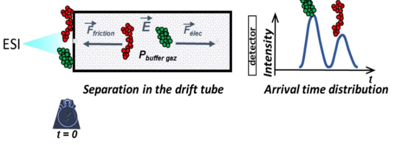
La spectrométrie de masse (SM) native permet d’obtenir des données structurales mais aussi thermodynamiques, ce qui en fait une technique de choix. La spectrométrie de masse est dite native quand elle préserve, de par des conditions d'ionisation douces, les structures non-covalentes de la solution à la phase gazeuse. Son utilisation permet d'identifier les espèces présentes en solution et donne accès aux stœchiométries des complexes étudiés. La spectrométrie de mobilité ionique (SMI), lorsqu’elle est couplée à la SM, apporte par ailleurs une nouvelle dimension qui est la séparation des molécules en fonction de leur conformation. La mobilité ionique nous permet d’obtenir une mesure de la surface exposée aux collisions, la Section Efficace de Collision (CCS). Cette mesure est reliée à la conformation des espèces présentes en phase gazeuse et nous permet d’établir des corrélations avec les structures grâce à l’utilisation complémentaire de la modélisation moléculaire.
Ce travail est donc divisé en deux parties. La première concerne le développement d’une méthode de caractérisation des équilibres de liaison entre le magnésium et des motifs d’ARN modèles. La seconde
8 partie est centrée sur l’analyse de complexes d’ARN par spectrométrie de mobilité ionique avec pour objectif de détecter des changements conformationnels dus à la liaison de ligands ou cations. L’intérêt ici est d’évaluer le potentiel de la SMI, technique en phase gazeuse, pour l’étude de structure en solution. Les structures d’ARN pouvant être très complexes et de tailles diverses, l’étude s’est portée sur l’analyse du motif appelé « kissing complexe ». Les kissing complexes sont formés par deux hairpins d'ARN, liées entre elles par liaisons Watson-Crick grâce à la complémentarité des bases de leurs boucles. Deux modèles bien caractérisés dans la littérature ont été utilisés : le système TAR-R06 et le système RNAIi-RNAIIi.
Résultats
Partie 1 : Nouveaux développements en spectrométrie de masse, nouveaux aspects sur la structures de kissing complexes
Dans un premier temps, nous avons développé une méthode afin de pouvoir obtenir des spectres reflétant directement les espèces présentes en solution et uniquement celles-ci. La force ionique est donc assurée par de l'acétate d'ammonium, classiquement utilisé en SM native. Nous avons pu doper ces solutions jusqu'à 1 mM en acétate de magnésium, Mg(OAc)2. Nos résultats ont aussi montré qu’une
optimisation des réglages instrumentaux par rapport à ceux habituellement utilisée par notre équipe était nécessaire. En effet, les conditions étaient trop douces et de nombreux adduits de NH4+ étaient retenus.
Pour notre étude, une réduction des adduits non-spécifiques d’ammonium est nécessaire, d’où l’utilisation de conditions plus dures. Il est à noter que les conditions optimisées activent les ions tout en conservant les liaisons non covalentes des complexes étudiés.
Dans un second temps, grâce au développement d’une méthode permettant de soustraire la contribution des adduits de magnésium non-spécifiques, les différentes espèces présentes ont pu être quantifiées. Les constantes d’équilibre individuelles de liaison de chaque magnésium spécifique ainsi que des pistes sur la localisation de ceux-ci ont pu être mis en évidence.
La première étape de notre méthode consiste en un traitement mathématique, utilisant les intensités mesurées sur une séquence de référence, qui permet de déterminer la part d’adduits spécifiques (liés au motif « kissing loop » avec une forte affinité) et de non-spécifiques (sites de liaison de faible affinité). Pour être une bonne référence, la séquence contrôle doit être de même type que la séquence d’intérêt (i.e. ADN ou ARN), de même taille et de même composition, mais ne pas posséder le motif auquel nous voulons attribuer les sites spécifiques. Par rapport à des duplex d’ARN choisis comme contrôles, nous avons
9 déterminé qu’il y a deux magnésiums spécifiques au kissing complexe TAR-R06, et seulement un pour le complexe RNAIi-RNAIIi.
La deuxième étape est la détermination des constantes apparentes d’équilibre de dissociation entre les deux hairpins de chaque complexe en fonction de la concentration en magnésium (KdKC,app). Afin de
pouvoir corréler l’intensité des pics détectés aux concentrations en solution, une correction utilisant les facteurs de réponse est utilisée. La détermination des constantes apparentes d’équilibre a permis de confirmer l’effet stabilisateur de Mg2+ puisque son ajout augmente l’affinité entre les deux hairpins du
kissing complexe, et ce quel que soit le kissing complexe étudié.
En combinant les données obtenues sur la stœchiométrie en adduits spécifiques et celles obtenues lors de la détermination de KdKC,app, il est possible de calculer les constantes d’équilibre de dissociation de
chaque Mg2+ spécifique au kissing complexe. Les constantes déterminées sont liées à la stœchiométrie en
cation et non à leur site de liaison. Nous avons montré que même si plusieurs cations sont spécifiques à un motif kissing complexe, comme c’est le cas pour TAR-R06, ces cations ne présentent pas nécessairement la même affinité. Cette étude est innovante puisque c’est la première fois que ce niveau de détail est atteint dans l’analyse des équilibres liés à l’interaction Mg2+-ARN.
Enfin, nous avons utilisé la spectrométrie de masse en tandem (SM/SM) afin d’obtenir des informations sur la localisation du magnésium le plus affin. Nos résultats montrent qu’il est possible de dissocier le kissing complexe en ces deux monomères. Lorsque du magnésium est ajouté, un Mg2+ est retrouvé sur
chacune des deux hairpins. Ces résultats ont permis d’émettre l’hypothèse d’une coopérativité négative entre deux sites de liaison du magnésium. Pour RNAIi-RNAIIi la coopérativité négative serait telle que les deux sites de liaison ne peuvent être peuplés simultanément. Pour TAR-R06, la liaison du premier Mg2+ à
l’un des deux sites de liaison, réduirait l’affinité du second Mg2+ pour le second site.
Dans un projet connexe, la spectrométrie de masse native est utilisée comme outil de criblage rapide pour classer des kissing complexes, différents par seulement une paire de base, en fonction de leur stabilité relative. L’avantage de la spectrométrie de masse est l’identification rapide des différentes espèces présentes en solution notamment la détection de complexes « inconnus » (i.e. formation d’homodimère par exemple). L’intensité relative des pics a été directement reliée à l’abondance en solution grâce à l’utilisation d’un standard interne et de simplifications.
10 Partie 2 : l’étude de complexes d’ARN et ADN par spectrométrie de masse couplée à la mobilité ionique révèle une compaction en phase gazeuse
En spectrométrie de masse les molécules sont désolvatées, ionisées et analysées en phase gazeuse. Il est souvent admis pour les protéines que celles-ci conservent leur forme globulaire de la solution à la phase gazeuse. Les acides nucléiques, quant à eux, ne sont pas globulaires. Plusieurs questions peuvent alors se poser : qu’advient-il des structures des acides nucléiques en phase gazeuse ? Est-ce qu’une technique en phase gazeuse peut sonder les structures en solution ? La spectrométrie de masse seule ne peut répondre à ces questions. En revanche, la SM couplée à la spectrométrie de mobilité ionique apporte une nouvelle dimension qui est la séparation des molécules en fonction de leur forme. Grâce à cette technique, la Section Efficace de Collision (CCS) des molécules peut être obtenue. La CCS est une grandeur physique liée aux frictions entre les ions et le gaz tampon présent dans la chambre de mobilité. La comparaison entre les valeurs de CCS expérimentales et les CCS théoriques obtenues par modélisation permet d’avoir une idée sur la structure des ions en phase gazeuse et d’établir un modèle. L’un des autres buts de cette partie est aussi de détecter des changements de conformation dus à la liaison de ligands et/ou cations sur des complexes d’intérêt. Pour répondre aux questions posées précédemment, nous avons analysé en premier lieu des duplexes d’ADN ou d’ARN, puis des kissing complexes.
Les résultats montrent qu’un ensemble de conformations est adopté par les duplexes d’ADN, d’ARN, et par les kissing complexes. Les structures étudiées étant flexibles, plusieurs conformations proches peuvent être adoptées, formant ainsi des distributions de CCS larges. De plus, les comparaisons entre duplexes d’ADN, d’ARN et kissing complexes de même taille et composition, ont révélé que ces structures couvrent la même gamme de CCS. Ces résultats montrent qu’une hélice B, une hélice A et kissing complexes ont des compacités très proches en phase gazeuse alors que les structures sont différentes en solution.
L’un des principaux résultats de notre étude est qu’il y a une compaction de la structure des acides nucléiques étudiés, i.e. duplexes et kissing complexes, du moins aux états de charge typiquement obtenus par spectrométrie de masse native, c’est-à-dire à force ionique physiologique. Notre hypothèse est qu’aux bas états de charge produits par SM native, les biomolécules ont tendance à devenir globulaires à cause de la formation de nouvelles liaisons hydrogène entre les phosphates. Cette compaction, d’environ 20% en termes de CCS, est de nature à masquer l’éventuel effet de cations ou ligands sur la conformation de l’acide nucléique en solution. Cette étude révèle donc une limitation quant à l’utilisation de la spectrométrie de mobilité ionique pour sonder des structures initialement présentes en solution.
11 Néanmoins, nous avons aussi vu que l’augmentation de l’état de charge grâce à l’utilisation d’un agent super-chargeant pourrait prévenir l’effet de compaction observé. Cette piste est donc à explorer. Plusieurs études ont été réalisées sur l’effet de l’activation des ions sur leur structure en phase gazeuse. Pour les protéines, l’effet dépend du type d’activation et de l’énergie donnée aux ions. Afin d’en savoir davantage sur le comportement des acides nucléiques en phase gazeuse, nous avons réalisé ce même type d’expériences, i.e. activer plus les ions en jouant sur divers paramètres instrumentaux. Les résultats montrent qu’une plus grande compaction encore est observée lors de l’activation des longues séquences analysées. D’après ces résultats, la compaction s’effectue jusqu’à un seuil où la structure ne peux pas se compacter plus. La taille de la séquence, son état de charge ainsi que la distribution microscopique des charges (influencée par les cations restant liés) vont influencer cette étape.
Conclusions et perspectives
En conclusion, la spectrométrie de masse native ainsi que la spectrométrie de mobilité ionique sont deux outils très polyvalents. Elles peuvent être utilisées pour cribler différents motifs car l’identification des différentes espèces présentes en solution, leur stœchiométrie ainsi qu’une analyse quantitative peuvent être effectuées. Notre méthode apporte de nouvelles connaissances sur l’interaction Mg2+-cations. Elle
peut être adaptée à différents motifs ou encore différents cations ou ligands. Nous avons aussi montrer que la spectrométrie de mobilité ionique ne peut être utilisée dans l’analyse de structures d’ARN qu’en tenant compte de la forte compaction qui se produit en phase gazeuse, aux états de charge obtenus par SM native.
Nos résultats ont aussi montré l’intérêt d’analyser des acides nucléiques en présence de sels. Une autre étape intéressante serait de trouver des additifs afin d’influencer le processus électrospray et de jouer sur la proportion en adduits.
Notre étude est portée uniquement sur de petits motifs tertiaires d’ARN. De nouvelles connaissances ont donc été amenées sur le comportement des ARN en phase gazeuse et permettent de faire un pas en avant vers l’étude de structures beaucoup plus complexes telles que les riboswitches.
13
ABBREVIATIONS
A Adenine/Adenosine
ATD Arrival Time Distribution
bp Base pair
C Cytosine/Cytidine
CCS Collision Cross Section
CCSD Collision Cross Section Distribution
CID Collision induced dissociation
DNA Deoxyribonucleic acid
ESI Electrospray ionization
G Guanine/Guanosine
HP Hairpin
IM-MS Ion mobility-mass spectrometry
IMS Ion mobility spectrometry
KC Kissing complex
L Ligands
m/z Mass-to-charge ratio
mRNA Messenger RNA
MS Mass spectrometry
MS/MS Tandem mass spectrometry
NH4OAc Ammonium acetate
NMR Nuclear magnetic resonance
PDB ID Protein data bank identification number
Q Quadrupole
RNA Ribonucleic acid
rRNA Ribosomal RNA
SPR Surface Plasmon resonance
T Thymine/Thymidine
TAR Trans Activating responsive RNA element
14
TOF Time-of-flight
tRNA Transfer RNA
15
1. INTRODUCTION
I.
Nucleic Acids
Nucleic acids, and more particularly Deoxyribonucleic Acid (DNA), can be considered as the unit of life. Indeed, DNA contains all the coding genetic information of each component of the cell and is transmitted through generations.1 Cells are using this genetic code to synthesize proteins needed for their good
functioning. As DNA is present in the nucleus of eukaryotic cells and proteins are present in the cytoplasm, an intermediary is needed to go from the first one to the other. This is where Ribonucleic Acid (RNA) plays an important role. DNA will be transcribed into messenger RNA (mRNA), which is itself translated into proteins thanks to other RNAs. The central dogma of molecular biology is based on this simple principle.2
As for proteins, nucleic acids are biopolymers. They are coded by a specific alphabet and, depending on this alphabet arrangement and the biological environment, nucleic acids can adopt different structures. General information on DNA and RNA structures are discussed in the following paragraphs. RNA is discussed in more detail afterwards.
I.1. Bases and primary structure
Nucleic acids are composed of a chain of nucleotides.1 Nucleotides are formed by three different parts:
the phosphate group, the sugar and the base (Figure 1, A). The first difference between DNA and RNA comes from the nature of the sugar that is a deoxyribose for DNA and a ribose for RNA. The five natural bases are: Adenine (A), Guanine (G), Cytosine (C), Thymine (T) and Uracil (U). The second difference between DNA and RNA is that thymines can only be found in DNA and uracil only in RNA. The purine bases (A and G) composed of two aromatic cycles and the pyrimidine bases (C, T and U) containing a single aromatic cycle (Figure 1, B).
The primary structure is made by the succession of nucleotides. The phosphate group is making an ester bound between the 3’-OH of the first nucleotide with the 5’-OH of the second one (Figure 1, C). The alphabet composing the genetic code is then composed of just 4 letters. One can write the sequence of DNA or RNA as the succession of A, C, G and T/U. The successive phosphate-sugar-phosphate moiety is called the backbone of the nucleic acid.
16
Figure 1: Bases and nucleic acids primary structure. A. Scheme of a nucleotide. B. Most abundant bases in DNA and RNA. C. Example of the primary structure of RNA (for DNA, the sugar is replaced by a Deoxyribose).
A.
B.
17
I.2. Secondary and tertiary structures
The secondary structure of DNA and RNA implies the formation of hydrogen bonds between two bases. The Watson-Crick base pairing is the most well-known hydrogen bonding. Adenine is binding to Thymine and Uracil via two hydrogen bonds, and Cytosine is binding to Guanine via three hydrogen bonds (Figure 2). Due to the number of H-bonds formed, a G-C base pair is more stable than an A-T or A-U base pair. In some cases, non-Watson-Crick pairing, or non-canonical base pair, can occur. The tertiary structure of nucleic acids derives from their secondary structure.
Figure 2: Watson-Crick base pairing between A-U, A-T and G-C (from left to right).
I.2.1. Most abundant three-dimensional structures based on Watson-Crick base pairs
B- and A-form duplex
The most famous structure is the double helix adopted by double stranded DNA and RNA. It was first described by J. Watson and F. Crick in 1953.3 The B-form duplex, which is mostly adopted by DNA, is a
right-handed double helix which consists of the binding of two antiparallel complementary strands held together by H-bonds between base pairs (Figure 3, left). The B-helix is defined by a turn composed of 10 residues and one can defined a major and a minor groove.
The other well-known structure is the A-Helix, which is mainly adopted by RNA and can be adopted by DNA depending on the hydration conditions4 (Figure 3, right). The A-helix is also a right-handed helix. The
main difference between the A and B form comes from the placement of the base pair along the axis of the helix. In A-helix, the bases are more compressed and deviate from the vertical axis. This results in a difference of the major and minor grooves between A and B-form. In the A-form, the major groove is deeper which leads to a shallower minor groove.5 Also in the A-form there are 11 residues per turn.
18
Figure 3: Example of a B-Helix on the left (PDB ID 1BNA6) and an A-Helix on the right (PDB ID 353D7).
I.2.2. Examples of structures based on non-Watson-Crick base pairs
Triplexes
A triplex DNA can be formed when a third strand is binding to the major groove of a duplex DNA via Hoogsteen bonding. This triplex can be intramolecular or an intermolecular triplex when the third strand comes from another DNA molecule. Triplexes can be found naturally but can also have potential therapeutic effects, for example as a molecular target for a specific duplex (Figure 4, A).8
G-quadruplexes
G-quadruplexes are formed by two or more quartets of Guanines (Figure 4, B). A quartet is formed by four guanines that are binding together via Hoogsteen H-bonds. G-quadruplexes are found in G-rich sequences and evidence has been brought that they are present in gene promoters or even in telomers.9
I-motifs
I-motifs can form in C-rich sequences and consist of the binding of two parallel stranded duplexes via C+
-C base pairs (Figure 4, -C). The -Cytosines involved are exchanging a proton. I-motifs are stabilized in acidic conditions but may be formed in in vivo conditions.10
19
Figure 4: Examples of tertiary structures. A) Triplex DNA and the example of the Hydrogen bonds for a T-A-T triplet (PDB:1D3X11). B) Example of a G-quadruplex and the hydrogen network between Guanines (PDB ID 1KF112). C)
Example of an i-motif and the C+-C pair formed (PDB ID 1YBL13).
The structures and folding state of DNA and RNA are important to assure their functions in the cells. The next part is focusing on RNA and its structure-function relationships.
20
II.
An RNA story
In the central dogma of molecular biology exposed by F. Crick in 1958, the RNA is only considered as an intermediate between DNA and protein.2 However, in 1970, F. Crick himself refines his own words about
the transfer of the information between DNA, RNA and proteins.14 He classified the transfer of information
in three classes: general transfers, special transfers and unknown transfers (Figure 5). General transfers are the one previously described and admitted by the Central dogma. The special transfers may somehow happen depending on the cell type, and the unknown transfers may occur, but no proof was existing.
Figure 5: Refinement of the Central Dogma of Molecular Biology (adapted from Crick F.14)
From this fact arises the question of the origins of life, “who came first?” between DNA, RNA and proteins. This problem has become a chicken-and-egg problem. Several review papers are explaining the term of “RNA world” and why RNA should be considered first.15–18 The term “RNA world” was used for the first
time by Gilbert W. in 1986.15 RNA was shown to be not only the intermediate between DNA and proteins,
but has many diverse roles. RNA has been discovered to present catalytic abilities in the form of ribozymes and is involved in genetic regulation through riboswitches. All these capabilities of RNA suggest that an RNA world has existed before proteins and now all the different forms of RNAs are remnants of this ancient time.
II.1. The biological roles of RNA
Here we present a list of the main biological roles of RNA and that constitute the vestiges of this so-called “RNA world”.
21 Main actors in protein synthesis: mRNA, tRNA and rRNA
In 1962, when J. Watson and F. Crick obtained the Nobel prize of medicine for the discovery of the DNA structure, three types of RNA had been discovered and identified as key actors in protein synthesis: messenger RNA, transfer RNA and ribosomal RNA.1
Proteins are oligomers formed by the succession of amino acids. Proteins are then based on their own alphabet which is the amino acids succession. DNA contains all the genetic information coded under the succession of nucleotides. The questions asked were “How to go from DNA to proteins? How to go from nucleotides to amino acids?”. Figure 6 is showing a schematic view of the transcription-translation process. As DNA cannot go outside the nucleus of eukaryotic cells, DNA is transcribed into messenger RNA (mRNA) thanks to several proteins including the RNA polymerase II. mRNA is then translated in the cytoplasm to proteins thanks to ribosomes and transfer RNAs. The mRNA is “read” 3 nucleotides by 3 nucleotides following a specific code. Each triplet, called codon, corresponds to a specific amino acid. The synthesis of the protein starts at a starting codon, usually the AUG start codon, corresponding to Methionine in Eukaryotes and N-methylmethionine in Prokaryotes. Transfer RNAs (tRNAs) recognize the correct codon on the mRNA and ribosomes is the machinery allowing the interaction between mRNA and tRNA and the formation of the polypeptide chain. Each tRNA is unique because it is binding to only one specific amino acid and corresponds to one specific codon. The attachment of the proper amino acid to its corresponding tRNA is performed by the aminoacyl-tRNA synthetase. When the tRNA binds to its amino acid it is directed to the A-site inside the ribosome. tRNA and mRNA are interacting together inside the ribosome. Peptide bond and elongation of the polypeptide chain happen also in ribosomes. Ribosomes are composed of two subunits. In prokaryotes the large subunit is made of 34 proteins and two rRNAs (23S and 5S rRNA) and the small subunit comprises 21 proteins and only one rRNA (16S).19 The small
subunit mediates the interaction between mRNA and tRNA. The large subunit contains all the machinery to form peptide bonds. tRNAs pass through 3 different sites inside the ribosome: the A-site to interact with the corresponding codon, the P-site containing the elongated protein and the E site where tRNAs are released.
22
Figure 6: Scheme of the transcription and translation step to illustrate the three fundamental roles of RNA in protein synthesis (adapted from Lodish H. et al1).
Crystal structures of tRNAs and part of ribosomes were obtained and have revealed that both RNAs are structured in a specific manner to ensure their function.19,20 Structural details will be given in part II.2 and
II.3.
RNA as catalyst: discovery of ribozymes
For a long time people thought that catalysis was only performed by enzymes. However, in the early 80’s, Sidney Altman and Thomas Cech had discovered independently that RNA can be involved in catalysis reactions. For their respective works they received the Nobel Prize in chemistry in 1989. They discovered respectively that Ribonuclease P (RNAse P)21 and the Tetrahymena Group I intron22 possess enzyme
properties. RNAse P processes tRNA precursor (pre-tRNA) by catalyzing the hydrolysis of pre-tRNA. RNAse P is present in prokaryotes and eukaryotes.23 The Tetrahymena Group I intron on rRNA precursor was
shown to cleave and ligate itself without the intervention of proteins, and has thus autocatalytic properties. Since then, Group I intron were found in various organisms, from bacteria to eukaryotic cells. They catalyze their own excision from RNA through successive transesterification reactions (Figure 7, A).
23 Group I intron are considered as self-splicing RNAs as they do not need enzymes to be removed from RNA during mRNA splicing.23
The name Ribozymes for “Ribonucleic Enzymes” was then given to RNA molecules that present catalytic properties. Since the discovery of the two first ribozymes (RNAse P and Group I intron), several other types of ribozymes emerged: the Hammerhead ribozyme which became the most studied and characterized ribozyme, the Hepatitis Delta Virus (HDV) ribozyme, hairpin ribozyme, Varkud Satellite (VD) ribozyme, the glmS ribozyme which is also a riboswitch and the rRNA in ribosomes which was found to catalyze the formation of the peptide bound.23–26 Ribozymes catalyze three main types of reactions:
transesterification, hydrolysis and peptidyl transfer. They are involved in many biological processes which may be important in the life of cells (RNA translation, RNA splicing, etc). As these RNA molecules are chemical catalysts and can act without the need of proteins, they may be a kind of proof of what is left from the RNA world where all the reactions were done without proteins.
Figure 7: A) Schematization of Group I intron cleavage by two successive transesterifications autocatalyzed by the ribozyme (adapted from Ramesh A. et al23).B) Three main reactions catalyzed by ribozymes: transesterification like
in Group I intron, hydrolysis like with RNAse P and peptidyl transfer for rRNA in ribosomes.
It has been shown that ribozymes adopt specific structures and that they are composed of key motifs like helical parts or pseudoknots. In some cases, metal ions like magnesium are also binding and help for the
24 catalytic properties. These different structural features will be discussed in part II.2 and II.3 of this introduction.
RNA as gene regulator: discovery of riboswitches
In 2002, researchers proved that vitamin derivatives like thiamine pyrophosphate (TPP, derivative of vitamin B1), Flavin mononucleotide (FMN, derivative of vitamin B2) and the coenzyme B12 (also called
AdoCbl, derivative of vitamin B12) interact with the mRNA controlling the expression of the corresponding
vitamin.27,28 They showed that each vitamin derivative was binding to a specific domain of the mRNA and
that, above a certain threshold, it conduces to the regulation of the relative gene. The name riboswitch was given by Ronald R. Breaker to the part of mRNA that regulate gene expression through the binding of a specific metabolite. In the last fifteen years, riboswitches have become a trendy subject. More than 20 classes of riboswitches were discovered in many different organisms. Many researches and reviews have been published since their discovery in 2002.29–35
Riboswitches are found at the 5’ untranslated region (UTR) of mRNA in eubacteria. However, a riboswitch binding to Thiamine Pyrophosphate (TPP) was found in plants, fungi and some other eukaryotic cells.36–38
Riboswitches are composed of two domains: the aptamer part where the specific ligand is binding and the expression platform, directly downstream the aptamer, which transduces the ligand binding event into a gene control response (Figure 8). The binding of the ligand to the aptamer part induces a conformational change in all the riboswitch and more particularly in the expression platform, which leads either to the repression or to the expression of the gene. Riboswitches regulate genes through various mechanisms. The most common ones are the transcription termination, the translation initiation (Figure 8) and splicing control. Depending on the configuration of the riboswitch, the regulation can be an activation of the gene or a repression.30,34,35,39
25
Figure 8: Mechanisms of gene regulation, examples with the transcription and translation (adapted from Serganov et al35). In red are the repression mechanisms and in green the activation mechanism. Transcription termination: no
binding of the polymerase due to the termination forming sequence. Transcription anti-termination: polymerase can bind and transcribe the gene. Translation inhibition: the binding of the ligand induce a conformational change that hide the ribosome binding site (RBS). Transcription activation: the RBS is accessible to the ribosome so translation can happen.
Riboswitches sense a large variety of ligands: coenzyme and vitamin derivatives as seen previously (TPP, FMN, coenzyme B12)27,28,40, purines and derivatives41,42, amino acids43,44 or even cations like
magnesium45,46.
As for ribozymes, the structure of riboswitches is well organized and is of great importance for their function as a conformational change is involved. We will discuss about these structural features in the following parts, II.2 and II.3.
II.2. RNA structures: from RNA motifs to more complex structures
As seen previously, RNA ensures many functions and is involved in various biological processes. To do so it must adopt specific secondary and tertiary structures. Several reviews are detailing the different types of building blocks from secondary to tertiary structure motifs.47–50 In this section, we will present the main
secondary motifs and tertiary motifs that lead to more complex three-dimensional structures.
II.2.1. Secondary structure motifs
The secondary structure of an RNA is defined by the interactions between nucleotides, which can bind through canonical or non-canonical base pairing, and by the succession of nucleotides. RNA adopts several
26 secondary structure motifs that are considered as building blocks for ribozymes or riboswitches structures.47,48 We present here the main building blocks found in RNA. Another important part in RNA
structure, which will not be described here, is base stacking.47
Single stranded RNA
In 3D RNA structures, some parts remain as a single strand (Figure 9, A). Duplex
Duplex parts correspond to double stranded RNA (Figure 9, B). Usually they form a right-handed A-helix. The A-helix has 11 residues per turn and the base pairs are tilted from the helical axis of about 18° so the bases are displaced of 4 Å from the axis. Those constrains make the major groove deeper and narrower and on the contrary the minor groove will be shallower and wider compare to a B-helix.
Bulges and internal loops
A bulge occurs when unpaired nucleotides are present on one of the two strands forming a double helix (Figure 9, C). The smallest bulge is composed of only one nucleotide but their size can go to several ones. The formation of bulges can bend the general A-form of the double helix, however the type of nucleotides, the number of nucleotides in the bulge, the nucleotide surrounding the bulge and the presence of cations can influence the percentage of bending of the helix. Bulges can be a preferred biding site for proteins, ligands or cations.51 One can quote the example of the HIV-1 TAR RNA-Tat protein system where the TAR
RNA sequence is forming a bulge and a hairpin and it was demonstrated that the bulge is involved in Tat recognition.52
Contrary to bulges, internal loops are formed when unpaired nucleotides are present on both strands of the double helix (Figure 9, D). When one or two nucleotides are involved it is called a mismatch, if more than three nucleotides are involved it is an internal loop. The nucleotides of an internal loop or mismatch form non Watson-Crick base pairs. One of the most widespread mismatches is the G•U wobble base pair. It often constitutes a recognition site for proteins, ligands or cations.53,54 Mismatches create a small
distortion of the A-helix but do not bend the helix.47 As internal loop geometry depends on the number
27 Hairpins
Hairpins are formed by a duplex part, the stem, and end by a loop of unpaired nucleotides (Figure 9, E). The size of the loop varies from two to several nucleotides. Hairpins are flexible depending on their composition, the size of the stem and loop. The hairpin is a motif of choice in RNA interactions and is important for RNA folding. For example, it is one of the main motifs involved in tRNA structure. Indeed, three hairpins are present on the structure and give this familiar structure of tRNA.20,55–57 The most
widespread loop is the tetraloop and more particularly the GNRA tetraloop (where N can be all nucleotides and R a purine). It is a motif naturally present in a variety of RNAs like rRNAs, group I introns or ribozymes like the Hammerhead.49,58,59 The formation of this tetraloop involves a network of hydrogen bonds
between the different bases, which is important for RNA structure stability. Junctions between helical parts
As said in their name, the junctions between helical domains are the regions where at least three different helical parts are connected (Figure 9, F). These regions are important for RNA folding as they determine the number of branches and so the global structure. For example, the three-way junction in the TPP-riboswitch60,61 or in the Hammerhead ribozyme59 gives the Y-shape of both structures. tRNAs are defined
by a four-way junction.62 In junctions, regions of unpaired nucleotides stabilize the overall structure. They
can constitute the site of base triples or binding pockets. For example, in the Hammerhead ribozyme, the cleavage site is located on the unpaired bases in the junction.47,59
28
29
II.2.2. Tertiary structure motifs
The secondary structure motifs described above associate and interact to form tertiary structure motifs, important for the global three-dimensional structure of RNA.
Base Triple interactions
A base triple happens when an unpaired nucleotide is making hydrogen bonds with a Watson-Crick base pair present in a duplex. It is a common motif found in most of complex structures of RNA because the formation of new hydrogen bonds helps to maintain and stabilize the RNA conformation. The known triples include (UA)U, (CG)C, (CG)G, A(GC), (UA)A, (CG)A and (AU)G.48 Base triples were shown to be
present in various RNA structures like tRNAPhe 55–57 , group I intron ribozymes63 or even riboswitches like
the PreQ1-riboswitch64. Figure 10 presents two examples of structures involving base triples: Group I
intron and the aptamer part of the PreQ1-riboswitch in T. Tengcongensis.
Figure 10: Example of base triple interactions involved in RNA structure. A. Group I intron RNA structure showing 6 base triples (adapted from Chastain et al63). B. Aptamer part of the PreQ1-riboswitch from T. Tengcongensis (adapted
30
Figure 11: Examples of RNA three dimensional structures based on the pseudoknot motif. A) Schematization of the pseudoknot fold. (B-C) X-ray structure of the Hepatitis Delta Virus ribozyme in complex with the spliceosomal protein U1A (PDB: 1DRZ).65 (D-E) X-ray structure of the SAM-II riboswitch (PDB: 2QWY).66 (F-G) NMR structure of the
31 Pseudoknots
A pseudoknot is formed when unpaired nucleotides in a loop make hydrogen bonds with nucleotides of a single stranded region. One can consider this motif as a fusion of two hairpins. It is one of the most widespread tertiary motifs present in RNA structures. When the region between the two stems is at least one unpaired nucleotide, it bends the overall structure of the pseudoknot. If there are no bases between the two stems, the stems are coaxially stacked and form a continuous helix. This bending on the pseudoknots can have an influence on the RNA function, for example it as an effect on the efficiency of frameshifting during retroviral RNA translation.68 The pseudoknot motif is easily found in
riboswitches66,69,70 or ribozymes65. Figure 11 presents some examples of RNA structures based on the
pseudoknot element. Kissing complexes
Kissing complexes are formed when the nucleotides in the loops of two RNA hairpins are complementary. The smallest loop-loop interaction is formed by only two nucleotides in each loop71, and can go to several
nucleotides. If the loop is too long, the probability to form other structures, like bulges or internal loops, is higher. Studies on the flanking bases at the interface between the loop and the stem show that this base pair is important for the kissing complex’s stability. For example, in 2000, Ducongé et al. have shown that a G-A closing base pair makes the kissing complex more stable than any other base pair.72 Study of
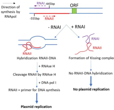
loop-loop interactions were extensively done on the biological RNAI-RNAII kissing complex involved in the replication of the ColE1 plasmid in Escherichia Coli and on the TAR RNA sequence from the mRNA of the HIV-1 virus. These two kissing complexes will be described in more detail in the scope of the thesis. Except these two kissing complexes, this motif is present in other biologically relevant systems: the dimerization initiation site (DIS) of the HIV-1 mRNA which induces the dimerization of the viral genome by forming a kissing complex73,74, a kissing complex formed in the Neurospora Varkud Satellite ribozyme involved in the
catalytic domain75,76, a kissing loop that improves the binding of the ligand and help for proper folding in
the btuB riboswitch in E.Coli77 or a loop-loop interaction formed in the add A-riboswitch after ligand
binding41. Based on the study of several kissing complexes, the structure of this type of motif is bent at
the junction between the two loops, and the formation of phosphate clusters in the major groove of the loop-loop interaction can possibly be metal binding sites.78,79 Figure 12 presents some examples of RNA
32
Figure 12: Examples of kissing loop structures. A) Schematization of the kissing complex fold. (B) X-ray structure of the HIV-1 DIS Lai kissing complex (PDB: 2B8R). (C) X-ray structure of the Neurospora Varkud Satellite kissing complex region (PDB: 2MI0). (D) X-ray structure of the add A-riboswitch from Vibrio Vulnificus (PDB: 1Y26)
II.3. RNA structure and Cations
Most of the tertiary motifs and three-dimensional final structures of RNA bind to cations.50,80 Several
reviews show the importance of cations, and more particularly magnesium, in the folding and stability of tertiary structures.80–85 In the following part, we will discuss about the importance of ions surrounding
33
II.3.1. The “ion atmosphere”
DNA and RNA are negatively charged polyelectrolytes: each phosphate is carrying a negative charge. In the cell, DNA and RNA are surrounded by ions that form the “ion atmosphere”. This “ion atmosphere” involves many monovalent and divalent cations depending on the ionic composition. This ionic environment reduces the electrostatic repulsion between all the negative charges present on nucleic acids. For example, without surrounding ions, the electrostatic repulsion in the folding of the 400 nucleotide-long Tetrahymena intron would correspond to ≈ 600 kcal/mol.86–88 The ion atmosphere is thus
of upmost importance in RNA folding and stabilization. Indeed, monovalent and divalent cations will help to maintain RNA in their functional structures by overcoming the electrostatic repulsion.83,84
The effect of the “ion atmosphere” on RNA folding structure will depend on its composition in monovalent and divalent cations. The main monovalent cations are K+ and Na+. It has been shown that without Mg2+
and with a high concentration of monovalent cations, some RNA structures can still fold. Also the size of the monovalent ion has an impact on RNA folding. For example the A-riboswitch is more stable with Li+
than Cs+ (ionic radii:Li+ < Cs+).89,90 Some tertiary structures form mostly in the presence of monovalent
cations; like G-quadruplexes DNA or RNA in which K+, Na+ or NH
4+ bind in-between each G-quartets to fold
G-quadruplexes.91
Several theoretical models are used to describe the interactions between the ion atmosphere and nucleic acids. They take into account electrostatic interactions between charged element. However, we will not describe the different models in this manuscript. A review written by Lipfert et al88 is explaining very well
the basics of the most popular models like the Counterions Condensation (based on ion condensation around nucleic acids until a certain charge critical value is reached) or the Poisson-Boltzmann theory (based on an average description of electrostatic interactions). Modelling is challenging because one needs to take into account a lot of charges and interactions between them.
The ion atmosphere is mostly invisible by X-ray crystallography because this technique does not consider all the layers of ions that are surrounding RNA or DNA in a non-periodical way. X-ray crystallography instead shows direct contacts between nucleic acids and ions. One of the main experimental techniques allowing to “count” the ion pertaining to the “ion atmosphere” is the buffer equilibration-atomic emission spectroscopy (BE-AES). Briefly, this technique “counts” how many ions are present in a solution by comparing the sample containing the nucleic acid and a control composed of only buffer. It is then possible to count the number of ions which are in excess in the “ion atmosphere”. The quantity of nucleic acid is determined by the quantification of the phosphorus atoms.87,88
34
II.3.2. Role of Magnesium ions on RNA folding and stability
It is now widely admitted that RNA folding and stability depend on the cation environment, and that among all ions the main cation involved is Mg2+.80,82–85 But one of the main challenges is to understand
how magnesium is stabilizing RNA structures.
In the cell, the total concentration of Magnesium is about 17 to 20 mM. However, magnesium is involved in a lot of biological processes, involving proteins or nucleic acids.92 Consequently, the free concentration
of magnesium is only around 0.25 to 1 mM, because most of the magnesium is used somewhere in the cell.93 Mg2+ has a small ionic radius (0.72 Ȧ94) and in pure water it is coordinated to 6 water molecules. The
dehydration of Mg2+ is energetically more costly than that of Na+ for example.85
Nowadays a lot of X-ray structures of complex RNA have been elucidated and present new insights into magnesium binding. One of the first structures obtained with Mg2+ binding, in the early 80’s, is that of the
tRNAPhe. From these structures, the number and location of magnesium ions were determined. The
number of Mg2+ binding sites depends on the type of crystal obtained: for the orthorhombic form 4 Mg2+
are binding95,96 to tRNAPhe whereas for the monoclinic form, 5 Mg2+ were thought to bind but after
refinement two more Mg2+ were found20. In both cases, Mg2+ are located in the D-stem and in the
anticodon-stem. All Mg2+ contribute to the stabilization of the global tRNA shape82,96. In 2006,
crystallization of the aptamer part of the TPP-riboswitch has revealed that two hexa-coordinated Mg2+
were interacting with both the RNA and TPP. They helped TPP to bind to the aptamer by stabilizing its interaction, and also stabilize the overall Y-shape of the activated aptamer.60,61 More recently, in 2013, a
study of SAM-I riboswitch and Mg2+ showed that the active structure of the aptamer is obtained only when
SAM interacts with magnesium.97 From all these studies, magnesium presents different roles from
conformation stabilization, interaction with specific ligands (i.e. riboswitches) or is involved in the cleavage reaction of ribozymes98. Figure 13 present the examples of the TPP and SAM-I riboswitches.
35
Figure 13: Examples of X-ray structures determined with Mg2+ ions. A) TPP riboswitch on top with a view of the location of specific
Mg2+ (PDB: 2GDI).61 B) SAM-I riboswitch extracted from Hennely et al97.
The acquisition of X-ray structures and the analysis of the interaction between Mg2+ and RNA have led to
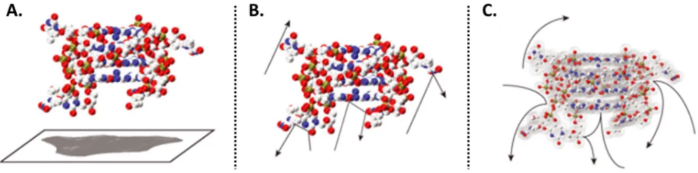
the classification of Mg2+ ions into two main groups: “diffuse ions” and “site-binding” ions.82,83,85 Schematic
representation of these two binding modes are shown on Figure 14. Diffuse ions bind to RNA through long-range electrostatic interactions. The first hydration shell of each molecules, i.e. ions and the RNA, are fully conserved and separated (Figure 14, A). Diffuse ions are moving around RNAs. On the contrary to diffuse ions, site-bound Mg2+ are closer to the surface of the RNA and are divided in two subgroups.
The first group, defined by the term of “outer-sphere” complex, is represented by ions and RNA that share their first hydration shell. This type of ions can be trapped into electrostatic pockets from which they cannot move. The second group, composed by “inner-sphere” complexes, is defined by RNA-ions direct contact. In this case, Mg2+ is directly coordinated to the RNA without H
2O intermediate binding (Figure 14,
B). Usually Mg2+ cations are binding to phosphates or oxygens. In some cases they are binding to
nitrogen.85,99 Experimentally it will be difficult to distinguish the different types of binding but several
techniques can be used to obtain various information on stoichiometries and locations. Combining all the data can possibly give information on the type of binding. Biophysical techniques are described in the section II.4. Several theoretical models based on electrostatics and thermodynamics have been proposed to understand what is behind these different binding modes82,83,88, but they will not be discussed in this
36
Figure 14: Representation of the different Mg2+ bindings to RNA. 1) Diffuse ions, 2) Site binding ions with the “outer
sphere” complex and the “inner sphere” complex. Adapted from Misra et al82.
II.4. Why and How to study RNA-cations structures and equilibria?
II.4.1. WHY?
We have seen that the three-dimensional structure of RNA is important to assure its function. Also magnesium was shown to be involved in RNA folding and stabilization, like in riboswitches or ribozymes.82,100 Understanding how ions are helping RNA to fold is of great importance, for example to
understand the folding pathways involved in riboswitches or ribozymes. As an example, Figure 15 shows the hypothesis of the folding pathway of the A-riboswitch which may depends on cations and ligand binding.
37
Figure 15: Hypothesis of folding pathways of an A-riboswitch aptamer. Several conformational states may exist and may depends on the binding of cations (blue dots) and/or ligands (green). The grey-shaded structures have been discussed in the literature.
Riboswitches and ribozymes are more and more studied nowadays and become interesting as drug targets or therapeutic tools.101 As riboswitches are involved in gene regulation of mostly bacteria, they become a
target of choice for new antimicrobial molecules.102 For example, the TPP-riboswitch recognizes not only
TPP as ligand but also Pyrithiamine pyrophosphate (PTPP) which is toxic for fungi and bacteria. PTPP blocks the binding of TPP and then induces gene repression.102 Engineered hammerhead ribozymes have been
created to cleave some specific parts of mRNA that are responsible of the expression of proteins important in the development of cancer cells.101
To engineer such structures, it is thus important to have as much information as possible on the folding and stabilization pathway. The fields of characterization of global structure of RNA complexes and RNA-Mg2+ interactions are still challenging as it is still difficult to obtain structures of big RNA complexes, to
understand clearly how magnesium is interacting with three-dimensional structures, where it is located and to quantify the effect of magnesium.
II.4.2. HOW?
To characterize RNA structures and interactions between RNA and Mg2+ several biophysical techniques
38 or even Mg2+ location. However, all the existing techniques present advantages and disadvantages. In this
section, we will describe some main techniques used to analyze RNA-Mg2+ interaction and structures,
their advantages and their limitations. X-ray Crystallography
X-ray crystallography is based on the analysis of the diffraction of X-rays by a crystal. By the acquisition of diffraction spectra, one can go back to electronic density map and then have access to the position of atoms and so to the structure and position of bound ions. Many structures of riboswitches61,66,103,
tRNA20,55,56 and ribozymes59,65 were obtained by X-ray diffraction. The pros of this technique are its high
resolution and the acquisition of atomistic details. Also, X-ray crystallography allows the identification of inner-sphere bound ions if resolution is good enough (≤ 3 Å). So we can have access to magnesium stoichiometry.99 However, in a recent study published in 2016, Auffinger and collaborators showed that
mistakes are often made on Mg2+ assignment and one should be careful when using and analyzing the
data.99 One of the main drawback of X-ray diffraction is the need for crystals. Finding the good conditions
can be time consuming, and not all conditions can be studied. In addition, by using X-ray diffraction one will have a rather static view of the structure, and no information about equilibria between RNA and cations is accessible.
Nuclear Magnetic Resonance spectroscopy (NMR)
NMR is based on the resonance frequency of atoms (H, C, N mostly) in the presence of a magnetic field. As a function of their chemical environment, the resonance frequency of the atoms will shift. Distances between atoms are determined using 2D and 3D experiments. When the signal between two atoms is strong, it means that they are close to one another. The possible structure is reconstructed based on the defined distances. NMR is a solution technique compared to X-ray crystallography, so physiological conditions are easier to mimic. Like X-ray diffraction, NMR is a high-resolution technique that allows the determination of RNA structures. For example, structures of kissing complexes78,79 and pseudoknots68
were obtained by NMR spectroscopy. The limitations of this technique are first that Mg2+ is invisible to
NMR, only assumptions on its position can be made. It is possible to do 25Mg-NMR104 but this isotope has
a low sensitivity so the experiments might be very long. Secondly, experiments can be long, costly, sample-consuming and the structure determination can be fastidious.