Basic science for the clinician
When are pro-inflammatory cytokines SAFE
in heart failure?
Sandrine Lecour
1
*
and Richard W. James
2
1
Hatter Cardiovascular Research Institute, Department of Medicine, Faculty of Health Sciences, University of Cape Town, Cape Town, South Africa and2
Clinical Diabetes Unit, Department of Internal Medicine, Faculty of Medicine, University of Geneva, Geneva, Switzerland
Received 29 September 2010; revised 2 December 2010; accepted 9 December 2010; online publish-ahead-of-print 7 February 2011
The cytokine hypothesis presently suggests that an excessive production of pro-inflammatory cytokines, such as tumour necrosis
factor alpha (TNF) and interleukin 6 (IL6), contributes to the pathogenesis of heart failure. The concept, successfully proved in
genetically modified animal models, failed to translate to humans. Recently, accumulation of apparently paradoxical experimental
data demonstrates that, under certain conditions, production of pro-inflammatory cytokines can initiate the activation of a pro-survival
cardioprotective signalling pathway. This novel path that involves the activation of a transcription factor, signal transducer and
activator of transcription 3 (STAT3), has been termed the survival activating factor enhancement (SAFE) pathway. In this review, we
will discuss whether targeting the SAFE pathway may be considered as a preventive and/or therapeutic measure for the treatment
of heart failure.
-Keywords
Heart failure † Pro-inflammatory cytokines † Tumour necrosis factor alpha † Signal transducer and activator of
transcription † Prosurvival pathways
Introduction
In the 1990s, the introduction of the cytokine hypothesis proposed
that an excessive production of pro-inflammatory cytokines
con-tributes to heart failure (HF).
1,2Despite encouraging animal
studies and small clinical trials, larger clinical trials targeting the
cytokines in HF have failed. The question as to whether the
increased levels of inflammatory cytokines observed with HF are
a cause or consequence of the disease still remains unresolved.
In the new millennium, data from numerous animal studies have
lead to a better understanding of the beneficial vs. deleterious
effect
of
pro-inflammatory
cytokines
in
pathophysiological
conditions. Recent experimental work currently suggests that
activation of these cytokines, including tumour necrosis factor
alpha (TNF) and the interleukin 6 family, can promote a
pro-survival signalling pathway termed the SAFE (survivor
activat-ing factor enhancement) pathway to protect against myocardial
infarction (MI).
3,4In this review, we will summarize evidence for
and against the cytokine hypothesis in both experimental models
and clinical conditions and we will discuss whether targeting the
SAFE pathway may be considered as a preventive and/or
thera-peutic measure for the treatment of HF.
Pro- and anti-inflammatory
cytokines imbalance in heart
failure
Pro-inflammatory cytokines in heart
failure
The interest of TNF (also known as cachectin) in HF evolved from the
observation that cachexia is a common phenomenon associated with
severe HF.
5In the 1990s, numerous studies evidenced an increase in
circulating TNF, function to the severity and the outcome of HF.
6,7The production of TNF in HF may originate from the periphery
(liver) or the myocardium (see review
8). In HF patients, cachexia
was associated with a further increase in circulating TNF receptors
type I and type II (TNFR1 and TNR2), through which TNF is believed
to exert its function.
6Tumour necrosis factor receptor type I is
*Corresponding author. Tel:+27 21 406 6278, Fax: +27 21 447 8789, Email:[email protected]
considered as a major predictor of mortality and new-onset HF in
patients with acute MI (C-ALPHA study).
9The main components of the interleukin 6 family cytokines are
interleukin 6 (IL6), leucaemia inhibitory factor (LIF), and
cardiotro-phin 1. In animal models and in man, these three cytokines are
increased in HF. Similar to TNF, the circulating levels of IL6 increase
with the severity of HF and it may be considered as a prognostic
marker in patients with congestive HF.
10,11The production of IL6
in the disease may originate from either the periphery or the
myo-cardium depending on the disease stage.
11,12In addition to TNF and
IL6, myocardial gene expression of both cardiotrophin 1 and LIF
also increase in the failing heart.
13,14Most of the effects associated
with these cytokines occur after binding to the receptor gp130
which is also increased in HF, to a greater extent in patients with
dilated cardiomyopathy than valvular or ischaemic cardiomyopathy.
This suggests that the role of gp130 in the pathogenesis of chronic
HF may vary as a function of the aetiology of HF.
10,14These studies demonstrate that pro-inflammatory cytokines can
be considered as a biomarker in HF. Unfortunately, levels of
cyto-kines measured from one study to another may vary considerably,
which is related essentially to the commercial kit/technique used
(Table
1
).
Anti-inflammatory cytokines in heart
failure
Overall, the production of anti-inflammatory cytokines has been
much less studied than the pro-inflammatory cytokines in the
human heart. The main anti-inflammatory cytokine studied in HF is
interleukin 10 (IL10). Produced in mononuclear cells (essentially
macrophages and T cells), IL10 inhibits the production of
pro-inflammatory cytokines as well as the matrix metalloproteinases
by activated monocytes.
15Compared with healthy control subjects,
plasma levels of IL10 are decreased in patients with HF, in particular,
in patients suffering from advanced HF NYHA class III and IV.
16Circulating thrombospondin-1, another anti-inflammatory
cyto-kine released upon activation of platelets, is also reduced in HF,
together with tumour growth factor b1.
17,18The cytokine hypothesis: evidence
for and against
Evidence for a contribution of cytokines
in the development and pathogenesis
of the disease
The adverse effect of pro-inflammatory cytokines in the heart has
been widely demonstrated in animal models, using mainly cytokine
depleted or overexpressing mouse models. In experimental
models, inflammatory cytokines promote left ventricular
remodel-ling, acute reversible contractile dysfunction, and uncouple
myo-cardial b-adrenergic receptors (see review
19). Cardiac specific
overexpression of TNF causes cardiac failure
20while
pathophysio-logically relevant concentrations of TNF promote progressive left
ventricular dysfunction and remodelling in rats.
21Interleukin 10
knockout animals exhibit increased mortality and larger infarct
size compared with their littermate controls.
22All these data
suggest that limiting the cytokine imbalance observed in
pathophy-siological conditions, such as HF may limit the development of the
disease and improve its outcome. Such modulation of the cytokine
balance in HF can be approached in two ways: (i) by enhancing the
level of anti-inflammatory cytokines and (ii) by targeting a decrease
in pro-inflammatory cytokines. These two approaches have been
considered, to varying degrees, in both experimental models and
clinical conditions.
Increase of anti-inflammatory cytokines
The concept of raising the anti-inflammatory cytokines in HF has
been poorly studied. In animal studies, treatment with recombinant
IL10 limits myocardial lesions in viral myocarditis in rats
23and
improves left ventricular function in rats with HF after
experimen-tal MI.
24This effect was associated with a decrease in circulating
inflammatory cytokines TNF and IL6 and a reduction in myocardial
macrophage infiltration.
24Similarly, chronic treatment with
ator-vastatin increases plasma levels of IL10, decreases the TNF/IL10
ratio, ameliorates left ventricular remodelling, and improves left
ventricular function in rats with HF subsequent to MI.
25In fact,
the simplest and safest way to increase IL10 is with physical
exer-cise. In a rat model of chronic HF, regular swimming improves
. . . .
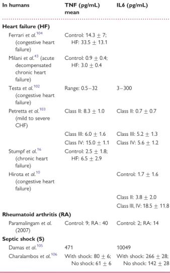
Table 1
Tumour necrosis factor and interleukin 6
levels in various pathophysiological conditions
In humans TNF (pg/mL) mean IL6 (pg/mL) Heart failure (HF) Ferrari et al.104 (congestive heart failure) Control: 14.3 + 7; HF: 33.5 + 13.1 Milani et al.43(acute
decompensated chronic heart failure) Control: 0.9 + 0.4; HF: 3.0 + 0.4 Testa et al.102 (congestive heart failure) Range: 0.5 – 32 3 – 300 Petretta et al.103 (mild to severe CHF)
Class II: 8.3 + 1.0 Class II: 0.7 + 0.7
Class III: 6.0 + 1.6 Class III: 5.2 + 1.3 Class IV: 15.0 + 1.1 Class IV: 5.6 + 1.2 Stumpf et al.16 (chronic heart failure) Control: 2.5 + 1.8; HF: 6.5 + 2.9 Hirota et al.10 (congestive heart failure) Control: 1.7 + 1.6 Class II: 3.8 + 2.0 Class III, IV: 18.5 + 11.8 Rheumatoid arthritis (RA)
Paramalingam et al. (2007)
Control: 9; RA : 40 Control: 2; RA: 14 Septic shock (S)
Damas et al.105 471 10049
Charalambos et al.106 With shock: 80 + 6;
No shock: 61 + 6
With shock: 266 + 28; No shock: 142 + 28
plasma levels of IL10 and cardiac function and reduces muscle
cel-lular damage.
26In human studies, specifically targeting an increase of IL10 in HF
has been essentially ignored. Intense treatment with atorvastatin in
patients with stable coronary disease reduces subsequent
hospital-ization for HF.
27Immunomodulation therapy with intravenous
immunoglobulin in patients with chronic HF improved the left
ventricular ejection fraction and increased plasma IL10 levels.
28A recent Cochrane systematic review and meta-analysis shows
that exercise training, known to increase IL10 levels, reduces
HF-related hospitalization and results in clinical improvement in
health-related quality-of-life.
29Similarly, the HF-ACTION study
demonstrates that participation in an exercise training programme
provides improvement in HF patients-reported health status
com-pared with usual care.
30Targeting the anti-inflammatory cytokines
to limit the development of the pathology in HF certainly merits
further investigation.
Decrease of pro-inflammatory cytokines
The concept of decreasing the pro-inflammatory cytokines has
been more readily explored in both clinical and experimental
set-tings but this has led to conflicting and confusing data.
In a viral myocarditis mouse model, anti-TNF improved survival
and myocardial lesions.
31Tumour necrosis factor neutralization
attenuated the impairment of left ventricular function 10 weeks
after MI in rats.
32Similarly, contractile dysfunction was attenuated
in TNF R1 knockout mice with MI
33or with TNF antibodies after
microembolization.
34Conversely,
adenoviral
transfection
of
TNFR1 increased contractile dysfunction following MI.
35In a small randomized preclinical trial with 18 patients of NYHA
class III, a single intravenous infusion of the TNF antagonist
Etaner-cept resulted in a decrease in TNF bioactivity and a significant
overall increase in quality-of-life scores.
36Similarly, pentoxifilline,
a putative TNF inhibitor, was beneficial in small clinical trials with
HF patients. Pentoxyfilline therapy of ischaemic cardiomypathy for
6 months improved the ejection fraction.
37The benefit with regard
to symptoms of HF and cardiac function was seen in all grades of
severity of HF (class I – IV) and in patients with ischaemic and
idio-pathic dilated cardiomyopathy.
37,38However, the beneficial effect
has not consistently been associated with a reduction in
pro-inflammatory cytokines, therefore suggesting that
pentoxyfil-line, which is a phosphodiesterase inhibitor, may protect
indepen-dently of an immunomodulatory effect (see review
39).
Diet enriched with omega-3 polyunsaturated fatty acids (v-3
PUFA) may decrease pro-inflammatory cytokines in patients with
HF but further studies are needed to determine the optimal
source and dosing of v-3 PUFA.
40Evidence against a contribution
of cytokines in the disease
In
contrast
to
the
preclinical
trials,
large
randomized
placebo-controlled clinical trials with anti-TNF therapies were
dis-appointing. No beneficial effect was observed with Etanercept in
patients with chronic HF (NYHA class II – IV) as reported in
RENEWAL, RENAISSANCE, and RECOVER (see review
41). The
ACCLAIM study tested the effect of immunomodulation therapy
in patients with chronic HF. The rationale of the study was to
have a non-specific but broad immunomodulatory effect by
redu-cing pro-inflammatory cytokines and increasing anti-inflammatory
cytokines.
42The trial did not show any significant reduction in
mortality or cardiovascular-related hospitalization. Unfortunately,
the levels of cytokine activation were not assessed in the study.
Of note, two specific sub-group of patients, those without a
pre-vious history of MI and those with NYHA II had significant
reduction in their primary endpoint therefore suggesting that this
therapy may benefit certain subgroups with HF.
The beneficial effect of several drugs in HF also brings into
ques-tion the contribuques-tion of pro-inflammatory cytokines in the disease.
In patients hospitalized with advanced acutely decompensated
con-gestive HF, traditional therapy leads to clinical improvement but no
reduction in pro-inflammatory cytokine levels.
43Similarly,
treat-ment with amiodarone in patients with ischaemic cardiomyopathy
is associated with an increase in circulating TNF levels but this
increase is not associated with an adverse effect on survival.
44Although the severity of HF correlates with an increase in
plasma inflammatory cytokines levels in patients, it is important
to mention that these levels are much lower compared with
inflammatory diseases, such as septic conditions (Table
1
). Does
this mean that their contribution to the disease, if any, may be
rela-tively modest? Or are there other explanations?
Pro-inflammatory cytokines
initiate a cardioprotective
signalling cascade in the heart:
the survival activating factor
enhancement pathway
With the new millennium, a large body of experimental work
con-ducted in various animal models has brought more insights with
respect to the disappointment of multiple clinical trials targeting
the inflammatory cytokines in HF. In fact, activation of the
immune system (with TNF and/or IL6) can promote the activation
of an intrinsic cell survival signalling pathway that involves
acti-vation of a transcription factor, the signal transducer and activator
of transcription 3 (STAT3). This path, recently discovered in MI,
has been termed the SAFE (survivor activating factor
enhance-ment) pathway.
4,45Tumour necrosis factor and interleukin 6
cytokines can protect against
ischaemia-reperfusion injury
As observed in HF, an increase in pro-inflammatory cytokines
occurs in patients with MI and circulating IL6, TNF, and their
respective receptors are increased further after reperfusion (see
review
46). Both cytokines are thought to contribute to contractile
dysfunction,
47–50most likely as a result of perturbation in calcium
homoeostasis and formation of free radicals.
51,52However, as early as 1998, there was experimental data
disput-ing the contribution of TNF or IL6 to the damage associated with
MI. Hence, exogenous addition of TNF protects against hypoxic
injury in cardiomyocytes
53and mice lacking both TNFR1 and
TNFR2 are more susceptible to MI than their littermate controls.
54Similarly, expression of IL6 occurs in the viable border zone of a
myocardial infarct.
55In fact, the cardioprotective effect of TNF is influenced by
several factors including dose, sex, time, and type of receptors
acti-vated. Hence, TNF protects against ischaemia-reperfusion in a
dose-dependent manner with small amounts of exogenous TNF
(0.5 ng/mL, in vitro) given prior to ischaemia-reperfusion enhancing
cell survival while higher concentrations (10 – 20 ng/mL, in vitro) are
cytotoxic.
3,56,57Deficiency of TNFR1 protects the myocardium
through IL6 following TNF infusion,
58and the activation of the
TNFR2 seems to convey the protective effect of TNF.
59–61Both cytokines are also important components in the powerful
protective effect of preconditioning, which fails in mice in the
absence of either TNF or IL6.
56,62–65Tumour necrosis factor
can mimic pre- or post-conditioning in vitro and in vivo.
47,56,66–68Signal transducer and activator of
transcription 3 is a downstream target of
tumour necrosis factor and interleukin 6
Once TNF and IL6 bind to their specific receptors (TNFR
and gp130), a common signalling path, called the Janus kinase
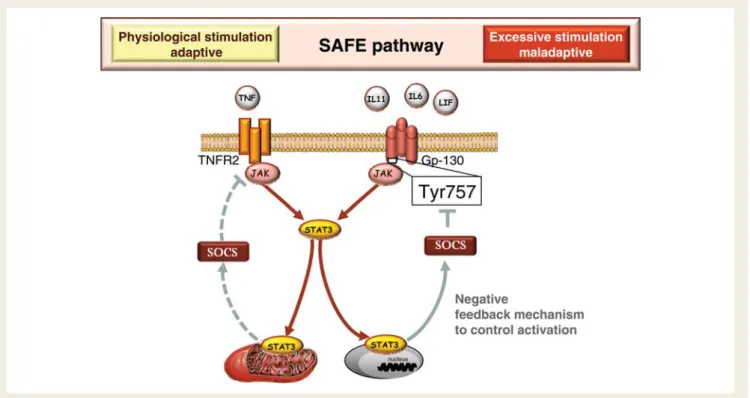
(JAK)/STAT3 pathway can be activated (Figure
1
). Janus kinases
are a family of tyrosine kinases that are associated with the
cyto-plasmic domain of cytokine and growth factor receptors (including
TNFR and gp130) and play a major role in transducing signals from
the cytosol to the nucleus (see reviews
69–71). Upon activation of
the receptors, JAK proteins phosphorylate and create a docking
site for STAT proteins, which in turn are activated by
phosphoryl-ation (Figure
1
). While the mechanisms involved in JAK activation
by the gp130 and TNFR1 have been clearly characterized, the
acti-vation of JAK 2 by TNFR2 still remains to be elucidated. Tumour
necrosis factor receptor II does not contain an intrinsic protein
tyrosine kinase but the phosphorylation of JAK2 by this receptor
may occur via TNF receptor-associated factor 2 (TRAF2) or
nuclear factor-kappaB activation, both components previously
implicated in TNF-mediated cardioprotection.
72,73The STAT
family of transcription factor proteins consists of seven identified
members: only STAT3 will be considered in this review. Tyrosine
phosphorylation of STAT3 enables it to homodimerize and
trans-locate to the nucleus. Both tyrosine and serine phosphorylated
STAT3 are also present within the mitochondria.
74,75Under
phys-iological conditions, this pathway can be regulated by the
suppres-sor of cytokine signalling (SOCS) proteins that serve as a negative
regulator of JAK/STAT signalling. Activation of STAT3 induces the
expression of SOCS which in turn binds to the tyrosine 757
residue of membrane receptor which is necessary for the
docking of JAK and STAT3 at the receptors to activate the JAK/
STAT3 pathway (see reviews
71,76).
Activation of the SAFE pathway with TNF/JAK/STAT3 or IL6/
JAK/STAT3 signalling is required for the cardioprotective effect
of ischaemic pre- and post-conditioning as neither TNF knockout,
IL6 knockout or cardiomyocyte STAT3 knockout mice could be
protected with a conditioning stimulus.
62,63,77–79Activation of
Figure 1
Survivor activating factor enhancement (SAFE) pathway activated in the heart. After binding onto their specific receptors, tumour
necrosis factor and interleukin 6 family cytokines activate the janus kinase/signal transducer and activator of transcription 3 (JAK/STAT3)
signal-ling path. After phosphorylation, signal transducer and activator of transcription 3 translocates either in the nucleus or in the mitochondria. The
activation of the survivor activating factor enhancement pathway is regulated by the activation of the suppressor of cytokine signalling (SOCS).
Moderate stimulation of the survivor activating factor enhancement pathway leads to cell survival but intense stimulation is detrimental.
the JAK/STAT3 pathway also occurs with many other
cardiopro-tective agents, such as erythropoietin, cannabinoid agonists,
insulin, prostaglandins, and high density lipoproteins (HDL).
4,80,81Downstream effectors of the SAFE pathway following cytokine
and STAT3 activation still need to be defined. Several targets of
STAT3 have been identified including proteins that are involved
in cell survival and proliferation, such as Bcl-2, Bcl-xL, Mcl-1, Fas,
cyclin D1, E1, p21, some growth factors (VEGF), and other
tran-scription factors (see review
4). In the preconditioning setting,
STAT3 plays a critical role by increasing the anti-apoptotic gene
Bcl-2 and reducing the pro-apoptotic gene bax.
82Also, STAT3
mediates cardioprotection via the phosphorylation and inactivation
of the pro-apoptotic factor Bad.
64In late preconditioning, STAT3
activates superoxide dismutase,
83inducible nitric oxide synthase,
62and cyclooxygenase 2
62expression. Similarly, the JAK/STAT
pathway protects in opioid-induced cardioprotection via
phos-phorylation and inactivation of glycogen synthase kinase 3b
(GSK3 b).
84Mitochondrial connexion 43, a key element of the
signal transduction cascade of the protection by ischaemic
precon-ditioning,
85may also be a downstream target of STAT3.
86By definition, STAT3 is a transcription factor but its effects
observed in a setting of ischaemia/reperfusion or classic
precondi-tioning do not seem to occur at a transcription level, as the
time-frame between the activation of STAT3 and its action is too short
to allow an effect at the gene level. This observation suggests that
STAT3 has additional effects, such as direct phosphorylation of
target molecules. With the recent discovery that STAT3
translo-cates to the mitochondria, the protective effect of STAT3 in
pre-and post-conditioning is likely to be mediated by modulating the
opening of the mitochondrial permeability transition pore.
75Tumour necrosis factor can also promote cardioprotection via
TRAF2, nuclear factor kappaB, reactive oxygen species, and
protein kinase C.
4,72,73With regards to sphingolipids, TNF-induced
cardioprotection is mediated via ceramide but sphingosine-1
phos-phate protects via TNF and STAT3.
3,87Whether these
intermedi-ates form part of the SAFE pathway or constitute an alternative
protective signalling path activated by TNF merits further
investigation.
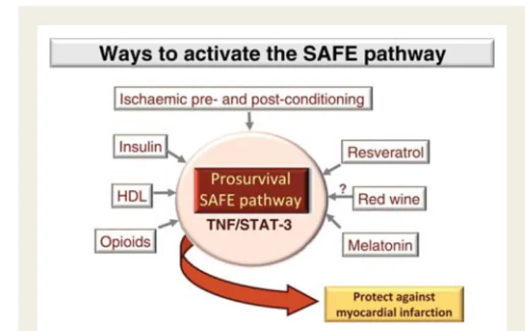
How to activate the survival activating
factor enhancement pathway?
The SAFE pathway being recently discovered, few upstream targets
have been delineated so far. Initially discovered as a downstream
target of ischaemic preconditioning and ischaemic
postcondition-ing
64,67,88(see Figure
2
), it is now recognized that various
cardio-protective agents can activate both TNF and STAT3, such as
bradykinin, adrenoreceptors, leptin, opioids, and cannabinoids.
89Similarly, HDL (whose low levels are associated with poor
progno-sis in patients with HF
90) or one of its components sphingosine-1
phosphate, confers protection via the activation of TNF and/or
STAT3.
80,87Moderate consumption of red wine may also confer
cardioprotection via the activation of the SAFE pathway. Hence,
resveratrol and two biogenic amines present in red wine
(ethanola-mine and melatonin) protect the ischaemic isolated mouse heart
against MI but this effect was lost in TNF knockout mice or
STAT3 knockout mice.
91,92Surprisingly, adenosine, a well-known
preconditioning mimetic does not seem to activate the SAFE
pathway, therefore confirming the existence of alternative
cardio-protective signalling paths, such as the well-described reperfusion
injury salvage kinase pathway or protein kinase G-dependent
pathway.
63,77,93Activation of the survival
activating factor enhancement
pathway in heart failure: a safe
therapy to consider?
Evidence for a survival activating factor
enhancement therapy in heart failure
As demonstrated in the previous section, experimental data
provide evidence that activation of the SAFE pathway is protective
in MI, but what about HF?
In humans, the concept that activation of the SAFE pathway may
protect against HF is supported by the alteration in left ventricular
STAT3 and gp130 proteins and phosphorylation of JAK observed
in patients with dilated cardiomyopathy.
14,94The expression of
SOCS is also attenuated in terminally failing human hearts.
94Simi-larly, women with postpartum cardiomyopathies have a decrease
in STAT3 levels compared with healthy women.
95In
experimental
studies, although
cardiomyocyte
STAT3
deficient mice do not develop any cardiac abnormalities of function
or morphology at a young age,
96abnormalities appear with aging
and severe cardiac fibrosis is observed at 6 months (see
review
70). Female cardiomyocyte STAT3 deficient mice develop
postpartum cardiomyopathy suggesting that activation of STAT3
is essential to protect the maternal heart from postpartum
oxidative stress.
95Mice with cardiomyocyte overexpression of
STAT3 activate a hypertrophic signal but also a protective signal
against
doxorubicin-induced
cardiomyopathy
by
inhibiting
reduction in cardiac contractile gene expression and inducing
cardiac protective factors.
97Similarly, mice with cardiac restriction
of gp130 display rapid onset of dilated cardiomyopathy and massive
Figure 2
Multiple ways of activating the SAFE pathway to
protect against myocardial infarction. SAFE, survivor activating
factor enhancement; HDL, high density lipoprotein cholesterol.
induction of myocyte apoptosis during aortic pressure overload vs.
the control mice that exhibit compensatory hypertrophy.
98Also,
activation of STAT3 by granulocyte colony stimulating factor in
dilated cardiomyopathy improves survival and cardiac function.
99Furthermore, activation of gp130/STAT3 with IL11 attenuates
cardiac fibrosis following MI.
100Evidence against a survival activating
factor enhancement therapy
in heart failure
These promising data in favour of a protective effect of the SAFE
activation in HF should however be considered with precaution
for several reasons. First, as mentioned earlier, TNF can activate
two types of receptors, TNFR1 and TNFR2. Activation of the
SAFE pathway involves the activation of TNFR2 only.
67In
exper-imental ischaemic HF, TNFR1 and TNFR2 have disparate and
opposing effects on remodelling, hypertrophy, inflammation and
apoptosis with TNFR1 exacerbating, and TNFR2 ameliorating
these effects.
59,61Potential drugs targeting the activation of the
SAFE pathway should therefore be specific to TNFR2. Second,
continuous gp130-mediated JAK/STAT3 activation obtained by
blockage of the tyrosine 757 on the gp130 site (and therefore
inef-ficacy of SOCS to regulate the pathway) promotes cardiac
inflam-mation, adverse remodelling and HF in mice with MI.
101These data
suggest that an overstimulation of the SAFE pathway would lead to
an adverse outcome. Third, the putative beneficial role of
cyto-kines/STAT3 in ischaemic HF may not translate in non-ischaemic
HF. The kinetics of production of cytokines varies as a function
of the aetiology of HF with, for instance, gp130 increased to a
greater extent in patients with dilated cardiomyopathy than
ischae-mic cardiomyopathy.
14Thus defining beneficial/deleterious roles of
the SAFE pathway in HF will require intensive investigation taking
into account the aetiology of the disease.
Conclusion
In HF, there is an undoubted imbalance between pro-inflammatory
and anti-inflammatory cytokines. This imbalance correlates with
the severity of the disease and the aetiology of HF. Animal
studies conducted essentially on ischaemic HF models with
over-stimulation or repression of the pro-inflammatory cytokines are
in favour of a causal role of these cytokines in the pathogenesis
of HF. In contrast, while human studies suggest that measuring
cytokines can be used as a biomarker for HF, they presently do
not support a contribution of pro-inflammatory cytokines in the
development of the disease. Over the last decade, new
experimen-tal data and human studies have demonstrated the Janus nature of
cytokines, being either cytoprotective or detrimental. It underlines
the dynamic aspect of cytokine action, where questions such as
concentration, the time when produced and the type of receptors
that they activate need to be addressed. The protective effect of
the cytokines has been clearly demonstrated when there is a
mod-erate activation of the SAFE pathway. Conversely, an uncontrolled
activation of the SAFE pathway may be detrimental. Targeting a
controlled activation of the SAFE pathway in HF is undoubtedly
a valid therapeutic objective but the precise kinetic and bioactivity
characteristics of pro-inflammatory cytokines in either chronic or
acute HF should be carefully delineated before this can be
considered.
Funding
S.L. is supported by a South African Medical Research Council (MRC)
Career Award, grants from the National Research Foundation, MRC
Cape Heart Group and the Swiss South African Joint Research
Pro-gramme (JRP-16) under the aegis of the Council for Scientific and
Industrial Research. R.W.J is supported by grants from the Swiss
National Research Foundation (No 31-118418) and the Swiss South
African Joint Research Programme (JRP-16) under the aegis of Swiss
State Secretariat for Education and Research.
Conflict of interest: none declared
References
1. Seta Y, Shan K, Bozkurt B, Oral H, Mann DL. Basic mechanisms in heart failure: the cytokine hypothesis. J Card Fail 1996;2:243 – 249.
2. Oral H, Kapadia S, Nakano M, Torre-Amione G, Lee J, Lee-Jackson D, Young JB, Mann DL. Tumor necrosis factor-alpha and the failing human heart. Clin Cardiol 1995;18:IV20 – IV27.
3. Lecour S, Smith RM, Woodward B, Opie LH, Rochette L, Sack MN. Identifi-cation of a novel role for sphingolipid signaling in TNF alpha and ischemic pre-conditioning mediated cardioprotection. J Mol Cell Cardiol 2002;34:509 – 518. 4. Lecour S. Activation of the protective survivor activating factor enhancement
(SAFE) pathway against reperfusion injury: does it go beyond the RISK pathway? J Mol Cell Cardiol 2009;47:32 – 40.
5. Levine B, Kalman J, Mayer L, Fillit H, Packer M. Elevated circulating levels of tumor necrosis factor in severe chronic heart failure. N Engl J Med 1990;323: 236 – 241.
6. McMurray J, Abdullah I, Dargie HJ, Shapiro D. Increased concentrations of tumour necrosis factor in ‘cachectic’ patients with severe chronic heart failure. Br Heart J 1991;66:356 – 358.
7. Deswal A, Petersen NJ, Feldman AM, Young JB, White BG, Mann DL. Cytokines and cytokine receptors in advanced heart failure : an analysis of the cytokine database from the Vesnarinone trial (VEST). Circulation 2001;103:2055 – 2059. 8. Kleinbongard P, Heusch G, Schulz R. TNFalpha in atherosclerosis, myocardial
ischemia/reperfusion and heart failure. Pharmacol Ther 2010;127:295 – 314. 9. Valgimigli M, Ceconi C, Malagutti P, Merli E, Soukhomovskaia O, Francolini G,
Cicchitelli G, Olivares A, Parrinello G, Percoco G, Guardigli G, Mele D, Pirani R, Ferrari R. Tumor necrosis factor-alpha receptor 1 is a major predictor of mortality and new-onset heart failure in patients with acute myocardial infarc-tion: the Cytokine-Activation and Long-Term Prognosis in Myocardial Infarction (C-ALPHA) study. Circulation 2005;111:863 – 870.
10. Hirota H, Izumi M, Hamaguchi T, Sugiyama S, Murakami E, Kunisada K, Fujio Y, Oshima Y, Nakaoka Y, Yamauchi-Takihara K. Circulating interleukin-6 family cytokines and their receptors in patients with congestive heart failure. Heart Vessels 2004;19:237 – 241.
11. Tsutamoto T, Hisanaga T, Wada A, Maeda K, Ohnishi M, Fukai D, Mabuchi N, Sawaki M, Kinoshita M. Interleukin-6 spillover in the peripheral circulation increases with the severity of heart failure, and the high plasma level of interleukin-6 is an important prognostic predictor in patients with congestive heart failure. J Am Coll Cardiol 1998;31:391 – 398.
12. Kubota T, Miyagishima M, Alvarez RJ, Kormos R, Rosenblum WD, Demetris AJ, Semigran MJ, Dec GW, Holubkov R, McTiernan CF, Mann DL, Feldman AM, McNamara DM. Expression of proinflammatory cytokines in the failing human heart: comparison of recent-onset and end-stage congestive heart failure. J Heart Lung Transplant 2000;19:819 – 824.
13. Eiken HG, Oie E, Damas JK, Yndestad A, Bjerkeli V, Aass H, Simonsen S,
Geiran OR, Tonnessen T, Christensen G, Froland SS, Gullestad L,
Attramadal H, Aukrust P. Myocardial gene expression of leukaemia inhibitory factor, interleukin-6 and glycoprotein 130 in end-stage human heart failure. Eur J Clin Invest 2001;31:389 – 397.
14. Kurdi M, Booz GW. Can the protective actions of JAK-STAT in the heart be exploited therapeutically? Parsing the regulation of interleukin-6-type cytokine signaling. J Cardiovasc Pharmacol 2007;50:126 – 141.
15. Silvestre JS, Mallat Z, Tamarat R, Duriez M, Tedgui A, Levy BI. Regulation of matrix metalloproteinase activity in ischemic tissue by interleukin-10: role in ischemia-induced angiogenesis. Circ Res 2001;89:259 – 264.
16. Stumpf C, Lehner C, Yilmaz A, Daniel WG, Garlichs CD. Decrease of serum levels of the anti-inflammatory cytokine interleukin-10 in patients with advanced chronic heart failure. Clin Sci (Lond) 2003;105:45 – 50.
17. Batlle M, Perez-Villa F, Lazaro A, Garcia-Pras E, Vallejos I, Sionis A, Castel MA, Roig E. Decreased expression of thrombospondin-1 in failing hearts may favor ventricular remodeling. Transplant Proc 2009;41:2231 – 2233.
18. Sakata Y, Chancey AL, Divakaran VG, Sekiguchi K, Sivasubramanian N, Mann DL. Transforming growth factor-beta receptor antagonism attenuates myocardial fibrosis in mice with cardiac-restricted overexpression of tumor necrosis factor. Basic Res Cardiol 2008;103:60 – 68.
19. Chen D, Assad-Kottner C, Orrego C, Torre-Amione G. Cytokines and acute heart failure. Crit Care Med 2008;36:S9 – 16.
20. Kubota T, McTiernan CF, Frye CS, Slawson SE, Lemster BH, Koretsky AP, Demetris AJ, Feldman AM. Dilated cardiomyopathy in transgenic mice with cardiac- specific overexpression of tumor necrosis factor-alpha. Circ Res 1997; 81:627 – 635.
21. Bozkurt B, Kribbs SB, Clubb FJ Jr, Michael LH, Didenko VV, Hornsby PJ, Seta Y, Oral H, Spinale FG, Mann DL. Pathophysiologically relevant concentrations of tumor necrosis factor- alpha promote progressive left ventricular dysfunction and remodeling in rats. Circulation 1998;97:1382 – 1391.
22. Yang Z, Zingarelli B, Szabo C. Crucial role of endogenous interleukin-10 pro-duction in myocardial ischemia/reperfusion injury. Circulation 2000;101: 1019 – 1026.
23. Nishio R, Matsumori A, Shioi T, Ishida H, Sasayama S. Treatment of experimental viral myocarditis with interleukin-10. Circulation 1999;100:1102 – 1108. 24. Stumpf C, Seybold K, Petzi S, Wasmeier G, Raaz D, Yilmaz A, Anger T,
Daniel WG, Garlichs CD. Interleukin-10 improves left ventricular function in rats with heart failure subsequent to myocardial infarction. Eur J Heart Fail 2008;10:733 – 739.
25. Stumpf C, Petzi S, Seybold K, Wasmeier G, Arnold M, Raaz D, Yilmaz A, Daniel WG, Garlichs CD. Atorvastatin enhances interleukin-10 levels and improves cardiac function in rats after acute myocardial infarction. Clin Sci (Lond) 2009;116:45 – 52.
26. Nunes RB, Tonetto M, Machado N, Chazan M, Heck TG, Veiga AB, Dall’Ago P. Physical exercise improves plasmatic levels of IL-10, left ventricular end-diastolic pressure, and muscle lipid peroxidation in chronic heart failure rats. J Appl Physiol 2008;104:1641 – 1647.
27. Khush KK, Waters DD, Bittner V, Deedwania PC, Kastelein JJ, Lewis SJ, Wenger NK. Effect of high-dose atorvastatin on hospitalizations for heart failure: subgroup analysis of the Treating to New Targets (TNT) study. Circulation 2007;115:576 – 583.
28. Gullestad L, Aass H, Fjeld JG, Wikeby L, Andreassen AK, Ihlen H, Simonsen S, Kjekshus J, Nitter-Hauge S, Ueland T, Lien E, Froland SS, Aukrust P. Immunomo-dulating therapy with intravenous immunoglobulin in patients with chronic heart failure. Circulation 2001;103:220 – 225.
29. Davies EJ, Moxham T, Rees K, Singh S, Coats AJ, Ebrahim S, Lough F, Taylor RS. Exercise training for systolic heart failure: Cochrane systematic review and meta-analysis. Eur J Heart Fail 12:706 – 715.
30. Flynn KE, Pina IL, Whellan DJ, Lin L, Blumenthal JA, Ellis SJ, Fine LJ, Howlett JG, Keteyian SJ, Kitzman DW, Kraus WE, Miller NH, Schulman KA, Spertus JA, O’Connor CM, Weinfurt KP. Effects of exercise training on health status in patients with chronic heart failure: HF-ACTION randomized controlled trial. Jama 2009;301:1451 – 1459.
31. Matsumori A, Sasayama S. Immunomodulating agents for the management of heart failure with myocarditis and cardiomyopathy—lessons from animal exper-iments. Eur Heart J 1995;16(Suppl. O):140 – 143.
32. Berthonneche C, Sulpice T, Boucher F, Gouraud L, de Leiris J, O’Connor SE, Herbert JM, Janiak P. New insights into the pathological role of TNF-alpha in early cardiac dysfunction and subsequent heart failure after infarction in rats. Am J Physiol Heart Circ Physiol 2004;287:H340 – 350.
33. Ramani R, Mathier M, Wang P, Gibson G, Togel S, Dawson J, Bauer A, Alber S, Watkins SC, McTiernan CF, Feldman AM. Inhibition of tumor necrosis factor receptor-1-mediated pathways has beneficial effects in a murine model of post-ischemic remodeling. Am J Physiol Heart Circ Physiol 2004;287:H1369 – 1377. 34. Thielmann M, Dorge H, Martin C, Belosjorow S, Schwanke U, van De Sand A,
Konietzka I, Buchert A, Kruger A, Schulz R, Heusch G. Myocardial dysfunction with coronary microembolization: signal transduction through a sequence of nitric oxide, tumor necrosis factor-alpha, and sphingosine. Circ Res 2002;90: 807 – 813.
35. Monden Y, Kubota T, Inoue T, Tsutsumi T, Kawano S, Ide T, Tsutsui H, Sunagawa K. Tumor necrosis factor-alpha is toxic via receptor 1 and protective via receptor 2 in a murine model of myocardial infarction. Am J Physiol Heart Circ Physiol 2007;293:H743 – 753.
36. Deswal A, Bozkurt B, Seta Y, Parilti-Eiswirth S, Hayes FA, Blosch C, Mann DL. Safety and efficacy of a soluble P75 tumor necrosis factor receptor (Enbrel, eta-nercept) in patients with advanced heart failure. Circulation 1999;99:3224 – 3226. 37. Sliwa K, Skudicky D, Candy G, Wisenbaugh T, Sareli P. Randomised investigation of effects of pentoxifylline on left-ventricular performance idiopathic dilated car-diomyopathy. Lancet 1998;351:1091 – 1093.
38. Sliwa K, Woodiwiss A, Kone VN, Candy G, Badenhorst D, Norton G, Zambakides C, Peters F, Essop R. Therapy of ischemic cardiomyopathy with the immunomodulating agent pentoxifylline: results of a randomized study. Cir-culation 2004;109:750 – 755.
39. Shaw SM, Shah MK, Williams SG, Fildes JE. Immunological mechanisms of pen-toxifylline in chronic heart failure. Eur J Heart Fail 2009;11:113 – 118. 40. Lavie CJ, Milani RV, Mehra MR, Ventura HO. Omega-3 polyunsaturated fatty
acids and cardiovascular diseases. J Am Coll Cardiol 2009;54:585 – 594. 41. Anker SD, Coats AJ. How to RECOVER from RENAISSANCE? The significance
of the results of RECOVER, RENAISSANCE, RENEWAL and ATTACH. Int J Cardiol 2002;86:123 – 130.
42. Torre-Amione G, Anker SD, Bourge RC, Colucci WS, Greenberg BH, Hildebrandt P, Keren A, Motro M, Moye LA, Otterstad JE, Pratt CM, Ponikowski P, Rouleau JL, Sestier F, Winkelmann BR, Young JB. Results of a non-specific immunomodulation therapy in chronic heart failure (ACCLAIM trial): a placebo-controlled randomised trial. Lancet 2008;371:228 – 236.
43. Milani RV, Mehra MR, Endres S, Eigler A, Cooper ES, Lavie CJ Jr, Ventura HO. The clinical relevance of circulating tumor necrosis factor-alpha in acute decom-pensated chronic heart failure without cachexia. Chest 1996;110:992 – 995. 44. Oral H, Fisher SG, Fay WP, Singh SN, Fletcher RD, Morady F. Effects of
amio-darone on tumor necrosis factor-alpha levels in congestive heart failure second-ary to ischemic or idiopathic dilated cardiomyopathy. Am J Cardiol 1999;83: 388 – 391.
45. Lecour S. Multiple protective pathways against reperfusion injury: a SAFE path without Aktion? J Mol Cell Cardiol 2009;46:607 – 609.
46. Kleinbongard P, Schulz R, Heusch G. TNFalpha in myocardial ischemia/reperfu-sion, remodeling and heart failure. Heart Fail Rev 2010.
47. Skyschally A, Gres P, Hoffmann S, Haude M, Erbel R, Schulz R, Heusch G. Bidir-ectional role of tumor necrosis factor-alpha in coronary microembolization: pro-gressive contractile dysfunction versus delayed protection against infarction. Circ Res 2007;100:140 – 146.
48. Yang S, Zheng R, Hu S, Ma Y, Choudhry MA, Messina JL, Rue LW III, Bland KI, Chaudry IH. Mechanism of cardiac depression after trauma-hemorrhage: increased cardiomyocyte IL-6 and effect of sex steroids on IL-6 regulation and cardiac function. Am J Physiol Heart Circ Physiol 2004;287:H2183 – 2191. 49. Finkel MS, Oddis CV, Jacob TD, Watkins SC, Hattler BG, Simmons RL. Negative
inotropic effects of cytokines on the heart mediated by nitric oxide. Science 1992;257:387 – 389.
50. Pagani FD, Baker LS, Hsi C, Knox M, Fink MP, Visner MS. Left ventricular systolic and diastolic dysfunction after infusion of tumor necrosis factor-alpha in con-scious dogs. J Clin Invest 1992;90:389 – 398.
51. Yokoyama T, Vaca L, Rossen RD, Durante W, Hazarika P, Mann DL. Cellular basis for the negative inotropic effects of tumor necrosis factor-a in the adult mammalian heart. J Clin Invest 1993;92:2303 – 2312.
52. Canton M, Skyschally A, Menabo R, Boengler K, Gres P, Schulz R, Haude M, Erbel R, Di Lisa F, Heusch G. Oxidative modification of tropomyosin and myo-cardial dysfunction following coronary microembolization. Eur Heart J 2006;27: 875 – 881.
53. Nakano M, Knowlton AA, Dibbs Z, Mann DL. Tumor necrosis factor-alpha confers resistance to hypoxic injury in the adult mammalian cardiac myocyte. Cir-culation 1998;97:1392 – 1400.
54. Kurrelmeyer KM, Michael LH, Baumgarten G, Taffet GE, Peschon JJ, Sivasubramanian N, Entman ML, Mann DL. Endogenous tumor necrosis factor protects the adult cardiac myocyte against ischemic-induced apoptosis in a murine model of acute myocardial infarction. Proc Natl Acad Sci U S A 2000; 97:5456 – 5461.
55. Gwechenberger M, Mendoza LH, Youker KA, Frangogiannis NG, Smith CW, Michael LH, Entman ML. Cardiac myocytes produce interleukin-6 in culture and in viable border zone of reperfused infarctions. Circulation 1999;99:546 – 551. 56. Deuchar GA, Opie LH, Lecour S. TNFalpha is required to confer protection in an in vivo model of classical ischaemic preconditioning. Life Sci 2007;80: 1686 – 1691.
57. Lacerda L, McCarthy J, Mungly SF, Lynn EG, Sack MN, Opie LH, Lecour S. TNFalpha protects cardiac mitochondria independently of its cell surface recep-tors. Basic Res Cardio 2010;105:751 – 762.
58. Wang M, Markel T, Crisostomo P, Herring C, Meldrum KK, Lillemoe KD, Meldrum DR. Deficiency of TNFR1 protects myocardium through SOCS3 and IL-6 but not p38 MAPK or IL-1beta. Am J Physiol Heart Circ Physiol 2007;292: H1694 – 1699.
59. Hamid T, Gu Y, Ortines RV, Bhattacharya C, Wang G, Xuan YT, Prabhu SD. Divergent tumor necrosis factor receptor-related remodeling responses in heart failure: role of nuclear factor-kappaB and inflammatory activation. Circula-tion 2009;119:1386 – 1397.
60. Crisostomo PR, Wang M, Herring CM, Markel TA, Meldrum KK, Lillemoe KD, Meldrum DR. Gender differences in injury induced mesenchymal stem cell apop-tosis and VEGF, TNF, IL-6 expression: role of the 55 kDa TNF receptor (TNFR1). J Mol Cell Cardiol 2007;42:142 – 149.
61. Schulz R, Heusch G. Tumor necrosis factor-alpha and its receptors 1 and 2: Yin and Yang in myocardial infarction? Circulation 2009;119:1355 – 1357. 62. Dawn B, Xuan YT, Guo Y, Rezazadeh A, Stein AB, Hunt G, Wu WJ, Tan W,
Bolli R. IL-6 plays an obligatory role in late preconditioning via JAK-STAT signal-ing and upregulation of iNOS and COX-2. Cardiovasc Res 2004;64:61 – 71. 63. Smith RM, Suleman N, McCarthy J, Sack MN. Classic ischemic but not
pharma-cologic preconditioning is abrogated following genetic ablation of the TNFa gene. Cardiovasc Res 2002;55:553 – 560.
64. Lecour S, Suleman N, Deuchar GA, Somers S, Lacerda L, Huisamen B, Opie LH. Pharmacological preconditioning with tumor necrosis factor-alpha activates signal transducer and activator of transcription-3 at reperfusion without invol-ving classic prosurvival kinases (Akt and extracellular signal-regulated kinase). Cir-culation 2005;112:3911 – 3918.
65. Smith RM, Lecour S, Sack MN. Innate immunity and cardiac preconditioning: a putative intrinsic cardioprotective program. Cardiovasc Res 2002;55:474 – 482. 66. Lecour S, Rochette L, Opie L. Free radicals trigger TNFalpha-induced
cardiopro-tection. Cardiovasc Res 2005;65:239 – 243.
67. Lacerda L, Somers S, Opie LH, Lecour S. Ischaemic postconditioning protects against reperfusion injury via the SAFE pathway. Cardiovasc Res 2009;84: 201 – 208.
68. Lacerda L, Smith RM, Opie L, Lecour S. TNFalpha-induced cytoprotection requires the production of free radicals within mitochondria in C(2)C(12) myo-tubes. Life Sci 2006.
69. Imada K, Leonard WJ. The Jak-STAT pathway. Mol Immunol 2000;37:1 – 11. 70. Boengler K, Hilfiker-Kleiner D, Drexler H, Heusch G, Schulz R. The myocardial
JAK/STAT pathway: from protection to failure. Pharmacol Ther 2008;120: 172 – 185.
71. Kurdi M, Booz GW. JAK redux: a second look at the regulation and role of JAKs in the heart. Am J Physiol Heart Circ Physiol 2009;297:H1545 – 1556.
72. Somers S, Lacerda L, Opie L, Lecour S. NFkB triggers TNF induced cardiopro-tection in C2C12. J Mol Cell Cardiol 2005;38:1040 – 1041.
73. Burchfield JS, Dong JW, Sakata Y, Gao F, Tzeng HP, Topkara VK, Entman ML, Sivasubramanian N, Mann DL. The cytoprotective effects of tumor necrosis factor are conveyed through tumor necrosis factor receptor-associated factor 2 in the heart. Circ Heart Fail 2010;3:157 – 164.
74. Wegrzyn J, Potla R, Chwae YJ, Sepuri NB, Zhang Q, Koeck T, Derecka M, Szczepanek K, Szelag M, Gornicka A, Moh A, Moghaddas S, Chen Q, Bobbili S, Cichy J, Dulak J, Baker DP, Wolfman A, Stuehr D, Hassan MO, Fu XY, Avadhani N, Drake JI, Fawcett P, Lesnefsky EJ, Larner AC. Function of mitochondrial Stat3 in cellular respiration. Science 2009;323:793 – 797. 75. Boengler K, Hilfiker-Kleiner D, Heusch G, Schulz R. Inhibition of permeability
transition pore opening by mitochondrial STAT3 and its role in myocardial ischemia/reperfusion. Basic Res Cardiol 2010;105:771 – 785.
76. Fischer P, Hilfiker-Kleiner D. Role of gp130-mediated signalling pathways in the heart and its impact on potential therapeutic aspects. Br J Pharmacol 2008; 153(Suppl. 1):S414 – S427.
77. Smith RM, Suleman N, Lacerda L, Opie LH, Akira S, Chien KR, Sack MN. Genetic depletion of cardiac myocyte STAT-3 abolishes classical preconditioning. Cardi-ovasc Res 2004;63:611 – 616.
78. Dawn B, Guo Y, Rezazadeh A, Wang OL, Stein AB, Hunt G, Varma J, Xuan YT, Wu WJ, Tan W, Zhu X, Bolli R. Tumor necrosis factor-alpha does not modulate ischemia/reperfusion injury in naive myocardium but is essential for the develop-ment of late preconditioning. J Mol Cell Cardiol 2004;37:51 – 61.
79. Xuan YT, Guo Y, Han H, Zhu Y, Bolli R. An essential role of the JAK-STAT pathway in ischemic preconditioning. Proc Natl Acad Sci U S A 2001;98: 9050 – 9055.
80. Frias MA, Lang U, Gerber-Wicht C, James RW. Native and reconstituted HDL protect cardiomyocytes from doxorubicin-induced apoptosis. Cardiovasc Res 2010;85:118 – 126.
81. Frias MA, James RW, Gerber-Wicht C, Lang U. Native and reconstituted HDL activate Stat3 in ventricular cardiomyocytes via ERK1/2: role of sphingosine-1-phosphate. Cardiovasc Res 2009;82:313 – 323.
82. Hattori R, Maulik N, Otani H, Zhu L, Cordis G, Engelman RM, Siddiqui MAQ, Das DK. Role of STAT3 in ischemic preconditioning. J Mol Cell Cardiol 2001; 33:1929 – 1936.
83. Jung JE, Kim GS, Narasimhan P, Song YS, Chan PH. Regulation of Mn-superoxide dismutase activity and neuroprotection by STAT3 in mice after cerebral ische-mia. J Neurosci 2009;29:7003 – 7014.
84. Gross ER, Hsu AK, Gross GJ. The JAK/STAT pathway is essential for opioid-induced cardioprotection: JAK2 as a mediator of STAT3, Akt, and GSK-3 beta. Am J Physiol Heart Circ Physiol 2006;291:H827 – 834.
85. Schulz R, Boengler K, Totzeck A, Luo Y, Garcia-Dorado D, Heusch G. Connexin 43 in ischemic pre- and postconditioning. Heart Fail Rev 2007;12:261 – 266. 86. Ozog MA, Bernier SM, Bates DC, Chatterjee B, Lo CW, Naus CC. The complex
of ciliary neurotrophic factor-ciliary neurotrophic factor receptor alpha up-regulates connexin43 and intercellular coupling in astrocytes via the Janus tyrosine kinase/signal transducer and activator of transcription pathway. Mol Biol Cell 2004;15:4761 – 4774.
87. Somers S, Opie L, Lecour S. Sphingosine-1-phosphate (S1P) can mimic ischaemic postconditioning via activation of the STAT-3 pathway. European Heart Journal 2009;30:729.
88. Suleman N, Somers S, Smith R, Opie LH, Lecour SC. Dual activation of STAT-3 and Akt is required during the trigger phase of ischaemic preconditioning. Car-diovasc Res 2008;79:127 – 133.
89. Hausenloy DJ, Lecour S, Yellon DM. RISK and SAFE pro-survival signalling path-ways in ischaemic postconditioning: two sides of the same coin. Antioxid Redox Signal 2010; doi:10.1089/ars.2010.3360. Published online ahead of print 26 October 2010.
90. Mehra MR, Uber PA, Lavie CJ, Milani RV, Park MH, Ventura HO. High-density lipoprotein cholesterol levels and prognosis in advanced heart failure. J Heart Lung Transplant 2009;28:876 – 880.
91. Kelly RF, Lamont KT, Somers S, Hacking D, Lacerda L, Thomas P, Opie LH, Lecour S. Ethanolamine is a novel STAT-3 dependent cardioprotective agent. Basic Res Cardiol 2010;105:763 – 770.
92. Lamont K, Opie LH, Lecour S. Sleep your way to cardioprotection:red wine induced cardioprotection is mediated via melatonin and STAT3. J Mol Cell Cardiol 2010;48:S158 – S159.
93. Heusch G, Boengler K, Schulz R. Cardioprotection: nitric oxide, protein kinases, and mitochondria. Circulation 2008;118:1915 – 1919.
94. Podewski EK, Hilfiker-Kleiner D, Hilfiker A, Morawietz H, Lichtenberg A, Wollert KC, Drexler H. Alterations in Janus kinase (JAK)-signal transducers and activators of transcription (STAT) signaling in patients with end-stage dilated cardiomyopathy. Circulation 2003;107:798 – 802.
95. Hilfiker-Kleiner D, Kaminski K, Podewski E, Bonda T, Schaefer A, Sliwa K, Forster O, Quint A, Landmesser U, Doerries C, Luchtefeld M, Poli V, Schneider MD, Balligand JL, Desjardins F, Ansari A, Struman I, Nguyen NQ, Zschemisch NH, Klein G, Heusch G, Schulz R, Hilfiker A, Drexler H. A cathepsin D-cleaved 16 kDa form of prolactin mediates postpartum cardiomyopathy. Cell 2007;128:589 – 600.
96. Jacoby JJ, Kalinowski A, Liu MG, Zhang SS, Gao Q, Chai GX, Ji L, Iwamoto Y, Li E, Schneider M, Russell KS, Fu XY. Cardiomyocyte-restricted knockout of STAT3 results in higher sensitivity to inflammation, cardiac fibrosis, and heart failure with advanced age. Proc Natl Acad Sci U S A 2003;100:12929 – 12934. 97. Kunisada K, Negoro S, Tone E, Funamoto M, Osugi T, Yamada S, Okabe M,
Kishimoto T, Yamauchi-Takihara K. Signal transducer and activator of transcrip-tion 3 in the heart transduces not only a hypertrophic signal but a protective signal against doxorubicin-induced cardiomyopathy. Proc Natl Acad Sci U S A 2000;97:315 – 319.
98. Hirota H, Chen J, Betz UA, Rajewsky K, Gu Y, Ross J Jr, Muller W, Chien KR. Loss of a gp130 cardiac muscle cell survival pathway is a critical event in the onset of heart failure during biomechanical stress. Cell 1999; 97:189 – 198.
99. Miyata S, Takemura G, Kawase Y, Li Y, Okada H, Maruyama R, Ushikoshi H, Esaki M, Kanamori H, Li L, Misao Y, Tezuka A, Toyo-Oka T, Minatoguchi S, Fujiwara T, Fujiwara H. Autophagic cardiomyocyte death in cardiomyopathic hamsters and its prevention by granulocyte colony-stimulating factor. Am J Pathol 2006;168:386 – 397.
100. Obana M, Maeda M, Takeda K, Hayama A, Mohri T, Yamashita T, Nakaoka Y, Komuro I, Matsumiya G, Azuma J, Fujio Y. Therapeutic activation of signal trans-ducer and activator of transcription 3 by interleukin-11 ameliorates cardiac fibrosis after myocardial infarction. Circulation 2010;121:684 – 691.
101. Hilfiker-Kleiner D, Shukla P, Klein G, Schaefer A, Stapel B, Hoch M, Muller W, Scherr M, Theilmeier G, Ernst M, Hilfiker A, Drexler H. Continuous glycoprotein-130-mediated signal transducer and activator of transcription-3 activation promotes inflammation, left ventricular rupture, and adverse outcome in subacute myocardial infarction. Circulation 2010;122:145 – 155. 102. Testa M, Yeh M, Lee P, Fanelli R, Loperfido F, Berman JW, LeJemtel TH.
Cir-culating levels of cytokines and their endogenous modulators in patients with mild to severe congestive heart failure due to coronary artery disease or hypertension. J Am Coll Cardiol 1996;28:964 – 971.
103. Petretta M, Condorelli GL, Spinelli L, Scopacasa F, de Caterina M, Leosco D, Vicario ML, Bonaduce D. Circulating levels of cytokines and their site of pro-duction in patients with mild to severe chronic heart failure. Am Heart J 2000; 140:E28.
104. Ferrari R, Bachetti T, Confortini R, Opasich C, Febo O, Corti A, Cassani G, Visioli O. Tumor necrosis factor soluble receptors in patients with various degrees of congestive heart failure. Circulation 1995;92:1479 – 1486.
105. Damas P, Ledoux D, Nys M, Vrindts Y, De Groote D, Franchimont P, Lamy M. Cytokine serum level during severe sepsis in human IL-6 as a marker of severity. Ann Surg 1992;215:356 – 362.
106. Gogos CA, Drosou E, Bassaris HP, Skoutelis A. Pro- versus anti-inflammatory cytokine profile in patients with severe sepsis: a marker for prognosis and future therapeutic options. J Infect Dis 2000;181:176 – 180.