HAL Id: tel-03128595
https://tel.archives-ouvertes.fr/tel-03128595
Submitted on 2 Feb 2021
HAL is a multi-disciplinary open access archive for the deposit and dissemination of sci-entific research documents, whether they are pub-lished or not. The documents may come from teaching and research institutions in France or abroad, or from public or private research centers.
L’archive ouverte pluridisciplinaire HAL, est destinée au dépôt et à la diffusion de documents scientifiques de niveau recherche, publiés ou non, émanant des établissements d’enseignement et de recherche français ou étrangers, des laboratoires publics ou privés.
Insights into the physiology of the gamma-delta T
Physiologie des lymphocytes T gamma-delta dans
l’interaction du cytomégalovirus avec son hôte
immunodéprimé
Hannah Kaminski
To cite this version:
Hannah Kaminski. Insights into the physiology of the gamma-delta T Physiologie des lymphocytes T gamma-delta dans l’interaction du cytomégalovirus avec son hôte immunodéprimé. Human health and pathology. Université de Bordeaux, 2020. English. �NNT : 2020BORD0328�. �tel-03128595�
UNIVERSITE DE BORDEAUX
Année 2020 thèse n°91917
THÈSE PRÉSENTÉE POUR OBTENIR LE GRADE DE
DOCTEUR DE L’UNIVERSITÉ DE BORDEAUX
École doctorale: Sciences de la Vie et de la Santé
Spécialité: Microbiologie Immunologie
Par Hannah KAMINSKI
Née le 10/02/1984 à Fontenay-sous-Bois (94)
Insights into the physiology of T lymphocytes through CMV/
immunocompromised host interaction study
Directeurs de thèse :
Docteur Julie DECHANET-MERVILLE et Professeur Pierre MERVILLE Soutenue le 22/12/2020
Membres du Jury
M. le Docteur David VERMIJLEN, DR Rapporteur
M le Professeur Olivier THAUNAT, PU-PH Rapporteur
Mme le Docteur Isabelle PELLEGRIN, PH Examinatrice
Mme le Docteur Christelle RETIERE, DR EFS Présidente
Mme le Docteur Julie DECHANET-MERVILLE, DR CNRS Directrice
À ma grand-mère, À mon grand-père, la blessure de ta disparition est toujours vive, tu me manques, j’espère que tu es fier de moi de là où tu es. À mon père, à la pudeur de ton affection que j’ai appris à décrypter et qui n’en empêche pas la grandeur, associée à un humour bien personnel et que j’aime tant. À ma mère, si discrète et si volontaire, j’admire tellement ton parcours dont pourtant tu parles peu, tu as su replanter tes racines, nous en sommes le fruit, et nous essaierons de continuer à les faire vivre. À ma sœur chérie, et à son cœur immense. À toi Julia, dont le sourire issu de ton regard neuf sur le monde m’émerveille tellement, j’ai hâte de continuer à te voir grandir À toi mon frère, qui me manque À toi Guillaume, avec toi, je regarde sereinement vers l’avenir, notre avenir, je t’aime.
REMERCIEMENTS
Au Docteur Julie Déchanet-Merville, ma directrice de thèse
Julie, merci de m’avoir accueillie à Immunoconcept durant mon master 2 puis durant ces quatre années de thèse. Merci d’avoir accepté d’encadrer ce travail, de l’avoir nourri de discussions formelles et informelles parfois en me laissant passer la porte de ton bureau à l’improviste. Merci de m’avoir fait tant progresser avec patience et enthousiasme. Je pense que tu essaies d’apporter à chacun ce dont il a besoin pour avancer. Apprendre à ton contact est riche d’enseignements, tant ta rigueur, tes connaissances et ton exigence sont grandes, j’ai fait de mon mieux pour y répondre. J’espère également que tu accepteras de m’accompagner encore un peu sur ce long chemin des découvertes scientifiques et que nos futurs travaux seront fructueux et passionnants.
Au Professeur Pierre Merville, mon directeur de thèse
Pierre, tu es bien plus qu’un directeur de thèse, toi qui m’accompagnes maintenant depuis plus de dix ans. Tu as su accepter mes défauts et me faire progresser, ce que tu aimes d’ailleurs le plus faire avec les personnes que tu formes. J’admire tellement la personne que tu es, l’une des plus complète à mes yeux dans les domaines du soin, de l’enseignement et de la recherche, en menant les actions avec efficacité, humilité et discrétion. Tu m’as notamment appris que la résistance aux échecs fait partie de l’apprentissage, que sortir de sa zone de confort est une des clés de la réussite, que le doute et l’incertitude permettent de progresser et font partie des outils de mesure d’une certaine forme d’intelligence. Avec toi, j’ai appris à chercher, enseigner et soigner ; et j’ai encore tant à apprendre, merci de ta confiance. J’espère que tu accepteras de poursuivre nos futurs projets collaboratifs avec autant de joie et d’enthousiasme.
Aux membres du jury
Je suis honorée que vous ayez accepté de juger ce travail et je vous remercie du temps que vous y avez consacré. Soyez assurés de ma reconnaissance et de mon respect.
Au Docteur David Vermijlen, je vous remercie pour votre relecture précise et vos
corrections détaillées du manuscrit, qui en améliorent considérablement la clarté. Je vous remercie également pour les premières discussions scientifiques sur le projet des lymphocytes T naïves qui répondent au CMV, et par avance pour les prochaines qui nous permettront d’avancer dans ce projet aux lumières de vos propositions et commentaires pertinents.
Au Docteur Olivier Thaunat, je te remercie d’avoir accepté de rapporter ce travail.
Je suis admirative de ton raisonnement et tes connaissances scientifiques et je sais qu’ils serviront une discussion passionnante et des questionnements pertinents même si le CMV n’est pas ton sujet de prédilection ; je me réjouis à l’avance du challenge que cela engagera et j’espère en être à la hauteur.
Au Docteur Christelle Rétière, je vous remercie de juger ce travail et de présider le
jury. Je me réjouis également de la discussion qui suivra la présentation de ce travail lors de la soutenance.
Au Docteur Isabelle Pellegrin, je te remercie d’avoir accepté de juger ce travail.
Nous allons désormais travailler ensemble dans ce nouveau groupe de recherche élargi, et le dynamisme et la vivacité avec lesquels tu nous rejoins laissent présager le meilleur !
Aux membres du groupe « Déchanet »
Tout d’abord un grand merci collectif, on est fier de porter le fanion « Déchanet » et je vous remercie pour tout ce que j’ai appris à vos côtés.
On apprend tout d’abord des plus jeunes, merci Yared et Gab, pour vos conseils toujours bienveillants, notamment dans la présentation des résultats, c’est en prenant exemple sur vous que j’espère m’être améliorée. Un merci spécial à Gab, avec qui j’ai (re)commencé au laboratoire en même temps, merci pour ton aide, ta gentillesse et ton écoute, et ton petit grain de folie !
Merci à toi Vincent, avec toi on file droit, mais toujours dans la bonne humeur ! Merci de ton expertise toujours précise et rigoureuse qui m’ont permis de mettre le pied à l’étrier au début de cette thèse. Merci aussi pour ta disponibilité, tu as toujours essayer de faire de ton mieux pour que les projets avancent.
Merci à toi John, collègue et ami depuis le M2RHG 2012-13, que de chemin parcouru depuis. Tu vas me manquer pendant tes 2 ans aux US, reviens nous vite !
Merci à toi Mimi, ton regard critique et bienveillant me permettent d’avancer !
Merci Maria, pour les discussions parfois improvisées dans ton bureau où on a refait le monde, et aussi sur ta franchise qui permet de se remettre en question et de s’améliorer. Merci Charlotte, collègue de bureau, amie dans la vie ! Ta gentillesse, ton écoute et ton affection m’ont accompagnées durant ce parcours de thèse et m’ont fait découvrir l’extraordinaire personne que tu es.
Merci à toi Florent, on se croise pour la thèse mais l’avenir amical, médical et scientifique est devant nous ! Ami depuis notre premier semestre ensemble en transplantation, nous avons tous les deux penché pour la Néphrologie sans oublier nos premiers sujets d’affection (toi les vieux, moi les microbes). Je me réjouis des prochains moments de vie, d’amitié et de travail qui nous attendent et j’espère que nous partagerons ensemble de beaux projets.
Aux membres de l’unité Immunoconcept
Merci à Thomas, pour nos discussions et ton initiation à l’aspect conceptuel de la science. J’espère que comme nous l’avons évoqué, nous réaliserons des projets dans lesquels cet aspect sera concrètement intégré.
Merci à Maël, pour nos discussions partagées avec Thomas, et le recul que tu m’as apporté, tu es le plus sage de nous trois, sans aucun doute, avec une pointe d’humour tout à fait décalée qui me fait rire à chaque fois.
Merci à Vanja, Dorothée, Nathalie S ; pour vos conseils experts et pour partager sur votre vision de la recherche, j’espère continuer à apprendre à votre contact ; notamment apprendre à toujours se poser des questions.
Merci à toute la bande de co-thésards, Andréa, Yared, Gab, Coco, Adrien, Melanie, Elena,
Marc, Damien, Gael, Céline, Amandine, vous êtes une sacrée équipe et passer « un peu »
(trop peu) de temps avec vous était une grande source de joie, merci d’avoir été là ! Je serais là aussi pour votre thèse pour la plupart l’année prochaine ! Et en dehors après le déconfinement pour l’apéro !
Merci à toi Isa D, qui me connaît depuis maintenant presque 8 ans, pour ton écoute, ta générosité et ton amitié. Merci à toi Séverine, toujours de bons conseils, et prête à rendre service quand on en a besoin. J’espère que nous continuerons à mieux nous connaître à l’avenir. Merci enfin à Anne, Nathalie M, Emeline, Atika, Xavier pour votre amitié et vos conseils techniques.
Enfin merci à vous, Jean-François, avec qui j’ai tant appris, notamment à avoir conscience qu’on a tant à apprendre, à comprendre et à découvrir.
À toi Isabelle, avec qui j’ai commencé à travailler les mains gantées à chercher des sérums dans le froid. Merci de m’avoir fait découvrir avec gentillesse douceur et bienveillance le monde de la virologie et en particulier celui du CMV.
Aux collègues et amis du service NDTA
À Lionel, ton enthousiasme, tes connaissances, tes capacités d’organisation et ta force de travail sont source d’inspiration. J’espère que nous ferons de beaux projets ensemble tant sur le plan du soin, de l’enseignement et de la recherche. J’espère que je serais à la hauteur de tes attentes.
À Karine, qui m’a appris la compétence associée à l’efficacité, et qui m’a donné tant d’amitié depuis mon arrivée à Bordeaux, show must go on !
À Benjamin, avec qui on a déjà tant fait ! Merci pour ton amitié sans faille
À Delphine, dont la force de travail et l’expérience est source d’admiration ; merci pour ta gentillesse et ton amitié qui guident aussi mes pas depuis 10 ans.
À Manon, Fred, Arthur, Pauline, Pierre P, Julie, Max, Seb, vos qualités et votre sens du travail d’équipe me rendent heureuse de travailler à vos côtés.
Au Professeur Combe, votre vision de la prise en charge des patients et de comment leur rendre service m’inspirera toujours, vous pour qui comprendre pour soigner se place au-dessus de tout le reste.
À Yahsou, ton travail précis et méticuleux me fascine. Merci également pour ton amitié, j’espère m’améliorer pour recommencer à vous suivre avec Flavien et Haute-Claire dans les sorties océan ! J’ai hâte également de pouvoir enfin vous inviter dans mon futur nid douillet ! À Valérie, femme de terrain au pragmatisme sans faille !
À Mathieu, Juliette, Julien M, Simon, Anaïs, Ludo, avec qui je commence à apprendre le rôle d’encadrement, j’ai essayé de vous apporter ce que je savais et j’espère être à la hauteur pour mener à bien nos projets.
À Féline, ta rigueur et ta maturité m’a tout de suite donné envie de travailler avec toi, j’espère être à la hauteur de tes attentes lors de ton Master 2.
Aux collègues du SMIT
Au Professeur Neau, vous qui me soutenez depuis mes débuts, merci d’avoir été à mon écoute et de continuer à m’encourager et à me guider, même de plus loin.
À Charles, Arnaud, Gaëtanne, Mathilde, Maïlys, Lisa, Thierry, Alexandre, Hervé,
Frédéric, merci pour tous vos apprentissage lors de mon passage de deux ans au 4ème qui restera pour moi une expérience incroyable, j’espère continuer à travailler avec vous.
À Benoît Pinson, pour nos discussions scientifiques passionnées au pretexte de quelques manipes de métabolique, qui seront à approfondir
À Rodolphe, pour ton accompagnement dans l’apprentissage des statistiques, tes conseils pertinents, et ton enthousiasme de combiner la biologie, l’immunologie et l’analyse statistique.
À mes amis
Laurence, mon amie d’enfance ; « qu’un ami véritable est une douce chose »
Benoît, à notre amitié qui a débuté sur les bancs de la fac en P1 et ne n’est jamais éteinte Aurélie, Constance, Mathilde, Lucie maintenant Lyonnaises, à notre amitié et aux futurs
nombreux moments qu’on partagera
Charlotte, Caro, Alice, mes amies de M2, qui le sont restées depuis, à très vite
Anna, trio avec Florent et à nos transmissions interminables du vendredi soir en
transplantation. Depuis, tu es une amie chère à mon cœur avec qui j’aime partager des moments de joie mais aussi de doute.
Table of Contents
I. OBJECTIVES OF THE WORK ... 10
II. VIRAL CHARACTERISTICS OF CMV ... 10
A. HERPES VIRAL FAMILIES AND THEIR HOST ... 10
B. VIRAL STRUCTURE AND CYCLE ... 14
i) Viral structure ... 14
ii) Viral dissemination ... 15
iii) Viral tropism ... 16
iv) Viral cycle ... 18
III. INTERACTION BETWEEN CMV AND ITS HUMAN HOST ... 19
A. CMV HAS OPTIMIZED THE PERSISTENCE IN ITS HOST... 21
i) By optimizing its viral replication ... 21
ii) By persisting under latent form ... 22
B. CMV HAS OPTIMIZED ITS EVASION OF THE IMMUNE SYSTEM ... 23
i) IFN I/PRR/inflammasome pathway ... 24
ii) Immunoglobulins ... 26
iii) NK cells: ... 27
iv) T lymphocytes ... 29
v) T lymphocytes ... 34
C. IMPROVING OUR UNDERSTANDING OF T CELL ROLE IN THE IMMUNE RESPONSE TO CMV ... 41
IV. FOCUS ON CMV INTERACTION WITH ITS HUMAN IMMUNOCOMPROMISED HOST: THE EXAMPLE OF KIDNEY TRANSPLANTATION ... 44
A. CLINICAL ASPECTS OF CMV DISEASE IN KIDNEY TRANSPLANT PATIENTS ... 46
i) CMV prevention ... 47
ii) Curative treatment ... 49
B. LONG-TERM KINETICS OF CMV IMMUNE RESPONSE: AN INFLATIONARY EVOLUTION ... 51
C. IMPROVING OUR MANAGEMENT OF CMV PREVENTIVE STRATEGY IN CMV SEROPOSITIVE PATIENTS. .... 53
i) mTOR signaling regarding the T cell differentiation status ... 56
ii) Clinical evaluation of mTOR pathway inhibition regarding CMV incidence ... 60
iii) Mechanistic evaluation of mTOR pathway inhibition regarding CMV incidence ... 62
V. PERSPECTIVES ... 67
A. BASIC SCIENCE PERSPECTIVES ... 67
i) better understanding of T cell role against CMV ... 67
ii) A better understanding of mTOR inhibition in CMV-specific T cells ... 68
iii) Evidence of dysfunctional CMV-specific T cells ... 69
B. CLINICAL PERSPECTIVES ... 70
i) Stratifying CMV risk based on immunomonitoring to propose news therapeutic options ... 70
C. CONCEPTUAL PERSPECTIVES ... 74
i) Operationalizing exhaustion ... 74
ii) Exhaustion seen as immunoregulation and its implication into the long-term equilibrium between CMV and specific immune response ... 76
VI. REFERENCES ... 78
VII. ANNEXES ... 92
A. ARTICLES ... 92
i) Article 1 Understanding human γδ T cell biology toward a better management of cytomegalovirus infection (accepted) ... 92 ii) Article 2 Sensing of cell stress by human γδ TCR-dependent recognition of annexin A2 (accepted) . 92
iii) Article 3 Characterization of a unique γδ T-cell Subset as a specific marker of cytomegalovirus
infection severity (accepted) ... 92
iv) Article 4 Single-cell RNA sequencing unveils the shared and the distinct cytotoxic hallmarks of human TCRVδ1 and TCRVδ2 γδ T lymphocytes (accepted) ... 92
v) Article 5 mTOR inhibitors prevent CMV infection through restoration of functional and T cells in kidney transplant recipients (submitted) ... 92
vi) Article 6 Effect of mTOR inhibitors during CMV disease in kidney transplant recipients: Results of a pilot retrospective study (accepted) ... 92
B. DRAFTS OF ARTICLE ... 92
i) Draft 1 Identification of a natural repertoire of innate-like human T cells reactive to CMV ... 92
ii) Draft 2 Characteristics and time course of CMV infections leading to antiviral drug resistance ... 92
iii) Draft 3 Evaluation of everolimus as a third preventive strategy of CMV disease in seropositive transplant recipients ... 92
iv) Draft 4 Immunological exhaustion: How to make a disparate concept operational? (submitted to the Journal of experimental medicine) ... 92
Table of Figures
Figure 1 Comparison of host and alpha herpesviruses phylogenetic trees. ... 11Figure 2 Estimation of a correlation between the timescale of virus and of host. ... 13
Figure 3 Structure of CMV particle ... 14
Figure 4 Alternative mechanisms of HCMV dissemination generated from animal model extrapolation (12). ... 16
Figure 5 Viral complexes and host-cell receptors involved in CMV entry ... 18
Figure 6 Life cycle of HCMV in a human cell. ... 19
Figure 7 The evolution of the immune system within vertebrates. ... 20
Figure 8 Modeling of latency/reactivation balance mechanism. ... 23
Figure 9 Interplay between CMV immune response and viral evasion ... 33
Figure 10 V9V2 waves production during fetal life and during post-natal period ... 36
Figure 11 Non-V9V waves production during fetal life and post-natal period... 38
Figure 12 Propositions for viral load and immune response kinetics. ... 45
Figure 13 CMV among opportunistic infections in kidney transplantation. ... 50
Figure 14 Viral kinetics supposed to induce inflationary (A) or exhausted (B) T cell response. ... 53
Figure 15 Signaling pathways of T cell activation and action of the different molecules used as immunosuppressive drugs in transplantation. ... 55
Figure 16 mTOR pathway in naive and memory CD8+ T cells. ... 59
Figure 17 mTOR pathway in effector memory CD8+ T cells ... 66
Figure 18 Proposition stratifying R+ patients based on CMV-immune monitoring when T cells are functional. 72 Figure 19 Proposition stratifying R+ patients based on CMV-immune monitoring when T cells are dysfunctional ... 73
Figure 20 Partial overlapping of the three criteria used to define exhaustion. ... 75
Tables
Table 1 Studies about preventive strategies in D+R- patients ... 49ABREVIATIONS
AIM2, Absent In Melanoma 2 CD, cluster of differentiation cDC , classic DC
CDR3, complementary-determining region 3 CMV, cytomegalovirus
DC, dendritic cell
DC-SIGN, dendritic cell-specific intercellular adhesion molecule-3-grabbing non-integrin E, early
EGF, Epidermal Growth Factor
EGFR, Epidermal Growth Factor receptor EPCR, endothelial protein C receptor ER, endoplasmic reticulum
EVR, everolimus FcR, Fc receptor
HCMV, human cytomegalovirus HLA, human leucocyte antigen HPC, haematopoietic progenitor cells ICAM, InterCellular Adhesion Molecule IE, immediate early
IFN, interferon Ig, immunoglobulin IL, interleukin
IRF, interferon-regulatory factor
LCMV, Lymphocytic choriomeningitis virus LFA-1, Lymphocyte function-associated antigen MPA, mycophenolic acid
mRNA, messenger RNA
mTOR, mammalian target of rapamycin mTORi, inhibitor of mTOR
NK, natural killer pAg, phospho-Antigen
pDC, plasmacytoid dendritic cell
PDGFR, Platelet-derived growth factor receptors PI3K, Phosphoinositide 3-kinase
pp65, phosphoprotein 65
PRR, Pattern recognition receptors TCR, T cell receptor
TGF, transforming growth factor TLR, toll like receptor
TNFtumor necrosis factor UL, unique long
US, unique short
I. Objectives of the work
1
Cytomegalovirus (CMV) is still responsible for the most common opportunistic infection in 2
immunocompromised hosts throughout the world (1). Latency after a first infection and the 3
mode of CMV transmission contribute to its large prevalence. On the other hand, some of its 4
viral characteristics which lead to its large cellular tropism contribute to its life-threatening 5
impact among immunocompromised hosts during both primary infection, reactivation or 6
superinfection as opposed for example to other herpesviruses. The symptoms associated with 7
the direct and indirect effects of CMV closely depend on the type and quality of interactions 8
between the host's defenses and the virus. In this work, we try to improve our understanding 9
about the immune arsenal that operate for virus control and in particular among the T cell 10
compartment, one of the most recently studied actors of immune response to CMV. Secondly, 11
we investigated the T cell response profile during viral reinfection in patients with a pre-12
established immune response. We attempt to understand why some patients might to be able 13
to perform strong viral control compared to others, giving additional clues to help them 14
restore a stronger host defense, especially with an immunosuppressive regimen including 15
mTOR inhibitors (mTORi). 16
17
II. Viral characteristics of CMV
18
A. Herpes viral families and their host 19
The family Herpesviridae is classified into three subfamilies, the Alpha-, Beta- and Gamma 20
Herpesviridae, which were originally defined by biological characteristics, but are now also
21
seen as major lineages according to criteria of gene content and sequence similarities (2, 3). 22
Fuller sequencing of seven mammalian herpesvirus genomes has shown that viruses from all 23
three subfamilies contain a subset of around 40 genes that, by the criteria of genomic position 24
and similarities in encoded amino acid sequences, are common to all the viruses. From these 25
data and many single gene sequences for other mammalian and avian herpesviruses, it is clear 26
that these viruses have a common evolutionary origin (4). 27
To go further, eight well conserved genes (UL2, UL5, UL15, UL19, UL27, UL28, UL29 and 28
UL30) that are common to mammalian herpesviruses were used to elaborate phylogenetic 29
trees and the alignments of their sequences with pairwise divergence favor a common herpes 30
virus ancestry which confirms the three different and subfamilies (5). 31
Next, the phylogenetic tree of the alphavirus virus was performed using estimates of the rate 32
of change for the a-herpesvirus UL27 (gB protein) gene sequences, which was the most 33
studied. A time scale of events was achieved, based on the proposition that most lineages 34
arose through ancient cospeciation with hosts. A comparison was made between the tree 35
branching model for viruses of different host species with the branching of host trees and 36
between the ranges of divergence of viral gB sequence pairs and paleontological estimates of 37
times (6) (Figure 1). 38
39
40
Figure 1 Comparison of host and alpha herpesviruses phylogenetic trees. 41
Phylogenetic trees of alpha-herpes viruses were constructed by alignments of 14 alphaherpesviral gB sequences. 42
Next, the branching pattern of the tree for viruses from different host species was compared to the branching of 43
the host’s tree. (6) 44
Thus, an approximate proportionality between magnitudes of pairwise divergences of viral 45
sequences (substitutions/site in one lineage) and times since lineages of corresponding pairs 46
of hosts split lead to establish the timescale of alpha herpes evolution and constitutes the first 47
evidence that evolution was linked to host evolution (6). Assuming a constant and uniform 48
molecular clock for all subfamilies, estimating the rate of change by comparing the 49
differences for the dataset with those for the UL27 dataset made it possible to extrapolate a 50
time scale for the other subfamilies. The graph below (Figure 2) compares divergence values 51
for composite tree characteristics (from 20 species, Figure 2A, from 46 species, Figure 2B) 52
with possible equivalent dates in host evolution (7) in millions of years (My) before the 53
present (Figure 2C). For the 2 and 1 examples noted above, selected pairs of divergence 54
values for virus lineages present in the species tree were plotted (i.e., those with the best 55
quantitative support) against host lineage divergence times (4). The straight line indicates an 56
overall consistency of pairwise divergences with cospeciation. Altogether, this high level of 57
congruence between the herpesvirus phylogenetic tree and that of the virus hosts’ lineages 58
together with comparison between divergences for branch points in the herpesvirus tree and 59
dates of corresponding events in mammalian evolution indicates that cospeciation has been a 60
prominent feature in herpesvirus evolution (5, 6, 8). These observations make it clear that the 61
three subfamilies were born around 180-220 million years ago. The major siblings of the 62
subfamilies were probably generated prior to the mammalian radiation from 80 to 60 million 63
years ago, and these specimens within the siblings occurred over the previous 80 million 64
years (5). We will see later, in particular, that the beta herpesviruses have emerged in host that 65
already presented all the actors of immune system. Given the length of this genetic 66
relationship, it is highly likely that adaptations in both host immunity and viral genomes 67
enhance the host capacity to coexist with these old residents. These complex interactions 68
between virus and host form an intricate network of interdependent genes and processes that 69
we tried to understand in molecular detail below. 70
71
Figure 2 Estimation of a correlation between the timescale of virus and of host. 72
Phylogenetic tree of 26-species of herpes families (A), of 46-species of herpes families (B) and comparison 73
between divergences for branch points in the herpesvirus tree and dates of corresponding events in mammalian 74
evolution (C).Figure 2C compares divergence values (substitutions/site in one lineage) features in the composite 75
tree for 2 sublineage (triangles) and1 sublineage with dates of possible equivalents in host evolution (10) in 76
millions of years before the present (My). The host datings are from a recent analysis using DNA sequences of
77
vertebrate genes Filled symbols, data from 20-species phylogenetic tree (A); open symbols, data from 46-species
78
tree (B). The line was drawn through the origin and the four highest-value filled symbols. For example the 79
divergence events and times for humans/chimpanzees is 5.5 My (8). 80
B. Viral structure and cycle 81
i) Viral structure 82
Human cytomegalovirus (HCMV) is a widespread beta human herpesvirus, also known as 83
human herpes type 5. Compared to other human herpesviruses, HCMV is the largest, with a 84
genome of 235 kb encoding 165 genes. The virion consists of a double-stranded linear DNA, 85
an icosahedral nucleocapsid, enveloped by a proteinaceous matrix (the tegument). These 86
components are enclosed in a lipid bilayer envelope. The tegument compartment contains the 87
majority of the virion proteins, the most abundant of which is the lower matrix 88
phosphoprotein 65 (pp65), also termed unique long 83 (UL83) (9). The tegument contains 89
also some cellular and viral mRNA (10) and enzymes such as the DNA polymerase, the 90
protein kinase and a cellular topoisomerase II, needed for viral replication (11). The host cell 91
endoplasmic reticulum Golgi intermediate compartment-derived lipid bilayer envelope 92
surrounding the tegument contains at least 20 virus-encoded glycoproteins that are involved 93
in host-cell attachment and entry. 94
95
Figure 3 Structure of CMV particle 96
97 98
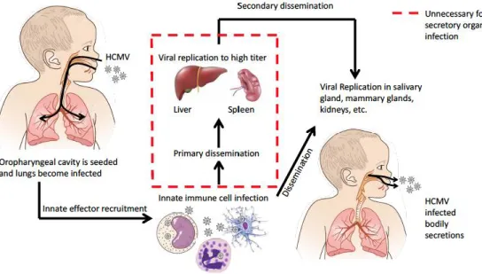
ii) Viral dissemination 99
Vertical and horizontal transmission of CMV have been described. Vertical transmission 100
occurs through transplacental and intrapartum transmission. Horizontal transmission is due to 101
organ transplantation from an infected organ or contact with infected body secretions. In a 102
first model, it is proposed that after an initial exposure, viremia associated with leukocytes 103
orchestrate a systemic diffusion in which organs such as the lung, spleen and liver are 104
infected. Finally, the virus undergoes a sequential dissemination in which the salivary glands, 105
breasts, and kidneys are infected (review (12)). Another model has shown how viral 106
transmission comes from upper respiratory tract (12). Alveolar macrophages and alveolar 107
epithelial cells of type 2 are first infected with endocytosis, that is facilitated by viral 108
glycoproteins gB, gH/gL/gO and by the pentameric complex gH/gL/UL128-UL130-UL131A. 109
The lytic cycle produces new infectious virus particles that infect other types of permissive 110
cells like fibroblasts, endothelial cells, dendritic cells and other innate immune cells, 111
including other alveolar macrophages. The dissemination occurs either through free-viral 112
particles infecting new cells or through cell-to-cell spread by direct cell-contact. Endothelial 113
cells play a crucial role in cell-to-cell spread and viral dissemination. They transmit thevirus 114
by direct cell contact and during trans endothelial migration (13). Furthermore, infected 115
endothelial cells regulate adhesion molecules such as ICAM- and VCAM-1 which facilitate 116
the extravasation of infected monocytes and neutrophils and the transfer of the productive 117
virus to improve haematogenic spread (14). Virus infected cells may directly sow secondary 118
organs that lead to secretion into body fluids, where draining lymph nodes and blood filtration 119
organs are not a necessary stage prior to secondary organ infection (Figure 4). 120
121
Figure 4 Alternative mechanisms of HCMV dissemination generated from animal model extrapolation (12). 122
In a first model, it is proposed that after an initial exposure, viremia associated with leukocytes orchestrate a 123
systemic diffusion in which organs such as the lung, spleen and liver are infected. Finally, the virus undergoes a 124
sequential dissemination in which the salivary glands, breasts, and kidneys are infected. Another model has 125
shown how viral transmission comes from upper respiratory tract and could through infected leucocytes 126
disseminate to all organs notably the secretory organs without a first step in the liver/spleen (12). 127
128
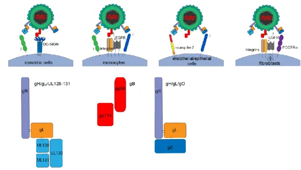
iii) Viral tropism 129
CMV has a very large tropism because it can infect a lot of human cell type, such as 130
monocytes/macrophages, endothelial cells, epithelial cells, smooth muscle cells, fibroblasts, 131
stromal cells, neuron cells, neutrophils and hepatocytes (15), thereby contributing to CMV's 132
high capacity to cause tissue damage in multiple organs. Heparan sulfate proteoglycans 133
function to tether the virus to the cell membrane, through complex gN/gM interaction (16). 134
Although strictly required, heparan sulfate proteoglycans are not solely sufficient for HCMV 135
infection (15). The question of whether EGFR was a (co) receptor with integrins capable of 136
recognizing gB when CMV entered fibroblasts was debated (17-19). Indeed, EGFR signaling 137
has been observed after CMV entry (17, 18) but the promotion of virus-cell fusion by EGFR 138
and the internalization of virion components to the cytoplasm linked to EGFR were not 139
demonstrated. The hypothesis was that integrins binding to gB participate in the activation of 140
EGFR signaling, since both 1 and 3 integrins have been shown to associate with EGFR and 141
activate it in a ligand-independent manner (20). Viral entry of CD34+ cells has been 142
associated with EGFR-gB interaction with EGFR signaling (21) but similarly to fibroblast, 143
direct binding and internalization has not been shown and one of the major mechanisms 144
highlighted for CMV entry in CD34+ cells was micropinocytosis (22, 23). Conversely, EGFR 145
signaling has been involved in CMV entry into monocytes (24) and the direct binding of 146
EGFR independently of integrin and its internalization has been recently demonstrated 147
(25). gH/gL/gO is required for the entry of CMV into endothelial/epithelial cells and 148
fibroblasts and PDGFR was identified as its ligand (with direct interaction between 149
PDGFR and gO) but is expressed only on fibroblasts (26), therefore the ligand(s) on 150
endothelial and epithelial cells remain(s) unknown. The pentameric gH/gL/UL128-131 151
complex has been involved in CMV entry in leukocytes, epithelial, endothelial (27) and 152
dendritic cells (28). Neuropilin-2 has recently been identified as a receptor of this pentameric 153
complex and its interaction have been shown to be necessary for the viral entry into epithelial 154
and endothelial cells (29) but the ligands of the pentameric complex host cell are not known 155
for the other cell types. Finally, our group has demonstrated that DC-SIGN binding gB also 156
contributes to the CMV entry in dendritic cells (30). The scheme below summarizes the type 157
of cells which may be infected and the viral-protein/host cell receptor known to be involved 158
in CMV entry (Figure 5). 159
160
161
Figure 5 Viral complexes and host-cell receptors involved in CMV entry 162
163
iv) Viral cycle 164
The viral cycle starts after infection of the host cell which may occur either by direct fusion or 165
through the endocytic pathway. The virus binds to the cell through interactions between viral 166
glycoprotein and specific surface receptors described earlier. Afterthis step, the fusion of the 167
envelope with the cell membrane occurs to release the nucleocapsids in the cytoplasm. These 168
nucleocapsids transfer to the nucleus, where viral DNA is released. This initiates Early 169
Immediate (IE)-1/ IE-2 gene expression. Viral replication and maturation are followed by the 170
stimulation and parallel accumulation of viral synthesis function. This process involves the 171
encapsulation of replicated viral DNA as capsids, which are then transported from the nucleus 172
to the cytoplasm. Secondary envelopment occurs in the cytoplasm at the mid-compartment of 173
the endoplasmic reticulum (ER)-Golgi. It is followed by a complex two-step final 174
envelopment and egress process that leads to virion release by exocytosis at the plasma 175
membrane (9). Three successive phases occur during CMV replications (Figure 6): very early 176
(or IE for Immediate Early, 0 to 2 hours), early (E, early, <24 hours), and late (L, >24 hours), 177
with each step controlling progress to the next. Schematically, IE proteins are responsible for 178
the positive and negative regulation of viral and cellular genes and control their own 179
expression. E proteins are critical for genome replication. The DNA is then replicated and 180
during the late phase, structural proteins are synthesized. The CMV replication cycle is slow; 181
in infected human fibroblasts, new virions are released after 48 to 72 hours. The maximum 182
quantity of virions is released within 72 to 96 hours. Viral production continues over several 183
days at a high level. The host cell eventually dies after 4 or 5 days (31). 184
185
Figure 6 Life cycle of HCMV in a human cell. 186
First, the virus enters into its host cell (direct fusion or endocytosis),the nucleocapsid from the cytoplasm is 187
translocated into the nucleus, where DNA is released. This initiates IE1-2 gene expression. Viral replication and 188
maturation involves the encapsulation of replicated viral DNA as capsids, which are then transported from the 189
nucleus to the cytoplasm. Secondary envelopment occurs in the cytoplasm at the endoplasmic reticulum (ER)-190
Golgi intermediate compartment. This is followed by a complex two-stage final envelopment and egress process 191
that leads to virion release by exocytosis at the plasma membrane (9). 192
193
III. Interaction between CMV and its human host
194
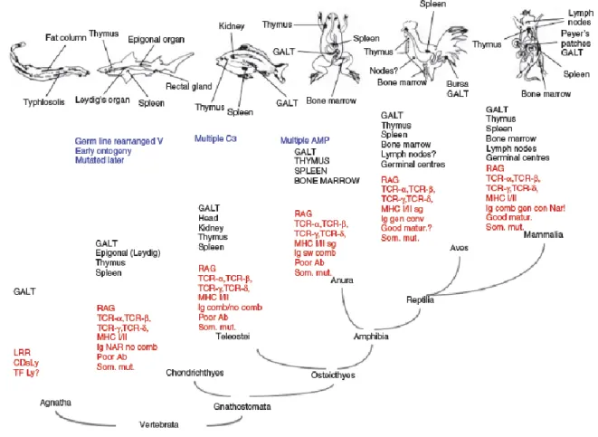
As mentioned earlier, CMV and its numerous hosts have evolved together for 80 millions of 195
years. CMV is only found in mammals, representing one of the classes with the most 196
advanced immune system (Figure 7). One of the specificity of CMV is its ability to use cell 197
host machinery to better replicate and encode components that help its viral escape and thus 198
promotes its host persistence. One can imagine that CMV replication and evasion qualities 199
have been acquired during its long-term co-evolution with the pressure selection of the virus 200
genes and with the integration of host genes into its own genome. This redesign of the viral 201
genome has led for example to the coding of “homologous viral-proteins” that are very 202
similar to host-proteins and are mostly involved in host evasion. 203
204
205
Figure 7 The evolution of the immune system within vertebrates. 206
AMP, antimicrobial peptides; C3, third component of the complement; CD ly, CD determinants of lymphocytes; 207
GALT, gut-associated lymphoid tissue; LRR, leucine-rich repeat; MHC, major histocompatibility complex; Poor 208
Ab, antibody response with no or weak affinity maturation in opposition to good maturation (good matur.); Som. 209
mut., somatic mutation; RAG, recombination associated gene; TCR, T-cell receptor; TF Ly, transcription factor 210
specifying lymphocyte development. In blue are represented new mechanisms or acquired function in the 211
immune system in the corresponding specie (ie Germlin rearranged V appeared in the first vertebrates 212
(agnathans). In black are represented the organs and in red the components of the immune system of each specie 213
(32) 214
215
A. CMV has optimized the persistence in its host 216
i) By optimizing its viral replication 217
HCMV encodes several proteins that contribute to maintain viral translation and replication in 218
its host cell in order to prevent spontaneous cellular response to stress induced by its 219
infection, normally resulting in decreased translation and cellular apoptosis (33). Indeed, 220
human cytomegalovirus shares a general lifecycle strategy with other mammalian double-221
stranded DNA viruses that replicate within the nucleus to accommodate host cell: increased 222
glucose uptake, metabolism and oxygen utilization; removal of cell growth controls (33). 223
They manipulate the cell cycle to an optimal point for virus growth; and inhibit apoptosis 224
during the productive phase of replication. These massive cell changes normally result in 225
reticulum stress and decrease translation capabilities, including translation. HCMV has been 226
shown to be able to maintain translation by modulating PI3K/AKT/mTOR signaling. HCMV 227
activates AKT (phosphorylation of Threonine 308 on AKT by PI3K) which in turn results in 228
the activation of mTORC1 with 4EBP and S6 phosphorylation. The virus also inhibits other 229
mechanisms in the cell stress response that may negatively control mTORC1 activation (33), 230
both of which contribute to cell survival during the viral replication phase. Moreover, 231
reticulum endoplasmic stress could be regulated by the modulation of the unfolded protein 232
response (UPR) (34), thereby contributing to the maintenance of protein translation. CMV 233
may also act on the post-translational stage by interacting with the cellular proteasome. 234
Indeed, CMV codes for proteins which catalyze the ubiquitylation of host proteins, thus 235
promoting their degradation (35). It also may encode proteins that indirectly alter the 236
conjugation of Ub or Ub-like proteins by altering the abundance and/or activity of cell 237
ubiquitin ligases (36); or remove existing Ub fragments by their deubiquitinase activity 238
(pUL48) (37). The biological relevance of deubiquitination for CMV replication was 239
highlighted when observing the impact of a mutation of this enzymatic center that 240
compromises the nearly tenfold replication capacity (37). Incorrect localization of checkpoint 241
proteins by HCMV also contributes to inhibit the DNA damage response (38). These different 242
functions could influence the cell cycle, the regulation of the response to DNA damage, and 243
finally the prevention of cell death/apoptosis (for review(39)). 244
245
ii) By persisting under latent form 246
One original way that CMV persists is its ability to become latent. Latency is the presence of 247
viral DNA without protein expression. CMV establishes latency in haematopoietic progenitor 248
cells (HPC) CD34+ and monocytes CD14+ (40), while endothelial cells as candidates for 249
latency for HCMV remain elusive (41). 250
Latency mechanisms have not been well understood. However, this knowledge was enhanced 251
by the use of recombinant viruses containing substitutions or stop codon insertions to disrupt 252
a single open reading frame (sORF). This has led to know that the growth restriction has been 253
conferred to UL138 and the replication advantage to UL135 and studies are helping to 254
understand their antagonistic role. Both genes are present on the same UL133-UL138 gene 255
locus and are separated by UL136. The three genes have a different kinetic expression as 256
UL135 is expressed in the early phase of infection and UL136 and 138 in the early and late 257
phase of infection, which may favor latency at the late phase (Figure 8). 258
In latently infected primary CD34+ HCPs, the expression of UL138 was detected, but 259
conversely that of UL135 (42) was downregulated. UL138 is needed for virus latency in 260
CD34+ HPC because its disruption results in failure of entering in latency (43). UL138 261
repressive capacity is partly due to prevent demethylation of histones. This subsequently 262
causes the viral genome to be silenced and in particular prevents the activation of the 263
immediate early promoter (44). UL138 acts also by activating EGFR, which promotes 264
EGFR/PI3K/AKT signaling and finally suppresses viral replication (45). Moreover, UL138 265
induces EGR1 through MEK/ERK signaling. As a result, UL138 via EGR-1/EGFR (46) 266
stimulates its own gene expression (47) creating a positive equilibrium from its antagonistic 267
counterpart UL135. Furthermore, increased levels of EGR-1 contribute to the maintenance of 268
the CD34+ progenitor phenotype (48). On the other hand, UL135 reduces the total and cell 269
surface levels of EGFR (47), thus downregulates EGFR signaling. The MiR-US22 (small 270
regulatory RNA) targets EGR-1 and induces its top-down regulation which reduces the 271
expression of UL138 and thus promotes reactivation (49). 272
Finally, UL136 codes for 5 isoforms: UL136p23/p19 are pro-latency proteins, while 273
UL136p33/p26 promote virus replication and UL136p25 isoform could be implicated in 274
either case depending on the context (50). 275
276
Figure 8 Modeling of latency/reactivation balance mechanism. 277
UL138 and UL135 are located on the same UL133-UL138 gene locus, separated by UL136. After CMV entry in 278
human cell, the expression of UL138 increase EGFR signaling which in turn activate MEK/ERK and PI3K/AKT 279
patways. EGFR/PI3K/AKT signaling suppresses viral replication.. MEK/ERK induce EGR-1activation which 280
induces in turn UL138 transcription. UL135 downregulates EGFR membrane expression which decrease EGFR 281
signaling. UL136 could express isoforms either involved in latency or reactivation 282
283
B. CMV has optimized its evasion of the immune system 284
CMV also optimized its persistence by evading the immune system. Here, we will review the 285
various players in the immune system, describe the mechanisms used by the CMV to escape 286
them and illustrate it in Figure 9. As discussed above, CMV infects species (mammals) that 287
have acquired the different types of immune actors and thus co-evolved together until now 288
(Figure 7). 289
Long-term cohabit could have promoted recombination with cellular DNA or post-integration 290
of cDNA derived from spliced cellular mRNA (51), which confer abilities for CMV to encode 291
homologous molecules able to interact with almost every immune actor. 292
We will see that studies have shown the mechanisms for evading CMV to counter all players 293
of the immune arsenal and we will discuss which mechanisms could alter T cell response, 294
for which only hypotheses can be made. 295
296
i) IFN I/PRR/inflammasome pathway 297
Pattern recognition receptors (PRR) act as the first line of defense because they can detect 298
pathogens at the cell surface or just after pathogen infection of their host cells, especially in 299
epithelial cells, in monocytes and dendritic cells. The PRR studies have demonstrated how 300
CMV can be recognized on the surface of cells, in cytosol, and in endosomes. The Toll like 301
receptors (TLR) are the best characterized PRR and recognize hydrophobic molecules in the 302
plasma membrane and nucleic acid in the endosomes and the cytoplasm. TLR2 recognizes gH 303
and gB in monocytes and fibroblasts (52, 53) but the exact phenomenon leading to its 304
downstream signaling is not fully understood. TLR2 activation leads to NF-κB activation, 305
type I IFN response by monocytes (54) and expression of pro-inflammatory cytokines such as 306
IL6 and TNFIn mice, TLR9recognizes CMV DNA in endosomes via the downstream
307
adaptor molecule myeloid differentiation factor 88 (MyD88). MyD88 activates IRF7 (56) and 308
induces the production of inflammatory cytokines, including IFN type I by plasmacytoid 309
dendritic cells (pDC) or classic DC (cDC) with a first peak 8 hours post-infection. In humans, 310
TLR9 is only expressed in B cells and pDC. This pDC TLR9/MyD88/IRF7 axis contributes to 311
activate NK cells (57, 58) and CD8+ T cells, involved in CMV clearance (56, 58). Finally, 312
DNA sensors such as DAI and IFI16 could recognized CMV DNA independently of TLR and 313
also lead to type I IFN production, which can explain why IFN I could be produced 314
dependently of MyD88 but independently of TLR (59). Cytosolic DNA could also be 315
detected by AIM2, involved in the activation of inflammasomes which results in caspase-316
dependent IL1/IL18 maturation in human and mouse macrophages (60, 61). In MCMV-317
infected macrophages, this sensor has a critical role in IL1 and IL18 production. Production 318
of IL-18 was demonstrated to be dependent on AIM2 during in vivo MCMV and directly 319
affects the production of interferon-gamma (IFN) by natural killer cells within 36 hours of 320
infection (62). Finally, the IFI16 sensor is also capable of binding CMV DNA in human 321
fibroblasts and induced anti-viral cytokines such as IFN (63). 322
CMV has developed a counterattack strategy that blocks these innate functions. pUL83 (pp-323
65) was identified for direct binding to IFI16 resulting in reduced cytokine production after 324
activation of the IFI16 pathway (63). pUL83 also binds AIM2 reducing similarly 325
inflammasomes-dependent AIM2 activation (64). IE86 inhibits the binding of NFB to the 326
IFN- promoter, inhibits TNF-induced NFB DNA binding (65) and also blocks NFκB-327
dependent transcription of pro-IL in macrophages (66). In addition to inflammatory signals 328
after TLR/DNA sensing, dendritic cells also play a role during CMV infection in producing 329
IL12 (which positively improves NK cell (67) and T cell response (68)) and in activating 330
the adaptive part of CMV immune responses (69, 70). UL111a codes for two IL-10 homologs 331
that modulate DC functions. It represses the maturation of monocytes to dendritic cells by 332
downregulating the expression of class II HLA, by decreasing co-stimulatory receptors such 333
as CD80, CD83, CD86, and CD40 (71, 72), CD11c expression, and by reducing the 334
acquisition of dendritic phenotype. UL111a also decreases TNF, IL6, IL1 gene 335
transcription and TGF, TNF and IL6 protein production by CD34+ cells (72, 73), and 336
promotes DC apoptosis as well (71, 74). Moreover, UL111a contributes to inhibit type I 337
interferon (IFN) production by pDC (75). Lastly, UL111a induces a M2 polarization of 338
monocytes with the downregulation of CD14, CD163 and class II HLA expression which 339
limits their CD4+T cell priming, and also inhibits their HO-I (Heme oxygenase) activity 340
which is involved in TNF and IL1 production (76). 341
342
ii) Immunoglobulins 343
Following B cell maturation, immunoglobulin G (IgG) are produced mainly against gB (9, 344
77) and less often against gH (78). Their role in controlling primary CMV infection is minor
345
since B-cell depleted mice can recover (79). However, anti-CMV IgG play a role in the 346
control of CMV reactivation since B-cell depleted mice are subjected to higher viral 347
load/dissemination during a CMV reactivation induced by immunodepression (79). The 348
transfer of immune serum demonstrates that the lack of antibodies allowed the virus spread in 349
deficient mice during reactivation (79). Anti-CMV IgG start to rise 7 days after the onset of 350
infection and reach neutralizing titers after two weeks (79). Anti-CMV IgG and IgM lead to 351
free virion opsonization/neutralization and target viral antigens on infected cells, with lysis 352
capacity of target cells in the presence of complement (80, 81). These IgG could also bind 353
cellular receptors for the Fc domain of IgG (FcR) on the cell surface of NK cells and V2neg 354
T cells which leads to antibody-dependent cell-mediated cytotoxicity (ADCC) (82) or 355
antibody-mediated cell inhibition by the production of IFN(68). UL118 and TRL11 genes of 356
HCMV have been identified as encoding gp68 and gp34 respectively as FcR counterparts 357
inhibiting IgG binding to NK cells (83). 358
iii) NK cells: 360
NK cells expand within few hours after CMV infection begins (62), with an initial type I IFN 361
and IL12 priming (84). They are involved in the disease recovery in mice (84, 85) and act on 362
target-infected cells by direct cytotoxicity and by activation and recruitment of other cells of 363
the immune system through their cytokines and chemokines secretion, including IFNγ and 364
TNFα (84). A specific phenotype of NKG2ChiCD57+ was described in the CMV response 365
and showed abilities to create a pool of memory NK cells (86-89). Their activation mode is 366
supported by cumulative signals from inhibitory and stimulatory receptors. Inhibitory 367
receptors with inhibitor tyrosine-based inhibitory motifs (ITIM) such as killer cell-Ig-like 368
receptors (KIRs), leukocyte Ig-like receptor 1 LILRB1 (LIR-1 also called CD85j), and C-type 369
lectin receptor CD94/NKG2A transmit a negative signal by recognizing class I HLA or class I 370
HLA homologs, such as MICA/MICB and HLA E. As will be further elaborated, HCMV 371
lowers the cell surface HLA molecule expression that would lead to decrease inhibitory 372
signals for NK cells. Interestingly, HCMV UL18 gene encodes a class I HLA homolog that 373
binds peptide and 2microglobulin and recognizes LIR-1/CD85j (90). Structural analyses 374
showed an increase in the contacts of UL18/peptide/LIR-1 complex and an optimal surface 375
complementarity in the LIR-1/UL18 interface, compared to the LIR/class I HLA interfaces, 376
resulting in a >1,000-fold higher affinity (90) and thus restoring LIR-1 inhibitory function. 377
LIR-1 is expressed on other cells of the immune system, including dendritic cells (91) and 378
CD8 T cells (92). For example, the binding of pUL18 to DC impairs cell migration and 379
CD40 ligand-induced maturation (93). 380
HLA-E is recognized by the inhibitory CD94/NKG2A dimer when it binds a peptide derived 381
from class I HLA molecules and by CD94/NKG2C, an activating receptor, when it binds 382
other kind of peptides (94). A nonameric peptide derived from pUL40 sequence (a class I 383
HLA homolog) is a ligand for HLA-E and promotes HLA-E expression on the cell surface, 384
facilitating interaction between HLA-E and CD94/NKG2A, and thus leading to inhibition of 385
NK cells (95). 386
On the other side, HCMV downregulates the ligands of stimulatory receptors to escape to NK 387
cell lysis. The ligands of NKG2D are the human major histocompatibility complex (MHC) 388
class I chain-related genes (MIC) A, MICB, and ULBP1-6 (UL Binding Protein) molecules, 389
which are particularly expressed during viral stress (96). pUL16 prevents cell surface 390
expression of both MICB and ULBP1-2. MICA and ULBP-3 expression are prevented by 391
pUL142 and UL122 which specifically blocks MICB (97) by sequestration into the 392
endoplasmic reticulum or Golgi (98). pUL141 blocks the adhesion molecules CD155 393
intracellularly (99), thereby preventing its recognition by DNAM-1 (100). Finally, pp65 394
dissociates the ζ-chain from NKp30 thus blocking its signaling (101). 395
Interactions between NK cells and CMV genes are the perfect example of host-virus co-396
evolution. Most viral proteins leading to the escape of NK cells are host-counterparts 397
suggesting that the replication of CMV in its host cells has borrowed genetic material that in 398
turn helps to encode viral counterparts, especially HLA or HLA-like molecules. The host has 399
selected counterattack strategies as well. For example, MICA*008 does not bind pUL142, so 400
it may persist on the surface of host membrane and be recognized by NKG2D (102). 401
Moreover, MICA and MICB downregulation are mediated by viral proteins produced during 402
intermediate early phase of CMV infection (103). As NK cells may be activated at an early 403
stage, this open a short “window” for NK cell control. Studying these mechanisms of host-404
virus interactions allowed the identification of new host proteins important during immune 405
response, as it is the case for LIR1 (discovered during studies on UL18) and ULBP proteins 406
(discovered during studies on UL16). 407
iv) T lymphocytes 409
T cells comprise CD4+ and CD8+ T cells. Their crucial role in controlling CMV has been 410
illustrated by clinical situations in which they are affected such as in HIV-induced acquired 411
immune deficiency syndrome or during immunosuppressive treatments, blocking activation 412
signals of T cells. Our understanding on the specific role of each subset is derived from 413
mouse models, which differentiate the role of CD4+ and CD8+ T cells from NK cells in 414
cell immunity. Polic et al. used sequential and additional depletion of CD4+, CD8+ T 415
cells and NK cells. They showed their partial redundant role with 5.6% of CMV recurrence 416
with NK depletion alone, 11.7% with CD4+ T cell depletion alone, 4% with CD8+ T cell 417
depletion alone, 24% with NK and CD4+ T cell depletion, 80% with NK and CD8+ T cell
418
depletion, 100% with the depletion of the three subsets in B-cell depleted mice (85). T 419
cells recognize peptides either presented by class II HLA molecules on antigen-presenting 420
cell (CD4+ T cells) or presented by class I HLA molecules expressed on target cells (CD8+ 421
T cells). The frequency of open reading frame (ORF) recognition by CD4+ T cells showed 422
that peptides from UL55 (gB), pp65, UL86 and UL99 (pp28) were the most commonly 423
recognized (104). Those from UL48, pp65, IE1 and IE2 were the most frequently identified 424
by CD8+ T cells. In peripheral blood, following CMV infection, CD4+ T cells (105) start 425
to rise 7 days later, while CD8+ T cells start to increase 20 days after CMV infection 426
begins (106). Kinetics of anti-CMV IgG, CD8+ and CD4+ T cells were compared in 427
symptomatic and asymptomatic transplant patients during CMV primary infection. It reveals 428
that anti-CMV IgG and CD8+ T cells increase similarly in both situations (106) while 429
CD4+ T cell expansion was delayed only in symptomatic patients, which shows the relevance 430
of CD4+ T cells in the control of CMV in immunocompromised hosts. Moreover, CMV-431
specific CD4+ T cells appear to have a specific function as compared to CD4+ T cell during 432
other antigen encounters. During primary CMV infection, a specific subset of CD4+CD28-433
granzyme B+Perforin+ T cells expands, and is found only in CMV+ individuals (107). They 434
can produce TNF, MIP-1 during CMV-peptides stimulation (107) and may lyse target cells 435
expressing CMV antigens in a class II-dependent manner (108). These observations suggest 436
that this specific effector memory CD4+ T cell subset is particularly important to control 437
CMV infection, compared to conventional IL2-producing memory CD4+ T cells that 438
remained in the lymph nodes (109). However, the lysis has not been consistently observed, 439
which suggests a CMV evasion effect even if antigen presentation remains (110). 440
Cytotoxic CD8+ T cells have also shown their protective effect, notably in mouse model 441
with CMV-specific CD8 T cells transfer (111). They produce massive amounts of IFN and
442
have cytotoxic activity on CMV-infected target cells (106). During the primary phase of 443
CMV infection, a phenotypic change is observed since they become CD27+, CD28+, 444
CD45RO+, CD45RA-, CCR7-, Perforin+, Granzyme B+, Ki67++ (106). Lately, in the 445
chronic phase of CMV infection, memory CMV-specific CD8+ T cells are separated into
446
two subsets: The central memory CD27+CD28-CD45RA+, with proliferation capacities after 447
TCR restimulation (also seen in other chronic viral infections) and the “terminally 448
differentiated effector memory cells”, also called TEMRA, CD27-CD28-CD45RA+ with low 449
proliferation capacity, but high cytotoxicity that appears to be highly present specifically 450
during CMV infection (112). 451
Even strongly efficient during CMV infection, CD4+ and CD8+ T cells could be 452
counterattacked by a CMV escape mechanism, targeting class II and class I HLA expression. 453
In the early phase of CMV replication, the peptide derived from IE1 transcription factor 454
presented in a class I HLA molecule is one of the first antigenic signal for CD8+ T cell 455
cytotoxicity (113). This could be blocked by pp65 phosphorylation of 1, preventing an IE-456
1-specific CTL response (114). 457
In addition US2, US3, US6, US10, and US11 block the generation and/or the export of HLA 458
class I/peptide complexes and induce rapid downregulation in class I HLA expression (for 459
review, (9)). US3 retains class I HLA into the endoplasmic reticulum (ER); US2 and US11 460
mediate cytosolic degradation of heavy chains by the proteasome (115), notably because 461
US11 in the ER membrane dislocates the new class I HLA from ER to cytosol (116). US6 462
interacts with transporters associated with antigen processing (TAP) in the endoplasmic 463
reticulum and interferes with the transport of class I molecules due to defective peptide 464
translocation (115). US10 delays the normal trafficking of class I HLA out of the ER (117). 465
US3 binds 2-microglobulin-associated heavy chains that are thus retained in the ER (118). 466
US2 also induces class II downregulation by promoting proteasome degradation, especially 467
HLA-DRand DM 468
469
471
472
Figure 9 Interplay between CMV immune response and viral evasion 473
9A. Full model of immune system/CMV interactions. 9B. Focus on and NK cell interactions. ADCC, antibody-dependent cell 474
cytotoxicity; ADCI, antibody-dependent cell inhibition; DC, dendritic cell; M, macrophage; IFI16, interferon inducible protein 16; 475
DNA-dependent activator of interferon; MICA/B, MHC class I polypeptide–related sequence A/B; ULBP, UL binding proteins; IL, 476
interleukin; TLR, toll like receptor; IFN, interferon; TNF, tumor necrosis factor. 1. NKG2C 2. NKG2D 3. NKp30 4. CD94/NKG2A 5. LIR-477
1 6. KIR 7. DNAM-1 8.EPCR, Annexin A2, unknown 9. IE1 peptide 10. IL18-receptor. 11. HLA-E. 478
34 479
v) T lymphocytes 480
The last identified player in cell-mediated immunity against CMV was the T lymphocytes 481
(120, 121). We have reviewed here their original development, their mode of recognition and 482
functions, their particularities regarding the evasion of CMV, and integrate them in the 483
specific immune response to CMV. 484
Although these criteria have been challenged, we are still attempting to classify the immune 485
response as “innate” or “adaptive”. “Innate” define immune actors that are present at the 486
antigen site of entry, that can get activated very quickly after antigen recognition but are 487
poorly specific with highly conserved and weakly diversified antigen receptors. Conversely, 488
the “adaptive” response is a term dedicated to actors who are not initially in the tissues, which 489
need a priming to activate, in this way more slowly but which express a specific antigen 490
receptor and develop long-term memory, providing long-term antigen-specific protection. 491
This memory fate has been well studied for B cells, T cells and more recently for NK cells 492
in the context of CMV, although they do not express a specific antigen receptor. 493
As they express a specific T cell receptor for antigen, T cells have therefore been 494
frequently described by comparison to B cells or conventional T cells. Like conventional 495
T cells, T cells differentiate in the thymus where they acquire TCR expression, V and 496
V chains, but TCR recognition is independent of the molecular histocompatibility complex 497
conversely to T cells. All these receptors present a common ability for “theoretical” 498
specificity and diversity obtained by somatic gene rearrangements from a limited number of 499
V (Variable), D (Diversity), J (Junction) and C (Constant) genes and by junctional diversity 500
with addition or deletion of nucleotides between the different gene segments when they are 501
joined together, also called somatic recombination. This diversity is mainly generated in the 502
complementary-determining region 3 (CDR3) of the T-cell antigen receptor (TCR) or B-cell 503