HAL Id: hal-01824375
https://hal-univ-tln.archives-ouvertes.fr/hal-01824375
Submitted on 27 Jun 2018HAL is a multi-disciplinary open access archive for the deposit and dissemination of sci-entific research documents, whether they are pub-lished or not. The documents may come from teaching and research institutions in France or abroad, or from public or private research centers.
L’archive ouverte pluridisciplinaire HAL, est destinée au dépôt et à la diffusion de documents scientifiques de niveau recherche, publiés ou non, émanant des établissements d’enseignement et de recherche français ou étrangers, des laboratoires publics ou privés.
Sr 1/2 Ce 5/14 � 1/7 WO 4 ; a new modulated ternary
scheelite compound
Rafael Hernandez Damascena dos Passos, Madjid Arab, Carlson Pereira de
Souza, Christine Leroux
To cite this version:
Rafael Hernandez Damascena dos Passos, Madjid Arab, Carlson Pereira de Souza, Christine Leroux. Sr 1/2 Ce 5/14 � 1/7 WO 4 ; a new modulated ternary scheelite compound. Acta Crystallographica Section B, Structural Science, Crystal Engineering and Materials, International Union of Crystallog-raphy, 2017, 73 (3), pp.466 - 473. �10.1107/S2052520617002827�. �hal-01824375�
Sr
1/2Ce
5/14□
1/7WO
4; a new modulated ternary scheelite
compound
Rafaël Hernandez Damasceno dos Passos,
a,bMadjid Arab,
aCarlson Pereira De
Souza,
band Christine Leroux
a,*a Université de Toulon, CNRS, IM2NP, BP 20132, F- 83957 La Garde Cedex, France
b Universidade do Rio Grande de Norte, DEQ/PPGEQ-LMNRC, Campus
Universitario, Lagoa Nova 59072-970 Natal, Brazil
Correspondence e-mail: [email protected]
Synopsis - A new ternary scheelite tungstate (Sr,Ce)WO4 phase was synthesized, with a 2D
incommensurately modulated structure linked to partial ordering of Sr, Ce cations and vacancies as was evidenced by high resolution electron microscopy. The bang gap of 3.2 eV makes it a promising violet emitting compound.
Abstract - type the Abstract here (Style: IUCr Abstract)
For the first time, a ternary tetragonal scheelite structure tungstate with strontium and cerium cations,(Sr,Ce)WO4, was synthesized. As much as 35 % Ce could be inserted in the
structure, leaving 1/7 of the (Sr,Ce) cation sites vacant. Partial ordering of Sr and Ce, with atomic displacements, were evidenced by high resolution electron microscopy. 2D incommensurate modulations occur in this material, in small domains 20 nm in size. The band gap of this compound is significantly lower than the band gap of SrWO4 and this was related
to the distortions of WO4 and (Sr,Ce)O8 polyhedra. The bang gap value of 3.2 eV makes
Sr1/2Ce5/14□1/7WO4 a promising candidate for violet luminescence.
Keywords: - scheelite, ordering, vacancies, incommensurate, high resolution electron microscopy, simulations, x ray diffraction, band gap.
1. Introduction
Scheelite molybdates or tungstates are known to be chemically stable luminescent compounds as well as good ionic conductors. In the last decade, their potential use as light emitting materials was explored and many new scheelite compounds were synthesized, by adding lanthanide cations in the structure (Abakumov et al., 2014; Morozov et al., 2013, Nogueira et al.,2013, Giang et al.,2015). The binary scheelite tetragonal structure of general formula ABO4, (space groupe I41/a) corresponds to (A2+,B6+) and the cation/anion ratio [A]/
the c axis built by [BO4] tetrahedra and [AO8] polyhedra sharing vertices. This structure
accommodates easily cations with different oxidation states for A and B as in BiVO4
(A3+,B5+), ZrGeO
4 (A4+, B4+) or AgIO4 (A+,B7+). When the cation/anion ratio is different from
1, the tetragonal structure of binary tungstate (B=W) or molybdate (B=Mo) scheelite can be distorted into a monoclinic structure as in Ce2W3O12 (A3+,B6+) (Arab et al., 2013) or an
orthorhombic one as in Ce10W22O81 (Patout et al., 2014). Ternary scheelites refer to
compounds with general formula (A,A')n(BO4)m. Most of them are built with a charge
difference of 2 between A et A' cations, as for example (Ag,RE)(MoO4)2 or (Li,RE)(MoO4)2
RE=rare earth elements (Abakumov et al., 2014). The charge difference is usually accommodated by cation vacancies on A sites, with or without cation ordering or incommensurate modulations (Abakumov et al., 2014). Ternary scheelite with a charge difference of 1 are uncommon, and apart from a lead-lanthanide based compound (Takai et
al., 2004), only (Ca,Eu)(BO4)2 and (Sr,Eu)(BO4)2 were extensively studied (Abakumov et al.,
2014; Iiang et al., 2015).
Among rare earth compounds with the scheelite structure, rare earth tungstates exhibit excellent thermal and chemical stability. Tungstates have been proven to be good host lattices for the luminescence of lanthanide ions (Zhou et al., 2014). Eu3+ is a commonly used
luminescent activator and Eu3+ activated SrWO
4 are efficient light emitting diodes (LED)
(Jiang et al., 2015). An interesting alternative to Eu 3+ is Ce3+ because of a potential tunable
light emission . The light emission of Ce3+ varies from ultraviolet to orange, depending on the
crystal field (Zhu et al., 2016; Li et al., 2008).The mechanism of Celight emission is usually linked to the interaction of the incomplete 4f shell energy levels of Ce with the band state of the host lattice (E. Roy et al., 2010). The determination of the band gap is one way to obtain information of the potential optical properties of one compound. As the band gap value is also linked to the structural defects around the lanthanide ions, which are promoted by the distortions of angles and bond distances within different polyhedra clusters, (Cavalcante et
al., 2012; Rosa et al., 2008; Lacomba-Perales et al., 2008), structural investigations have to
linked to optical characterizations.
The aim of this work was to synthesize a (Sr,Ce)WO4 ternary scheelite compound with
different optical properties compared to those of the binary scheelite SrWO4. A complexing
method was used to synthesized powders of binary SrWO4, and (Sr,Ce)WO4 ternary tungstate
scheelites. Rietveld refinements of the X ray diffraction patterns were performed to obtain accurate cell parameters and atomic distances. Electron diffraction and high resolution electron microscopy gave access to the incommensurate modulations and cation ordering. The chemical composition was determined by Energy Dispersive Spectroscopy (EDS) and the
cation valence by Electron Energy Loss Spectroscopy (EELS). UV-Visible Diffuse Reflectance Spectra (DRS) was used to determine the energy band gap.
We showed that the (Sr,Ce)WO4 has an incommensurate modulated structure linked to partial
ordering of Sr2+ and Ce3+. The influence of the structure on the band gap values was discussed
in terms of bonds distances and angles splitting of the polyhedra WO4 and Sr(Ce)O8.
2. Experimental procedures 2.1. Synthesis
The powders were synthesized by the so called EDTA-citrate complexing method, EDTA accounting for Ethylene-Diamine-Tetracetic-Acid. This method was already used to obtain binary tungstates SrWO4 and Ce2W3O12, (Arab et al., 2013) and was adapted in this work to
synthesize ternary tungstates. Strontium nitrate Sr(NO3)2, cerium(III) nitrate hexahydrate
Ce(NO3)3.6H2O and tungsten oxide WO3were used as cation source. EDTA was diluted in
ammonium hydroxide (27% vol.) NH4OH at 40°C with a constant stirring during 15 min.
Solutions 0.3 M of strontium and cerium nitrates and WO3 were added in the solution of the
EDTA/ NH4OH in various proportions and stirred 15 minutes again. Then 0.45 M citric acid
was added to initiate the complexation reaction at a temperature of 80°C and at pH =9. The reaction time lasted 3h until the appearance of a gel-like precipitate. This gel was subjected to a heat pretreatment at a temperature of 230°C for 3h to remove waste liquids and volatiles, leading to the formation of a black powder. The final annealing was performed at 1000°C for 5h, a temperature ensuring the crystallization of the gel, leading to yellowish-white powders.
2.2. Structural and chemical characterization
Transmission Electron Microscopy (TEM) and High Resolution Electron Microscopy (HREM) were performed with a conventional Tecnai 200 kV and with a Titan FEG 300 kV Cs corrected, equipped with a Gatan Imaging Filter (GIF). Simulated HREM images were
calculated using the multislice program JEMS. The parameters used for simulations of images taken with the 300 kV microscope were Cs=-1.9µm, Cc=2 mm, 1/2 α=1 mrad, ∆E=0.7eV. The
chemical composition of the ternary tungstate was determined by Energy Dispersive spectroscopy (EDS). Binary compounds SrWO4 and Ce2W3O12 were used as standards for the
determination of the KSr,W and KCe,W factors. The statistical study of powder's chemical
homogeneity consisted in 20 EDS analyses on randomly chosen individual grains. Electron Energy Loss Spectroscopy (EELS) gave access to the valence of cerium in the ternary compound. Spectra were acquired in the diffraction mode, with a 0.03 eV/channel resolution . X rays powder diffraction (XRD) patterns were collected in the classical θ−2θ mode, with an Empyrean Panalytical diffractometer, with a Cu anti cathode (λKα1 = 0.154056 nm),. Rietveld
refinements with the software Fullprof were performed, starting from data cif files found in literature, and cell parameters, atomic positions, as well as site occupations, were extracted. The experimental profiles were fitted with the most suitable pseudo-Voigt analytical function taking into account the line broadening function and the symmetric part of instrumental function.
2.3. Optical properties
The band gap values were obtained using the UV-visible diffuse reflectance spectroscopy (DRS). The spectra of the different samples were recorded using a Perkin Elmer LAMBDA 190s UV/Vis/NIR spectrometer along with a 150 mm integrating sphere. A given powder was compacted in a manual press into a 1.2 cm powder cup, clamped on the external port of the integrating sphere. The measurements were performed in the range of 250 to 800 nm at room temperature with a resolution of 0.08 nm. The baseline was determined using a calibrated reflectance standard to allow achievement of a reflectance accuracy of 0.5%.
3. Results
3.1. Chemical composition
The mean composition of the ternary compound was found to be Sr0.50Ce0.35WO4, which
can also be written Sr1/2Ce5/14□1/7WO4, emphasizing the occurrence of cation vacancies.
Although the majority of ternary tungstate or molybdate are stoichiometric, ternary scheelite compounds with cations vacancies were already mentioned (Abakumov et al., 2014). Ce3+
may partially oxidized into Ce4+, thus the valence state of cerium in Sr
0.50Ce0.35WO4 was
determined by EELS. Ce4+ and Ce3+ M
4,5 core loss edges can unambiguously be distinguished
due to differences in shape, intensities and energies (Garvie et al., 1999). The Ce4+ M
4,5 edges
consist in two main symmetrical peaks situated at 884 eV and 901.6 eV and two satellites peaks of lower intensities at 889.2 eV and 906.7 eV. On the contrary, the Ce3+ M
4 edge is
asymmetrical with features at 896.6, 898.4 and 899.7 eV and the Ce3+ M
5 edge shows a broad
maximum from 881.7 to 882.3 eV. Apart from the shape and energy positions of the energy loss peaks, the branching ratio, defined as the intensity ratio I(M5)/[I(M4)+I(M5)], can be used
to distinguish between Ce3+ (branching ratio 0.55) and Ce4+ (branching ratio 0.49). Fig. 2
shows a characteristic EELS spectrum, acquired on a single grain of the ternary powder. The best fit was obtained with two peaks at 882 and 883 eV for the M5 edge and three peaks at
respectively 896.9, 898.5 and 899.8 eV for the M4 edge, indicating the cerium is in a 3+
around 883 eV, and a branching ratio of 0.53, indicate the presence of Ce4+. Assuming the
linear relation between branching ratio and relative amount of Ce4+ proposed by Gravie et al.
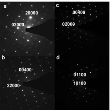
3.2. Electron diffraction
The main spots of the electron diffraction (ED) pattern, taken under different zone axes,
could all be indexed in the tetragonal scheelite structure.
Fig.3 presents high symmetry ED patterns, with respectively [001], [100], [1-10] and [111] zones axes, indexed in the tetragonal structure. In the ED pattern taken along [001] (Fig.3a), the main spots exhibit the characteristic four fold symmetry of the tetragonal structure along c, but the weaker spots cannot be indexed with integer indices. They correspond to two incommensurate vectors, q1=0.54 a*+0.82 b*, q2= -0.82 a*+0.54 b*, thus to a two dimensional structure modulation,which led to an indexation of the different ED patterns in a (3+2)D superspace group with 5 indices (Van Smaalen et al., 2007). Spots, indexed (00l00) with l=2n, that are forbidden in the space group I41/a of the tetragonal scheelite structure, can be observed in Fig 3b and 3c.
During tilt experiments, these spots do not vanish, excluding a double diffraction phenomenon. A reduction in symmetry must then be considered from I41/a to I-4 (82). This
symmetry loss was already observed for ternary scheelite molybdate like (Na,Gd)MoO4
(Abakumov et al., 2012) or (Li1Yb)MoO4 (Volkov et al., 2005). Thus, the superspace group
for Sr0.50Ce0.35WO4 was identified as I-4 (α,β,0, −βα0)00, with α= 0.54 and β=0.82. Fig.4 is
part of the [001] pattern, where the two incommensurate vectors q1 and q2 are drawn: all the
weak spots in the ED pattern can be indexed using q1 and q2. Modulated structures are not
uncommon in (A,A')n(BO4)m scheelite compounds (B=Mo or W) and an exhaustive overview
of them can be found in (Abakumov et al., 2014). This overview was realized in order to study the origin of these modulations, which were linked to the difference in cation sizes, the ordering of the A, A' cations and the presence of cations vacancies in the A sublattice. With a radius difference of ∆r=0.1 Å between Sr2+ and Ce3+, and 1/7 of vacancies, Sr
1/2Ce5/14□1/7WO4
belongs to the same group of partially ordered scheelites as Na2/7Gd4/7□1/7MoO4 (Abakumov et
al., 2012). The same modulations vectors q1=0.54 a*+0.82 b*, q2= -0.82 a*+0.54 b* are
observed for these structures, but the charge difference ∆q between A and A' cations is ∆q =2 (Na+,Gd3+) in one case, ∆q=1 in the other one (Sr2+,Ce3+). Another difference is that
two-dimensional incommensurate modulations were observed in molybdates with tetragonal structure but not in tungstate scheelites. On the contrary, ternary tungstate scheelites usually adopt a (3+1) D modulated monoclinic structure (Morozov et al., 2013). Thus, one can conclude that 2D modulations are linked to a small ∆r and vacancies on A sites, but not to ∆q, the charge difference between A and A' cations. Moreover, these modulations can occur in tungstates, not only in molybdates.
3.3. High resolution electron microscopy
High resolution electron microscopy (HREM) observations were performed along the [001] direction, where the projected structure corresponds to mixed columns of Sr, Ce and W cations, (see Fig5a) and along [111], where the projected structure corresponds to distinct atomic columns for the (Sr,Ce) cations and for W (Fig 5b).
Fig6 shows a HREM image taken along [001], along with a Fast Fourier Transform (FFT) and filtered images. The FFT image is similar to the diffraction pattern Fig4, with the same incommensurate spots. FFT performed on different regions of the HREM image do not always exhibit these spots, which indicate that modulations are localised in domains. The filtered image using the incommensurate spots confirms a 10-20 nm domain-type structure. The filtered image, using Bragg spots 20000 and 02000, has an enhanced contrast compared to the original image, shows no defects and can be interpreted in terms of atomic columns of cations.
In order to study a possible ordering of Sr and Ce, HREM images were taken along [111], using a 300 KV microscope Cs corrected. This projection direction allows to interpret HREM images in terms of atomic columns of W, Sr, Ce (Fig5.b). In particular, along the trace of (112) planes, there will be successively two W columns, two (Sr,Ce) columns. Fig.7 shows a HREM image taken along [111] were thickness variations give rise to different motifs. The two insets correspond to filtered images from a thin part and a thick part of the sample. In the thin part of the sample, the position of the bright spots as well as their intensity clearly varies along the trace of the (112) planes, suggesting different atomic occupation. Simulated HREM images were calculated using two structural models and compared to the filtered images. Both structural models were based on the I-4 space group deduced from the ED study. The first model (model 1) corresponds to a disordered scheelite structure; Sr, Ce cations occupy randomly the same 2(b) and 2(c) sites, and W is on the 2(a) and 2(d) sites. This structural model was already used for Na2/7Gd4/7MoO4 (Morozov et al., 2012). In the second model
(model 2), the Sr cations occupy the 2(b) site while the Ce cations are on 2(c) sites. The chemical composition was taken into account, thus vacancies were statistically distributed on 2(b) and 2(c) sites for both models. The atomic positions are summarized in Table 1.
The pattern observed in the thick part of the sample can solely be simulated with the structural model where Ce and Sr are ordered, for a thickness of 5-7 nm (Fig.8). The pattern observed in the thin part can, at first sight, be simulated for both disordered and ordered model, but the intensity variation observed along one row corresponds strictly to the different site occupation for strontium and cerium, as can be seen in Fig.8 where the projected structure is superimposed to a simulated one for a weak thickness of 3 nm. Thus, HREM allowed to prove experimentally the ordering of strontium and cerium atoms in Sr0.50Ce0.35WO4.
3.4. Crystal structure refinements (X ray diffraction)
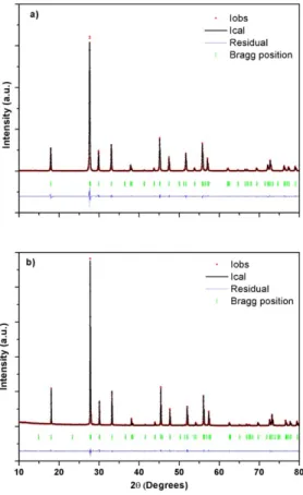
Fig.9 reports the XRD patterns of the binary SrWO4 (Fig.9a) and of the new ternary
Sr0.50Ce0.35WO4 (Fig.9b) compounds. The diagrams correspond to well crystallized single
phases without residual compounds. The phase identification showed that both tungstates exhibit XRD diagrams characteristic of the tetragonal scheelite structure. In particular, the splitting of the high angle peaks induced by a monoclinic structure, was not observed.
We performed different Rietveld refinements of the XRD patterns, according to electron microscopy results. The binary compound SrWO4 was refined in the I41/a space group. The
ternary compound Sr0.50Ce0.35WO4 was refined in the I41/a space group and in the I-4 space
group, with a random occupation of A sites by Ce3+ and Sr2+ (the so called disordered
scheelite). The occupation rate of the A sites was fixed to 50% for Sr and 35% for Ce, according to EDS results. In both space groups, the cations occupy fixed Wyckoff positions (see Table 1) and only the coordinates of oxygen atoms were refined . The refinements were initiate using cif files already published (Pereira et al., 2015; Abakumov et al., 2012). The refined cell parameters, oxygen coordinates and reliability factors are reported in Table 2. The refinements in the I41/a space group provides a good agreement with the experimental
data for the binary and the ternary compounds, with acceptable reliability factors. The cell parameters of the ternary scheelite Sr0.50Ce0.35WO4 are smaller than those of the binary SrWO4,
with a shrinkage of the unit cell of about 1,4% in volume. This is in agreement with the substitution of Sr2+ by Ce3+, which has a smaller radius (∆r=-0.1 Å) and the induced cation
vacancies. The loss of symmetry from the I41/a space group to the I-4 one, corresponds to a
splitting of each atomic position (see Table 1). Thus, the cations were distributed on the different sites according to model 1 (no ordering of Sr and Ce). Again, the reliability factors show a good agreement between the calculated and the experimental diffraction patterns.The differences in the reliability factors for the two refinements of the Sr0.50Ce0.35WO4 structure are
too small to discriminate between the two possible space groups (N° 82 and 88). This can be related to the polycrystalline character of our samples with heterogeneous size distribution in the nanometer range. Thus, investigations of a possible cation ordering through Rietveld refinements of XRD diagrams were meaningless.
The variation in the atomic coordinates observed for oxygen atoms, gives rise to different distortions of the polyhedra as can be seen in Table 3, which summarizes the A–O and W–O bond lengths and angles. The cerium insertion in the SrWO4 scheelite structure leads to a
contraction of WO4 tetrahedra and an expansion of AO8 polyhedra.
Basically, the SrWO4 binary compound presents a main W–O distance of 1,8408 Å and two
characteristic O–W–O angles values of 107° and 114°. The W–O distance decreases to 1.7903 Å in the Sr0.50Ce0.35WO4 structure with I41/a space group, but the O-W-O angles
remain the same, 107.7°and 113°. The A-O bond length increases, showing the WO4
contraction is accompanied by a AO8 expansion, and the O-A-O angles do not change
significantly. On the contrary, the refined structure with the I-4 space group shows much more WO4 and AO8 distortions. In this space group there are two W-O distances of 1.7964
and 1.7554 Å. The O-W-O angles deviate much more from the ideal tetrahedral angle of 109.9°, with values of 104°, 105°, 111°, 120° respectively. Similarly, the AO8 polyhedron has
four different A–O bond lengths and different O–A–O angles, as reported in Table 3. Thus, the structure refined with a space group I-4 is much more distorted than the one corresponding to I41/a.
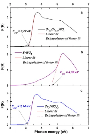
3.5. Optical properties
For potential optical applications (luminescence and photo-catalysis), the band-gap energies of the ternary scheelite structure, as well as those of the two binary compounds SrWO4 and
Ce2(WO4)3, were measured by UV-Vis diffuse reflectance spectra DRS at room temperature.
This allowed us to discuss the contribution of the insertion of cerium in the scheelite structure Fig.10 shows the UV-Vis spectra of Sr0.50Ce0.35WO4, SrWO4 and Ce2(WO4)3. Large bands
were observed for all spectra after 3 eV. According to theoretical calculations reported in the literature, tungstates scheelite materials like SrWO4 exhibit an optical absorption band due to
direct electronic transition (Zhang et al., 1998), which occurs through excitation of electrons from the valence band to the conduction band (Hwang et al., 2003). UV-Vis spectra show that the absorption band obtained for Ce2(WO4)3 is more intense than that of Sr0.50Ce0.35WO4 and
SrWO4. The intense band observed in the visible region is attributed to the electronic
transition of the cerium cation present in the sample.
The energy band gap is obtained by using the Kubelka – Munk method which consists in plotting F(R∞) versus photon energy. The band-gap is deduced by extrapolating the F(R∞)
profile using a linear fitting method. R∞ designates the reflectance of the sample at infinite
thickness and F(R∞) the absolute diffuse reflectance of the sample, which corresponds to the
conversion of the reflectance measurements R∞ according to the following equation:
F ( R
∞)=
(1−R
∞)
2
2 R
∞=
K
S
where K is the molar absorption coefficient and S the scattering coefficient.
The absorption band gap values for Sr0.50Ce0.35WO4, SrWO4 and Ce2(WO4)3 were found to be
3.22, 4.59 and 3.14 eV respectively. The energy gap of the SrWO4 is well in accordance with
the theoretical result of 4,51 eV (Song et al., 2009). In the case of Ce2(WO4)3, the band gap
system based on polyoxometalate [Ce(W5O18)2]8- cluster with CeO8 polyhedra (Roy et al.,
2010). The calculated molecular orbital diagram shows that the electron density consists on
the O2p atomic orbital for the HOMO and the Ce 4f orbitals for the LUMO, with a band gap of 3.28 eV. Variations of the gap values can be linked to different factors such as shape and crystallite size (Alivisatos et al., 1996), lattice parameters (Yang et al., 2015), degree of structural disorder in the lattice [Rosa et al., 2008; Lacomba-Perales et al., 2008).For SrWO4,
the band gap value shows that the maximum of the valence band can be mainly attributed to O2p levels, while the bottom of the conduction band is mainly composed of W 5d levels. The Sr 5s levels are mainly distributed in the conduction band , thus do not contribute to the band gap. (Song et al., 2009 ; Arora et al., 2006). The insertion of cerium in the host structure of SrWO4 can promote different energy distribution at intermediate levels, linked to the 4f, 5d
and 6s atomic orbitals, in the band gap near the valence and conduction bands. Combining all these elements, the band structure of Ce0.35Sr0.50WO4, would consist in a valence band with
mainly O2p states, and the bottom of the conduction band mainly composed with Ce 4f levels.
To further understand the optical properties evolution of the scheelite structures, the band gap is discussed in terms of microstructural parameters such as bond distance and angles of the polyhedra and the crystallite size.
Generally, it is admitted that the band gap of semiconductors increases when the particle size decreases (Lacomba-Perales et al., 2008). According to a statistical study of grain size distribution based on microscopy images (not reported here), the mean grain size is the same for the different powders, the low band gap value of the ternary compound cannot be attributed to a size effect. Concerning the influence of cell parameters on the band-gap value, the contraction of 1,4% in volume of the cell from SrWO4 to Sr1/2Ce5/14□1/7WO4 should induce
a small decrease of the band gap (Lacomba-Perales et al., 2008).
According to the data of Table 3, the band-gap decreases with decreasing W-O bond lengths and increasing of A(Sr,Ce)–O bonds lengths. In the same way, all O-W-O and O-A-O angles are much more distorted in the ternary compound refined in the I-4 space group. These changes in bonds and angles correspond to the W-O and A(Sr, Ce)–O stretching and twisting motions. They can be described as a combination of the symmetric and antisymmetric stretches of the oxygen atom bonds in the polyhedra WO4 and AO8. In addition to the
vacancies, the presence of cerium ions Ce4+ in the structure should induce even more
distortions, favouring the appearance of intermediate levels in the band gap. As a consequence, the Ce0.35Sr0.50WO4 scheelite structure gap energy decreases from 4.59 to 3.22
4. Conclusion
In the present work, a ternary (Sr,Ce)WO4 compound has been synthesized using a
modified EDTA-Citrate method. The structure was identified as a tetragonal scheelite and the XRD patterns showed well crystallized single phases up to 35%at. of cerium. Electron diffraction and high resolution electron microscopy evidenced the incommensurate modulations linked to partial ordering of Sr2+ and Ce3+, and the loss of symmetry from the
I41/a space group to the I-4 one. The Rietveld refinement results with the I-4 space group
revealed distortions of the AO8 polyhedra, ie. the local environment of Ce3+. The bang gap of
the ternary compound is significantly lower than the band gap of SrWO4 and this was related
to the 4f atomic orbitals of Ce and the induced crystallographic distortions linked to the incommensurate modulations. The bang gap value of 3.2 eV makes Sr1/2Ce5/14□1/7WO4 a
promising candidate for violet luminescence in the 380 nm range.
Acknowledgements - This study was done in the general framework of the french-brazilian cooperation project CAPES COFECUB PHC 77-113.
References
Abakumov, A.M., Morozov, V.A., Tsirlin,A.A., Verbeeck, J., Hadermann, J., (2014) Inorg. Chem., 53, 9407−9415
Albino, M., Pechev, S., Veber,P., Velazquez M., Josse, M., (2012) Acta Cryst., C68, i7–i8 Alivisatos, A. P., (1996) Science, 271, 933–937.
Arab, M., Lopes-Moriyama, A. L., Renovato dos Santos, T., Pereira de Souza, C., Gavarri, J. R., Leroux, Ch. (2013). Catalysis Today, 208, 35-41
Arora, S.K., Chudasama, B., (2006) Cryst. Res. Technol., 41, 1089–1095.
Cavalcante, L. S., Almeida, M. A. P., W. Avansi Jr., Tranquilin, R. L., Longo, E., Batista, N. C., Mastelaro, V. R., Siu Li, M., (2012) Inorg. Chem., 51 (20), pp 10675–10687
Culver, S.P., Greaney, M.J., Tinoco, A., Brutchey, R.L., (2015). Dalton Trans.,44,15042–15048 Garvie, L.A.J., Buseck, P.R., (1999), J. Phys. Chem. Sol., 60, 1943-1947
Hwang, D.W., Jae Sung, L., Wei, L., Se Hyuk, O., (2003) J. Phys. Chem. B., 107, 4963 4970 Jiang, P., Gao, W., Cong, R., Yang, T., (2015). Dalton Trans., 44, 6175-6183
Lacomba-Perales, R., Ruiz-Fuertes, J., Errandonea, D., Martiınez-Garcia, D., Segura, A., (2008) Europhys. Lett., 83, 37002.
Li, Y. Q., Hirosaki, N., Xie, R. J., Takeda, T., Mitomo, M., (2008), Chem. Mater., 20,6704-6714 Longo, V., Orhan, E., Cavalcante, L., Porto, S., Espinosa, J., Varela, J. A., Longo, E., (2007) Chem. Phys., 334, 180.
Morozov ,V.A., Arakcheeva, A.V., Chapuis, G., Guiblin, N., Rossell, M.D., Van Tendeloo, G., (2006).Chem. Mater., 18, 4075-4082
Morozov, V., Arakcheeva, A., Redkin, B., Sinitsyn,V., Khasanov, S., Kudrenko, E., Raskina, M., Lebedev, O., Van Tendeloo, G., (2012). Inorg. Chem, 51, 5313-5324
Morozov, V., Bertha, A., Meert, K.W., Van Rompaey, S., Batuk, D., Martinez, G.T., Van Aert, S. Smet, P.F., Raskina, M.V., Poelman, D. Abakumov, A.M., Hadermann J., (2013), Chem. Mat.,25, 4387-4395
Nogueira I.C., Cavalcante L.S., Pereira P.F.S., de Jesus M.M., Rivas mercury J.M., Batista N.C., Siu Li M., Longo E., (2013) J.Appl.Cryst., 46,1434-1446
Patout, L., Jacob, D., Arab, M., Pereira De Souza, C., Leroux, Ch., (2014). Acta Cryst B, 70, 268-274 Pereira, P.F.S. , Nogueira, I.C., Longo,E. , Nassar,E.J., Rosa, I.L.V.,Cavalcante, L.S., (2015) J. Rare Earths, 33, 113-128
Pinheiro, C.B., Abakumov, A.M., (2015), IUCRJ, 2, 137-154
Rosa, I. L., Marques, A. P. A., Tanaka, M. T., Melo, D. M., Leite, E. R., Longo, E., Varela, J. A., (2008) J. Fluoresc., 18, 239
Song, M., Zhang, Q., Liu, T., Yin, J., Guo, X., Zhang, H., Wang, X., (2009) Curr. Appl. Phys, 9, 812– 815.
Takai, S., Nakanishi, T., Oikawa, K., Torii, S., Hoshikawa, A., Kamiyama, T., Esaka, T., (2004) Solid State Ionics, 170,297-304
Van Smaalen S.,Incommensurate Crystallography, IUCr Monographs 21, 2007, Oxford Science Publications,
Volkov, V., Cascales, C., Kling, A., Zaldo, C., (2005), Chem. Mat., 17,291-300
Yang, X., Fu, Z. Liu, G., Zhang, C., Y. Wei, Wu, Z., Sheng, T., (2015) RSC Adv., 5,70220–70228. Zhang,Y., Holzwarth, N., William, RT., (1998) Phys. Rev B., 57, 12738-12750
Zhou, Y., He, X.H., Yan, B., (2014). Opt. Mater., 36, 602–607
Figure 1 ABO4 scheelite structure, a) drawing of the unit cell showing the piling up along the c axis
of edges-linked AO8 polyhedra. b) drawing of the unit cell cell along [001] showing the BO4 tetrahedra
and AO8 polyhedra are linked by vertices.
Figure 2 Cerium M4,5 core loss edge of Sr0.50Ce0.35WO4 .
Figure 3 High symmetry electron diffraction patterns of the incommensurately modulated Sr0.50Ce0.35WO4 ,along the zone axes [001] (a), [1-10] (b), [100] (c), [11-1] (d)
Figure 4 Part of [001] electron diffraction pattern, showing that all supplementary spots are due to the incommensurate vectors q1 and q2.
Figure 5 Projection of the ABO4 structure along two different crystallographic a) [001] and b) [111].
A and B cations are drawn as light blue (A) and dark blue (B), and the A-O and B-O links are drawn as sticks.
Figure 6 HREM image taken along [001], along with FFT and filtered images.
Figure 8 Map of images simulated (model 2). The defocus d varies from 2.5 nm to -1.5 nm, the thickness t from 1.4 nm to 7 nm. Projected atoms are represented as dots, Sr = blue,Ce = red, W= green
Figure 10 UV-Vis spectra of Sr0,5Ce0,35WO4, SrWO4 and Ce2(WO4)3 powders
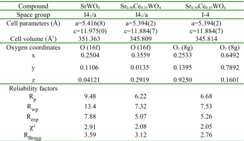
Table 1 atom coordinates of Sr, Ce , W and O in I41/a and I-4 space groups
Space group atome site coordinate
Sr,Ce 4b 0,0,1/2 0,1/2,3/4 I41/a W 4a 0,0,0 0,1/2,1/4 O 16 f x,y,z Sr,Ce 2b 0,0,1/2 I-4 Sr,Ce 2c 0,1/2,1/4 W 2a 0,0,0 W 2d 0,1/2,3/4 O1 O2 8 g x,y,z
Table 2 Refinement results for the binary SrWO4 and the ternary Ce0.35Sr0.50WO4 compounds using
two different space groups.
Compound SrWO4 Sr0.50Ce0.35WO4 Sr0.50Ce0.35WO4
Space group I41/a I41/a I-4
Cell parameters (Å) a=5.416(8) a=5.394(2) a=5.394(2)
c=11.975(0) c=11.884(7) c=11.884(7) Cell volume (Å3) 351.363 345.809 345.814 Oxygen coordinates O (16f) O (16f) O1 (8g) O2 (8g) x 0.2504 0.3559 0.2533 0.6492 y 0.1106 0.0135 0.1395 0.7892 z 0.04121 0.2919 0.9250 0.1601 Reliability factors Rp 9.48 6.22 6.68 Rwp 13.4 7.32 7.53 Rexp 7.88 5.07 5.26 χ² 2.91 2.08 2.05 RBragg 3.59 3.12 2.76 Table 3 Compound SrWO4 Sr0.50Ce0.35WO4 Sr0.50Ce0.35WO4
Space group I41/a I41/a I-4
Polyhedra WO4 Bond (Å) d: W-O 1.840(8) 1.790(4) 1.755(4) 1.796(4) Angle (º) O-W-O 106.9 114.6 107.7 113.1 105.0 111.7 104.2 120.5 AO8 Bond (Å) A-O1 2.533(4) 2.561(4) 2.519(5) 2.581(2) A-O2 2.571(6) 2.585(0) 2.588(9) 2.675(1) Angle (º) O-A-O 68.8 67.9 68.3 65.4 74.8 73.5 70.2 72.6 75.6 77.5 79.5 76.8 76.1 78.5 85.38 77.7 98.6 98.4 97.2 99.2 128.3 126.8 122.7 130.5 134.5 135.1 138.5 132.9 150.5 151.0 152.8 148,9
![Figure 4 Part of [001] electron diffraction pattern, showing that all supplementary spots are due to the incommensurate vectors q 1 and q 2 .](https://thumb-eu.123doks.com/thumbv2/123doknet/14580580.729137/15.892.268.626.103.466/figure-electron-diffraction-pattern-showing-supplementary-incommensurate-vectors.webp)
![Figure 6 HREM image taken along [001], along with FFT and filtered images.](https://thumb-eu.123doks.com/thumbv2/123doknet/14580580.729137/16.892.277.616.780.1064/figure-hrem-taken-along-along-with-filtered-images.webp)