HAL Id: tel-01310147
https://tel.archives-ouvertes.fr/tel-01310147
Submitted on 2 May 2016HAL is a multi-disciplinary open access archive for the deposit and dissemination of sci-entific research documents, whether they are pub-lished or not. The documents may come from teaching and research institutions in France or abroad, or from public or private research centers.
L’archive ouverte pluridisciplinaire HAL, est destinée au dépôt et à la diffusion de documents scientifiques de niveau recherche, publiés ou non, émanant des établissements d’enseignement et de recherche français ou étrangers, des laboratoires publics ou privés.
Design of mechanoresponsive surfaces and materials
César Rios Neyra
To cite this version:
César Rios Neyra. Design of mechanoresponsive surfaces and materials. Polymers. Université de Strasbourg, 2013. English. �NNT : 2013STRAE019�. �tel-01310147�
TH RA EX IN
É
pD
HÈSE dirig M. SCHAA APPORTE Mme. COC M. NARDIN XAMINAT M. SCHLA NVITES : M. JIERRY M. LAVALÉCOLE D
pour obtenDESIG
SU
gée par : AF Pierre EURS : CHE-GUER N Michel EUR : ATTER Guy Y Loïc LE PhilippUNIVE
DOCTORA
INS
Soute ir le grade DiscipGN OF
URFA
RENTE Lilia y eERSIT
RALE DE
STITUT C
T
préCésar
enance pré de :Doct
pline/ SpécF MEC
ACES
Pro ane Ma Dir Mu Pro Ma DirTÉ DE
PHYSIQ
CHARLE
THÈS
ésentée paRIOS N
évue le : 26teur de l’
cialité: Ch
CHAN
AND
ofesseur, U aître de Con recteur de re ulhouse ofesseur, U aître de Con recteur de reSTRA
QUE ET C
ES SADR
E
ar :
NEYRA
6 septembr’Univers
imie-Phy
NORE
MATE
niversité de nférences, U echerches, niversité de nférences, U echerches,ASBOU
CHIMIE P
ON
A
re 2013ité de St
ysique
SPON
ERIAL
e Strasbourg Université J Institut des e Strasbourg Université d INSERMURG
PHYSIQU
trasbour
NSIVE
LS
g oseph Four s sciences d g de StrasbouUE
g
E
rier, Grenob des matéria rg ble 1 aux dei
ii
Acknowledgements
This PhD work was conducted at the laboratories of the Institut Charles Sadron, UPR 22 CNRS within the “Ingénierie Macromoléculaire aux Interfaces” team and at the INSERM Unit UMR-S 1121 “Biomatériaux et Bioingénierie” in Strasbourg, France.
I would like to express my deepest appreciation to all those that helped and supported me, thus contributing to the accomplishment of my PhD.
I would like to start by expressing my sincere gratitude to Professor Pierre Schaaf, my PhD advisor, for giving me the opportunity to be part of his research group. His continuous support and guidance during my doctoral research was highly appreciated and helpful to work in this challenging topic.
I would like to express my entire gratitude to my co-advisor, Dr. Loïc Jierry, for the interesting scientific discussions, personal advices, motivation and patience during my research work.
I would like to extend my entire gratitude to Dr. Philippe Lavalle, concerning the fluorescence experiments on the confocal microscope, fluorescence microscope and spectrofluorimeter that I performed at INSERM labs. His quality of work, scientific discussions, patience, time and motivation were very appreciated during my thesis.
I also want to thank Dr. Fouzia Boulmedais and Dr. Jean-Claude Voegel for their scientific discussions during the group meetings, advice and patience during my doctoral thesis.
I would also like to thank Dr. Liliane Coche-Guerente and Dr. Michel Nardin for having accepted to act as referees for my thesis work and for providing interesting suggestions. I am also grateful to Professor Guy Schlatter for his participation as president of the advisory committee.
iii
I would also want to thank the directors of the Institut Charles Sadron, Professor Jean-François Legrand and Dr. Jean-Michel Guenet for accepting me as a PhD student at the institut.
I express my sincere gratitude to Dr. Joseph Hemmerlé and Monsieur Karim Benmligh for the design and implementation of the different stretching devices used during my uncountable experiments.
I would not forget the important contribution of Dr. Bernard Senger for the scientific discussions during the group meetings, the FRAP analysis and data treatment of the fluorescence experiments.
I would like to thank Dr. Joseph Selb for his great help during the fluorescence and rheological experiments together with the interpretations of the results and scientific discussions.
I would also like to thank Dr. Jacques Drutz for his time, for teaching me how to use the UV and IR machines as well as the discussion of the results.
I would like to thank Dr. Pascal Marie for the interesting scientific discussions about polydimethylsiloxane substrates and polymerization techniques.
I would like to thank Dr. Andreas Reisch and Dr. Benoît Frisch for teaching me the chemical modification of enzymes and polyelectrolytes and their respective analysis used during my experiments.
I would like to thank all my colleagues that I met during my thesis work with whom I worked or shared the fume hoods and had some fun:
My colleagues from my former lab at ISIS: Patty, Jonathan, Adrian, Anton and Vincent.
My colleagues from INSERM lab: Armelle, Géraldine, Christophe, Morgane, Julien, Engin, Christian, Cédric, Damien, Dominique and Vincent.
iv
My colleagues from IMI lab: Lydie, Johan, Eloïse, Tony, Elodie, Alexandre, Fabien, Thanh, Clément, Cécile, Gaulthier, Mathias, Gwenaëlle, Audrey, Emmanuelle, Nathalie, Jean-Nicolas and Prasad.
I would like also to thank my great friends for all the good times in and out of the ICS:
Maria, Diana, Lara, Johanna, David, Patty, Martin, Gaby, Christi, Andru, Franck, Stéphanie, Akkiz, Dominique, Rémi, Alliny, Nathalie, Anna, Mirella, Olga, Paul, Marek, Souvik, Christophe, Joseph, Gladys, Tam, Hatice, Christiane, Francine, Heveline, Yasmine, Patrycja, Arnaud, Camille, Laure, Sisouk and Daniel, I would never forget that good friends are there when we need them.
Finally, I would like to express my sincere gratefulness to the people who motivate me and trust me during all this time in France, my family:
My parents Maria and Alberto (R.I.P), my brothers P. Alberto Cristian and Carlos and my aunt Carmen for the support, advice and enthusiasm during this adventure since it started in 2006 in France for my studies, especially my PhD.
My dear Rebecca, for sharing my life every day, for the support I get from you when I’m down, the jokes you tell that makes me laugh and for just being like you are.
v
Abstract
In nature, there are plants that can be folded when being touched by animals or insects. At a lower scale, cells are known to probe the mechanical properties of the surface and adhere on it if it is appropriate for them. It is also well described that some kinds of proteins can exhibit hidden cryptic sites to specific ligands when a mechanical stretch is undergone by these proteins. In all these three examples, a mechanical stimulus starts a cascade of chemical transformations leading to the response of the living organism, the cell, or the protein, respectively.
In the last decade, these mechanosensitive transduction processes have inspired the scientific community and the design of smart materials and surfaces has emerged as a growing and very active field of research. Yet the work described up to now does concern mainly the color switching of materials when stretched or squeezed. In this context, the goal of my thesis was to develop new routes to design chemo-mechanoresponsive materials, i.e. materials that respond chemically to a mechanical stress, in a reversible way.
All the systems designed during my PhD thesis were based on the functionalization of silicone sheets: a first way was to create cryptic site surfaces by embedding ligands (biotin) into PEG brushes. The couple streptavidin/biotin was used as a model system. At rest, the surface so-prepared was antifouling and biotin ligands were specifically recognized by the streptavidin when the surface was stretched at 50%. Unfortunately, in this first approach, the mechanosensitive surface did not lead to a reversible process. In a second approach, we modified the silicone surface by using the polyelectrolyte multilayer (PEM) film deposition. This strategy was based on the covalent cross-linking of modified enzyme, the β-galactosidase, into the PEM. We succeeded in modulating the enzyme activity in the film under stretching and this approach appears as partially reversible under stretching/unstretching cycles. This work represents the first reported system where enzymatic activity can be modulated by stretching due to modulation of the enzyme conformation. In a last approach, we also designed a mixed system consisting of a silicone sheet onto which a polyacrylamide hydrogel is covalently attached with the goal to create a stretchable gel into which one can covalently attach enzymes or chemical mechanophores. These enzymes or mechanophores can thus be put under mechanical stress. We succeeded in creating a system that can be stretched up to 150% without detachment of the gel from the silicone and without inducing cracks in the gel.
vi
Résumé
Dans la nature, il existe des plantes capables de se mouvoir quand elles sont touchées par des animaux ou des insectes. A une échelle plus petite, il est connu que les cellules sondent leur environnement pour estimer les propriétés mécaniques de celui-ci et décider d’y adhérer ou non. Il a même été démontré que lorsque certaines protéines sont étirées sous l’effet d’un stress mécanique, elles exhibent des sites cryptiques permettant l’adhésion spécifique de ligands. Dans chacun de ses trois exemples, une cascade de transformations chimiques est amorcée par un stimulus mécanique qui permet une réponse de la plante, de la cellule ou d’une protéine, respectivement.
Au cours des dix dernières années, ces processus de transduction mécano-sensible ont inspiré la communauté scientifique et ont participé au développement d’une nouvelle classe de matériaux dits « intelligents », un domaine très actif et en constante évolution. Il faut cependant noter que dans le cas des matériaux mécano-sensibles, seul des matériaux capables de changer de couleur sous l’effet d’une contrainte mécanique ont été décrits. Dans ce contexte, le but de ma thèse a été de mettre au point de nouvelles voies d’accès à des matériaux chimio-mécano répondants, autrement dit, des matériaux capables de permettre une transformation chimique réversible lorsqu’ils sont soumis à un stress mécanique.
Tous les systèmes conçus au cours de ma thèse ont été développés sur des substrats en silicone : une première approche a consisté à créer des surfaces à sites cryptiques où une biotine est enfouie dans des brosses de chaines de poly(éthylène glycol) (PEG). Le système streptavidine/biotine a été utilisé comme modèle. Ces surfaces sont anti-adsorbantes à la streptavidine sauf lorsqu’elles sont étirées à hauteur de 50% où cette fois la biotine est reconnue. Ces surfaces mécanorépondantes se sont révélées non réversibles. Dans une seconde approche, nous avons modifiés la surface du silicone par adsorption d’une multicouche de polyélectrolytes. Cette stratégie est basée sur la réticulation covalente du film par une enzyme préalablement modifiée. L’enzyme choisie est la β-galactosidase. Nous sommes ainsi parvenus à créer une surface présentant une activité catalytique modulable par l’étirement mécanique, et ce, d’une façon partiellement réversible. Ce travail représente le premier exemple d’un système où une contrainte mécanique imposée à un matériau permet la déformation conformationnelle d’une enzyme, à l’origine de la diminution de l’activité catalytique du matériau. Dans une dernière approche, nous avons conçu un système mixte composé d’un substrat de silicone sur lequel une couche d’épaisseur macroscopique d’un gel de polyacrylamide est greffée de façon covalente. Des enzymes ou d’autres mécanophores pourront ainsi être inclus dans le réseau polymérique du gel de polyacrylamide et ainsi être étirés. Nous sommes parvenus à préparer de tels systèmes où l’hydrogel reste solidaire du film de silicone, sans apparition de craquelures jusqu’à 150% d’étirement.
vii
Table of contents
Abbreviations
1
General Introduction
3
Chapter 1: State of the art
1.1 A historical perspective 9
1.2 Mechanochemistry in biological systems 13
1.2.1 Enhancement of mechanical forces due to allosteric regulation 14
1.2.2 Cryptic site proteins 16
1.3 Cryptic site substrates 18
1.3.1 Polyelectrolyte multilayer based mechanoresponsive systems 18 1.3.2 Responsive systems based on poly(ethylene oxide) brushes 23
1.4 Summary 24
1.5 References 25
Chapter 2: Materials and methods
2.1 Materials 31
2.1.1 Solutions of polyelectrolytes 31
2.1.2 Fluorescence probes 31
2.1.2.1 Fluorescence polyelectrolytes 31
2.1.2.2 Fluorescence protein 31
2.1.3 Support for construction of films 33
2.1.3.1 Glass slides 33
2.1.3.2 Silicone 33
2.1.4 Stretching devices 34
2.1.4.1 Large stretching device 34
viii
2.1.5 Modification of silicon surfaces 35
2.1.5.1 UV-ozone (UVO) 35
2.1.6 Procedure to prepare PDMS Sylgard-184 36 2.1.6 Silanization procedure of the oxidized silicone sheet by using the
3-mercaptopropyltrimethoxysilane. 36
2.2 Methods 37
2.2.1 Contact angle (CA) measurements 37
2.2.2 Infrared spectroscopy (FTIR) 38
2.2.3 Atomic force microscopy (AFM) 38
2.2.4 Optical microscopy 38
2.2.5 Nuclear magnetic resonance (NMR) 38
2.2.6 Fluorescence based microscopy methods 38 2.2.6.1 Principles of fluorescence 39
2.2.6.2 Fluorescence microscope 40
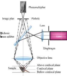
2.2.6.3 Confocal laser scanning microscope (CLSM) 41 2.2.6.4 Fluorescence recovery after photobleaching (FRAP) 42
2.3 References 45
Chapter 3: Investigation of a bispyrene unit used as a mechanical
sensor or a pH probe
3.1 Introduction 48
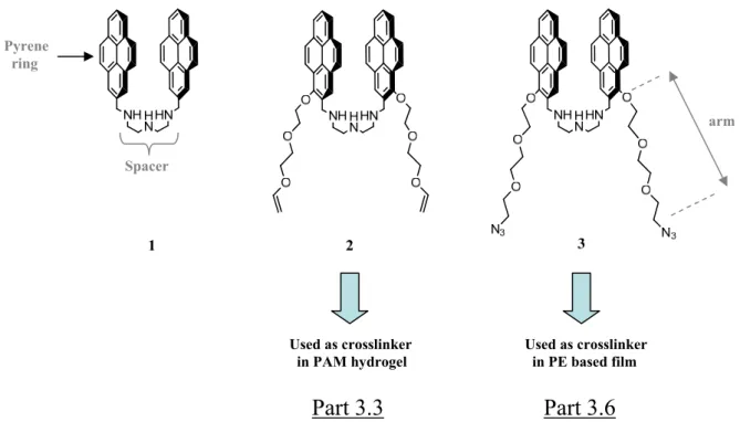
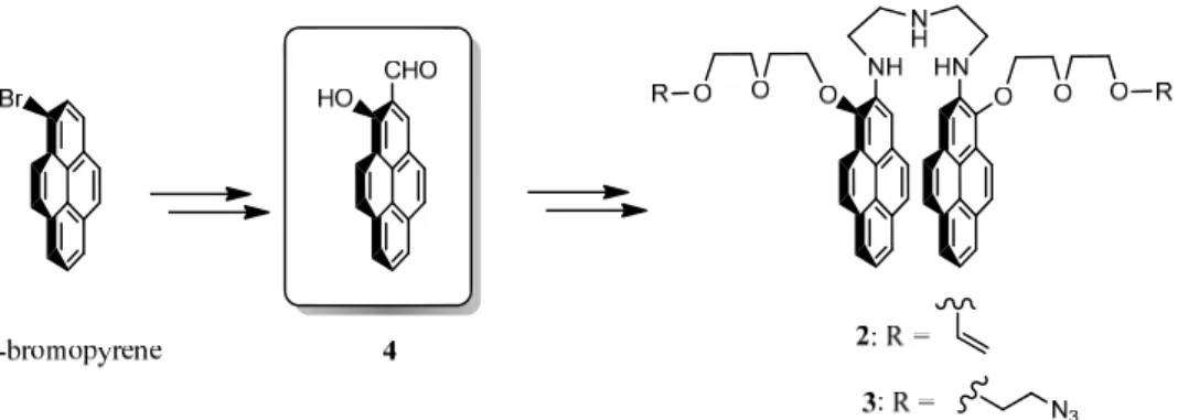
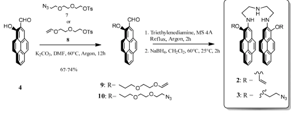
3.2 Design and synthesis of the bispyrenes 2 and 3 50
3.3 Bispyrene derivative 2 used as a mechanical sensor included in
poly(acrylamide) hydrogels 52
3.3.1 Optimization to get a viscous-elastic and stretchable PAM hydrogel 53 3.3.2 Bispyrene 2 cross-linked in the poly(acrylamide) hydrogel 55
3.4 Conclusion 57
3.5 References 58
3.6 Investigations of bispyrene 3 as homobifunctionnal cross-linker and
ix
Article 1: “Morphogen-driven self-construction of covalent films built from polyelectrolytes and homobifunctional spacers: buildup and pH response”
3.6.1 Supporting Information to article 1 69
Chapter 4: Covalent modifications of poly(dimethylsiloxane)
substrate to design reversible chemo-mechanoresponsive surfaces
4.1 Introduction 83
4.2 Choice of the elastomeric substrate: the PDMS 86
4.2.1 The PDMS Sylgard-184: presentation, principle of synthesis and preparation
4.2.1.1 Introduction 87
4.2.1.2 Methods and mechanism of PDMS reticulation 87
4.2.1.3 The PDMS Sylgard-184 89
4.3 Chemical modification of the PDMS surface 90
4.3.1 Principle of the UVO oxidation process of silicone surface 91 4.3.2 Results of the chemical modifications and characterization of the
silicone surface 92
4.3.2.1 UVO oxidation of silicone SMI and PDMS Sylgard-184 92 4.4 Silanization step: introduction of thiol groups onto oxidized silicone surface 97
4.4.1 Chemical modification process and characterization of the PDMS
surface with thiols groups 98
4.5 Grafting of biotine groups and PEG brushes on PDMS surfaces 99
4.5.1 Covalent grafting of Biotin groups on silanized PDMS surfaces 101
4.5.1.1 Experimental procedure 101
4.5.1.2 Protein adsorption 101
4.5.1.3 Fluorescence measurements 101
4.5.2 Effect of stretching and irreversibility of the Streptavidin anchored 103 4.5.3 Simultaneous grafting of PEG2000 chains and biotin groups on
the substrate 106
x
4.5.4.1 Covalent grafting of PEG chains and biotin groups on
stretched silicone 110
4.5.4.2 Effect of the PEG chain length 113
4.5.4.3 Sequential grafting of PEG and biotin in a stretched state. 114
4.5.4.4 Sequential grafting of biotin and PEG 115
4.6 Conclusion 116
4.7 References 117
Chapter 5: Polyelectrolyte multilayers with mechanically
modulable enzymatic activity (article in preparation)
5.1 Introduction 1225.2 Results 124
5.3 Conclusion 133
5.4 References 134
5.5 Supporting information 136
Chapter 6: Stretchable polyacrylamide hydrogel covalently
supported onto a silicone substrate: design of an ideal 3D
polymeric network for mechanotransductive investigations
(article in preparation)
6.1 Abstract 1536.2 Introduction 154
6.3 Material and methods 155
6.3.1 Materials 155
6.3.2 Methods 156
6.4 Results and discussion 158
6.4.1 Design of the PAM hydrogel supported covalently on silicone sheet 158
xi
6.4.3 Silicone sheet modifications 162
6.5 Silanization and PAM hydrogel formation 167
6.6 Conclusion 169
6.7 References 169
6.8 Supporting information 172
Conclusions
and
perspectives
179
Annex I
182
1
Abbreviations
β-Gal β-Galactosidase
β-GalFITC β-Galactosidase labeled with fluorescein β-Gal-mal β-Galactosidase-maleimide
β-Gal-malFITC β-Galactosidase-maleimide labeled with fluorescein Biotin-Mal biotine modified with maleimide groups
CA contact angle
CLSM confocal laser scanning microscope DCC N, N'-Dicyclohexylcarbodiimide
EDC N-(3-Dimethylaminopropyl)-N’-ethylcarbodiimide hydrochloride FDG fluorescein di(β-D-galactopyranoside)
FITC fluorescein isothiocyonate FTIR fourier transform infrared spectroscopy
HA hyaluronic acid
LbL layer by layer
MPS 3-mercaptopropyltrimethoxysilane NHS N-Hydroxysuccinimide sodium salt NMR nuclerar magnetic resonance spectroscopy PBS phosphate buffered saline
PEG poly(ethylene glycol)
PEG-Mal poly(ethylene glycol) modified with maleimide groups PEM polyelectrolyte multilayers
2
PLL poly(L-lysine)
PLLFITC poly(L-lysine) labeled with fluorescein
PLL-S-TP poly(L-lysine) modified with thiopyridone groups PDMS poly(dimethylsiloxane)
QCM quartz crystal microbalance StreptaFITC Streptavidin labeled with fluorescein
TCEP tris(2-carboxyethyl)phosphine hydrochloride TRIS tris(hydroxymethyl)aminomethane UVO ultraviolet ozone
3
General introduction
Material science has received over the last years considerable attention due, in particular, to potential technological applications. Enormous amount of progress has been made in the improvement in materials of high yield strength, materials with high electrical storage capacity or in bioactive biomaterials. One also assists in the emergence of new type of materials called "smart materials", "adaptative materials" or "stimuli responsive materials". These are materials whose properties adapt to changes of their environment1. In the area of responsive materials aimed at biomedical applications, the most studied stimuli are temperature, pH, ionic strength and protein concentration. Also investigated are materials that respond to an applied magnetic or electric field. A particularly illustrative example of this type of material is that of surfaces covered by lower critical solution temperature (LCST) polymers such as poly(N-isoprolacrylamide) (PNIPAM). For this polymer, water is a good solvent at low temperature (T < LCST 30°C) and becomes a poor solvent at temperatures above this value. PNIPAM chains thus adopt an extended conformation at low temperature and a globular conformation above the LCST. Surfaces covered by such a polymer can thus be used to adhere cells at 37°C, a temperature at which water is a poor solvent and to release the cells when the temperature is lowered below the LCST, restoring the initial surface.2 Another example of application of these PNIPAM polymers is by modifying them chemically by anchoring crown ethers onto them. The interaction of these polymers with different ions changes their LCST. Thus, when working at constant temperature, an increase of ion concentration can render water a good solvent, decreasing the ion concentration can render water a poor solvent. This then directly affects the polymer conformation which adopts an extended coil at high ion concentration and a globule conformation at low ion concentration. By anchoring these polymers onto porous materials one can create an ion gate that closes as soon as the concentration of specific ions reaches a critical concentration.3
Using proteins that change conformation in the presence of specific ligand molecules allows creating another type of responsive materials namely materials that change shape in the presence of the ligands. Sui et al. used for example calmodulin, a protein that undergoes a rapid but reversible transformation from an extended dumbbell to a collapsed conformation in response to binding of small drug or peptide molecules. They created a material where calmodulin molecules were linked by poly(ethyleneglycol) chains and that changes shape reversibly in the presence of calmodulin ligands.4 Recently the group of Kumacheva reported
4 a hydrogel that can undergo two different shape changes when submitted to two different triggers namely a change in temperature and a change in the CO2 concentration of the
contacting liquid.5 These examples represent only a few of a numerous list of reported stimuli responsive materials.
Despite the great activity in this field, only very few works report materials that respond to a mechanical stress such as a stretching. Such materials are called
mechanoresponsive. Most of the mechanoresponsive materials reported so far change color
under a mechanical stimulus. This color change can result from structural changes of the material6 or chemical reactions induced by the mechanical stress7. Since more than 8 years the group of P. Schaaf at the Institut Charles Sadron in collaboration with the Unité INSERM "Biomatériaux et bioingénierie" and the Institut des Surfaces et Matériaux de Mulhouse tries to develop systems that respond chemically or biologically by promoting cell adhesion triggered by a mechanical stimulus, namely triggered by stretching. Such systems can be called chemo and/or cyto-mechanoresponsive. Two types of systems can be envisioned: "bulk" systems and responsive surfaces. In "bulk" systems the active molecules are embedded in the bulk of the material. These molecules either change their chemical nature or their conformation during stretching or interact with other molecules that diffuse from outside into the material, these processes taking place only when the material is under stretching. As far as surfaces are concerned they can be rendered mechanically active either by becoming accessible to "ligands" with which they interact or by changing conformation during stretching.
In order to develop such systems one relies on elastomers. Their surface can be functionalized to become mechanically responsive or the elastomer can play the role of mechanical support for another material which is more fragile. Cross-linked poly(dimethylsiloxane) (PDMS) sheets (silicone) constitute elastomers that appear at first sight as a good choice to develop such systems: silicones are highly elastic with a high ultimate strain, they are fairly transparent which is of importance for microscopy techniques and they are biocompatible which can be of interest for cellular experiments. Yet silicones have also some drawbacks: they are chemically inert and thus difficult to functionalize. Even when functionalized, their properties change slowly with time due to diffusion of low molecular weight PDMS chains out of the material. Nevertheless, we have chosen this material as substrate to develop mechanically responsive systems. Two main strategies were followed in the past in our laboratory to modify the silicone surface. The first strategy consists in creating systems based on polyelectrolyte multilayers that are deposited onto the silicone
5 sheets. By stretching the silicone sheet, the multilayers are also stretched. If active molecules are embedded in these films they can become accessible to their environment. The other strategy consists of modifying the silicone sheets by polymer plasma in order to be able to further functionalize them with polymer brushes. By stretching one modifies the brush density and eventually exhibits active sites.
The goal of my thesis was to develop new strategies and processes to design chemo-mechanoresponsive surfaces. Plasma polymerization is a very powerful technique for surface modification of polymeric materials. Yet it requires a special experimental set-up and an experience that is not usually available in research laboratories. Our first task was thus to find an alternative of surface treatment of the silicone substrates that allows grafting polymer brushes and that leaves the silicone surface elastic (no cracks while stretching). We have developed a method based on UV-Ozone (UVO) surface exposition and further chemical surface modification leading to the introduction of versatile chemical groups on substrate: thiol or methacrylate groups. This way of chemical grafting has been done through a silanization step realized immediately after the UVO treatment. Several techniques like IR, contact angle measurements, optical, electronic and atomic force microscopies were investigated to characterize the resulting modified surface at rest and under stretching. Thus, by anchoring maleimide-poly(ethylene glycol) (Mal-PEG) chains and maleimide-biotin ligands onto thiols-covered substrates (by using the maleimide-thiol click reaction) we created substrates that become adsorptive to streptavidin under stretching (and antifouling at rest). Yet, our systems were not reversible when returning to the non-stretched state. Moreover, we could not obtain surfaces that were fully homogeneous. This work is described in chapter 4.
Next we wanted to develop bulk materials that respond to stretching. We selected poly(acrylamide) (PAM) hydrogels into which we wanted to covalently attach enzymes or mechanophore molecules. This kind of chemical gel is a very common hydrogel that has been intensively used to introduce covalent entities in its polymeric network. When stretching the PAM gel, the enzymes or the mechanophore molecules should change conformation and thus change their activity or emission spectrum respectively. Polyacrylamide gels prove to be very fragile and are thus difficult to stretch. For this reason we tried to covalently anchor PAM gels onto silicone substrates which provide the mechanical resistance. Based on the UV-ozone silicone treatment introduced to create brush systems, we thus developed a method that allows getting covalently attached PAM gels onto silicone sheets. A layer of methacrylate groups was introduced on the silicone substrate through chemical silanization. Then, in presence of all the constituting partners of the PAM in water, and the methacrylate-modified silicone
6 substrate, the radical polymerisation is initiated. The resulting PAM hydrogel is thus covalently and strongly attached on the silicone sheet. Under stretching, the gels do not crack and remain firmly attached to the substrate. This is, to our knowledge, the first reported method that allows to covalently attach a macroscopic sized hydrogel onto a silicone substrate. This work is presently under preparation for publication and will be described in details in chapter 6.
Next we wanted to incorporate mechanophores into this gel. As mechanophore model we selected bispyrene molecules which can be found in two conformations characterized by two separate fluorescent emissions: a "closed" or stacked conformation with a characteristic excimer emission spectrum and an "open" conformation with a characteristic monomer spectrum. We thus synthesized bispyrene molecules which can both play the role of mechanophore and pH sensor. These molecules are constituted of two pyrene rings linked through a triamine linker and flanked by two arms that allow linkage to the polyacrylamide or eventually to another polymer matrix. The idea was that when the polyacrylamide gel is in the non-stretched state, the bispyrene molecules adopt a stacked conformation and when brought under stretching, they adopt an open conformation. Unfortunately our model of mechanophore has adopted an open conformation already rest. They could thus not be used as an effective mechanophores. However, in order to value our molecules we used their properties to be bifunctional and pH sensitive. By ending their two lateral arms with azide moieties, we could use them as bifunctional linkers to be clicked with alkyne bearing polyelectrolytes. We made use of such a system to investigate the behaviour of the one-pot morphogen film driven constructions, a concept recently introduced by our group and that allows building films at an electrode by generating Cu(I) from Cu(II). Cu(I) acts as a catalyst for the azide/alkyne click reaction and allows films to build under the presence of an electrochemical trigger, exclusively from the substrate. Using our bispyrene molecules which can be found in two conformations, open and closed according to pH, we have shown that poly(acrylic acid) based film is not sensitive to the external pH on a large scale and has an interesting swelling/deswelling behaviour during pH variation. This work let to a publication in "Soft Matter" which is presented in chapter 3.
We also modified enzymes, β-Galactosidase, by grafting on their outer lysine groups, arms that allow covalently attaching them in a polyacrylamide hydrogel. Unfortunately we did not achieve creating an enzymatically active polyacrylamide gel. One of the reasons that we envision for this failure is that the exothermic reaction that takes place during the gel formation denatures the enzymes. We thus finally decided to use reticulated
poly(L-7 lysine)/hyaluronic acid (PLL/HA) polyelectrolyte multilayers as gel and to covalently attach the enzymes inside of it, in order to create an enzymatically-mechanoresponsive systems (chapter 5). An orthogonal bis-reticulation must be used to cross-link the PLL/HA multilayer on one side, and then, to graft later the modified enzymes into the multilayer film by using another kind of coupling chemistry. In order to achieve this goal, one has to proceed in three steps: first, cross-linking the film by using the carbodiimide chemistry, let diffusing the enzymes into the film and finally cross-linking the enzymes into the film through the maleimide-thiol click chemistry already used in a previous part of my manuscript (chapter 4). With this system we achieved creating enzymatically active films whose activity decreases when stretched. This system appears to be partially reversible when returning to the non-stretched state. The results relative to this system are currently under preparation for publication. These results will be presented in details.
Finally, in the chapter 1 will describe the context of my PhD work, including a short historical development of the mechanoresponsive systems since the end of the 19th century up to now. A description of the mechanosensitive processes in biological systems and the contribution of my group in the field of mechanoresponsive systems will complete this first chapter.
The material and the general methods used to realize the work presented in the chapters 3, 4, 5 and 6 are reported in chapter 2. It must be noted that to avoid repeating experimental sections in this chapter, the Material and Methods already described in the drafts of publication (chapter 5 and 6) or in published work (chapter 3) have not been repeated in chapter 2.
8
Chapter 1
State of the art
9 1.1 A historical perspective
Mechanical force is a notion that is taught in physics courses but that is rarely mentioned in chemistry. Does it mean that mechanical forces have no influence on chemical reactions or chemical processes? Biology proves the contrary. At the end of the 19th century, Julius Wolff made the first observation that bones develop their structure along force lines.8 This means that cells involved in bone development must be able to sense forces. More recently researchers became aware of the fact that stem cells develop different phenotypes depending on the mechanical properties of their environment.9 Again, this requires that stem cells are able to sense the mechanical properties of a substrate and transduce mechanical information into intra-cellular chemical signals. The understanding of the molecular mechanisms behind these sensing processes of cells has become a very active field of research and these mechanisms can be a source of inspiration to develop mechanoresponsive systems. We will shortly discuss some of these mechanisms later. Yet, even if chemists seldom refer to mechanical forces, as early as 1930 Staudinger mentioned a reduction in the molecular weight of polymers in response to mastication and he suggested that this might be due to homolytic carbon-carbon bond cleavage induced by mechanical forces.10 Later, in 1940, Kauzmann and
Eyring predicted that stretching specific bonds within a molecule could result in lowering the activation barrier associated with homolytic dissociation by altering the potential energy landscape of the reaction coordinates.11 This prediction was further validated experimentally since and it was found that bond cleavage induced by mechanical stress exerted on polymer chains occurs more easily for certain chemical bonds than for others. For example, by applying ultrasounds onto polymeric systems Encina and co-workers have shown that peroxide links break ten times faster than carbon-carbon bonds.12 Very recently the group of Fernandez showed, through elegant AFM force experiments, that reduction of disulfide bonds catalyzed by Escherichia coli thioreredoxin is enhanced when the bond is subjected to strong enough a pulling force.13 The enhancement of the bond breakage seems to follow Bell type law as a function of the applied force, r
F A.exp
F.xEa
/kBT
, where r
F represents the unbinding rate as a function of the applied pulling force F, A represents a natural frequency of attempt to rupture the bond, x is the distance from an equilibrium position to the transition state of the reaction, E is the activation energy of the reaction, a k and T are Brespectively the Boltzmann constant and the temperature.14 This law was introduced by Bell in 1978 to describe ligand/receptor forces acting during cell adhesion.15 Yet it is found that the reduction of disulfide bonds in proteins does not always increase with the applied tensile
10 force. In the presence of thioredoxin it is decelerated at low pulling forces (below 200 pN) and it is almost insensitive to the pulling force when the reduction reaction is catalyzed by metal ions.16 Baldus and Gräter predicted, through hybrid quantum and molecular mechanical calculations that redox potentials of disulfide bonds increase when pulling forces are increased from 30 to 3300 pN.17
Ultrasounds applied on polymer chains have proven to be a very efficient source for activating reactions through mechanical force. The application of ultrasounds generates cavitation bubbles. During their creation, these bubbles violently aspirate the polymer chains which are then subject to strong mechanical stress.18 Moore, Sottos and White started exploring this field by incorporating benzocyclobutene into poly(ethylene glycol) chains such that the relative stereochemistry of the polymer attachment points on the benzocyclobutene were either cis or trans.19 These chains were than subjected to ultrasounds and the putative
ortho-quinodimethide products of the mechanical activation were selectively trapped with a
maleimide derivative through a [4+2] cycloaddition. Interestingly, only a single isomer was observed, regardless of the stereochemistry of the benzocyclobutene starting material. This indicates that the cis and trans substituted benzocyclobutene underwent a disrotatory electrocyclic ring opening reaction to afford the same ortho-quinodimethide isomer. Yet this result is in violation with the Woodward – Hoffmann rule. It thus seems that thermal activation and mechanical activation reactions do not always follow the same reaction path.
Piermattei et al. used also ultrasounds to show that mechanical forces can be used to break bonds between a metal and one of its ligands when incorporated into polymer chains. This can be used to activate a latent organocatalyst which catalyzes a transesterification reaction for example.20 But most of the advances in this field concern materials that change colour upon stretching. This property is particularly interesting since it allows developping materials that report their points of weakness. This is achieved in different ways. The groups of Sottos and of Moore have incorporated mechanophores into polymer chains that under tensile stress undergo an internal chemical reaction that leads to a change of colour.21 More precisely they have introduce spiropyran molecules into poly(methyl acrylate) chains that were cross-linked into a poly(methyl methacrylate) matrix. Under stress, spiropyran undergoes an electrocyclic ring-opening reaction and transforms into merocyanine leading to a strong colour change of the material (figure 1.1).
11 Figure 1.1: Polymethylacrylate sample into which were incorporated chains containing the mechanophore spiropyrane (left) that reacts internally to become merocyanine under stretching (right). Images taken from ref 7.
Since this pioneering work many more polymeric systems incorporating mechanophores based on electrocyclic ring opening reactions have been reported. For example, in 2009, Craig and co-workers reported that gem-dichlorocyclopropanes incorporated into polymer backbones could also undergo force-induced electrocyclic ring opening reactions.22 Using a gem-dichlorocyclopropanated indene incorporated into a cross-linked poly(methylacrylate) material, the group of Moore reported recently a new type of material which under compression releases protons (mechanogenerated acid). This is due to a force dependent rearrangement that results in a proton elimination.23
Clark and co-workers reported a polymer-protein hybrid material which changes its fluorescence properties when submitted to stress (figure 1.2).24 This material relies on a new type of protein-based nano-sensor able to report a matrix deformation. This nano-sensor is composed of two cavities that are covalently coupled one to each other and that permanently entrap a pair of donor-acceptor fluorescent proteins giving the possibility for FRET to occur. Under stress, the matrix deforms and these deformations induce a nanometer size separation of the two cavities. This then leads to a FRET reduction. This reduction is very sensitive to Angstrom separations in the material.
12 Figure 1.2: Concept of mechano-sensors based on FRET between two fluorescent proteins encapsulated in two cavities of a thermosome. Deformation increases the distance between the two fluorescent molecules resulting in a reduced FRET process. Image taken from ref. 25
As already mentioned, AFM offers a unique opportunity to sense the effect of mechanical force onto single molecules. Gaub was the first to perform single molecule AFM experiments in which he pulled on single titin proteins. By repeatedly stretching the protein he showed the reversible unfolding of individual immunoglobulin domains (figure 1.3).26
Figure 1.3: AFM stretching curve of four immunoglobulin domain. One observes the unfolding of the different domains during stretching. Image taken from ref. 26
This work showed that one can reversibly change the conformation of proteins by AFM and opened the route towards single molecule stretching experiments. Later on Gaub and coworkers used this same technique to apply forces onto an enzyme, lipase B from
13
Candida Antartica. They applied a periodic stretching-release protocol on the enzyme and
found that not the force but the release of the force triggers the enzymatic activity.27 This seems to be due to the release of the constraint on the enzyme allowing the proteins to rearrange along a given path which goes through activation sites in the free energy landscape.
In 2010 Tseng at al. addressed the problem of the coupling between mechanics and chemistry in an enzyme through equilibrium experiments.28 The enzyme was deformed through a DNA molecular spring method (figure 1.4) and the effect on the chemical reaction it catalyzes was determined.
Figure 1.4: Representation of the DNA molecular spring method. A single stranded DNA is covalently coupled on an enzyme at two defined positions. When interacting with the complementary strand, the DNA stiffens inducing tension in the enzyme. The enzymatic activity of the enzyme is then measured. Image taken from ref. 28
As enzyme they used guanylate kinase that was modified such as to create a protein-DNA chimera. Depending of the ligation position of the protein-DNA and its length, they found that the enzyme activity was reduced and the reduction was stronger for shorter DNA (larger strength exerted by the DNA on the enzyme) even if the effect remains moderate (less than 10% reduction on the kinetic parameters).
1.2 Mechanochemistry in biological systems
One of the great differences between proteins and small molecules is their secondary, ternary and quaternary structure and their flexibility. These properties allow conformational changes of the proteins in a predetermined way under external stimuli and in particular under external forces. There are several mechanisms by which cells transform a force into a
14 chemical signal. One of them is based on force-induced conformational changes in proteins, and in particular in adhesive proteins. These then modify the interactions between these proteins and their ligands. There are several different ways in which a force can modify the affinity of a protein towards its ligand: (1) by modifying the conformation of a domain of the protein that allosterically regulates the binding site; (2) by exposing active sites that are buried in the native conformation (cryptic sites).
1.2.1 Enhancement of mechanical forces due to allosteric regulation
Cellular adhesion is mediated through adhesion proteins that bind ligands often in an external force responsive way. One prominent example is provided by leukocytes which, in response to an inflammatory process, are recruited from the bloodstream. The adhesion of leukocytes on blood vessels takes place through a variety of interactions, among them selectin-ligand and a subset of integrin-ligand interactions support rolling. The whole process takes place under hydrodynamic shear forces and nature has optimised the structure of the involved proteins to respond to this stress. Whereas for most ligand-receptor complexes, the lifetime decreases as a tensile force is applied on them (slip bonds), for selectin and integrins this bond lifetime first increases when the force strength increases, up to a critical force before decreasing at even higher forces (catch bonds). Selectins contain a ligand-binding lectin domain that binds in particular to the primary ligands P-selecting glycoprotein ligand 1 (PSGL-1), an epidermal growth factor-like (EGF) domain, multiple short consensus repeat domains, a transmembrane domain and a short cytoplasmic domain. They are found in two different conformations: a bent conformation in which the lectin and the EGF domains form an angle lying between 116° and 121° corresponding to a low affinity state towards PSGL-1 binding and an extended conformation in which the two domains form an angle lying between 149° and 154° and which corresponds to a high affinity state towards PSGL-1 binding 29. Forces exerted throughout the length of the receptor/ligand complex favour energetically the extended conformation and thus induce a transition from the bent to the extended conformation and thus from the low to the high affinity state (figure 1.5). This explains the increase of the lifetime of the complex as the force increases.29
15 Figure 1.5: Model for the conformational change of P-selectin under tensile force. In the absence of force the protein is in a bent conformation. Under tensile force the protein takes an extended conformation. The binding to receptors is different in these two conformations. Image taken from ref. 29
A similar force regulated binding strength takes place on integrins binding to intercellular adhesion molecule-1 (ICAM-1). Integrins are heterodimer molecules constituted of two units, and , which mediate cell-cell and cell-extracellular matrix interactions. The integrin subunit contains a domain known as the integrated (I) domain which is a major binding site. This domain is constituted of seven helices surrounding a central, six-stranded sheet. The C- and N- termini of the I domain are close one to each other on the "lower" face whereas the upper face is a metal-ion dependent adhesion site (MIDAS). Domain I can be found in two different states, a high-affinity and a low affinity state towards ligand.30 When an integrin binds an ICAM-1 on a substrate in shear flow, a tensile force tends to lengthen the molecules. Due to this force, one of the helices, 7, of the integrin is displaced axially towards its C-terminus which stabilizes the high affinity conformation of domain I.31 A general picture of conformational changes induced by mechanical forces leading to higher receptor affinity is presented in figure 1.6.
16 Figure 1.6: (left) schematic representation of the conformational change of domain I of L2
binding to intercellular adhesion molecule-1 (ICAM-1). (right) Representation of the conformational changes of Integring domain I under tensile force : (A) Bent conformation of the L2 integrin extracellular domain I with the closed headpiece and low affinity I domain;
(B) Extended conformation with the open headpiece high affinity I domain and bound ICAM-1. Images taken from ref. 30, 31
Another example of catch bonds is provided by the bacterial adhesive protein, FimH, which is the terminal adhesin on type 1 fimbriae, and which binds carbohydrate mannose and mediates weak bacterial adhesion at low flow and strong adhesion at high flow. FimH has two domains, a pilin domain that integrates FimH into the fimbriae and a lectin domain that binds to mannose. A linker chain connects the two domains. Steered Molecular Dynamics suggest that tensile forces applied on the N and C terminus of the lectin domain induce an extension in the linker domain.32 It was also shown that the disruption between the lectin and the pilin domains by a structural mutation of the interdomain increases the affinity of the lectin for mannose by up to 300 times.33 The interdomain thus regulates, in an allosteric way, the mannose binding domain and this also explains the increase of binding under shear force: as for selectins, the application of a force induces a conformational change in the protein which switches allosterically the binding domain from a low to a high affinity state.
1.2.2 Cryptic site proteins
Integrins play a crucial role in mechanostransduction as they are one of the major adhesion components that transfer a signal from the extracellular matrix to the cytoplasm. Outside of the cell, integrins are coupled to extracellular matrix proteins such as fibronectin;
17 inside of the cell they are coupled to proteins such as talin or filamins that make the link with the cytoskeleton. One way in which a force is converted into a biochemical signal, a stronger bonding for example, is by exhibiting cryptic sites. These sites are buried inside the protein in its unconstrained state whereas they become accessible when the protein is stretched under a tensile force. Fibronectin, for example, is constituted of more than 50 module repeats, many of them possessing an RGD adhesion sequence buried in their interior. Under tensile force, these modules partially unfold, exposing their adhesion site which is recognized by integrins. Moreover, the sequential unfolding of the modules can signal the magnitude of the stress that is acting on the protein.34
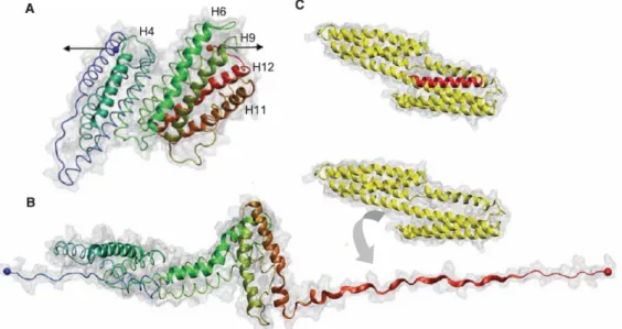
Inside of the cell one important adhesion player is talin. Talin couples the cytoskeleton to the extracellular matrix through integrins. It can bind to vinculin for assembly and reorganization of the actin cytoskeleton. Talin is constituted of a head that binds and activates integrins and of a rod that contains bundles of -helices with up to 11 buried vinculin binding sites. These sites become exposed by the application of a tensile force that stretches the molecule by a sequential unfolding of the helical bundles (figure 1.7).35
Figure 1.7: Representation of a domain of Talin (A) at rest; (B) under stretch. Under stretch the domain unfolds exhibiting an interaction domain with Vinculin (C). Image taken from ref 36.
Filamins constitute another family of proteins that connect transmembrane receptors such as integrins to actin. Filamins are dimeric proteins that consist of an N-terminal actin binding domain followed by 24 immunoglobulin-like domains (FLNa1-24).37 Filamins bind the integrins through interactions between the cytoplasmic tails of the integrin chains and
18 CD faces of FLNa19 and FLNa21. However, these CD faces are masked by the A-strand of the preceding FLNa18 and FLNa20 domains, thus inhibiting integrin binding.14 Steered Molecular Dynamics (SMD) simulations suggest that the CD faces of FLNa19 and FLNa21 become exposed as a force applies to the filamin rod. The force induces an unraveling of the filamin fragment structure via a displacement of the A-strand and a dissociation of the domain-domain interactions. Folding of the integrin binding domains FLNa19 and FLNa21 remain intact. This process exposes the binding of filamin to the integrin cytoplasmic tail allowing for the interaction to take place.38
1.3 Cryptic site substrates
It thus clearly appears that the first steps of the mechanism by which cells transform mechanical information into a chemical signal involve conformational changes of proteins by which cryptic sites become accessible for interaction with other proteins. Exhibiting crytic sites or molecules by stretching a material or a surface is the strategy followed by the group in which I performed my PhD work to create chemo or cyto mechanoresponsive substrates. We will now briefly review the research done in this field since 2005 by our group. Two routes were followed to develop such substrates: one route was based on polyelectrolyte multilayers and one route was based on grafting polymer brushes directly on silicone sheets.
1.3.1 Polyelectrolyte multilayer based mechanoresponsive systems
Polyelectrolyte multilayers are obtained by bringing a substrate alternately in contact with a polyanion and a polycation solution.39 This concept was introduced in 1991 by Decher.40 There exist two kinds of polyelectrolyte multilayers. The first multilayers that were discovered correspond to films whose thickness increases linearly with the number of deposition steps. These films are nicely stratified39 and no polyelectrolyte can diffuse through the film. The two most studied linearly growing polyelectrolyte multilayers are poly(styrene sulfonate)/poly(allylamine hydrochloride) (PSS/PAH) and poly(acrylic acid)/poly(diallyl dimethyl ammonium chloride) (PAA/PDADMA). In 1999 Elbert et al. discovered a second kind of multilayers, namely films whose thickness increases exponentially with the number of deposition steps.41 Picart et al. showed that this growth behaviour is due to the fact that at least one of the two polyelectrolytes participating in the film construction diffuses in and out of the film at each bilayer deposition step.42 These films correspond in fact to gel-like structures into which one can incorporate proteins and enzymes which can diffuse in the
19 film.43 It was also found that polyelectrolyte multilayers preserve the enzymatic activity. Garza et al.44 showed that one can deposit linearly growing films on top of exponentially growing ones and that the linearly growing film then acts as a barrier towards the diffusion of polyelectrolytes from the solution into the exponentially growing multilayer. This type of architecture is called a reservoir-barrier architecture.
In a first study two linearly growing multilayers were deposited one on top of the other, namely a poly(acrylic acid)/poly(allylamine hydrochloride) film on top of a Nafion/ poly(allylamine hydrochloride). This structure was deposited on a silicone substrate that could be stretched. Nafion being hydrophobic and the (PAA/PAH) film being hydrophilic, one observed a reversible hydrophobicity change during stretching/unstretching cycles. The amplitude of the hydrophobicity changes, measured by changes in the water contact angle, were important (variations of the water contact angle between 110 and 50°) when the ending (PAA/PAH) multilayer was constituted of two bilayers whereas changes of less than 10° were observed when the (PAA/PAH) film was constituted of 4 bilayers. The reversible change in hydrophobicity was interpreted as being due to a thinning of the ending multilayer under stretching, leading to the appearance of Nafion that comes in contact with water. Yet when the outer multilayer becomes too thick, Nafion remains always embedded under the PAA/PAH multilayer, whatever the stretching degree.45
Next the behaviour under stretching of reservoir-barrier structures was investigated with the idea to open the barrier under stretching and to close it again when returning to the non-stretched state. Two types of architectures were investigated (HA/PLL)-(PSS/PAH) and (HA/PLL)-(PSS/PDADMA) (HA: hyaluronic acid; PLL: poly(L-lysine)) where (HA/PLL) is an exponentially growing film which plays the role of reservoir and (PSS/PAH) and (PSS/PDADMA) are two linearly growing films which play the role of barrier. Whereas stretching the film induced cracks in the (PSS/PAH) multilayer which appears brittle, holes in the nanometer range formed with the (PSS/PDADMA) multilayer. When returning to the non-stretched state, the cracks closed and the two sides of the cracks became adjacent for (PSS/PAH) leading to a partial healing (figure 1.8).
20 Figure 1.8: (left) Confocal laser microscopy image of a (PLL/HA)30/PLLRho/(HA/PLL)/
(PSS/PAH)30/(HA/PLL)30(HA/PLLFITC) under stretching (top) and after coming back to rest
(bottom). Image taken from ref.46 (right): Fluorescence microscopy image of a (PLL/HA)30/PLLRho/(HA/PLL)/(PSS/PDADMA)5/(HA/PLL)30(HA/PLLFITC) film under
stretching. One observes nanovalves all over the surface. Image taken from ref. 47
For the (PSS/PDADMA) case, the holes disappeared so that the system healed totally.46, 47 These results were obtained by bringing the film in contact with the polyelectrolyte solutions during 10 minutes during each deposition step. Yet the linearly growing film is deposited on top of an exponentially growing one into which polyelectrolytes can diffuse and even exchange with polyelectrolyte chains already present in the film. This renders the buildup process sensitive to the contact time between the film and the polyelectrolyte solutions during the deposition steps. It was shown more recently that by reducing this contact time, no holes forms anymore under stretching for the (PSS/PDADMA) barrier and the barrier can remain tied under stretching when the number of bilayers constituting the barrier exceeds four.48, 49
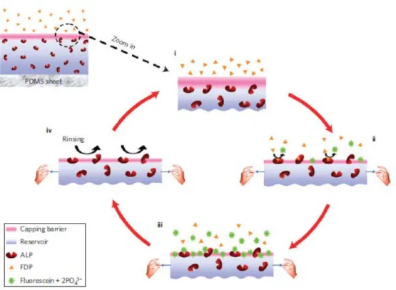
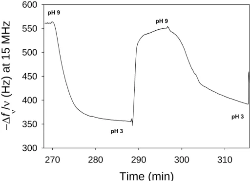
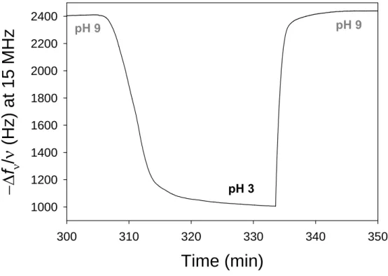
This observation led to the development of the first substrate that became enzymatically active under stretching in a partially reversible way.48 This substrate was constituted of a (HA/PLL)-(PSS/PDADMA) architecture where an enzyme, alkaline phosphatase, was incorporated in the reservoir. The film was deposited on a silicone sheet. Alkaline phosphatase is a hydrolase enzyme responsible for dephosphorylation of many types of molecules, including fluorescein diphosphate (FDP). FDP is a non-fluorescent substrate and hydrolysis of its two phosphate substituents mediated by alkaline phosphatase yields weakly fluorescent fluorescein monophosphate followed by strongly fluorescent fluorescein. FDP was present in the solution in contact with this film. In the absence of stretching, the
21 (PSS/PDADMA) multilayer played the role of barrier towards FDP. When the film was stretched above 70% a strong fluorescence increase was observed in the solution indicating that the enzymes became accessible to the substrate and remained active. When returning to the non-stretched state, the enzymatic activity was again strongly (but not totally) inhibited indicating reversibility of the system. It was shown that the (PSS/PDADMA) multilayer remained tied to FDP and that the enzymes present in the reservoir were exhibited through the barrier. This system can thus be qualified as the first cryptic site substrate. It is schematically represented in figure 1.9.
Figure 1.9: Schematic representation of the first enzymatic mechano-responsive film. Enzymes are embedded in a (HA/PLL) multilayer that is capped by a (PSS/PDADMA) multilayer playing the role of barrier towards the substrate of the enzyme. Under stretching the enzymes becomes accessible to its substrate and the reaction takes place. This system is close to reversible. Image taken ref.48
More recently the same concept was applied but in the reverse way, namely by incorporating the substrate FDP in the reservoir and by anchoring, on top of the (PSS/PDADMA) barrier the enzyme alkaline phosphatase.50 At rest the film was inactive and by stretching fluorescein diffused from the film towards the solution. This was due to the fact that by stretching the FDP substrate could come in contact with the enzymes and that
22 fluorescein, the product of the reaction, diffused into the solution. Unfortunately this system was not reversible.
Polyelectrolyte multilayers were also used in another way to create chemoresponsive substrates. Poly(acrylic acid) chains were chemically modified by grafting onto them phosphorylcholine moieties through short ethylene oxide linkers (EO)3 (PAA-PC chains). The
grafting ratio was about 25%. Phosphorycholine groups cover the outer membrane of red blood cells and are responsible for their non-thrombogenic character.51 These chains were then used to built PEI/(PSS/PAH)3/PSS/(PAH/PAA-PC)2 multilayer architectures which were
deposited on silicone sheets. These films proved to be antifouling (no protein adsorption and no cell adhesion) at rest and under stretching at least up to a stretching degree of 50%.52 These films were further functionalized by incorporating, between the (PSS/PAH)3 and the
(PAH/PAA-PC)2 multilayers one layer of PAA chains modified by grafting biotin or
(arginine-glycine-aspartactic acid) (RGD) peptides again linked to the PAA chains through an (EO)3 linker. Biotin is a ligand of streptavidin and the biotin/streptavidin bond is one of the
strongest non-covalent bonds reported in biology (for a schematic representation see figure 1.10).53
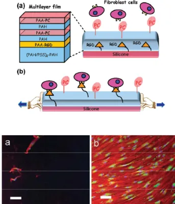
Figure 1.10: (top) Schematic representation of the fist cryptic surface film based on embedding ligands (her RGD peptides) under a multilayer (PAH/PAA-CP). (bottom left) Cells deposited on a non stretched films do not adhere; (bottom right) When stretched the film becomes cell adherent due to RGD peptide exhibition. Images taken from ref54.
23 RGD peptides are known to promote cell adhesion through interactions with integrins. At rest these architectures remain antifouling and no interaction is observed with streptavidin (in the case of biotin) or cells in the case of RGD. By stretching these films up to 50%, they become specifically interacting with streptavidin and adherent to cells. The amount of streptavidin depositing on a stretched film increases linearly with the stretching degree up to 50% of stretching.54 This shows that stretching these architectures renders ligands accessible to their receptors. Yet this system was not reversible: when returning to the non-stretched state the substrate remained interactive with streptavidin or cells.
1.3.2 Responsive systems based on poly(ethylene oxide) brushes55
The latter system we just described was based both on polyelectrolyte multilayers and on short PEO-PC chains extending into the solution. Another strategy to built cryptic site substrates was based on poly(ethylene oxide) chains of mass of about 2000 (45 monomers) and NH2-(EO)3-biotin or NH2-(EO)3-RGD peptides that are directly anchored onto silicone
sheets. In order to achieve the grafting onto the silicone sheets, the substrate was first modified by plasma polymerization of maleic anhydride resulting in an anhydride maleic polymer film deposited on the silicone substrate. The anhydride maleic groups were then hydrolysed through contact with water resulting in a surface covered by carboxylic groups. The difficulty of this type of functionalization is to sufficiently cover the surface yet not to render the functionalized surface brittle which would result in cracks during stretching.56 Once this functionalization was achieved, PEO chains ending by an amine group were covalently fixed onto the surface through EDC/NHS chemistry performed under stretching at 60°C. In a second step, ligands, biotin or RGD peptides, terminated by an amine were covalently anchored onto the remaining non-reacted carboxylic groups under stretching. When returning to the non-stretched state, this system appeared anti-fouling, non-interacting with streptavidin and non cell adherent. Under stretching streptavidin interacted with the surface and cells adhered readily. When returning to the non stretched state the system returned to its initial state. The postulated mechanism is schematically represented in figure 1.11. The mechanoresponsive process appears that totally reversible and this appears to be the first reversible cyto and chemo mechanoresponsive system. One of our goals was to achieve a similar system, yet without using plasma polymerisation, a technique that is readily available in most chemistry laboratories.
24 Figure 1.11: Schematic representation of the first fully reversible cyto and chemo mechano-responsive film. The grafting of the PEG chains and the biotin (or RGD) ligands is based on a first functionalization of the silicone substrate (yellow) by polymer plasma treatment. Image taken from ref. 55
1.4 Summary
In the last decade, important breakthroughs have been reported in the field of mechanoresponsive material design. Most of these works does concern exclusively the transduction of a mechanical force into a changing of colour (or fluorescence emission) of the material. The reason for this is not a lack of originality but rather due to the impossibility to get another outcome. Indeed, the mechanophore-contained materials investigated, such as polyethylene or polymethylmethacrylate, are mainly inert to their environment: they cannot communicate with the surrounding media through exchange with solution (aqueous or organic) or air, except displaying a colour. Thus, the transformation of the mechanical stress undergone by the material cannot lead to a change of a catalytic reaction, a releasing of compounds or a recognition process: only the emission of light seems to be the response of these kinds of systems. In all these mecanoresponsive material, the strategy systematically involved is founded on the molecular incorporation of the mecanophore into the polymeric architecture of the material. Thus, the host material chosen is of crucial importance because it must be both resistant to stress and elastic to be reversibly mechanoresponsive. It most cases, this last feature is not met.
The contribution of my group is based on another strategy inspired from nature: use cryptic site pocket to design a new kind of mechanoresponsive systems. An important contribution has been reported since 2005 in the literature, as described in this first chapter. In this strategy, only the material will undergo the mechanical force. The consequence of this
25 force is to reveal active sites, which were not accessible before the imposition of the stress. By using multilayer films or PEG brushes, these aqueous based systems in essence can exchange and thus communicate with their environment through water. This property let hopes to design a new class of 2D or 3D mechanoresponsive systems able to recognize molecules specifically and have catalytic activity triggered by a mechanical force. This was the aim of my PhD project: “Design of mechanoresponsive surfaces and materials”.
1.5 References
1. Stuart, M. A. C.; Huck, W. T. S.; Genzer, J.; Muller, M.; Ober, C.; Stamm, M.; Sukhorukov, G. B.; Szleifer, I.; Tsukruk, V. V.; Urban, M.; Winnik, F.; Zauscher, S.; Luzinov, I.; Minko, S., Emerging applications of stimuli-responsive polymer materials.
Nature Materials 9, 101-113.
2. Yamada, N.; Okano, T.; Sakai, H.; Karikusa, F.; Sawasaki, Y.; Sakurai, Y., Thermoresponsive Polymeric Surfaces - Control of Attachment and Detachment of Cultured-Cells. Makromol. Chem-Rapid 1990, 11, 571-576.
3. Ito, T.; Hioki, T.; Yamaguchi, T.; Shinbo, T.; Nakao, S.; Kimura, S., Development of a molecular recognition ion gating membrane and estimation of its pore size control. Journal of
the American Chemical Society 2002, 124, 7840-7846.
4. Sui, Z. J.; King, W. J.; Murphy, W. L., Dynamic materials based on a protein conformational change. Advanced Materials 2007, 19, 3377-3380.
5. Lukach, A.; Liu, K.; Therien-Aubin, H.; Kumacheva, E., Controlling the Degree of Polymerization, Bond Lengths, and Bond Angles of Plasmonic Polymers. Journal of the
American Chemical Society 2013, 134, 18853-18859.
6. Lott, J.; Weder, C., Luminescent Mechanochromic Sensors Based on Poly(vinylidene fluoride) and Excimer-Forming p-Phenylene Vinylene Dyes. Macromolecular Chemistry and
Physics 2010, 211, 28-34.
7. Davis, D. A.; Hamilton, A.; Yang, J.; Cremar, L. D.; Van Gough, D.; Potisek, S. L.; Ong, M. T.; Braun, P. V.; Martinez, T. J.; White, S. R.; Moore, J. S.; Sottos, N. R., Force-induced activation of covalent bonds in mechanoresponsive polymeric materials. Nature 2009, 459, 68 - 72.
8. Wolff, J., The law of bone remodelling, Berlin, Heidelberg, New York, Springer, 1986 (translation of the German 1892 edition)
26 9. Engler, A. J.; Sen, S.; Sweeney, H. L.; Discher, D. E., Matrix elasticity directs stem cell lineage specification. Cell 2006, 126, 677-689.
10. Staudinger, H.; Bondy, H. F., Uber Isopren und Kautschuk 19. Mitteil.): Uber die Molekuhlgrosse des Kautschuks und der Balata. Ber. Dtsch. Chem. Ges. 1930, 63, 734.
11. Kauzmann, W.; Eyring, H., The Viscous Flow of Large Molecules. J. Am. Chem. Soc. 1940, 62, 3113-3125.
12. Encina, M. V.; Lissi, E.; Sarasua, M.; Gargallo, L.; Radic, D., Ultrasonic Degradation of Polyvinylpyrrolidone - Effect of Peroxide Linkages. Journal of Polymer Science Part
C-Polymer Letters 1980, 18, 757-760.
13. Wiita, A. P.; Perez-Jimenez, R.; Walther, K. A.; Grater, F.; Berne, B. J.; Holmgren, A.; Sanchez-Ruiz, J. M.; Fernandez, J. M., Probing the chemistry of thioredoxin catalysis with force. Nature 2007, 450, 124-130.
14. Alegre-Cebollada, J.; Perez-Jimenez, R.; Kosuri, P.; Fernandez, J. M., Single-molecule Force Spectroscopy Approach to Enzyme Catalysis. Journal of Biological Chemistry 2010,
285, 18961-18966.
15. Bell, G. I., Models for Specific Adhesion of Cells to Cells. Science 1978, 200, 618-627. 16. Perez-Jimenez, R.; Li, J. Y.; Kosuri, P.; Sanchez-Romero, I.; Wiita, A. P.; Rodriguez-Larrea, D.; Chueca, A.; Holmgren, A.; Miranda-Vizuete, A.; Becker, K.; Cho, S. H.; Beckwith, J.; Gelhaye, E.; Jacquot, J. P.; Gaucher, E.; Sanchez-Ruiz, J. M.; Berne, B. J.; Fernandez, J. M., Diversity of chemical mechanisms in thioredoxin catalysis revealed by single-molecule force spectroscopy. Nature Structural & Molecular Biology 2009, 16, 890-U120.
17. Baldus, I. B.; Grater, F., Mechanical Force Can Fine-Tune Redox Potentials of Disulfide Bonds. Biophysical Journal 2012, 102, 622-629.
18. Kryger, M. J.; Munaretto, A. M.; Moore, J. S., Structure-Mechanochemical Activity Relationships for Cyclobutane Mechanophores. Journal of the American Chemical Society 2011, 133, 18992-18998.
19. Hickenboth, C. R.; Moore, J. S.; White, S. R.; Sottos, N. R.; Baudry, J.; Wilson, S. R., Biasing reaction pathways with mechanical force. Nature 2007, 446, 423-427.
20. Piermattei, A.; Karthikeyan, S.; Sijbesma, R. P., Activating catalysts with mechanical force. Nature Chemistry 2009, 1, 133-137.
21. Davis, D. A.; Hamilton, A.; Yang, J. L.; Cremar, L. D.; Van Gough, D.; Potisek, S. L.; Ong, M. T.; Braun, P. V.; Martinez, T. J.; White, S. R.; Moore, J. S.; Sottos, N. R.,