O R I G I N A L A R T I C L E – G Y N E C O L O G I C O N C O L O G Y
Long-term Outcome and Late Side Effects in Endometrial Cancer
Patients Treated with Surgery and Postoperative Radiation
Therapy
Fernanda G. Herrera, MD1, Olalla Santa Cruz, MD1, Chahin Achtari, MD2, Jean Bourhis, MD1, and Mahmut Ozsahin, MD, PhD1
1Department of Radiation Oncology, Centre Hospitalier Universitaire Vaudois (CHUV), Lausanne, Switzerland; 2Department of Gynecology Oncology, Centre Hospitalier Universitaire Vaudois (CHUV), Lausanne, Switzerland
ABSTRACT
Background. We retrospectively reviewed the long-term outcome and late side effects of endometrial cancer (EC) patients treated with different techniques of postoperative radiotherapy (PORT).
Methods. Between 1999 and 2012, 237 patients with EC were treated with PORT. Two-dimensional external beam radiotherapy (2D-EBRT) was used in 69 patients (30 %), three-dimensional EBRT (3D-EBRT) in 51 (21 %), and intensity-modulated RT (IMRT) with helical Tomotherapy in 47 (20 %). All patients received a vaginal brachytherapy (VB) boost. Seventy patients (29 %) received VB alone. Results. After a median of 68 months (range, 6–154) of follow-up, overall survival was 75 % [95 % confidence interval (CI), 69–81], disease-free survival was 72 % (95% CI, 66–78), cancer-specific survival was 85 % (95 % CI, 80–89), and locoregional control was 86 % (95 % CI, 81–91). The 5-year estimates of grade 3 or more toxicity and second cancer rates were 0 and 7 % (95 % CI, 1–13) for VB alone, 6 % (95 % CI, 1–11) and 0 % for IMRT ? VB, 9 % (95 % CI, 1–17) and 5 % (95 % CI, 1–9) for 3D-EBRT ? VB, and 22 % (95 % CI, 12–32) and 12 % (95 % CI, 4–20) for 2D-EBRT ? VB (P = 0.002 and P = 0.01), respectively.
Conclusions. Pelvic EBRT should be tailored to patients with high-risk EC because the severe late toxicity observed might outweigh the benefits. When EBRT is prescribed for
EC, IMRT should be considered, because it was associated with a significant reduction of severe late side effects.
Endometrial cancer (EC) is the most common gyneco-logic malignancy in developed countries.1,2At the time of diagnosis, most patients present with early-stage disease, and low-risk patients have a risk of locoregional recurrence (LRR) of 5–10 %. However, high- to intermediate-risk patients harbor a combination of high-grade, deep myo-metrial invasion and/or lymphovascular space invasion (LVSI), with a risk of LRR of up to 27 %.3Randomized studies have shown that postoperative radiotherapy (PORT) decreases the LRR rate without affecting overall survival (OS). However, two-thirds of the patients in those trials had low-risk disease and a substantial risk of dying as a result of competing hazards.3–8The long-term outcome of these trials also confirmed the morbidity risks of adju-vant PORT using mainly two-dimensional (2D) external beam radiotherapy (EBRT) techniques.9The postoperative radiotherapy in EC (PORTEC-2) randomized trial showed that the toxicity of PORT using three-dimensional (3D)-EBRT techniques outweighs the benefits and that vaginal brachytherapy (VB) alone can be enough to avoid local recurrences in the subgroup of patients with high- to intermediate-risk factors (grade 1–2 tumors, [50 % myo-metrial invasion, endometrioid type, age [60 years, and no LVSI).10 Pelvic radiotherapy (RT) has changed dramati-cally over the last few decades with the introduction of intensity-modulated RT (IMRT). The question that remains open is whether IMRT will lead to a reduced rate of severe side effects. Studies have found evidence of an increased risk of secondary neoplasms after PORT, and some investigators have recently postulated that IMRT can potentially increase the risk of second cancers.9,11,12 We
Ó Society of Surgical Oncology 2014 First Received: 3 January 2014; Published Online: 7 March 2014 M. Ozsahin, MD, PhD
e-mail: [email protected] DOI 10.1245/s10434-014-3622-9
aimed in this study to assess a population-based cohort who received adjuvant PORT over a 12-year period with dif-ferent technique of RT. In our series, IMRT was delivered with helical Tomotherapy (Accuray, Madison, WI). To-motherapy is a new generation 6-MV photon accelerator that allows helical delivery of highly conformal and homogeneous doses associated with daily image-guided RT. We assessed the severe late toxicity (grade 3 or more) and the incidence of second cancers.
METHODS Study Population
After approval by the local ethics committee, we retro-spectively reviewed the charts of 237 eligible patients from a total of 245 EC patients who received adjuvant PORT between 1999 and 2012 at the Lausanne University Hos-pital. Inclusion criteria were a pathologic diagnosis of EC, stage I–IVA according to the International Federation of Gynecology and Obstetrics (FIGO) 1988 definition, and a minimum of 6 months of follow-up.13 One patient was excluded from the analysis because of disseminated dis-ease, and 7 patients were excluded for incomplete follow-up. Data were obtained from the electronic and written medical records and included age at diagnosis, date of diagnosis, date of surgery, type of surgical procedure, number of pathologically examined lymph nodes, surgical margins, histology, grade, depth of myometrial invasion, stage, LVSI, type and dose of PORT, date and location of recurrence, date of last follow-up, date of death, incidence and types of second cancers, and late side effects (grade 3 or more) based on common terminology criteria for adverse events version 4.0, which were confirmed from follow-up records and surgical and/or procedural inter-ventions. Sites of failure were grouped into isolated vaginal recurrence, LRR (pelvic and/or paraaortic), and distant metastases (extraabdominal sites and positive peritoneal cytology).
Statistical Considerations
Proportions were compared by using the Chi square test for values of 5 or higher and with Fisher’s exact test for values of less than 5. Survival curves were estimated by using the Kaplan–Meier method. Time to any event was measured from the day RT started. Death certificates confirmed date of deaths. If clinical or pathologic evidence of active, recurrent disease was present, deaths were attributed to EC. The events were death (all causes) for OS, EC-related mortality for cancer-specific survival (CSS), and death (all causes) or relapse for disease-free survival
(DFS). For the locoregional control (LRC) rate, the event consisted of local or regional relapse. Confidence intervals (CI) were calculated from standard errors. In univariate analyses, differences between groups were assessed by using the log-rank test. All obtained significant P values were corrected for multiple comparisons with the Bonfer-roni correction method in which the P values are multiplied by the number of comparisons. In multivariate analyses, we screened for prognostic factors with a P value of less than 0.05 in univariate analyses by using the Cox regression analysis to define the independent contribution of each prognostic factor. A P value of \0.05 was considered to be statistically significant. All data were examined using JMP version 9.0.1 (SAS Institute Inc., Cary, NC).
RESULTS
Patient Characteristics
A total of 237 patients with EC were identified with complete follow-up. Patients’ surgical, pathologic, and treatment characteristics are detailed in Table1. The median age was 69 years (range, 37–90 years).
Surgery
All patients underwent total abdominal hysterectomy and bilateral salpingo-oophorectomy (TAH-BSO) includ-ing abdominal washinclud-ing, with (n = 126) or without (n = 111) lymphadenectomy.
Radiotherapy
RT started 6–8 weeks after surgery and was delivered by using a two-dimensional four-field technique (2D-EBRT) in 69 patients (30 %), four-field conformal RT (3D-EBRT) in 51 (21 %), and Tomotherapy in 47 (20 %). All of the patients treated in the Tomotherapy group had daily image-guided RT using helical megavoltage-based computed tomography. All patients treated with different techniques of EBRT also received a VB boost. Seventy patients (29 %) were treated with postoperative VB alone. Patients treated with 2D-EBRT had the radiation portals determined using a kilovoltage simulator. From the intro-duction of 3D-EBRT, target volumes and organs at risk were contoured on a computed tomography image, and personalized shielding was applied by using the multileaf collimator. For both 2D-EBRT and 3D-EBRT, the radia-tion fields extended from the anterior aspect of the pubic symphysis to the L5–S1 interspace and laterally posteriorly up to the middle sacrum. With the introduction of helical Tomotherapy, the radiation volumes were based on the
radiation therapy oncology group consensus guidelines, including the irradiation of common iliac nodes.14 The median EBRT dose was 45 Gy (range, 41.4–50.4 Gy) in fractions of 1.8 Gy. VB was administered to the upper 3 cm of the vagina by using a high-dose rate technique delivered via vaginal cylinders; the dose was prescribed to the vaginal mucosa (0.5 cm from the cylinder surface). The median number of VB fractions was 3 (range, 2–6), and the median VB dose was 5 Gy per fraction (range, 3–7.5 Gy). The most frequent VB regimen was 10 Gy in 2 fractions for those previously receiving EBRT (VB boost) and 20 Gy in 4 fractions for those only receiving VB.
Disease Outcome
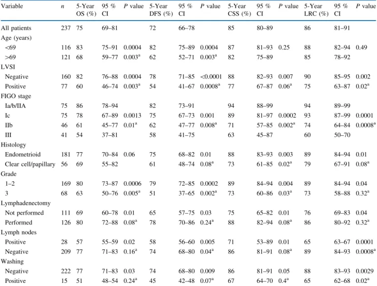
In a median follow-up of 68 months (range, 6–154 months), the 5-year OS was 75 % (95 % CI, 69–81), DFS was 72 % (95 % CI, 66–78), CSS was 85 % (95 % CI, 80–89), and LRC was 86 % (95 % CI, 81–91). By the end of follow-up, 161 of 237 (68 %) patients were alive without disease, and 12 of 237 were alive with recurrent disease. Thirty-three of 237 (14 %) patients died of EC, and 31 of 237 (13 %) died of intercurrent diseases (24 cardiovascular, 6 s tumors, and 1 treatment-related toxicity). A total of 47 patients (20 %) experienced a recurrence. In univariate analyses, statistically significant factors unfavorably influencing OS and DFS were patient age ([69 years), presence of LVSI, advanced stage, pap-illary-serous or clear-cell histology, grade 3 tumors, absence of lymphadenectomy, presence of positive lymph nodes, and positive abdominal washing. For CSS and LRC, the previously mentioned parameters applied, except for age (Table2). After multivariate analyses, the remaining independent prognostic factors unfavorably influencing OS and DFS were age ([69 years), advanced stage, posi-tive LVSI, absence of lymphadenectomy, and grade 3 histology. For CSS and LRC, the previously mentioned variables applied except for age (Table3).
The 5-year LRC rate was 89 % (95 % CI, 80–96) for patients treated with VB alone versus 85 % (95 % CI, 79–91) for those treated with EBRT ? VB (P = 0.5). Among those treated with VB alone, there was an increased proportion of patients with endometrioid-type histology [endometrioid type (n = 62) versus serous type or clear cell (n = 8); P = 0.0026], grade 1–2 [grade 1–2 (n = 67) versus grade 3 (n = 3), P \ 0.0001], earlier-stage tumors [Ia, b, and IIa (n = 23), Ic (n = 42), and IIb (n = 5); P\ 0.0001], and absence of LVSI [LVSI absent (n = 64), LVSI present (n = 6); P \ 0.0001]. When comparing patients (n = 60) who fit the PORTEC-2 inclusion criteria, i.e., stage I tumors, patients [60 years old (Ic and grade 1 or 2), or Ib (grade 3) or stage IIa tumors at any age excluding grade 3 or [50 % myometrial invasion, with
those without the PORTEC-2 criteria (n = 177), the 5-year LRC was 96 % (95 % CI, 91–100) versus 83 % (95 % CI, 77–89; P = 0.02), respectively.10
Toxicity
By the end of follow-up, 24 patients (9.7 %) developed severe late toxicity (grade 3 or more). Two patients developed urethral stenosis requiring surgery, resulting in permanent urinary incontinence. Three patients developed synchronous urethral stenosis and rectovaginal fistulas. Sixteen patients had intestinal toxicity (bowel stenosis and/ or rectovaginal fistulas). One patient died because of
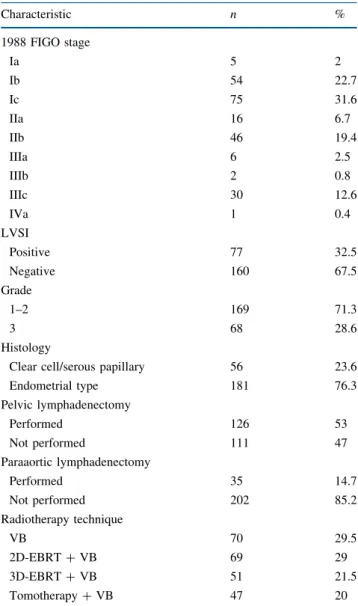
small-TABLE 1 Patients’ surgical, pathologic, and treatment characteris-tics (n = 237) Characteristic n % 1988 FIGO stage Ia 5 2 Ib 54 22.7 Ic 75 31.6 IIa 16 6.7 IIb 46 19.4 IIIa 6 2.5 IIIb 2 0.8 IIIc 30 12.6 IVa 1 0.4 LVSI Positive 77 32.5 Negative 160 67.5 Grade 1–2 169 71.3 3 68 28.6 Histology
Clear cell/serous papillary 56 23.6 Endometrial type 181 76.3 Pelvic lymphadenectomy Performed 126 53 Not performed 111 47 Paraaortic lymphadenectomy Performed 35 14.7 Not performed 202 85.2 Radiotherapy technique VB 70 29.5 2D-EBRT ? VB 69 29 3D-EBRT ? VB 51 21.5 Tomotherapy ? VB 47 20 FIGO international federation of gynecology and obstetrics staging system, LVSI lymphovascular space invasion, VB vaginal cuff brachytherapy, 2D-EBRT two-dimensional external beam radiother-apy, 3D-EBRT three-dimensional external beam radiotherapy
TABLE 2 Univariate analysis Variable n 5-Year OS (%) 95 % CI P value 5-Year DFS (%) 95 % CI P value 5-Year CSS (%) 95 % CI P value 5-Year LRC (%) 95 % CI P value All patients 237 75 69–81 72 66–78 85 80–89 86 81–91 Age (years) \69 116 83 75–91 0.0004 82 75–89 0.0004 87 81–93 0.25 88 82–94 0.49 [69 121 68 59–77 0.003a 62 52–71 0.003a 82 75–89 85 78–92 LVSI Negative 160 82 76–88 0.0004 78 71–85 \0.0001 88 82–93 0.007 90 85–95 0.002 Positive 77 60 46–74 0.003a 54 41–67 0.0008a 77 67–87 0.06a 75 63–87 0.02a FIGO stage Ia/b/IIA 75 86 78–94 82 73–91 94 88–99 94 89–99 Ic 75 78 67–89 0.0013 75 67–73 0.001 89 81–97 0.0002 93 87–99 0.0001 IIb 46 61 45–77 0.01a 62 47–77 0.008a 71 57–85 0.002a 74 64–84 0.0008a III 41 54 37–81 58 41–75 63 45–87 60 50–70 Histology Endometrioid 181 77 70–84 0.06 75 68–82 0.01 88 83–93 0.003 89 84–94 0.01 Clear cell/papillary 56 69 55–82 61 48–74 0.08a 73 61–85 0.02a 79 67–91 0.08a Grade 1–2 169 80 73–87 0.0006 79 72–85 0.0002 89 84–94 0.004 89 84–94 0.04 3 68 63 50–76 0.005a 51 37–65 0.002a 73 60–86 0.03a 73 58–88 0.32a Lymphadenectomy Not performed 111 69 60–78 0.01 65 57–75 0.03 75 65–82 0.01 76 69–83 0.04 Performed 126 80 72–88 0.08a 78 70–86 0.24a 88 82–94 0.08a 86 80–92 0.32a Lymph nodes Positive 28 57 55–59 0.02 58 56–60 0.005 71 53–89 0.01 65 63–67 0.0001 Negative 209 77 71–83 0.16a 74 68–80 0.04a 86 81–91 0.08a 89 84–93 0.0008a Washing Negative 222 77 71–83 0.03 74 68–80 0.009 86 81–91 0.05 88 83–93 0.0029 Positive 15 51 48–54 0.24a 45 42–48 0.07a 67 64–70 0.4a 65 62–68 0.02a
LVSI lymphovascular space invasion, OS overall survival, DFS disease-free survival, CSS cancer-specific survival, LRC locoregional control, CI confidence interval, FIGO international federation of gynecology and obstetrics staging system
a P values after Bonferroni multiple correction analysis
TABLE 3 Multivariate Cox analysis
Variable OS DFS CSS LRC
RR P value RR P value RR P value RR P value Age [69 years 1.04 0.0006 1.11 0.0003 – NS – NS Stage
Ia/b/IIa vs. 1.53 0.0001 1.99 0.001 1.03 \0.0001 1.12 0.005 Ic vs. IIb vs. III
LVSI positive 1.72 0.04 1.63 0.01 1.51 0.01 1.02 0.01 Lymphadenectomy (not performed) 1.08 0.02 1.75 0.02 1.13 0.01 1.09 0.01 Grade 3 histology 1.38 0.01 1.35 0.005 1.33 0.04 1.34 0.01 LVSI lymphovascular space invasion, OS overall survival, DFS disease-free survival, CSS cancer-specific survival, LRC locoregional control, NS nonsignificant, RR relative risk
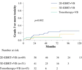
bowel obstruction; she developed an acute abdomen requiring emergency surgery and died 1 week later. We registered two patients with severe chronic lymphedema, one of whom had severe intestinal toxicity requiring sur-gery. One patient had a radiation-induced sacral fracture. According to the RT technique, the 5-year Kaplan–Meier estimate of grade 3 or more toxicity was 0 % for VB alone, 6 % (95 % CI, 1–11) for helical Tomotherapy ? VB, 9 % (95 % CI, 1–17) for 3D-EBRT ? VB, and 22 % (95 % CI, 12–32) for 2D-EBRT ? VB (P = 0.002; Fig.1).
Second Cancers
In a follow-up period of 10–151 months, 24 patients (10 %) were diagnosed with second cancers after PORT. The 5- and 10-year estimated second-cancer incidence was 9 % (95 % CI, 5–13) and 23 % (95 % CI, 13–33), respectively, for the whole population. The 10-year esti-mated second-cancer rate was 19 % (95 % CI, 10–29) in patients younger than 60 years at diagnosis versus 15 % (95 % CI, 9–21) compared with those 60 years or more (P = 0.46). In 4 of 24 women, the malignancy was situated inside or in close proximity to the irradiated volume. The most common in-field second cancers were bladder (n = 1) and colorectal (n = 3) cancer. The most common out-of-field malignancy was breast cancer (n = 12). According to the RT technique, the 5-year estimated second-cancer incidence was 0 % for helical Tomotherapy, 5 % for 3D-EBRT (95 % CI, 1–9), 7 % for VB alone (95 % CI, 1–13), and 12 % (95 % CI, 4–20) for 2D-EBRT (P = 0.01).
DISCUSSION
It is recognized that EBRT in patients with EC leads to better pelvic control compared with surgery alone. A favorable influence on survival, however, has failed to be shown in several randomized trials and meta-analyses.3–6,8 Therefore, the life expectancy of these patients should be taken into consideration to tailor adjuvant RT with the final aim of keeping good LRC while preventing RT-related side effects. Depending on the treatment technique, prognostic factors, and median follow-up time, in early stage disease LRR rates are in the range of 5–15 %.15,16 Our results compare well with the literature (14 %; 95 % CI, 9–19) and confirm good LRC with PORT (Table4). Patients treated with VB alone had a 5-year LRC of 89 %. Well-selected high- to intermediate-risk patients according to the PORTEC-2 inclusion criteria had an LRC rate of 96 % at 5 years. These results are in line with those obtained in PORTEC-2, suggesting the feasibility of such an approach in an appropriately selected subgroup of patients.10 It is important to note that in the PORTEC-2 study, there was also a significant quality-of-life advantage for patients receiving VB alone.17Serious complications in 3–5 % of patients after PORT have been reported in various ran-domized trials and, as in our patients, concerned mainly the gastrointestinal tract.3,4,6,9 In our study, severe complica-tions were diagnosed in nearly 10 % of the patients. These higher-than-expected rates of severe late complications are in line with what is reported in other studies using EBRT ? VB.18We agree that the benefit of VB as a boost after EBRT is questionable.19 The increased incidence of injury to the bowel might be explained by the fact that after hysterectomy, the small-bowel loops occupy the place of the uterus, thereby receiving high doses of EBRT and remaining close to the VB source. We have recently abandoned the systematic use of the VB boost, offering this additional treatment only to patients with cervical invasion or positive vaginal margins. In our series, we showed that the change from 2D- to 3D-EBRT and to helical Tomo-therapy significantly decreased the incidence of severe side effects. The use of IMRT for gynecologic cancers is still a matter of debate. The largest prospective study comparing non-IMRT versus IMRT showed a reduction in grade 3 or more long-term toxicity from 17 to 6 %.20 Other retro-spective series comparing 3D-EBRT versus IMRT after TAH-BSO have failed to show any benefit.21 A recent phase II feasibility trial by the radiation therapy oncology group reported a nonsignificant 12 % reduction in grade 2 or more bowel adverse events in patients treated with IMRT after TAH-BSO.22 A phase III randomized trial is warranted to confirm the potential benefits of IMRT in EC. In our series, the 5-year second-cancer incidence was 0 % for helical Tomotherapy, 5 % for 3D-EBRT, and 12 % for 0.0 0.2 0.4 0.6 0.8 1.0 0 p=0.002 24 58 2D-EBRT+VB (n=69) Number at risk 3D-EBRT+VB (n=51) Tomotherapy+VB (n=43) 2D-EBRT+VB 3D-EBRT+VB Tomotherapy+VB 41 32 46 25 6 34 16 2 24 13 3 48 72 96 120 Months
Grade 3 or more toxicity
FIG. 1 Grade 3 or more late toxicity by radiotherapy technique. 2D-EBRT two-dimensional external beam radiation therapy, 3D-2D-EBRT three-dimensional external beam radiation therapy, VB vaginal cuff brachytherapy
TABLE 4 Randomized trials evaluating the role of adjuvant radiotherapy in endometrial cancer Study Eligibility Arms No of patients LRR rate (%) DFS rate (%) OS rate (%) Number of EC related deaths Median follow-up (months)
Comments Aalders et al. 4 , 11 Stage I V B ? pelvic RT 260 2 8 8 8 9 2 8 [ 60 Lymphadenectomy required VB alone 277 7 8 7 9 1 2 5 Creutzberg et al. PORTEC 1 5 , 14 Stage I, G1, [ 50 % invasion; or G2 any invasion; or G3, \ 50 % invasion Pelvic RT 354 6* 50 52 37 156 No lymphadenectomy Observation 361 15 54 60 30 Keys et al. GOG 99 3 G2/3 ? LVSI ? [ 2/3 myometrial invasion, [ 50 years of age with 2 o f these features; or [ 70 years of age with any of these features Pelvic RT 190 1.6* 93 92 19 69 Lymphadenectomy required Observation 202 9 8 5 8 6 1 5 ASTEC NCIC CTG EN5 6 FIGO (1988) I o r IIA with either G3 (serous included) or [ 50 % myometrial invasion Pelvic RT 452 3.2 85 84 37 58 Lymphadenectomy not required; VB given to 53% of the patients in the observation arm Observation 453 6.1 86 84 41 Nout et al PORTEC 2 9 FIGO (1988) I o r IIA & G1-2, [ 60 years, \ 50 % myometrial invasion; or G3 with \ 50 % myometrial invasion; or IIA, G1-2; or G3 \ 50 % invasion Pelvic RT 214 2.1 83 80 10 45 Lymphadenectomy not required VB 213 5.1 78 85 15 Sorbe et al. 7 FIGO (1988) stage I, endometrioid type and G1-2, or G3 with FIGO Ic ([ 50 % invasion), or aneuploidy Pelvic RT ? VB 224 1.5* 87 89 15 62 Lymphadenectomy not required (only sampling of enlarged lymph nodes) VB alone 223 5 8 7 9 0 2 7 Randall et al. GOG 122 23 FIGO III/IV, maximum residual disease \ 2 cm, no LVSI WART ? pelvis 202 NR 38 42* 100* 74 Node sampling optional Chemotherapy (CAP) 194 NR 42 53 78 Susumu et al. 25 FIGO (1988) Ic-IIIc with [ 50 % myometrial invasion Pelvic RT 193 7 8 4 8 5 2 1 6 0 9 6 % of patients had lymphadenectomy Chemotherapy (CAP) 193 7 8 2 8 7 1 3 Maggi et al. 24 FIGO I/II with [ 50 % myometrial invasion, G3 or IIIc, high risk histology excluded Pelvic RT 166 11 63 69 9 9 5 Pelvic and lumbo-aortic node sampling Chemotherapy (CAP) 177 14 63 66 10 VB vaginal cuff brachytherapy, RT radiation therapy, LRR locoregional recurrence, DFS disease-free survival, OS overall survival, EC endometrial cancer, G grade, LVSI lymphovascular space invasion, FIGO international federation of gynecology and obstetrics, PORTEC the postoperative radiotherapy in EC study group, GOG gynecology oncology group, NCIC CTG EN5 national cancer institute of Canada clinical trials group EC trial 5, ASTEC a surgical trial in EC, WART whole abdominal radiation therapy, AP doxorubicin and cisplatin, CAP cyclophosphamide, doxorubicin, cisplatin a Differences between arms were statistically significant (P \ 0.05)
2D-EBRT, keeping in mind the shorter follow-up in Tomotherapy patients. Onsrud et al.11 analyzed the long-term outcome of a randomized trial of postoperative VB ? EBRT versus VB alone. After a median follow-up of 20.5 years, women younger than 60 years treated with VB ? EBRT had a significantly higher mortality rate due to second malignancies (hazard ratio, 2.02; 95 % CI, 1.3–3.1). It should be noted that the RT delivery in that study was performed mainly with cobalt-60 using a two-field technique. This technique fully exposes the bladder and the bowel to high doses of radiation. The 15-year rates of second cancers in the PORTEC-1 randomized trial were 22 % in patients treated with adjuvant EBRT versus 16 % in the observational group (P = 0.10). The incidence rates were compared with those of an age-and sex-matched population. The observed versus expected ratios were 1.40 for the total group (1.62 for EBRT and 1.2 for the obser-vational group, P not significant). The predominant cancer types were gastrointestinal cancer (6.2 % in the EBRT group vs. 3.2 % in the observational group) and breast cancer (4.8 % in the EBRT group vs. 6.6 % in the obser-vational group). These differences did not reach statistical significance.15 Chaturvedi et al.23 reported on a series of 101,760 cervical cancer patients with more than 40 years of follow-up who were treated (n = 52,613) or not treated (n = 27,382) with RT. They observed 12,496 incidents of second cancers [standard incidence ratio (SIR) = 1.30, 95 % CI, 1.28–1.33]. Compared with the general popula-tion, the excess absolute risk was 22.7 per 10,000 person-years. Cervical cancer patients treated with RT as opposed to those not treated with RT were at increased risk of second cancers at any site, and the SIR was dependent on the amount of RT administered. Heavily irradiated organs located in the irradiated field and receiving[3 Gy (average radiation dose was 10–66 Gy depending on the location of the organ) had an SIR of 1.59 (95 % CI, 1.16–1.26), compared with moderately (1–3 Gy) and lightly (\1 Gy) irradiated sites (SIR = 1.30, 95 % CI, 1.54–1.66, and SIR = 1.21, 95 % CI, 1.16–1.26, respectively). This study demonstrates that the risk of RT-induced second cancers increases with time and that there is an RT dose effect.
Our study adds information to the ongoing discussion in prescribing adjuvant EBRT versus VB and the technology of EBRT. Nevertheless, because of its retrospective approach, it has several limitations. We could not obtain information regarding urinary incontinence or fecal leak-age, which are well-known side effects particularly after EBRT and are better assessed in quality-of-life studies. Lymphadenectomy was heterogeneously performed. Finally, we believe that RT and chemotherapy should be considered for patients with locally advanced disease and those with clear-cell or serous-papillary histology on the basis of the high frequency of distant recurrence and LRR
observed.24–27 The role of chemotherapy in stage I–II disease with high-risk pathologic features is under evalu-ation in the ongoing PORTEC-3 trial.
CONCLUSION
For patients with high- or intermediate-risk EC, VB alone offers high rates of local control with no severe complications. EBRT should be tailored to patients with high-risk features because the possible severe late toxicity may outweigh the benefits. The addition of a VB boost after EBRT is associated with higher-than-expected late severe complications. The correlation between severe late toxicity and RT techniques is observed in our study. When EBRT is indicated, IMRT and daily image-guided RT should be considered as a viable treatment option to min-imize severe late toxicity. Patients should be informed about the potential increased risk of second malignancies after the diagnosis of EC and PORT because 2D-EBRT techniques significantly increase the risk of second tumors and the severity of side effects. Longer follow-up and more patients are needed to confirm the lowest second cancer rates obtained with IMRT in this study. The potential benefits observed with IMRT should be confirmed in a randomized trial.
ACKNOWLEDGMENT The authors are grateful to Julia Rengiers for her excellent help in the preparation of the manuscript.
CONFLICT OF INTEREST Authors declared no conflicts of interest.
REFERENCES
1. Ferlay J, Shin HR, Bray F, Forman D, Mathers C, Parkin DM. Estimates of worldwide burden of cancer in 2008: GLOBOCAN 2008. Int J Cancer. 2010;127:2893–917.
2. Ferlay J, Parkin DM, Steliarova-Foucher E. Estimates of cancer incidence and mortality in Europe in 2008. Eur J Cancer. 2010;46:765–81.
3. Keys HM, Roberts JA, Brunetto VL, Zaino RJ, Spirtos NM, Bloss JD, et al. A phase III trial of surgery with or without adjunctive external pelvic radiation therapy in intermediate risk endometrial adenocarcinoma: a gynecologic oncology group study. Gynecol Oncol. 2004;92:744–51.
4. Aalders J, Abeler V, Kolstad P, Onsrud M. Postoperative external irradiation and prognostic parameters in stage I endometrial carcinoma: clinical and histopathologic study of 540 patients. Obstet Gynecol. 1980;56:419–27.
5. Creutzberg CL, van Putten WL, Koper PC, Lybeert ML, Jobsen JJ, Wa´rla´m-Rodenhuis CC, et al. Surgery and postoperative radiotherapy versus surgery alone for patients with stage-1 endometrial carcinoma: multicentre randomised trial. PORTEC study group. Post operative radiation therapy in endometrial carcinoma. Lancet. 2000;355(9213):1404–11.
6. Group AES, Blake P, Swart AM, Orton J, Kitchener H, Whelan T, et al. Adjuvant external beam radiotherapy in the treatment of
endometrial cancer (MRC ASTEC and NCIC CTG EN.5 ran-domised trials): pooled trial results, systematic review, and meta-analysis. Lancet. 2009;373(9658):137–46.
7. Sorbe B, Horvath G, Andersson H, Boman K, Lundgren C, Pettersson B. External pelvic and vaginal irradiation versus vaginal irradiation alone as postoperative therapy in medium-risk endometrial carcinoma: a prospective randomized study. Int J Radiat Oncol Biol Phys. 2012;82:1249–55.
8. Kong A, Johnson N, Kitchener HC, Lawrie TA. Adjuvant radiotherapy for stage I endometrial cancer: an updated Cochrane systematic review and meta-analysis. J Natl Cancer Inst. 2012; 104:1625–34.
9. Creutzberg CL, van Putten WL, Koper PC, Lybeert ML, Jobsen JJ, Wa´rla´m-Rodenhuis CC, et al. The morbidity of treatment for patients with stage I endometrial cancer: results from a ran-domized trial. Int J Radiat Oncol Biol Phys. 2001;51:1246–55. 10. Nout RA, Smit VT, Putter H, Ju¨rgenliemk-Schulz IM, Jobsen JJ,
Lutgens LC, et al. Vaginal brachytherapy versus pelvic external beam radiotherapy for patients with endometrial cancer of high-intermediate risk (PORTEC-2): an open-label, non-inferiority, randomised trial. Lancet. 2010;375(9717):816–23.
11. Onsrud M, Cvancarova M, Hellebust TP, Trope CG, Kristensen GB, Lindemann K. Long-term outcomes after pelvic radiation for early-stage endometrial cancer. J Clin Oncol. 2013;31:3951–6. 12. Zwahlen DR, Ruben JD, Jones P, Gagliardi F, Millar JL,
Schneider U. Effect of intensity-modulated pelvic radiotherapy on second cancer risk in the postoperative treatment of endo-metrial and cervical cancer. Int J Radiat Oncol Biol Phys. 2009;74:539–45.
13. Morrow CP, Bundy BN, Kurman RJ, Creasman WT, Heller P, Homesley HD, et al. Relationship between surgical-pathological risk factors and outcome in clinical stage I and II carcinoma of the endometrium: a gynecologic oncology group study. Gynecol Oncol. 1991;40:55–65.
14. Small W Jr, Mell LK, Anderson P, Creutzberg C, De Los Santos J, Gaffney D, et al. Consensus guidelines for delineation of clinical target volume for intensity-modulated pelvic radiother-apy in postoperative treatment of endometrial and cervical cancer. Int J Radiat Oncol Biol Phys. 2008;71:428–34. 15. Creutzberg CL, Nout RA, Lybeert ML, Wa´rla´m-Rodenhuis CC,
Jobsen JJ, Mens JW, et al. Fifteen-year radiotherapy outcomes of the randomized PORTEC-1 trial for endometrial carcinoma. Int J Radiat Oncol Biol Phys. 2011;81:e631–8.
16. Klopp AH, Jhingran A, Ramondetta L, Lu K, Gershenson DM, Eifel PJ. Node-positive adenocarcinoma of the endometrium: outcome and patterns of recurrence with and without external beam irradiation. Gynecol Oncol. 2009;115:6–11.
17. Nout RA, Putter H, Jurgenliemk-Schulz IM, Jobsen JJ, Lutgens LC, van der Steen-Banasik EM, et al. Five-year quality of life of endometrial cancer patients treated in the randomised post
operative radiation therapy in endometrial cancer (PORTEC-2) trial and comparison with norm data. Eur J Cancer. 2012;48: 1638–48.
18. Irwin C, Levin W, Fyles A, Pintilie M, Manchul L, Kirkbride P. The role of adjuvant radiotherapy in carcinoma of the endome-trium: results in 550 patients with pathologic stage I disease. Gynecol Oncol. 1998;70:247–54.
19. Crosby MA, Tward JD, Szabo A, Lee CM, Gaffney DK. Does brachytherapy improve survival in addition to external beam radiation therapy in patients with high risk stage I and II endo-metrial carcinoma? Am J Clin Oncol. 2010;33:364–9.
20. Kidd EA, Siegel BA, Dehdashti F, Rader JS, Mutic S, Mutch DG, et al. Clinical outcomes of definitive intensity-modulated radia-tion therapy with fluorodeoxyglucose-positron emission tomog-raphy simulation in patients with locally advanced cervical can-cer. Int J Radiat Oncol Biol Phys. 2010;77:1085–91.
21. Wright JD, Deutsch I, Wilde ET, Ananth CV, Neugut AI, Lewin SN, et al. Uptake and outcomes of intensity-modulated radiation therapy for uterine cancer. Gynecol Oncol. 2013;130:43–8. 22. Jhingran A, Winter K, Portelance L, Miller B, Salehpour M, Gaur
R, et al. A phase II study of intensity modulated radiation therapy to the pelvis for postoperative patients with endometrial carci-noma: radiation therapy oncology group trial 0418. Int J Radiat Oncol Biol Phys. 2012;84:e23–8.
23. Chaturvedi AK, Engels EA, Gilbert ES, Chen BE, Storm H, Lynch CF, et al. Second cancers among 104,760 survivors of cervical cancer: evaluation of long-term risk. J Natl Cancer Inst. 2007;99:1634–43.
24. Randall ME, Filiaci VL, Muss H, Spirtos NM, Mannel RS, Fowler J, et al. Randomized phase III trial of whole-abdominal irradiation versus doxorubicin and cisplatin chemotherapy in advanced endometrial carcinoma: a gynecologic oncology group study. J Clin Oncol. 2006;24:36–44.
25. Maggi R, Lissoni A, Spina F, Melpignano M, Zola P, Favalli G, et al. Adjuvant chemotherapy vs radiotherapy in high-risk endo-metrial carcinoma: results of a randomised trial. Br J Cancer. 2006;95:266–71.
26. Susumu N, Sagae S, Udagawa Y, Niwa K, Kuramoto H, Satoh S, et al. Randomized phase III trial of pelvic radiotherapy versus cisplatin-based combined chemotherapy in patients with inter-mediate- and high-risk endometrial cancer: a Japanese gynecologic oncology group study. Gynecol Oncol. 2008;108: 226–33.
27. Boruta DM II, Gehrig PA, Fader AN, Olawaiye AB. Management of women with uterine papillary serous cancer: a society of gynecologic oncology (SGO) review. Gynecol Oncol. 2009; 115:142–53.