ORIGINAL PAPER
Minimally invasive versus open transforaminal lumbar
interbody fusion: evaluating initial experience
Constantin Schizas&Nicolas Tzinieris&
Elefterios Tsiridis&Victor Kosmopoulos
Received: 10 August 2008 / Accepted: 30 September 2008 / Published online: 21 November 2008
# Springer-Verlag 2008
Abstract The aim of this study was to compare our experience with minimally invasive transforaminal lumbar interbody fusion (MITLIF) and open midline transforaminal lumbar interbody fusion (TLIF). A total of 36 patients suffering from isthmic spondylolisthesis or degenerative disc disease were operated with either a MITLIF (n=18) or an open TLIF technique (n=18) with an average follow-up of 22 and 24 months, respectively. Clinical outcome was assessed using the visual analogue scale (VAS) and the Oswestry disability index (ODI). There was no difference in length of surgery between the two groups. The MITLIF group resulted in a significant reduction of blood loss and had a shorter length of hospital stay. No difference was observed in postoperative pain, initial analgesia consump-tion, VAS or ODI between the groups. Three pseudarthroses were observed in the MITLIF group although this was not statistically significant. A steeper learning effect was observed for the MITLIF group.
Résumé Le but de cette étude est de comparer notre expérience de l’arthrodèse lombaire intercorporéale trans-foraminale par voie mini invasive (MITLIF) ou par voie sanglante classique (TLIF). 36 patients présentant un spondylolisthésis isthmique ou discopathie dégénérative ont été traités soit par MITLIF (n=18) soit par voie sanglante TLIF (n=18), le suivi moyen étant respectivement de 22 et 24 mois. Le devenir clinique a été évalué selon l’échelle visuelle analogique (VAS) et le score d’Oswestry (ODI). Il n’y a pas de différence sur la durée opératoire dans les deux groupes. Le groupe MITLIF a des pertes sanguines et une durée moyenne d’hospitalisation inférieures au groupe TLIF. Il n’y a aucune différence observée sur les douleurs postopératoires, dans la consommation d’analgésiques, le score VAS ou le score ODI. Trois pseudarthroses ont été observées avec la technique MITLIF mais la différence n’est pas significative. Une courbe d’apprentissage plus pentue a été observée avec le groupe MITLIF.
Introduction
Transforaminal lumbar interbody fusion (TLIF) was popu-larised by Harms et al. as an alternative to posterior lumbar interbody fusion (PLIF) [5]. TLIF offers several advantages including decreased retraction of the dural sac, lessening the risk of postoperative radiculitis [7]. Nevertheless, similar to other open posterior procedures, it involves stripping of the paravertebral muscles resulting in long lasting sequelae and thus affecting patient outcome [10].
In an attempt to minimise trauma, a minimally invasive transforaminal lumbar interbody fusion (MITLIF) approach has been described [8, 19]. Clinical results on this new technique are just recently being reported in the literature. Only two studies are currently available comparing MITLIF
C. Schizas (*)
:
N. TzinierisHôpital Orthopédique de la Suisse Romande, Centre Hospitalier Universitaire Vaudois and The University of Lausanne, Lausanne, Switzerland
e-mail: [email protected] E. Tsiridis
Orthopaedic Department, St. James’ University Hospital, Leeds, UK
V. Kosmopoulos
Bone and Joint Research Center, Department of Orthopaedic Surgery, University of North Texas Health Science Center, Fort Worth, TX, USA
V. Kosmopoulos
Department of Orthopaedic Surgery, John Peter Smith Hospital, Fort Worth, TX, USA
to open techniques [8,18]. No study to date, however, has compared the MITLIF approach to the classical midline open TLIF. The aim of this study was to prospectively evaluate and compare the classical midline open TLIF to the MITLIF performed by a single surgeon.
Material and methods
Treatment groups
This study includes a total of 36 patients who underwent either a MITLIF (n=18) or an open TLIF (n=18) procedure. These patients were the first to undergo either MITLIF or open TLIF procedures consecutively operated on by the same surgeon at a single institution. The MITLIF technique was favoured in grade I spondylolisthesis, whereas the open technique was favoured in foraminal stenosis with asymmetrical disc disease or degenerative disc disease.
The average patient age was 45.5 years for the MITLIF group and 48.1 years for the open TLIF group. There was no statistical difference between patients in the MITLIF and open TLIF groups receiving disability pension (9 and 6, respectively). In the MITLIF group, the preoperative diagnoses were isthmic spondylolisthesis (n=15), asym-metrical disc disease with foraminal stenosis (n=2), and iatrogenic spondylolysis (n=1). In the TLIF group the preoperative diagnoses were isthmic spondylolisthesis (n= 6), and foraminal stenosis with asymmetrical disc disease or symptomatic degenerative disc disease (n=12). Preopera-tive symptoms consisted of radicular pain in 13 of the MITLIF cases and 12 of the open TLIF cases with the remaining patients suffering from low back pain. All patients failed conservative measures. Surgery at the
L5-S1 level was performed in 12 patients in the MITLIF group and 11 in the open TLIF group. In the MITLIF group the Sextant pedicle screw system (Medtronic, USA) was used in the first 11 cases, while the remaining seven received the Viper cannulated screw system (DePuy Spine, USA). All patients received a single PEEK cage (Medtronic, USA).
Operative techniques MITLIF
Using an image intensifier, four Kirschner wires were percutaneously introduced into the pedicles. Following the manufacturer’s recommendations and using the appropriate instrumentation two pedicle screws and a connecting rod were initially introduced on one side. The side first instrumented was the one opposite to the symptomatic nerve root or the right side if there were no radicular symptoms. On the opposite side a tubular retractor was introduced through an incision joining the two remaining Kirschner wires and aimed at the intervertebral foramen. A complete facetectomy was performed and the lateral border of the dura was identified. After identification of the nerve root, an annulotomy was performed in the triangular area bordered medially by the dura and laterally by the exiting nerve root (Kambin’s triangle) [9]. Interbody distractors were introduced in the disc space using an image intensifier, and progressive distraction was performed and held by temporarily tightening the nuts of the contralateral pedicle screws. Following preparation of the end plates and curettage of the disc, morcelised bone graft obtained from the removed facet was augmented with iliac crest and packed in the anterior disc space. A single PEEK cage was introduced diagonally across the disc space and filled with the remaining bone graft (Fig.1). Following removal of the
Fig. 1 Intraoperative lateral views before (left) and after (right) unilateral instrumentation and interbody distraction showing reduction of the spondylolisthesis
tubular retractor, pedicle screws were introduced on the foraminotomy side through the Kirschner wires already in situ. After introduction of the second rod, compression was applied on both sides. All patients of the MITLIF group underwent immediate postoperative computed tomography (CT) in order to document each implant’s position. Open TLIF
The open TLIF approach was performed as described by Harms et al. using a midline open approach [5]. In addition to the interbody grafting, intertransverse fusion was carried out on the contralateral side using the bone obtained from the facetectomy additionally augmented with autologous bone from the iliac crest.
Suction drains were introduced in all patients and removed when they drained less than 50 cc in a 24-hour period. All patients used a soft lumbar support for three months. Patients were reviewed clinically and radiological-ly at regular intervals. The average follow-up time for all the patients in both groups was 22 months (range 12–39) and was similar between the MITLIF and TLIF groups (22 and 24 months, respectively).
Outcome measures
Parameters studied included length of surgery, intraoper-ative and total blood loss, radiation received by the patient (expressed in dose area product), and pain which was recorded daily on a visual analogue scale (VAS) from the first postoperative day until discharge. Analgesia intake was classified according to the WHO guidelines [21] (class I non-opioids, class II weak opioids, class III strong opioids) and recorded on a daily basis until discharge. VAS and Oswestry disability index (ODI) [3] scores were recorded preoperatively and at the latest follow-up. Plain anteropos-terior (AP) and lateral radiographs were examined by an independent observer for nonunion as evidenced by implant loosening or rupture. In the case of suspected loosening or implant failure, a CT scan was used to confirm the diagnosis. Statistical analyses were performed using an independent sample Student t test and Fisher’s exact test. The learning effect was determined for both procedures using linear regressions for operative time vs. case number,
as well as data splitting in three equal parts, and comparison of complications according to Health Technology Assessment (HTA) [17] guidelines.
Results
None of the cases in the MITLIF group needed to be converted to the open TLIF technique. There was no statistical difference in duration of surgery between the two groups or in intraoperative radiation exposure as expressed in dose area product (2.7 cGy/cm2for the MITLIF group as compared to 1.8 cGy/cm2 for the open TLIF group) (Table 1). The average intraoperative blood loss was 456 ml and 961 ml and total blood loss was 551 ml and 1438 ml for the MITLIF and open TLIF groups, respectively. These differences were highly significant (p<0.01).
There was a significant difference (p<0.05) in length of stay between the MITLIF (6.1 days) and the open TLIF (8.2 days) groups (Table 2). All patients from both groups having radicular type pain had their symptoms improved. In terms of changes in VAS and ODI, there were no statistically significant differences between the two groups. In addition, there was no difference (p=0.3) in the average morphine sulphate intake between the MITLIF (19.2 mg/day) and the TLIF (12.1 mg/day) groups.
There was no difference in WHO class I, II and III analgesia intake between the two groups, neither on a daily basis nor in the total (corrected for length of stay).
Three immediate complications occurred in the MITLIF group. The complications were a dural tear (fourth consecutive case) requiring fibrin glue application, a brachial plexus palsy (11th consecutive case) due to arm positioning, and an L5 root paresis (16th consecutive case). All cases made a full recovery. In the open TLIF group, one patient suffered a transient L3 radicular pain (first case), and in another patient the PEEK cage ruptured during introduction in the disc space due to inadequate disc distraction (sixth consecutive case) requiring replacement with a new implant.
No pedicle screw was found to encroach the spinal canal on the immediate postoperative CT scan performed on the 18 MITLIF cases. Three cases of obvious nonunion were observed in the MITLIF group (second, 11th, and 14th
Table 1 Intraoperative parameters used to compare the MITLIF and open TLIF groups
Treatment group Length of surgery (h) Intraoperative blood loss (cc) Drains (cc) Total blood loss (cc) Radiation (cGy/cm2)
MITLIF 5.8 456* 95* 551* 2.7
TLIF 5.2 961 477 1438 1.8
MITLIF minimally invasive transforaminal lumbar interbody fusion, TLIF transforaminal lumbar interbody fusion *p<0.01
consecutive patients) witnessed by plain radiographs showing loosening of pedicle screws one year post surgery in two patients and screw breakage 36 months postsurgery in the third patient. In all three cases a CT scan confirmed implant failure or loosening although it was still difficult to comment on interbody bone bridging. Nonunions were not related to the use of a particular implant. One patient underwent revision, due to increasing pain through a posterior approach (Fig. 2), whereas another underwent posterior revision followed by an anterior lumbar interbody fusion. At revision surgery, both patients were found to have loose implants posteriorly but only one had an obvious loose interbody cage. Both patients improved clinically following revision surgery. The patient presenting with screw failure refused revision surgery despite persistence of painful symptoms. No screw loosening or failure was observed in the TLIF group. The difference in fusion rates between groups was not statistically significant (p=0.2).
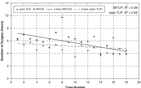
A learning effect was observed in the MITLIF group with the average duration of surgery dropping 1.8 hours from the first third (6.1 hours) to the last third (4.3 hours) of the 18 consecutive cases performed. The overall linear correlation between duration of surgery and case number for the MITLIF group was however weak (r2
=0.38) (Fig.3). The change of instrumentation on the 11th case coincided
with an average two hour reduction in operating time. The first 11 cases had a weak linear learning correlation (r2
= 0.01) whereas the last seven cases had a modest linear correlation (r2
=0.56). The learning effect was also ob-served in the open TLIF group but at a lesser extent with the average duration of surgery dropping only half an hour from the first third (5.6 hours) to the last third (5.1 hours) of the 18 consecutive cases performed. The overall linear correlation between duration of surgery and case number for the open TLIF group was also weak (r2
= 0.08). Comparing the slopes of the linear regressions we observed a more rapid decrease in duration of surgery with increasing case number for the MITLIF group as compared to the open TLIF group. There was no pattern of increased incidence of complications during the early part of the learning curve for either technique.
Discussion
The advent of percutaneous screw systems has allowed the development of less invasive fusion techniques [4,6, 12]. MITLIF is a relatively new technique attracting increased interest in less invasive surgical fusion techniques [14–16]. Schwender et al. presented the first clinical series reporting on their initial experience with 49 patients during a 22-month follow-up [19]. In this series of patients they reported two screw malpositions, two new radiculopathies, and a 100% fusion rate based on plain radiographs. Noteworthy was that in some cases bone morphogenetic protein (BMP) was used to augment local autograft. A subsequent study presented a series of 20 cases using a similar MITLIF operating technique [8]. The authors reported less blood loss and analgesia for their MITLIF group as compared to a historical PLIF group of patients. A combination of local bone with allograft was used but no details on fusion rates and clinical results were reported.
More recently a unilateral pedicle screw fixation tech-nique has been described by two different groups [1, 2]. Both reported series using BMP but their follow-up was too short to draw conclusions on the long-term safety of such an approach. The most recent study available to date emanates from a group not involved in the initial development of the technique [18]. This retrospective study included a series of
Fig. 2 Anteroposterior view of a minimally invasive transforaminal lumbar interbody fusion (MITLIF) case showing loosening of three out of the four screws (seearrows)
Table 2 Comparison of length of stay and of pain and function between the MITLIF and open TLIF groups
Treatment group Length of stay (days) Preoperative VAS Latest VAS ΔVAS Preoperative ODI Latest ODI ΔODI
MITLIF 6.1* 7.7 3.5 4.2 55 33 22
TLIF 8.2 5.0 2.8 2.2 53 26 27
MITLIF minimally invasive transforaminal lumbar interbody fusion, TLIF transforaminal lumbar interbody fusion, VAS visual analogue scale, ODI Oswestry disability index
53 patients undergoing MITLIF, ten having a multilevel procedure, with a 16-month follow-up. A historical control group of 67 patients who underwent an open TLIF procedure was used for comparison. Bone union was analysed using CT for 87% of the patients in the MITLIF group with a resulting 94% fusion rate. Even though the overall clinical results were similar between groups, less pain was reported in the MITLIF group after the second postoperative day. Blood loss was also significantly reduced in the MITLIF group. Nevertheless, this study included multilevel procedures in both the open TLIF and MITLIF groups. Furthermore, a mini-open technique was performed through a Wiltse muscle splitting approach which could be regarded as a lesser invasive technique than the midline approach.
The results from this study compare well with those presented by others as we also found a significant difference in blood loss between our two groups. In contrast to the two aforementioned studies [8,18] however, our MITLIF patient group reported similar amounts of postoperative pain as the open TLIF group. It could be that this difference between outcomes relates to the fact that PLIF, a technique needing more dissection than TLIF, was chosen as the control group in one of these two studies [8], or to the fact that several multilevel cases were included in the open TLIF comparison group in the other study [18]. Our patient sample may not be representative of surgical practice in other countries, in particular since a significant proportion were receiving disability pension.
The three pseudarthrosis cases in the MITLIF group have raised our awareness of the importance of good preparation of the disc space as well as perhaps the dilemma with the routine use of BMP as described in other studies [1,2,19]. The advantages of lessened blood loss and shorter hospital stay may nevertheless be outweighed by the cost of routine BMP use.
The pseudarthrosis in the MITLIF group could be due to the more difficult disc space preparation and grafting using tubular retractors, especially for the spondylolisthesis patients, which is a biomechanically different and more challenging group to fuse [13, 20]. Even though some surgeons reported high fusion rates in open surgery for spondylolisthesis using decompression material, their results cannot be transposed to the minimally invasive setting [11].
At the time this study was initiated, MITLIF was a relatively new technique with no long-term results. We thus opted to reserve the minimally invasive approach to isthmic spondylilisthesis cases, a classical TLIF indication, since the necessary facet joint excision would not further destabilise the affected level. In terms of clinical outcome comparison, which was not the main goal of this study, our nonrandomised approach with differences in diagnosis between groups may limit the strength of the resulting outcomes. This would however, play a lesser role as far as blood loss, duration of surgery, postoperative pain, and analgesia consumption are concerned, since the surgical technique was exactly the same on all patients, irrespective of diagnosis.
We also found that the MITLIF technique had a steeper curve even though the interacting progressive experience (single surgeon) with the open technique may have positively influenced the learning effect of the percutaneous technique.
Conclusions
The MITLIF approach had a steeper learning effect as compared to the open TLIF approach but does not appear to be linked with increased morbidity. Even though there was
MITLIF, R2 = 0.38 open TLIF, R2 = 0.08 0 2 4 6 8 10 12 0 2 4 6 8 10 12 14 16 18 20 Case Number
Duration of Surgery (hours)
open TLIF MITLIF Linear (MITLIF) Linear (open TLIF) Fig. 3 Duration of surgery in
hours demonstrating a weakly correlated learning effect for the minimally invasive transforami-nal lumbar interbody fusion (MITLIF) and open transfora-minal lumbar interbody fusion (TLIF) groups
a significant difference in blood loss and shorter length of hospital stay in the MITLIF group, we found no difference in analgesia consumption or pain perception during the early postoperative period. The occurrence of pseudarthrosis in our MITLIF group is of concern and suggests that extra care should be taken while preparing and grafting the disc space, especially with isthmic spondylolisthesis patients. Larger prospective randomised trials, including patients operated after the learning curve has stabilised, would be needed in order to confirm our findings.
Acknowledgments Financial support was received in the form of a research grant from DePuy Spine, Inc., Raynham, Massachusetts, USA.
References
1. Beringer WF, Mobasser JP (2006) Unilateral pedicle screw instrumentation for minimally invasive transforaminal lumbar interbody fusion. Neurosurg Focus 20:E4
2. Deutsch H, Musacchio MJ Jr (2006) Minimally invasive trans-foraminal lumbar interbody fusion with unilateral pedicle screw fixation. Neurosurg Focus 20:E10
3. Fairbank JC, Couper J, Davies JB, O’Brien JP (1980) The Oswestry low back pain disability questionnaire. Physiotherapy 66:271–273
4. Foley KT, Gupta SK (2002) Percutaneous pedicle screw fixation of the lumbar spine: preliminary clinical results. J Neurosurg 97:7–12
5. Harms JG, Jeszenszky D (1998) The unilateral transforaminal approach for posterior lumbar interbody fusion. Orthop Traumatol 6:88–89
6. Holly LT, Foley KT (2003) Three-dimensional fluoroscopy-guided percutaneous thoracolumbar pedicle screw placement. Technical note. J Neurosurg 99:324–329
7. Humphreys SC, Hodges SD, Patwardhan AG, Eck JC, Murphy RB, Covington LA (2001) Comparison of posterior and transforaminal approaches to lumbar interbody fusion. Spine 26:567–571
8. Isaacs RE, Podichetty VK, Santiago P, Sandhu FA, Spears J, Kelly K, Rice L, Fessler RG (2005) Minimally invasive microendoscopy-assisted transforaminal lumbar interbody fusion with instrumentation. J Neurosurg Spine 3:98–105
9. Kambin P (1992) Arthroscopic microdiscectomy. Arthroscopy 8:287–295
10. Kawaguchi Y, Matsui H, Tsuji H (1994) Back muscle injury after posterior lumbar spine surgery. Part 2: histologic and histochemical analyses in humans. Spine 19:2598–2602 11. Kho VK, Chen WC (2008) Posterolateral fusion using
laminec-tomy bone chips in the treatment of lumbar spondylolisthesis. Intern Orthopaed 32(1):115–119
12. Khoo LT, Palmer S, Laich DT, Fessler RG (2002) Minimally invasive percutaneous posterior lumbar interbody fusion. Neurosurgery 51: S166–S181
13. Kim SS, Denis F, Lonstein JE, Winter RB (1990) Factors affecting fusion rate in adult spondylolisthesis. Spine 15(9):979–984 14. Mummaneni PV, Rodts GE Jr (2005) The mini-open
trans-foraminal lumbar interbody fusion. Neurosurgery 57:256–261 15. Ozgur BM, Hughes SA, Baird LC, Taylor WR (2006) Minimally
disruptive decompression and transforaminal lumbar interbody fusion. Spine J 6:27–33
16. Ozgur BM, Yoo K, Rodriguez G, Taylor WR (2005) Minimally-invasive technique for transforaminal lumbar interbody fusion (TLIF). Eur Spine J 14:887–894
17. Ramsay CR, Wallace SA, Garthwaite PH, Monk AF, Russell IT, Grant AM (2002) Assessing the learning curve effect in health technologies. Lessons from the nonclinical literature. Int J Technol Assess Health Care 18:1–10
18. Scheufler KM, Dohmen H, Vougioukas VI (2007) Percutaneous transforaminal lumbar interbody fusion for the treatment of degenerative lumbar instability. Neurosurgery 60:203–212 19. Schwender JD, Holly LT, Rouben DP, Foley KT (2005)
Minimally invasive transforaminal lumbar interbody fusion (TLIF): technical feasibility and initial results. J Spinal Disord Tech 18 Suppl:S1–S6
20. Suk SI, Lee CK, Kim WJ, Lee JH, Cho KJ, Kim HG (1997) Adding posterior lumbar interbody fusion to pedicle screw fixation and posterolateral fusion after decompression in spondy-lolytic spondylolisthesis. Spine 22(2):210–219, discussion 219–20 21. Ventafridda V, Saita L, Ripamonti C, De CF (1985) WHO guidelines for the use of analgesics in cancer pain. Int J Tissue React 7:93–96