HAL Id: hal-02931618
https://hal.archives-ouvertes.fr/hal-02931618v2
Submitted on 9 Sep 2020HAL is a multi-disciplinary open access
archive for the deposit and dissemination of sci-entific research documents, whether they are pub-lished or not. The documents may come from teaching and research institutions in France or abroad, or from public or private research centers.
L’archive ouverte pluridisciplinaire HAL, est destinée au dépôt et à la diffusion de documents scientifiques de niveau recherche, publiés ou non, émanant des établissements d’enseignement et de recherche français ou étrangers, des laboratoires publics ou privés.
Contributions of Electrophysiology for Identifying
Cortical Language Systems in Patients with Epilepsy
Agnès Trébuchon, Catherine Liegeois-Chauvel, Jorge Gonzalez Martinez,
F.-Xavier Alario
To cite this version:
Agnès Trébuchon, Catherine Liegeois-Chauvel, Jorge Gonzalez Martinez, F.-Xavier Alario. Contri-butions of Electrophysiology for Identifying Cortical Language Systems in Patients with Epilepsy. Epilepsy & Behavior, [San Diego CA]: Elsevier B.V., In press. �hal-02931618v2�
This is the author’s final version, and that the article has been accepted for publication in the journal Epilepsy and Behavior. CC BY-NC-ND 4.0 by the authors. DOI: pending.
Contributions of Electrophysiology for Identifying Cortical Language Systems
in Patients with Epilepsy
Short title: Language Systems in Patients with Epilepsy
Agnès Trebuchon1,Catherine Liégeois-Chauvel1,2 Jorge Gonzalez Martinez2, F.-Xavier Alario2,3 *
1: Aix-Marseille Univ, INSERM, INS, Inst Neurosci Syst, Marseille, France
2: Department of Neurological Surgery, School of Medicine, University of Pittsburgh (PA), USA
3: Aix-Marseille Univ, CNRS, LPC, Marseille, France
* Corresponding author:
F.-Xavier Alario, Cortical Systems Laboratory, Department of Neurological Surgery, University of Pittsburgh, 200 Lothrop Street, A526 Scaife Hall, Pittsburgh, PA 1521; email:
Key questions
1. What are the specificities of the cortical organization of language?
2. Can magneto-encephalography (MEG) be routinely used for clinical language localization?
3. What information can be derived from invasive electrophysiological recordings of neural activity?
4. What information can be derived from the electrical disruption of language networks?
5. How can converging evidence be used for language lateralization and localization?
Abstract
A crucial element of the surgical treatment of medically refractory epilepsy is to delineate cortical areas that must be spared in order to avoid clinically relevant neurological and neuropsychological deficits post-operatively. For each patient, this typically necessitates determining the language lateralization between hemispheres and language localization within hemisphere. Understanding cortical language systems is complicated by two primary challenges: the extent of the neural tissue involved, and the substantial variability across individuals, especially in pathological populations. We review the contributions made through the study of electrophysiological activity to address these challenges. These contributions are based on the techniques of magnetoencephalography, intracerebral recordings, electrical cortical stimulation, and the electro-video analyses of seizures and their semiology. We highlight why no single modality alone is adequate to identify cortical language systems and suggest avenues for improving current practice.
Keywords
1. Magnetoencephalography 2. Electrical cortical stimulation
3. Stereotactic electroencephalography 4. Electrocorticography
5. Seizure semiology 6. Aphasia
Word count without references
1. Introduction: What are the specificities of the cortical
organization of language?
A crucial element of the surgical treatment of medically refractory epilepsy is to delineate cortical areas that must be spared in order to avoid clinically relevant neurological and neuropsychological deficits post-operatively [1,2]. Primary visual, motor, sensory and auditory areas have relatively predictable localization and function so are more easily identified [for a review, see 3]. The characterization of associative areas is more challenging, and the cortical organization of language is possibly the most difficult to understand.
The first challenge lies in the extent and intricacy of language related cortical systems. In current cognitive neuroscientific models, language processing is not described as a monolithic function, or a as dual perceptual-productive function [4]. A more accurate picture can only emerge if regions and functions are considered beyond the classical “Broca and Wernicke’s” model [5]. Language is thought to emerge from the combination of a variety of functions that are widely distributed, organized in anatomo-functional pathways, and to some extent, spatially segregated [6–8]. Language processed from auditory input is thought to recruit two main pathways [9], based on a similar functional architecture to the visual system [10]. The ventral stream propagates from auditory cortex to anterior, middle and inferior temporal regions in the temporal lobe, where speech is processed for meaning (lexico-semantic processing). This pathway culminates with sentence comprehension, involving the anterior temporal pole and inferior frontal gyrus. The dorsal stream propagates from auditory cortex towards the temporo-parietal junction, onto the inferior frontal gyrus, pre-motor and motor areas, processing the phonological form and articulatory programs for speech [e.g. 9,11–16]. A similar distinction has been proposed and debated for the visually prompted production of language, such as when reading words or naming objects, recruiting the fusiform gyrus in the baso-temporal area [17,18]. The meaning of words (and statements) is thought to be processed by a network predominantly involving the angular gyrus and the anterior temporal pole [e.g. 19–21]. Writing abilities recruit this network in connection with posterior parts of the middle frontal gyrus involved in sequence encoding and programming [22]. This summary is too short, if anything because we have focused predominantly on neo-cortical regions. The richness of current models of language processing can be explored in Hickock and Small’s “Neurobiology of Language” [23] across 88 chapters organized in sections labelled A to O over more than 1100 pages.
The second challenge lies in the inter-individual variability of language related cortical systems. The broad description above highlights that one hemisphere is typically “dominant”, but often not exclusive. This is largely valid in most right-handed individuals and a large proportion of left-handed individuals. Still, it is well established that there can be substantial variability of lateralization, and across language processes. This diversity is present across healthy speakers and is thought to be increased in neurological pathologies [24]. Substantial variability in spatial localization within a hemisphere has been reported in patients with epilepsy [25–28]. Certain lesions underlying drug resistant epilepsy such as focal cortical dysplasias or
nodular heterotopias may result in a different functional organization compared to healthy speakers [29,30].
In essence, language function strongly substantiates the hypothesis that the brain is a complex and adaptive system in which a vast range of (sub-)functions arise from coordinated neural activity across diverse spatial and temporal scales [31]. The extent and variability of language functional anatomy across healthy individuals [quantified in 32], which is increased in pathologies such as epilepsy, adds to the challenge of mapping cortical systems involved in language at the individual level.
Language lateralization vs language localization
Our brief sketch shows that the classical clinical question of “language lateralization” [33] is too coarse compared to current neuroscientific descriptions. In principle, each function could primarily recruit the “dominant” (typically, left) hemisphere, the “non-dominant” hemisphere, or both [24], although not all possible combinations of function and lateralization are expected to occur. A clinical discussion of lateralization should rather be framed in terms of specific functional sub-units such as lexical selection [34], or anatomo-functional subdivisions such as the dorsal pathway of processing [11,15,35]. The surgical disruption of any component, be it core or peripheral to the language function, could have distinct consequences for language processing and performance.
In this framework, language lateralization refers to determining whether “language” or, more appropriately (see above), a language sub-function is preferentially processed by the left, the right, or both hemispheres. Language localization (or “mapping”) refers to determining, generally within one hemisphere, the anato-functional relationships that best describe language processing.
Techniques currently available for determining language lateralization and
language localization in patients with focal epilepsy
Ideally, this up-to-date conceptualization of the cortical language systems should guide and optimize the clinical management of patients with medically refractory epilepsy, where surgical interventions could affect regions that are in proximity to or directly involve the language network. Determining language lateralization and localization for a given individual can be based on a variety of sources.
Function perturbation as an index of lateralization is the core functional hypothesis of the invasive intra-carotid amobarbital test traditionally used in pre-surgical settings (ICA or Wada test) [36,37]. A non-invasive alternative to the Wada test is functional magnetic resonance imaging (fMRI). There have been a substantial number of studies comparing lateralization assessed by the Wada test and fMRI [38,39], summarized in various literature reviews [most recently 40, see also 41,42, or sections 5.2 and 5.3 in 24], and the research continues [e.g. 25,43]. Nowadays, fMRI assessment of language lateralization is a common and well-established procedure in clinical practice that has largely replaced the Wada test [44–46]. Beyond mere
language lateralization, fMRI is also widely used clinically for localization. as it provides a broad picture of the functional anatomy of language. The analyses of distinct activation patterns allows identification of areas that participate in different language functions. Benjamin et al. [40] provides a thorough review of fMRI language mapping in pre-surgical epilepsy management, from functional task design to data analysis and interpretation. fMRI activations recorded during language tasks are typically located in the inferior frontal gyrus, the posterior part of the middle frontal gyrus, the angular gyrus, posterior superior temporal and supra marginal gyri (“Wernicke’s area”). Demonstrating activation of the infero-temporal areas is notoriously difficult in fMRI due to MR field inhomogeneities. This issue affects all activation in the inferior temporal gyrus which is critical in naming (e.g. the baso-temporal language area, located at the temporal-occipital border on the inferior to lateral surface, lateral to the fusiform gyrus), as well as mesial structures such as the hippocampus, all of which are often central to discussions about epilepsy patient management. fMRI has many strengths for the localization of neural functions: non-invasiveness, whole brain functional view, standardized signal processing, wide-spread use, etc. Despite these advantages, its sub-optimal yield in certain language relevant cortical areas imposes practical limitations on its use in planning surgical interventions.
Beyond fMRI, electrophysiological data can help define the anatomo-functional organization of the language network. The principle elements of electrophysiological data are derived from recordings of brain activation during verbal tasks, using either non-invasive methods such as magnetoencephalography (MEG) or invasive procedures such as electro-corticography (ECoG) or stereo-tactic electro-encephalography (SEEG). Signals recorded from chronically implanted epilepsy monitoring probes (ECoG or sEEG; e.g. broadband High gamma activity, HGA), are increasingly utilized, as they have been shown to produce reliable measures of time-resolved anatomo-functional correlations during cognitive tasks [47–51]. The second element of electrophysiological data is derived from the observation of disruptions within the language network. Electrical cortical stimulation (ECS) is often cited as the gold standard for localization of so-called “eloquent areas”, or of specific functional sub-units involved in the language network. ECS of a brain area during a verbal task may, or may not produce a transient language deficit, thus identifying "positive” areas as a specific functional sub-unit underlying language processing. In addition, language disturbances can be observed during or shortly after seizures, referred to as ictal- or post-ictal aphasia, respectively. The analysis of these electro-clinical correlations can provide additional information about the degree of asymmetry between left and right language networks.
The different approaches mentioned in this section are schematically represented in Figure 1 according to their relative merits for identifying language lateralization and localization. In the remainder of this targeted review, we outline the current methods of so-called “language mapping” at the interface between clinical management and research endeavors, focusing on the specific insights provided by electrophysiological recordings.
Figure 1 - Different techniques used to assess language lateralization and
language localization.
The different techniques are schematically located along two axes describing their relative merits for identifying language lateralization between hemispheres (x-axis) and language localization within hemisphere (y-axis). The vignettes on the right illustrate different electrophysiological recordings, from top to bottom: significant high gamma band activity (HGA) during picture naming (computed with the software MIA [111]), analysis of temporal processing during syllable perception, and magnetoencephalography (MEG) with atypical vs. typical lateralization. fMRI: functional Magnetic Resonance; Wada: intra-carotid amobarbital test.
2. Can magnetoencephalography be routinely used for
clinical language localization?
Neurophysiological activity can be localized across hemispheres with magnetoencephalography (MEG). This is a noninvasive method that allows co-registration of MRI with the evoked magnetic fields generated by intracellular electric current flows in the brain, thus yielding an estimate of the anatomical localization of neural sources. The analysis of these MEG sources during functional tasks is increasingly used to assess language lateralization.
Early MEG studies demonstrated that certain language tasks (e.g. visual or auditory word recognition) activate areas that are more lateralized than areas activated by less verbal tasks (e.g. face recognition) [52,53], thus revealing the individual’s putative language lateralization. With further analysis, these lateralization estimates were in some cases shown to be consistent with intra-operative mapping [54] or with surgical outcomes favorable for language [55]. A pivotal study involving a cohort of 100 surgical candidates confirmed the computed MEG lateralization indexes showed high levels of specificity and selectivity when compared to the Wada test [56]. In 2010, Pirmoradi et al. reviewed the available literature on pre-surgical language assessment using MEG [57], concluding that word recognition was the task yielding the highest concordance between MEG and Wada lateralization. In contrast, expressive language function was more difficult to lateralize, owing to the use of covert tasks. The diversity of source reconstruction techniques and potential variability in their outcomes; and the ensuing need for standardized protocols and procedures were also highlighted [57].
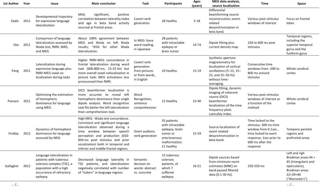
Since 2010, over 20 studies pertaining to language lateralization based on MEG signals have been published. Their main features are summarized in Table 1. These studies were published by various research groups, demonstrating ongoing and substantial heterogeneity in their core features: the extent to which lateralization is compared to Wada or fMRI indexes; the language tasks used and often advocated for; the MEG source localization methods; the regions on focus; the time windows used to compute laterality indexes; and the number of dipoles or source amplitude threshold. Some of these spatial, temporal, or task variations are intended to maximize the reliability of the lateralization index to be computed.
Our interpretation is that language lateralization is predominantly determined by evoked, rather than induced, magnetic field responses. Emphasis has not been on the precise source localization (e.g. of dipoles), but rather on determining which hemisphere generates more activity. The time window of analysis appears to be a crucial parameter to disentangle neural sources linked to sensory components from later cognitive components (e.g. between 300-600 milliseconds). The most effective lateralization and localization of language processing seems to be derived from the thorough analysis of neural activity, separately for frontal and temporal sources, within this time window. Importantly we suggest a cautious interpretation as these studies typically do not compare their relative merits with one another.
The results of various studies reveal that lateralization may evolve in younger subjects (between ~5 and 18 years), in ways that are very relevant for pre-surgical assessment in pediatric
patients. Various studies point to potential differences in lateralization between "receptive" and "expressive" language. However, these observations have not been thoroughly replicated. Additionally, most of these studies report analyses where MEG activity was time-locked to stimulus appearance. Time-locking MEG to the verbal response was seldom performed, despite its importance in the assessment of functional activity linked to expressive language [except in 58][for response-locked analysis in ECoG see 59,in EEG, see 60,in MEG, see 61].
Table 1 around here
Two complementary perspectives are derived from the literature summarized in Table 1 and, previously, in [57]. Bagić et al. [62] argue, notably on the basis of repeated observations of good to very good consistency between MEG-derived indexes and the Wada test taken as gold-standard, that language lateralization based on MEG is ready to play a routine role in clinical pre-surgical evaluations. In contrast, Loring et al. [63; see also 64] highlight the technically demanding nature of acquisition, analysis, and interpretation of MEG data. Especially in clinical settings, there may be substantial differences across medical and research centers in terms of expertise and experience. Part of the variability in the published results is likely driven by procedural inconsistencies. Gauging the locally available expertise is critical when deciding whether MEG based assessment of language areas should inform clinical decisions. Our interpretation is that after more than 40 papers have been published on determining language lateralization using MEG, clarification cannot be achieved with yet another empirical study based on a specific task and specific data-analysis choices. A better approach might be a collaborative multi-centric effort, where blindly collected data is analyzed across centers to assess, contrast, and compare different tasks and source localization methods [65]. Additionally, increasing focus on the links between cognitive processes and neural activity will further shape the question of lateralization. Improved “cognitive resolution” may help focus analyses on the time windows, localizations, and signal processing methods that render language processing particularly salient.
3. What information can be derived from invasive
electrophysiological recordings of neural activity?
Invasive electrophysiological explorations have contributed less than Wada or fMRI procedures to the determination of language lateralization. ECoG or sEEG implantations are often unilateral, but even when bilateral , typically involve only a subset of the probes present in the hemisphere of primary interest [66]. In this context, the issue of lateralization has been addressed mostly across patients.a) Temporal processing
Temporal course of evoked responses in auditory cortices were asymmetric during syllable perception, such that only left structures contributed to processing the temporal acoustic structure of the heard syllables, in right-handed left language dominant patients. It was
therefore concluded that the temporal processing characteristic of auditory structures, while not restricted to language, could indicate language lateralization [see 67 for details, and 68 for a replication in a bilaterally implanted patient]. In our view, however, ECoG and SEEG do not provide the primary or most powerful means to assess language hemispheric specialization. Rather, they provide an opportunity to assess the degree of symmetry among specific functional sub-units of the language network within the implanted areas.
b) Oscillatory activities
In addition to event related potentials, electrical activity synchronized across neurons generates oscillations that are recorded by depth probes. The frequencies and amplitudes of these oscillations vary according to task demands. High gamma amplitude modulations have shown better functional specificity and favorable spatio-temporal profiles for language tasks compared to lower frequency energy modulations [48–51,69]. These indexes provide a complementary method for detecting cortical activation and for mapping functional regions with high spatial and temporal resolution. They are becoming a systematic tool for functional language mapping [e.g. 51,70–74].
Visual confrontation naming is one the most frequently used tasks during presurgical language mapping. The risk of decline in visual confrontational naming following left temporal lobe epilepsy surgery, is perceived by the patients and reflected in their complaints regarding word finding difficulties[75–78]. Overt visual naming elicits significant HGA modulations in peri-rolandic cortex with a wider extent over Broca’s areas and premotor cortex in adults and children [79,80,for a review, see 81]. The analysis of neural spatio- temporal dynamics recorded from EcoG during visual naming has explored how parallel processing occurs along the ventral and dorsal streams [82,83].
Beyond areas located in the core of the language network, the hippocampus has been identified as a potentially important node [84]. This is partly due to clinical observations linking healthy hippocampi with word production performance [77]. Increased focus on the hippocampus is also related to the incidental observation that HGA was greatly attenuated in trials where naming failed [85], or from the covariation of various metrics of hippocampal activity and linguistic manipulations in word production tasks [86,87]. This function of the hippocampal node has been tentatively linked to the implicit learning of the object and its name [86] or the online retrieval of semantic association knowledge [87]. The importance of the hippocampus for visual naming performance is an issue that remains debated [88].
To create a more ecologically valid measure of the conversational challenges encountered by patients, an auditory descriptive naming task was developed by Marla Hamberger and collaborators [89]. Rather than visual stimuli, the tasks involves spoken sentences that describe specific objects whose names the patient must retrieve [89,90]. This task appears to be more sensitive than visual naming for the identification of left frontal regions involved in language function, eliciting extensive HGA in the left inferior, middle, and superior frontal gyri as well as premotor regions [72,91]. To our knowledge, no SEEG study has used this
protocol, so questions regarding the involvement of the hippocampus in auditory word retrieval remain unanswered.
Besides the visual and auditory tasks routinely used in the clinic, HGA is a powerful tool to distinguish the spatio-temporal course of different language sub-processes, and to more accurately map the role of each region within the network [92]. Auditory word-related HGA flows from the posterior superior temporal gyrus (pSTG), encoding low-level acoustic properties of verbal sounds, to the middle STG and then to the superior temporal sulcus (STS), presumably involved in lexical encoding [93]. Visual semantic and phonological tasks disentangle the role of the Pars triangularis and Pars Opercularis in semantic and phonological processes, respectively [94].
4. What information can be derived from the electrical
disruption of language networks?
Strengths and limitations of electrical-cortical stimulation (ECS), the “gold
standard” method
Electrical-cortical stimulation (ECS) has been performed intra-operatively for decades during awake surgery [2]. ECS is also performed during prolonged epilepsy monitoring based on electrocorticography (ECoG) [95] and stereo-electro-encephalography (sEEG) [96]. ECS is often considered to be the gold standard to localise specific functional sub-units in the language network, that would result in language deficits after resection. The correlation between a type of error (e.g. lexical access deficit) and the stimulated area can inform clinicians about the role of such areas within the entire language network (e.g. lexical selection). Despite its proven utility, and the fact that functional mapping is increasingly performed during SEEG or ECoG pre-surgical investigations, the methodology of the procedure varies substantially between clinical centers. Furthermore, ECS remains challenging and susceptible to a variety of pitfalls stemming from certain technical constraints, and the need to individualize the procedure.
Indeed, the major pitfall, with clinical consequences for the patient, is “false negative ECS”, where there is failure to identify an area as being a specific sub-unit of the language network despite its involvement the network. The sensitivity and specificity of sEEG-ECS has been assessed against reference meta-analytic fMRI studies. It was concluded that ECS can reliably identify contacts with/without language function but may under-detect all language sites [82] partly due to the task performed (or process targeted) during ECS. As outlined in the previous sections, language processing should not be understood as a monolithic function but rather as emerging from the combination of a variety of functions that are widely distributed, organized in anatomo-functional pathways, and spatially segregated around the Sylvian valley (see Section 1). Language function testing should integrate this complexity. The choice of task to be used during ECS and the interpretation of ECS effects on behavioral performance should be tailored to specific hypotheses about the role of the stimulated area [3,95–99].
The information derived from electrophysiological measures (e.g. HGA) and manipulations (e.g. ECS) can be combined to better understand each of the two methods, and to better define cortical networks involved in language. Their comparison has revealed useful, if imperfect, agreements [100–103]. A meta-analysis showed that HGA measures were specific but not sensitive to localize language areas, compared to the ECS [104]. This conclusion was confirmed in a sEEG study [102]. In addition, this study revealed a larger number of regions activated by language tasks compared to those identified by positive ECS. To assess the clinical relevance of ECS, does one rely only on the sites which are positive for both methods, reflecting crucial nodes of the language network? A straightforward rule of thumb could be that only ECS+ nodes are essential for the tasks, the others (i.e. HGA+ and ECS-) merely reflecting activations collateral to the task. In other words, ECS supersedes HGA for clinical decisions. However, given the stated limitations of the ECS exploration technique, such conclusion would be simplistic. Once again, the different sources of information should be combined to arrive at a tentative interpretation.
It is notable that the apparent “discrepancies” depend on the stimulation site. This observation provides some clues to explain why HGA sensitivity is so low. For instance, a high concordance between ECS(+) and HGA(+) is observed along the fusiform gyrus, where stimulation typically entails anomia. Comparing visual and auditory naming tasks, Forseth et al [105] concluded that the mid fusiform gyrus is a hetero-modal lexical semantic hub since it supported object semantic information irrespective of the sensory modality (auditory or visual input). A significant increase in HGA was observed in auditory and visual naming, compared to reverse speech or scrambled pictures, respectively. Electrical stimulation of this area disrupted both naming tasks. The authors underscored the risk for semantic processing deficit after left temporal lobe resection [105]. Kojima et al [91] demonstrated that post-operative/acute language deficits could be predicted by the extent of resection of sites with picture-naming related HGA (+) along left medial/inferior temporal regions. For reliable localization of the frontal regions, they suggested using auditory naming. Conversely, Cervenka et al [72] showed that the odds of identifying cortical activations in “dominant" anterior temporal neo-cortex were significantly greater during auditory than visual naming. Furthermore, the areas active during auditory naming were at an even greater risk of causing post-operative deficit relative to visual naming, even during more conservative temporal lobe resections (only 3.5 cm from the anterior temporal tip). This finding is in agreement with Hamberger’s studies that demonstrated ANT sites perturbed by stimulation were located anteriorly to VNT sites. Additionally, hippocampal resection has been shown to produce naming deficits independent of the degree of neocortical resection [76,90,106].
Can ictal and post-ictal language deficits be accurately assessed and are they
of any value for language localization?
When evaluating the link between language and electrophysiology in epilepsy, it is important to consider seizures and their semiology, to gauge how they might reveal additional information about language network organization. Language disturbances can be observed
during or shortly after seizures and are referred to as ictal- or post-ictal aphasia, respectively. This is particularly relevant in cases of temporal lobe epilepsy.
Regarding hemispheric language lateralization, post-ictal aphasia has long been recognized as a good marker to identify the “dominant” hemisphere [for reviews, see 107,108]. Assessing language abilities during the post-ictal periods can sometimes be challenging, but a simple timing of when language recovery occurs can be sufficient to provide crucial information. The "Cincinnati method" is a good example. It consists of a single sentence reading test, and a measure of recovery delay, defined as the moment when the sentence is read correctly. Such language recovery delay has been used for more than 20 years and reveals valuable information about language lateralization [109].
Beyond that, the joint and precise analysis of video and intra-cranial electrophysiological signals during seizures can be a valuable investigative tool for language if it is tailored to the individual patient. A specialized clinical setup must be performed systematically, and not all seizures are easily analyzed due to variations in the patients level of peri-ictal awareness or consciousness. Nonetheless, a thorough analysis of language disturbance at seizure onset has been shown to be reliable for language localization [110]. In that study, impaired speech comprehension was associated with posterior lateral temporal involvement, anomia and reduced verbal fluency with anterior medio-basal temporal structures, and jargon aphasia with basal temporal involvement. Furthermore, the ictal language production deficits, such as anomia and reduced fluency, could not simply be explained by an involvement of Broca’s area, since this region was not affected by seizure discharge. In short, early ictal examination of language function may help to understand sub-lobar seizure organization in addition to language lateralization [110], contrary to what has been described when analyses were performed globally during seizures or limited to only post ictal aphasia [107].
5. How can converging evidence be used for language
lateralization and localization?
As discussed in the preceding sections, the identification of cortical systems linked to language processing is a challenging endeavor. Determining language lateralization and localization for a given individual should be based on a variety of sources. We illustrate the approach with two representative clinical cases.
The first patient, a 44-year-old right-handed male, presented with a late onset drug-resistant epilepsy at the age of 30. Seizures occurred weekly and consisted of a negative sensation (anxiety, fear, etc.) with early ictal aphasia followed by impairment of consciousness and prolonged post ictal aphasia. Ictal scalp EEG revealed delayed rhythmic discharges in the left temporo-frontal region. MRI was negative, and FDG-PET showed hypometabolism in the left temporal lobe (mesial and pole), as well as in left temporal peri-sylvian regions including the insula. SEEG demonstrated a large epileptogenic zone including anterior parts of the left temporal lobe, left insula and right hippocampus. Due to the extent of the EZ and high risk of
post-operative cognitive deficit, no surgical resection has been proposed. Figure 2 illustrates the stepwise clinical reasoning to resolve the patient’s language network. In this case, the conclusion is arguably simple because all techniques converged on the same response, left-lateralization of the tested language functions.
The second patient, a 17-year-old right-handed male, had suffered with epilepsy since the age of 2. No auras were reported; his seizures started with impairment of consciousness, leftward eye and head version, sometimes followed by a hyperkinetic period. Post-ictally, there was no language deficit. Ictal scalp EEG revealed a brief left temporo-parietal attenuation followed by a rhythmic discharge in the same region. MRI was negative, and FDG-PET showed hypometabolism in the lateral and posterior regions of the left temporal lobe. SEEG recordings confirmed an interictal pattern typical of cortical dysplasia in the posterior part of the superior and middle temporal gyri, delineating the epileptogenic zone to the lateral part of these structures. Figure 3 illustrates the stepwise clinical approach to resolving the language network. This second case is arguably more complex, because seemingly diverging results across methods suggest a more intricate inter-hemispheric language network. An awake resection was performed in 2012. Histological examination confirmed type IIb cortical dysplasia. In his latest examination, the patient was not completely seizure free (Engel II).
Post-operative language outcome
The integration of multiple sources of evidence to guide surgery remains to be evaluated thoroughly. There are still too few studies reporting post-operative outcomes in relation to HGA status, especially after the resection of HGA(+) sites. Based on the pioneering study by Sinai et al [103],drawing firm conclusions is difficult because data is available for only 5 out of 13 patients. Yet, the available data suggests that the resection of HGA positive sites produces a language deficit (3 patients) while no change in naming performance was observed if these sites were spared during surgery. More recently, [72] reported unanticipated post-operative language deficits based on ECS results, in patients whose resection included HGA(+) sites. The number of resected HGA(+) sites recorded from standard language areas are likely to be very relevant in predicting the need for post-operative speech therapy [91]. Arya et al [82] addressed the question of neuropsychological outcome in a population of 17 children, by administering a standard neuropsychological battery pre- and 12 months post-operatively. They highlighted that the resection of HGA(+) sites elicited during visual naming, some of which were located outside of anatomic language areas, was associated with poorer overall neuropsychological outcome, particularly a significant decrease in working verbal memory.
Figure 2 – Clinical step-wise exploration of the language network reveals
coherent observations across methods
See main text for clinical details. HGA: High Gamma Activity; pSTG: posterior Superior Temporal Gyrus; EZ: Epileptogenic Zone; ECS: Electrico-Cortical Stimulation.
Figure 3 – Clinical step-wise exploration of the language network reveals
seemingly differing patterns across techniques, leading to a complex
conclusion
See main text for clinical details. HGA: High gamma activity; [a/p]BTA: [anterior/posterior] Basal-Temporal Area; IFG: Inferior Frontal Gyrus; pSTG: posterior Superior Temporal Gyrus; SMG: Supra-Marginal Gyrus; AG: Angular Gyrus; ERP: Event-Related Potentials; EZ: Epileptogenic Zone; ECS: Electrico-Cortical Stimulation.
6. Summary
Describing cortical language systems at an individual level is complicated by two challenges: the extent of the associative cortex involved, and the inter-individual variability in anatomo-functional relations. It is crucial to go beyond the simple question of language lateralization to understand the organization of language processing. Owing to its ability to directly record neural activity and its corresponding high temporal resolution, electrophysiology can play a prominent role in this task. MEG is a potentially powerful method, but many protocol features (e.g. signal processing options or cognitive tasks) remain to be objectively optimized to reach a broader clinical consensus. Despite their limited sampling, intra-cerebral recordings are a rich source of information when their location permits appropriate cognitive testing. While ECS often enjoys a “gold standard” status, it should not be used alone to ascertain language function. Ideally, all of these measures should be combined, along with clinical observations of ictal and post-ictal states, to arrive at a fine-grained description of the cortical systems for language that is specific to each patient.
Key questions (answered)
1. What are the specificities of the cortical organization of language?
Language lateralization seems to imply a monolithic function when in fact language processing emerges from a variety of (sub-)functions whose localization is widely distributed. Localization of language (sub-)functions is thus the central and challenging issue, owing to observed interindividual variability, especially in patients with epilepsy.
2. Can magneto-encephalography (MEG) be routinely used for clinical language localization?
MEG recordings can be used to assess specific processes and their deployment in sub-second timescales, but they involve substantial technical skills and consensus regarding cognitive tasks or signal processing procedures is lacking.
3. What information can be derived from invasive electrophysiological recordings of neural activity?
Invasive explorations are often unilateral so may not adequately resolve lateralization. Both perceptive and productive tasks have been successfully used to investigate language localization and to partly constrain electrical stimulation explorations. Various metrics (local field potentials, power in various frequency bands) should in principle be considered. Establishing precise links between these activities and processing models requires ongoing investigations.
4. What information can be derived from the electrical disruption of language networks?
Cognitive tasks and stimulation sites should be carefully paired and tailored to each individual. A low stimulation intensity may generate false negatives whereas a high intensity may generate discharges and disrupt the network beyond the stimulation site (non-specific positives). Analysis of ictal and post-ictal language requires a well-trained and dedicated team and can potentially contribute to understanding sub-lobar seizure organization.
5. How can converging evidence be used for language lateralization and localization?
Determining language lateralization and localization for a given individual should be based on as many diverse sources of information as possible, including those discussed here as well as fMRI and thorough neuropsychological assessment
7. References
[1] Lüders HO. Textbook of epilepsy surgery. London: Informa Healthcare; 2008.
[2] Penfield W, Jasper HH. Epilepsy and the functional anatomy of the human brain. Boston: Little Brown & Co; 1954.
[3] Selimbeyoglu A. Electrical stimulation of the human brain: perceptual and behavioral phenomena reported in the old and new literature. Front Hum Neurosci 2010. https://doi.org/10.3389/fnhum.2010.00046.
[4] Geschwind N. The Organization of Language and the Brain 1970;170:940–4.
[5] Tremblay P, Dick AS. Broca and Wernicke are dead, or moving past the classic model of language neurobiology. Brain Lang 2016;162:60–71. https://doi.org/10.1016/j.bandl.2016.08.004.
[6] Ardila A, Bernal B, Rosselli M. How Localized are Language Brain Areas? A Review of Brodmann Areas Involvement in Oral Language. Arch Clin Neuropsychol 2016;31:112–22.
https://doi.org/10.1093/arclin/acv081.
[7] Poeppel D. The maps problem and the mapping problem: Two challenges for a cognitive neuroscience of speech and language. Cogn Neuropsychol 2012;29:34–55.
https://doi.org/10.1080/02643294.2012.710600.
[8] Price CJ. A review and synthesis of the first 20 years of PET and fMRI studies of heard speech, spoken language and reading. NeuroImage 2012;62:816–47.
https://doi.org/10.1016/j.neuroimage.2012.04.062.
[9] Hickok G, Poeppel D. Dorsal and ventral streams: a framework for understanding aspects of the functional anatomy of language. Cognition 2004;92:67–99.
https://doi.org/10.1016/j.cognition.2003.10.011.
[10] Kravitz DJ, Saleem KS, Baker CI, Mishkin M. A new neural framework for visuospatial processing. Nat Rev Neurosci 2011;12:217–30. https://doi.org/10.1038/nrn3008.
[11] Friederici AD, Gierhan SM. The language network. Curr Opin Neurobiol 2013;23:250–4. https://doi.org/10.1016/j.conb.2012.10.002.
[12] Giraud A-L, Poeppel D. Cortical oscillations and speech processing: emerging computational principles and operations. Nat Neurosci 2012;15:511–7. https://doi.org/10.1038/nn.3063.
[13] Hartwigsen G, Weigel A, Schuschan P, Siebner HR, Weise D, Classen J, et al. Dissociating Parieto-Frontal Networks for Phonological and Semantic Word Decisions: A Condition-and-Perturb TMS Study. Cereb Cortex 2016;26:2590–601. https://doi.org/10.1093/cercor/bhv092.
[14] Hickok G. Computational neuroanatomy of speech production. Nat Rev Neurosci 2012;13:135–45. https://doi.org/10.1038/nrn3158.
[15] Murakami T, Kell CA, Restle J, Ugawa Y, Ziemann U. Left Dorsal Speech Stream Components and Their Contribution to Phonological Processing. J Neurosci 2015;35:1411–22.
https://doi.org/10.1523/JNEUROSCI.0246-14.2015.
[16] Saur D, Kreher BW, Schnell S, Kummerer D, Kellmeyer P, Vry M-S, et al. Ventral and dorsal pathways for language. Proc Natl Acad Sci 2008;105:18035–40.
https://doi.org/10.1073/pnas.0805234105.
[17] Lüders HO, Lesser RP, Hahn J, Dinner DS, Morris HH, Wyllie E, et al. Basal temporal language area. Brain 1991;114:743–54. https://doi.org/10.1093/brain/114.2.743.
[18] Trébuchon-Da Fonseca A, Guedj E, Alario F-X, Laguitton V, Mundler O, Chauvel P, et al. Brain regions underlying word finding difficulties in temporal lobe epilepsy. Brain 2009;132:2772–84. https://doi.org/10.1093/brain/awp083.
[19] Binder JR, Desai RH, Graves WW, Conant LL. Where Is the Semantic System? A Critical Review and Meta-Analysis of 120 Functional Neuroimaging Studies. Cereb Cortex 2009;19:2767–96. https://doi.org/10.1093/cercor/bhp055.
[20] Lambon Ralph MA, Jefferies E, Patterson K, Rogers TT. The neural and computational bases of semantic cognition. Nat Rev Neurosci 2017;18:42–55. https://doi.org/10.1038/nrn.2016.150. [21] Mesulam M-M, Wieneke C, Hurley R, Rademaker A, Thompson CK, Weintraub S, et al. Words and
objects at the tip of the left temporal lobe in primary progressive aphasia. Brain 2013;136:601– 18. https://doi.org/10.1093/brain/aws336.
[22] Rapp B, Purcell J. Understanding How We Produce Written Words: Lessons from the Brain. In: de Zubicaray GI, Schiller NO, editors. Oxf. Handb. Neurolinguistics, Oxford University Press; 2019, p. 424–48. https://doi.org/10.1093/oxfordhb/9780190672027.013.17.
[23] Hickok G, Small SL. Neurobiology of Language. San Francisco: Academic Press; 2016. https://doi.org/10.1016/c2011-0-07351-9.
[24] Tzourio-Mazoyer N, Perrone-Bertolotti M, Jobard G, Mazoyer B, Baciu M. Multi-factorial
modulation of hemispheric specialization and plasticity for language in healthy and pathological conditions: A review. Cortex 2017;86:314–39. https://doi.org/10.1016/j.cortex.2016.05.013. [25] Berl MM, Zimmaro LA, Khan OI, Dustin I, Ritzl E, Duke ES, et al. Characterization of atypical
language activation patterns in focal epilepsy. Ann Neurol 2014;75:33–42. https://doi.org/10.1002/ana.24015.
[26] Flinker A, Chang EF, Barbaro NM, Berger MS, Knight RT. Sub-centimeter language organization in the human temporal lobe. Brain Lang 2011;117:103–9.
https://doi.org/10.1016/j.bandl.2010.09.009.
[27] Leonard MK, Cai R, Babiak MC, Ren A, Chang EF. The peri-Sylvian cortical network underlying single word repetition revealed by electrocortical stimulation and direct neural recordings. Brain Lang 2019;193:58–72. https://doi.org/10.1016/j.bandl.2016.06.001.
[28] Ojemann G, Ojemann J, Lettich E, Berger M. Cortical language localization in left, dominant hemisphere: An electrical stimulation mapping investigation in 117 patients. J Neurosurg 1989;71:316–26. https://doi.org/10.3171/jns.1989.71.3.0316.
[29] Duchowny M. Clinical, functional, and neurophysiologic assessment of dysplastic cortical networks: Implications for cortical functioning and surgical management. Epilepsia 2009;50:19– 27. https://doi.org/10.1111/j.1528-1167.2009.02291.x.
[30] Souci S, Petton M, Jung J, Bourdillon P, Bouet R, Richard-Mornas A, et al. Task-induced gamma band effect in type II focal cortical dysplasia: An exploratory study. Epilepsy Behav 2018;85:76– 84. https://doi.org/10.1016/j.yebeh.2018.05.017.
[31] Sporns O, Betzel RF. Modular Brain Networks. Annu Rev Psychol 2016;67:613–40. https://doi.org/10.1146/annurev-psych-122414-033634.
[32] Mahowald K, Fedorenko E. Reliable individual-level neural markers of high-level language processing: A necessary precursor for relating neural variability to behavioral and genetic variability. NeuroImage 2016;139:74–93. https://doi.org/10.1016/j.neuroimage.2016.05.073. [33] Eling P, editor. Reader in the history of aphasia: from Franz Gall to Norman Geschwind.
Amsterdam ; Philadelphia: J. Benjamins Pub. Co; 1994.
[34] Riès S, Dronkers NF, Knight RT. Choosing words: left hemisphere, right hemisphere, or both? Perspective on the lateralization of word retrieval. Ann N Y Acad Sci 2016;1369:111–31. https://doi.org/10.1111/nyas.12993.
[35] Schwartz MF, Faseyitan O, Kim J, Coslett HB. The dorsal stream contribution to phonological retrieval in object naming. Brain 2012;135:3799–814. https://doi.org/10.1093/brain/aws300. [36] Loring DW, Meador KJ, Lee GP, Murro AM, Smith JR, Flanigin HF, et al. Cerebral language
lateralization: Evidence from intracarotid amobarbital testing. Neuropsychologia 1990;28:831–8. https://doi.org/10.1016/0028-3932(90)90007-B.
[37] Wada J, Rasmussen T. Intracarotid Injection of Sodium Amytal for the Lateralization of Cerebral Speech Dominance. J Neurosurg 1960;17:266–82. https://doi.org/10.3171/jns.1960.17.2.0266. [38] Binder JR, Swanson SJ, Hammeke TA, Morris GL, Mueller WM, Fischer M, et al. Determination of
language dominance using functional MRI: A comparison with the Wada test. Neurology 1996;46:978–84. https://doi.org/10.1212/WNL.46.4.978.
[39] Springer JA, Binder JR, Hammeke TA, Swanson SJ, Frost JA, Bellgowan PSF, et al. Language dominance in neurologically normal and epilepsy subjects. Brain 1999;122:2033–46.
https://doi.org/10.1093/brain/122.11.2033.
[40] Benjamin CFA, Gkiatis K, Matsopoulos GK, Garganis K. Presurgical Language fMRI in Epilepsy: An Introduction. In: Argyropoulos GPD, editor. Transl. Neurosci. Speech Lang. Disord., Cham: Springer International Publishing; 2020, p. 205–39. https://doi.org/10.1007/978-3-030-35687-3_10.
[41] Bauer PR, Reitsma JB, Houweling BM, Ferrier CH, Ramsey NF. Can fMRI safely replace the Wada test for preoperative assessment of language lateralisation? A meta-analysis and systematic review. J Neurol Neurosurg Psychiatry 2014;85:581–8. https://doi.org/10.1136/jnnp-2013-305659. [42] Dym RJ, Burns J, Freeman K, Lipton ML. Is Functional MR Imaging Assessment of Hemispheric
Language Dominance as Good as the Wada Test?: A Meta-Analysis. Radiology 2011;261:446–55. https://doi.org/10.1148/radiol.11101344.
[43] Janecek JK, Swanson SJ, Sabsevitz DS, Hammeke TA, Raghavan M, E. Rozman M, et al. Language lateralization by fMRI and Wada testing in 229 patients with epilepsy: Rates and predictors of discordance. Epilepsia 2013;54:314–22. https://doi.org/10.1111/epi.12068. [44] Bobholz JA, Rao SM, Saykin AJ, Pliskin N. Clinical Use of Functional Magnetic Resonance
Imaging: Reflections on the New CPT Codes. Neuropsychol Rev 2007;17:189–91. https://doi.org/10.1007/s11065-007-9022-1.
[45] Szaflarski JP, Gloss D, Binder JR, Gaillard WD, Golby AJ, Holland SK, et al. Practice guideline summary: Use of fMRI in the presurgical evaluation of patients with epilepsy: Report of the Guideline Development, Dissemination, and Implementation Subcommittee of the American Academy of Neurology. Neurology 2017;88:395–402.
https://doi.org/10.1212/WNL.0000000000003532.
[46] Binder JR. FMRI Is a Valid Noninvasive Alternative to Wada Testing. Epilepsy Behav EB 2011;20:214–22. https://doi.org/10.1016/j.yebeh.2010.08.004.
[47] Jacobs J, Kahana MJ. Direct brain recordings fuel advances in cognitive electrophysiology. Trends Cogn Sci 2010;14:162–71. https://doi.org/10.1016/j.tics.2010.01.005.
[48] Lachaux J-P, Fonlupt P, Kahane P, Minotti L, Hoffmann D, Bertrand O, et al. Relationship between task-related gamma oscillations and BOLD Signal: New insights from combined fMRI and intracranial EEG. Hum Brain Mapp 2007;28:1368–75. https://doi.org/10.1002/hbm.20352. [49] Lachaux J-P, Axmacher N, Mormann F, Halgren E, Crone NE. High-frequency neural activity and
human cognition: Past, present and possible future of intracranial EEG research. Prog Neurobiol 2012;98:279–301. https://doi.org/10.1016/j.pneurobio.2012.06.008.
[50] Parvizi J, Kastner S. Promises and limitations of human intracranial electroencephalography. Nat Neurosci 2018;21:474–83. https://doi.org/10.1038/s41593-018-0108-2.
[51] Wang Y, Fifer MS, Flinker A, Korzeniewska A, Cervenka MC, Anderson WS, et al. Spatial-temporal functional mapping of language at the bedside with electrocorticography. Neurology 2016;86:1181–9. https://doi.org/10.1212/WNL.0000000000002525.
[52] Simos PG, Breier JI, Zouridakis G, Papanicolaou AC. Identification of Language-Specific Brain Activity Using Magnetoencephalography. J Clin Exp Neuropsychol 1998;20:706–22.
[53] Zouridakis G, Simos PG, Breier JI, Papanicolaou AC. Functional Hemispheric Asymmetry Assessment in a Visual Language Task Using MEG. Brain Topogr 1998;11:57–65.
https://doi.org/10.1023/A:1022270620396.
[54] Simos PG, Breier JI, Maggio WW, Gormley WB, Zouridakis G, Willmore LJ, et al. Atypical temporal lobe language representation: MEG and intraoperative stimulation mapping
correlation. Neuroreport 1999;10:139–42. https://doi.org/10.1097/00001756-199901180-00026. [55] Castillo EM, Simos PG, Venkataraman V, Breier JI, Wheless JW, Papanicolaou AC. Mapping of
Expressive Language Cortex using Magnetic Source Imaging. Neurocase 2001;7:419–22. https://doi.org/10.1076/neur.7.5.419.16249.
[56] Papanicolaou AC, Simos PG, Castillo EM, Breier JI, Sarkari S, Pataraia E, et al.
Magnetocephalography: a noninvasive alternative to the Wada procedure. J Neurosurg 2004;100:867–76. https://doi.org/10.3171/jns.2004.100.5.0867.
[57] Pirmoradi M, Béland R, Nguyen DK, Bacon BA, Lassonde M. Language tasks used for the presurgical assessment of epileptic patients with MEG. Epileptic Disord 2010;12:97–108. https://doi.org/10.1684/epd.2010.0314.
[58] Traut T, Sardesh N, Bulubas L, Findlay A, Honma SM, Mizuiri D, et al. MEG imaging of recurrent gliomas reveals functional plasticity of hemispheric language specialization. Hum Brain Mapp 2019;40:1082–92. https://doi.org/10.1002/hbm.24430.
[59] Edwards E, Nagarajan SS, Dalal SS, Canolty RT, Kirsch HE, Barbaro NM, et al. Spatiotemporal imaging of cortical activation during verb generation and picture naming. NeuroImage 2010;50:291–301. https://doi.org/10.1016/j.neuroimage.2009.12.035.
[60] Riès S, Janssen N, Burle B, Alario F-X. Response-Locked Brain Dynamics of Word Production. PLoS ONE 2013;8:e58197. https://doi.org/10.1371/journal.pone.0058197.
[61] Salmelin R, Schnitzler A, Schmitz F, Freund H-J. Single word reading in developmental stutterers and fluent speakers. Brain 2000;123:1184–202. https://doi.org/10.1093/brain/123.6.1184.
[62] Bagić AI, Bowyer SM, Kirsch HE, Funke ME, Burgess RC. American Clinical MEG Society (ACMEGS) Position Statement #2: The Value of Magnetoencephalography (MEG)/Magnetic Source Imaging (MSI) in Noninvasive Presurgical Mapping of Eloquent Cortices of Patients Preparing for Surgical Interventions. J Clin Neurophysiol 2017;34:189–95.
https://doi.org/10.1097/WNP.0000000000000366.
[63] Loring DW, Gaillard WD, Bookheimer SY, Meador KJ, Ojemann JG. Cortical cartography reveals political and physical maps. Epilepsia 2014;55:633–7. https://doi.org/10.1111/epi.12553.
[64] Mégevand P, Seeck M. Electroencephalography, magnetoencephalography and source localization: their value in epilepsy. Curr Opin Neurol 2018;31:176–83.
[65] Botvinik-Nezer R, Holzmeister F, Camerer CF, Dreber A, Huber J, Johannesson M, et al. Variability in the analysis of a single neuroimaging dataset by many teams. Nature 2020:1–7. https://doi.org/10.1038/s41586-020-2314-9.
[66] Chauvel P, Gonzalez-Martinez J, Bulacio J. Presurgical intracranial investigations in epilepsy surgery. Handb. Clin. Neurol., vol. 161, Elsevier; 2019, p. 45–71. https://doi.org/10.1016/B978-0-444-64142-7.00040-0.
[67] Liégeois-Chauvel C, de Graaf JB, Laguitton V, Chauvel P. Specialization of Left Auditory Cortex for Speech Perception in Man Depends on Temporal Coding. Cereb Cortex 1999;9:484–96. https://doi.org/10.1093/cercor/9.5.484.
[68] Morillon B, Liégeois-Chauvel C, Arnal LH, Bénar CG, Giraud A-L. Asymmetric Function of Theta and Gamma Activity in Syllable Processing: An Intra-Cortical Study. Front Psychol 2012;3. https://doi.org/10.3389/fpsyg.2012.00248.
[69] Crone NE, Boatman D, Gordon B, Hao L. Induced electrocorticographic gamma activity during auditory perception. Clin Neurophysiol 2001;112:565–82. https://doi.org/10.1016/S1388-2457(00)00545-9.
[70] Arya R, Wilson JA, Vannest J, Byars AW, Greiner HM, Buroker J, et al. Electrocorticographic language mapping in children by high-gamma synchronization during spontaneous conversation: Comparison with conventional electrical cortical stimulation. Epilepsy Res 2015;110:78–87. https://doi.org/10.1016/j.eplepsyres.2014.11.013.
[71] Asano E, Gotman J. Is electrocorticography-based language mapping ready to replace stimulation? Neurology 2016;86:1174–6. https://doi.org/10.1212/WNL.0000000000002533. [72] Cervenka MC, Corines J, Boatman-Reich DF, Eloyan A, Sheng X, Franaszczuk PJ, et al.
Electrocorticographic functional mapping identifies human cortex critical for auditory and visual naming. NeuroImage 2013;69:267–76. https://doi.org/10.1016/j.neuroimage.2012.12.037. [73] Kambara T, Sood S, Alqatan Z, Klingert C, Ratnam D, Hayakawa A, et al. Presurgical language
mapping using event-related high-gamma activity: The Detroit procedure. Clin Neurophysiol 2018;129:145–54. https://doi.org/10.1016/j.clinph.2017.10.018.
[74] Wu HC, Nagasawa T, Brown EC, Juhasz C, Rothermel R, Hoechstetter K, et al.
Gamma-oscillations modulated by picture naming and word reading: Intracranial recording in epileptic patients. Clin Neurophysiol 2011;122:1929–42. https://doi.org/10.1016/j.clinph.2011.03.011. [75] Busch RM, Floden DP, Prayson B, Chapin JS, Kim KH, Ferguson L, et al. Estimating risk of
word-finding problems in adults undergoing epilepsy surgery. Neurology 2016;87:2363–9. https://doi.org/10.1212/WNL.0000000000003378.
[76] Busch RM, Hogue O, Kattan MW, Hamberger M, Drane DL, Hermann B, et al. Nomograms to predict naming decline after temporal lobe surgery in adults with epilepsy. Neurology 2018;91:e2144–52. https://doi.org/10.1212/WNL.0000000000006629.
[77] Ives-Deliperi VL, Butler JT. Naming outcomes of anterior temporal lobectomy in epilepsy patients: A systematic review of the literature. Epilepsy Behav 2012;24:194–8.
https://doi.org/10.1016/j.yebeh.2012.04.115.
[78] Miller M, Hogue O, Hogan T, Busch RM. Naming decline after epilepsy surgery is associated with subjective language complaints. Epilepsy Behav 2019;99:106484.
https://doi.org/10.1016/j.yebeh.2019.106484.
[79] Arya R, Wilson JA, Fujiwara H, Rozhkov L, Leach JL, Byars AW, et al. Presurgical language localization with visual naming associated ECoG high- gamma modulation in pediatric drug-resistant epilepsy. Epilepsia 2017;58:663–73. https://doi.org/10.1111/epi.13708.
[80] Nakai Y, Jeong JW, Brown EC, Rothermel R, Kojima K, Kambara T, et al. Three- and
four-dimensional mapping of speech and language in patients with epilepsy. Brain 2017;140:1351–70. https://doi.org/10.1093/brain/awx051.
[81] Llorens A, Trébuchon A, Liégeois-Chauvel C, Alario F-X. Intra-cranial recordings of brain activity during language production. Front Psychol 2011;2:1–12.
https://doi.org/10.3389/fpsyg.2011.00375.
[82] Arya R, Babajani-Feremi A, Byars AW, Vannest J, Greiner HM, Wheless JW, et al. A model for visual naming based on spatiotemporal dynamics of ECoG high-gamma modulation. Epilepsy Behav 2019;99:106455. https://doi.org/10.1016/j.yebeh.2019.106455.
[83] Dubarry A-S, Llorens A, Trébuchon A, Carron R, Liégeois-Chauvel C, Bénar CG, et al. Estimating Parallel Processing in a Language Task Using Single-Trial Intracerebral Electroencephalography. Psychol Sci 2017;28:414–26. https://doi.org/10.1177/0956797616681296.
[84] Covington NV, Duff MC. Expanding the Language Network: Direct Contributions from the Hippocampus. Trends Cogn Sci 2016;20:869–70. https://doi.org/10.1016/j.tics.2016.10.006. [85] Hamamé CM, Alario F-X, Llorens A, Liégeois-Chauvel C, Trébuchon-Da Fonseca A. High
frequency gamma activity in the left hippocampus predicts visual object naming performance. Brain Lang 2014;135:104–14. https://doi.org/10.1016/j.bandl.2014.05.007.
[86] Llorens A, Dubarry A-S, Trébuchon-Da Fonseca A, Chauvel P, Alario F-X, Liégeois-Chauvel C. Contextual modulation of hippocampal activity during picture naming. Brain Lang 2016;159:92– 101. https://doi.org/10.1016/j.bandl.2016.05.011.
[87] Piai V, Anderson KL, Lin JJ, Dewar C, Parvizi J, Dronkers NF, et al. Direct brain recordings reveal hippocampal rhythm underpinnings of language processing. Proc Natl Acad Sci 2016.
https://doi.org/10.1073/pnas.1603312113.
[88] Drane DL, Pedersen NP. Knowledge of language function and underlying neural networks gained from focal seizures and epilepsy surgery. Brain Lang 2019;189:20–33.
https://doi.org/10.1016/j.bandl.2018.12.007.
[89] Hamberger MJ, Seidel WT, Mckhann GM, Perrine K, Goodman RR. Brain stimulation reveals critical auditory naming cortex. Brain 2005;128:2742–9. https://doi.org/10.1093/brain/awh621.
[90] Hamberger MJ, Seidel WT. Auditory and visual naming tests: Normative and patient data for accuracy, response time, and tip-of-the-tongue. J Int Neuropsychol Soc 2003;9:479–89. https://doi.org/10.1017/S135561770393013X.
[91] Kojima K, Brown EC, Matsuzaki N, Rothermel R, Fuerst D, Shah A, et al. Gamma activity modulated by picture and auditory naming tasks: Intracranial recording in patients with focal epilepsy. Clin Neurophysiol 2013;124:1737–44. https://doi.org/10.1016/j.clinph.2013.01.030. [92] Flinker A, Piai V, Knight R. Intracranial electrophysiology in language research. Oxf Handb
Psycholinguist 2 Ed 2018. https://doi.org/10.1093/oxfordhb/9780198568971.001.0001.
[93] Canolty RT, Soltani M, Dalal SS, Edwards E, Dronkers NF, Nagarajan SS, et al. Spatiotemporal dynamics of word processing in the human brain. Front Neurosci 2007;1.
https://doi.org/10.3389/neuro.01.1.1.014.2007.
[94] Mainy N, Jung J, Baciu M, Kahane P, Schoendorff B, Minotti L, et al. Cortical dynamics of word recognition. Hum Brain Mapp 2008;29:1215–30. https://doi.org/10.1002/hbm.20457.
[95] Hamberger MJ. Cortical Language Mapping in Epilepsy: A Critical Review. Neuropsychol Rev 2007;17:477–89. https://doi.org/10.1007/s11065-007-9046-6.
[96] Trébuchon A, Chauvel P. Electrical Stimulation for Seizure Induction and Functional Mapping in Stereoelectroencephalography: J Clin Neurophysiol 2016;33:511–21.
https://doi.org/10.1097/WNP.0000000000000313.
[97] Bédos Ulvin L, Jonas J, Brissart H, Colnat-Coulbois S, Thiriaux A, Vignal J-P, et al. Intracerebral stimulation of left and right ventral temporal cortex during object naming. Brain Lang
2017;175:71–6. https://doi.org/10.1016/j.bandl.2017.09.003.
[98] Quigg M, Fountain NB. Conduction aphasia elicited by stimulation of the left posterior superior temporal gyrus. J Neurol Neurosurg Psychiatry 1999;66:393–6.
https://doi.org/10.1136/jnnp.66.3.393.
[99] Schwartz TH, Devinsky O, Doyle W, Perrine K. Function-Specific High-Probability “Nodes” Identified in Posterior Language Cortex. Epilepsia 1999;40:575–83.
https://doi.org/10.1111/j.1528-1157.1999.tb05559.x.
[100] Arya R, Crone NE. Electrocorticographic high-gamma language mapping: Limitations of comparisons with electrocortical stimulation. Epilepsy Behav 2018;82:200–1.
https://doi.org/10.1016/j.yebeh.2018.02.017.
[101] Babajani-Feremi A, Narayana S, Rezaie R, Choudhri AF, Fulton SP, Boop FA, et al. Language mapping using high gamma electrocorticography, fMRI, and TMS versus electrocortical stimulation. Clin Neurophysiol 2016;127:1822–36. https://doi.org/10.1016/j.clinph.2015.11.017. [102] Cuisenier P, Testud B, Minotti L, El Bouzaïdi Tiali S, Martineau L, Job A-S, et al. Relationship
between direct cortical stimulation and induced high-frequency activity for language mapping during SEEG recording. J Neurosurg 2020:1–11. https://doi.org/10.3171/2020.2.JNS192751.
[103] Sinai A, Bowers CW, Crainiceanu CM, Boatman D, Gordon B, Lesser RP, et al.
Electrocorticographic high gamma activity versus electrical cortical stimulation mapping of naming. Brain 2005;128:1556–70. https://doi.org/10.1093/brain/awh491.
[104] Arya R, Horn PS, Crone NE. ECoG high-gamma modulation versus electrical stimulation for presurgical language mapping. Epilepsy Behav 2018;79:26–33.
https://doi.org/10.1016/j.yebeh.2017.10.044.
[105] Forseth KJ, Kadipasaoglu CM, Conner CR, Hickok G, Knight RT, Tandon N. A lexical semantic hub for heteromodal naming in middle fusiform gyrus. Brain 2018;141:1–15.
https://doi.org/10.1093/brain/awy120.
[106] Hamberger MJ. Object naming in epilepsy and epilepsy surgery. Epilepsy Behav 2015;46:27–33. https://doi.org/10.1016/j.yebeh.2014.12.019.
[107] Loesch AM, Steger H, Losher C, Hartl E, Rémi J, Vollmar C, et al. Seizure-associated aphasia has good lateralizing but poor localizing significance. Epilepsia 2017;58:1551–5.
https://doi.org/10.1111/epi.13835.
[108] Privitera M, Kim KK. Postictal language function. Epilepsy Behav 2010;19:140–5. https://doi.org/10.1016/j.yebeh.2010.06.028.
[109] Ramirez MJ, Schefft BK, Howe SR, Hwa-Shain Y, Privitera MD. Interictal and Postictal Language Testing Accurately Lateralizes Language Dominant Temporal Lobe Complex Partial Seizures. Epilepsia 2008;49:22–32. https://doi.org/10.1111/j.1528-1167.2007.01209.x.
[110] Trébuchon A, Lambert I, Guisiano B, McGonigal A, Perot C, Bonini F, et al. The different patterns of seizure-induced aphasia in temporal lobe epilepsies. Epilepsy Behav 2018;78:256–64.
https://doi.org/10.1016/j.yebeh.2017.08.022.
[111] Dubarry A-S. MIA: Multi-patient Intracerebral data Analysis. Neurotrack 2020. http://www.neurotrack.fr/neurophysiology/seeg/mia/ (accessed August 3, 2020).
Authors’ note
The authors thank Dr Lisa Gillinder for her detailed comments on a previous version of the manuscript. This work was carried out in part within the “Institut Convergences ILCB” (ANR-16-CONV-0002) thus benefiting from support from the French government, managed by the French National Agency for Research (ANR) and the Excellence Initiative of Aix-Marseille University (A*MIDEX), and was also supported by the National Science Foundation (NSF) through grant number 1835323.
Competing interests statemen
Table 1 -
Extended summary of studies reporting an assessment of language lateralization based on MEG data published after 2010. The list was constructed by searching the terms “MEG or “Magnetoencephalography” and “language lateralization” in PubMed on March 2020; it wascomplemented by manual examination of the reference list in each article. Only empirical articles are listed; review articles are cited in the text.
1st Author Year Issue Main conclusion Task Participants (years) Ages MEG data analysis, source localization Time Space
Kadis 2011 Developmental trajectory for expressive language lateralization
Mild, significant, positive correlation between laterality index and age in beta band activity sourced at frontal areas.
Covert verb generation 28 healthy 5-18 Differential beamforming source reconstruction; event-related desynchronization in beta band. Various post-stimulus
windows of interest Focus on frontal lobes
Ota 2011
Comparison of language lateralization assessed by Wada test, fMRI, NIRS, and MEG
About 100% agreement between MEG and Wada on left Wada results; ~65% for other Wada lateralization. In MEG: Kana word reading, in Japanese 28 patients with intractable epilepsy or brain tumor
14-74 Dipole fitting plus current density map. 250 to 600 ms post stimulus
Temporal regions, including the superior temporal gyrus and the fusiform gyrus
Pang 2011
Lateralization during expressive language plus fMRI-MEG voxel co-localization during tasks
Higher fMRI-MEG concordance in frontal lateralization during word task (600-800 ms; 15-25 Hz) but more overall voxel-colocalization in picture task; MEG activations less pronounced than fMRI.
Covert verb generation from pictures or from words, in English 10 healthy 15-19 Synthetic aperture magnetometry for localization of cortical oscillations (5–15, 15– 25, and 25–50 Hz) without time-averaging. Consecutive time windows from -200 to 800 ms around stimulus Whole cerebral cortex Passaro 2011
Optimizing the estimation of hemispheric
dominance for language using MEG
DICS beamformer localization is more accurate to reveal left hemispheric dominance than single dipole analysis. Word recognition task fits better the left lateralization than comprehension task.
Word Recognition, sentence comprehension
12 Healthy 23-40
Dipole fitting, dynamic imaging of coherent source (DICS) beamformer
localization of the time frequency plots. Laterality index. Various post-stimulus windows of interest as a function of the method Whole cerebral cortex
Findlay 2012 Dynamics of hemisphere dominance for language assessed by MEG
High MEG - Wada test concordance. Consistent and significant language lateralization observed during a time window between speech perception and production (650-850 ms post stimulus and prior vocalization) both in temporal and inferior and middle frontal regions.
Overt auditory verb generation 35 patients with intractable epilepsy, brain tumor or arteriovenous malformation; 21 healthy 15-59 Source localization of event related desynchronization in beta band.
Time locked to the stimulus: 300 ms time window from 0-1sec , time locked to overt response, 1sec prior to 600 ms after the response Temporo-parietal regions and prefrontal areas Gallagher 2012 Language lateralization in patients with tuberous sclerosis complex (TSC), a population with a high occurrence of refractory epilepsy
Decreased language laterality in TSC patients, and lateralization negatively correlated with number of “tubers” in language regions
Semantic decision on words: abstract vs. concrete 15 tuberous sclerosis patients, of which 7 suffered epilepsy 16-51
Dipole sources based from minimum-norm estimates (MNE) on band passed filtered data (0.1-30 Hz).
250-550 ms
Left and righ Brodman areas 44 + 45 (triangularis and opercularis), Brodman areas 22+39+40 ("Wernicke's") … /… … /…