HAL Id: hal-02196756
https://hal.archives-ouvertes.fr/hal-02196756
Submitted on 12 Feb 2020HAL is a multi-disciplinary open access archive for the deposit and dissemination of sci-entific research documents, whether they are pub-lished or not. The documents may come from teaching and research institutions in France or abroad, or from public or private research centers.
L’archive ouverte pluridisciplinaire HAL, est destinée au dépôt et à la diffusion de documents scientifiques de niveau recherche, publiés ou non, émanant des établissements d’enseignement et de recherche français ou étrangers, des laboratoires publics ou privés.
Benjamin Reeves, Maria Rosa Beccia, Pier Lorenzo Solari, Danil Smiles,
David Shuh, Catherine Berthomieu, Didier Marcellin, Nicolas Bremond, Luisa
Mangialajo, Sophie Pagnotta, et al.
To cite this version:
Benjamin Reeves, Maria Rosa Beccia, Pier Lorenzo Solari, Danil Smiles, David Shuh, et al.. Uranium Uptake in Paracentrotus lividus Sea Urchin, Accumulation and Speciation. Environmental Science and Technology, American Chemical Society, 2019, 53 (14), pp.7974-7983. �10.1021/acs.est.8b06380�. �hal-02196756�
This document is confidential and is proprietary to the American Chemical Society and its authors. Do not copy or disclose without written permission. If you have received this item in error, notify the sender and delete all copies.
Uranium uptake in Paracentrotus lividus sea urchin, accumulation and speciation
Journal: Environmental Science & Technology Manuscript ID es-2018-06380c.R1
Manuscript Type: Article Date Submitted by the
Author: 10-May-2019
Complete List of Authors: Reeves, Benjamin; Universite Cote d'Azur, ICN
Beccia, Maria Rosa; Universite Cote d'Azur, Institut de Chimie de Nice Solari, Pier Lorenzo; Synchrotron Soleil,
Smiles, Danil; Chemical Sciences Division, Glenn T. Seaborg Center Shuh, David; Chemical Sciences Division, Glenn T. Seaborg Center Berthomieu, Catherine; CEA-Cadarache, Laboratoire des Interactions Protéine Métal
Marcellin, Didier; CEA-Cadarache, Laboratoire des Interactions Protéine Métal
Bremond, Nicolas; CEA, DRF/BIAM
Passeron Mangialajo, Luisa; COMUE Sorbonne Universites; Universite Cote d'Azur, Ecomers
Pagnotta, Sophie; Universite Cote d'Azur, CCMA Monfort, Marguerite; CEA DAM Ile de France Moulin, Christophe; CEA DAM Ile de France
Uranium uptake in Paracentrotus lividus sea urchin, accumulation and
1
speciation
2
3
Benjamin Reeves
1,7, Maria Rosa Beccia
1, Pier Lorenzo Solari
2, Danil E. Smiles
3, David K. Shuh
3,
4
Catherine Berthomieu
4, Didier Marcellin
4, Nicolas Bremond
4, Luisa Mangialajo
5,6, Sophie
5
Pagnotta
8, Marguerite Monfort
7, Christophe Moulin
7, Christophe Den Auwer
16
7
8
9
10
Uranium uptake in Paracentrotus lividus sea urchin, accumulation and
11
speciation
12
13
Benjamin Reeves
1,7, Maria Rosa Beccia
1, Pier Lorenzo Solari
2, Danil E. Smiles
3, David K. Shuh
3,
14
Catherine Berthomieu
4, Didier Marcellin
4, Nicolas Bremond
4, Luisa Mangialajo
5,6, Sophie
15
Pagnotta
8, Marguerite Monfort
7, Christophe Moulin
7, Christophe Den Auwer
116
17
(1) Université Côte d’Azur, CNRS, Institut de Chimie de Nice, UMR 7272, 06108 Nice, France
18
(2) Synchrotron Soleil, L’Orme des Merisiers, Saint-Aubin, BP 48, F-91192 Gif-sur-Yvette Cedex,
19
France
20
(3) Chemical Sciences Division, Lawrence Berkeley National Laboratory, Berkeley, California
21
94720, USA
22
(4) CEA, CNRS, Aix Marseille University, BIAM UMR7265, Saint Paul-Lez-Durance, France
23
(5) Université Côte d’Azur, Université Nice Sophia Antipolis, CNRS, FRE 3729 ECOMERS, 06108
24
Nice, France
25
(6) Sorbonne Universités, UPMC Univ. Paris 06, INSU-CNRS, Laboratoire d’Océanographie de
26
Villefranche, Villefranche sur mer, France
27
(7) CEA, DAM, DIF, F-92297 Arpajon, France
28
(8) Université Côte d’Azur, Centre Commun de Microscopie Appliquée, 06108 Nice France
29
30
Abstract
31
Uranium speciation and bioaccumulation were investigated in the sea urchin Paracentrotus lividus. Through
32
accumulation experiments in a well-controlled aquarium followed by ICP-OES analysis, the quantification
33
of uranium in the different compartments of the sea urchin was performed. Uranium is mainly distributed
34
in the test (skeletal components), as it is the major constituent of the sea urchin, but in terms of quantity of
35
uranium per gram of compartment, the following rating: intestinal tract > gonads >> test, was obtained.
36
Combining both extended X-ray Absorption Spectroscopy (XAS) and time resolved laser induced
37
fluorescence (TRLFS) spectroscopic analysis, it was possible to identify two different forms of uranium in
38
the sea urchin, one in the test, as a carbonato-calcium complex, and the second one in the gonads and
39
intestinal tract, as a protein complex. Toposome is a major calcium-binding transferrin–like protein
40
contained within the sea urchin. EXAFS data fitting of both contaminated organs in vivo and the
uranium-41
toposome complex from protein purified out of the gonads revealed that it is suspected to complex uranium
in gonads and intestinal tract. This hypothesis is also supported by the results from two imaging techniques
43
i.e. Transmission Electron Microscopy (TEM) and Scanning Transmission X-ray Microscopy (STXM).
44
This thorough investigation of uranium uptake in sea urchin is one of the few attempts to assess the
45
speciation in a living marine organism in vivo.
46
47
48
INTRODUCTION
49
Uranium is a natural radioelement present in the earth's crust under its natural isotope distribution (NatU:
50
238U = 99.275%, 235U = 0.719% and 234U = 0.0057%). It is a very weak radiotoxin (NatU specific activity =
51
25767Bq/g) but most importantly, a chemical toxin, as it is able to interact with various biological targets
52
resulting in heavy metal poisoning. Its crustal concentration ranges between 0.3 and 12 mg/kg, depending
53
on the geological composition. Due to anthropogenic activities, uranium is also present in the environment
54
as technologically enhanced naturally occurring radioactive materials (TENORM) where mining activities
55
are or have been implemented, mostly for nuclear fuel applications. Additional anthropogenic origins of
56
uranium in the environment may also result from nuclear power accidents and nuclear weapons activities.
1-57
3 Last, in some particular areas of military conflict, the use of depleted uranium in munition components has
58
resulted in the dispersion of uranium metal into the environment.4 Most importantly, because uranium is a
59
limited issue of public health to date (except in some specific mining or contaminated zones as mentioned
60
above), it serves as a model (uranium is easy to manipulate in the laboratory) for more radioactive actinyls
61
of the early actinide family, i.e., neptunyl and plutonyl, predominantly in pentavalent oxidation state (+V)
62
or, in specific oxidative conditions, as hexavalent (+VI).
63
In most environmental and biological conditions, uranium mainly occurs in its hexavalent oxidation
64
state, in the form of the di-oxo uranyl cation {UO22+}. Uranyl, if bioavailable, may compete with essential
65
biological metal cations in binding proteins, affecting all the biological processes that depend on them.5 For
66
example, the coordination mechanisms of uranyl with the iron binding protein transferrin has been explored
67
several times, by Pible et al. in 2006 ,6 by Vidaud et al.7 in 2007, by Hemadi et al. in 2009,8 and more
68
recently by theoretical approaches by Wang et al.9 It was shown that the uranyl ion can compete with iron,
69
which could potentially lead to the internalization of uranium in the cytoplasm of cells. However, the
70
bioavailability and potential transfer of uranyl strongly depends on its physico-chemical speciation. For
71
instance, several studies showed that uranyl bioavailability decreases when it is bound to some inorganic
72
ligands (e.g., phosphate, carbonate) or adsorbed on colloidal and particulate matter.10 This is why it is
73
essential to deeply understand its speciation in the biosphere and biocycles, to evaluate the health risk
74
engendered on living organisms and potentially humans, through the trophic chain.
75
76
Seawater comprises the largest percentage of the hydrosphere (ca. 96.5 %) and covers about 71% of the
77
earth’s surface.11 It is also the final environmental repository for contaminated waters from rivers and basins.
78
In oceans and seas, uranium is naturally occurring at an average concentration of around 10-8 M although,
79
as for the earth's crust, heterogeneities apply.12 In 1956, Rona et al. reported a concentration of uranium
80
between 3.1 and 3.5 µg/kg in sea water at different locations, i.e., in the North Atlantic, the Gulf of Mexico,
81
and in the Straits of Florida.13 Ku et al. reported similar values with a mean concentration of uranium of
82
about 3.3 µg/L.14 Altogether uranium represents about 1% of the total radioactivity in seawater (the major
83
contributor being 40K accounting for more than 90%).15 In seawater, the accumulation of several heavy
84
metals in marine organisms has been widely studied at all trophic levels.16-18 Indeed, a wide diversity of
85
organisms has been investigated, from simple organisms like algae to more complex ones like fish. It is far
86
beyond the scope of this introduction to make an exhaustive report on this topic. Concerning radionuclides
87
specifically, the IAEA (International Atomic Energy Agency) has continuously updated Concentration
88
Factor (CF) values that could be used for impact calculations.19-20 The CF is defined as the ratio between
89
the concentration of the element of interest in the studied organism and the concentration of the element in
90
the surrounding medium. The IAEA reported values of CF for 137Cs and 90Sr in different species of the biota,
91
from algae to fish, and evaluated the distribution inside the organism, in multiple locations in the Baltic
92
Sea.19 Jeffree et al. recently studied the accumulation and the speciation of 241Am, 109Cd, 57Co, 51Cr, 134Cs,
93
54Mn and 65Zn in spotted dog fish and turbot.21 The distribution inside the organism was determined, and
94
even though similarities were observed between some of the elements, the distribution is still
element-95
dependent. Recently, Maloubier et al. studied the bioaccumulation of 241Am and 152Eu in the marine sponge
96
Aplysina Cavernicola and reported speciation data for europium.22Some recent work has also focused only
97
on uranium. Barillet et al. showed that uranium is highly bioaccumulated in Zebrafish Danio Rerio, and
98
also that it can affect some of the biological functions, like hepatic defences.23 Eb-Levadoux et al. also
99
showed that in Zebrafish, uranium is reprotoxic due to a potential interaction with proteins.24However, to
100
the best of our knowledge, there is no other data on uranium speciation inside marine organisms, probably
101
because of the very low concentration of this element in seawater (ppb levels), which challenges the use of
102
spectroscopic methods for speciation assessment. Nonetheless, speciation data obtained in vivo are essential
103
to shift from a large-scale descriptive approach and inventories to a well–informed biochemical mechanistic
104
approach.
105
106
In previous work, we investigated the uranium speciation in seawater, showing that in these conditions,
107
it is mainly present as a dicalcium uranyl tricarbonate complex, Ca2UO2(CO3)3.25 This form of uranyl has
108
already been reported in aqueous natural systems.26 It has also been shown not to be bioavailable when it
109
occurs in natural drinking waters.27 In the present report, we are addressing the question of uranium
110
speciation upon bioaccumulation in sea urchin Paracentrotus lividus (Figure 1) by proceeding to in vivo
111
contamination experiments in a simplified and model biotope. We chose P. lividus because it is widely
112
distributed throughout the Mediterranean Sea and the north-eastern Atlantic. Moreover, P. lividus is often
113
used as a biochemical indicator of local pollution because of its sedentary habits and well-known sensitivity
114
to pollutants. It is known to accumulate heavy metals like zinc, lead, copper, iron or cadmium.28-29 For
115
instance, Warnau et al. reported the concentration of several heavy metals (Zn, Pb, Cd, Fe, Cu, Cr and Ti)
116
in the sea grass Posidonia oceanica and in the sea urchin Paracentrotus lividus, from three different
117
locations in the Mediterranean Sea.30 They also measured the quantity of each studied metal in the three
118
different compartments of the sea urchins; the test (skeleton = shell + spines), the intestinal tract, and the
119
gonads. They reported that the metal ion accumulation changes with the body compartment. For most metal
120
ions, accumulation is ranked in the following order: digestive tube and gonads ≥ test. This is also what was
121
observed one year later with other elements, i.e., Ag, Cs and Am.31 Our objective here was to assess the
122
speciation of uranium in the different compartments of P. lividus in order to decipher the accumulation
123
biomechanisms. To do so, we first described the bio-distribution of uranium in the aquarium and within the
124
sea urchin. We have then assessed the uranium speciation in the main organs of the sea urchin (test, gonads
125
and intestinal tract) with two spectroscopic X-ray probes that are complementary, namely X-ray Absorption
126
Spectroscopy (XAS under both XANES and EXAFS regimes) and Scanning Transmission X-ray
127
Microscopy (STXM) elemental imaging. They have been combined with Time Resolved Laser
128
Fluorescence Spectroscopy (TRLFS) data and Transmission Electron Microscopy (TEM) images. In a last
129
step, the toposome protein, which is the main protein present in the sea urchin organs, was extracted and
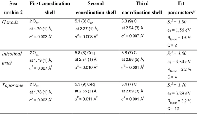
130
purified out of the gonads.32 As the toposome is a transferrin like protein and is acting as a Ca reservoir for
131
sea urchins, it should be considered as a potential candidate for uranyl binding in the gonads. The speciation
132
of uranyl in a solution containing the purified toposome was investigated.
133
134
EXPERIMENTAL SECTION
135
Seawater, sea urchin collection and aquarium setup
136
Seawater was collected in the Mediterranean Sea at the Environmental Laboratory of the International
137
Atomic Energy Agency (IAEA) at 30 m from the coast of Monaco, 50 m deep (43° 43’ 49” N, 7° 25’ 40”).
138
The seawater was filtered at 0.2 μm (Whatman, GF/C grade) and sterilized by UV treatment to eliminate
139
particles and microorganisms. Commercial silica gravel (850 g) was placed at the bottom of the aquarium
140
filled with 10 L of seawater. The silica gel was needed to ensure the survivability of the sea urchins inside
141
the aquarium, and played no chemical role, nor interfered significantly with the experiments. Only one sea
142
urchin was placed in the aquarium at the same time. The aquarium was equipped with a filter and an air
pump that were turned on 7 days before placing the sea urchins inside to equilibrate the whole system. The
144
seawater temperature was maintained at 16°C using a water-cooling system during the experiments.
145
Paracentrotus lividus sea urchins (Figure 1) were collected in Villefranche-sur-mer, by the Laboratoire
146
Océanographique de Villefranche (UMR 7093, Mediterranean Sea, France) and were fed with native algae
147
until 3 days before contamination. The food was then removed from the aquarium, and the water was cleaned
148
of any remnants before the first uranium spike. Specimens with similar size were chosen (average diameter
149
= 7-8 cm, average total dry mass = 20 g). All results reported in this work concern experiments performed
150
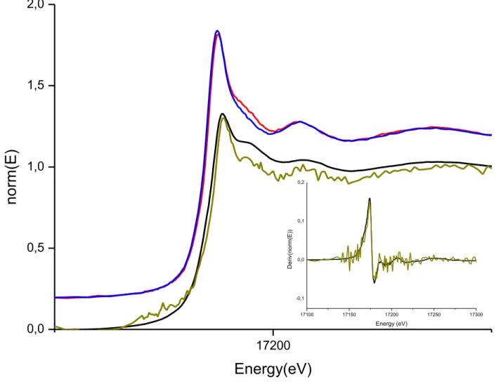
on female specimens. Each specimen used in this report is described in Table S1 of Supplementary
151
Information (SI) file.
152
153
Spiking procedure and uranium distribution
154
Uranium nitrate UO2(NO3).5H2O was directly dissolved in diluted nitric acid (0.1 M) to obtain the 0.375 M
155
uranium solution spike for aquarium use. Both nitric acid and uranium nitrate were of reagent grade, and
156
deionized water was used to dilute the nitric acid. Every 24 hours, 500 µL of this solution were introduced
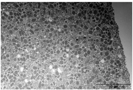
157
in the aquarium, to reach a theoretical final concentration of [U] = 1.88.10-4 M after 10 days. Prior to the
158
spiking (around 10 min before), 500 µL of a 2.10-4 M solution of sodium hydroxide were introduced in the
159
aquarium to avoid any modification of the pH. The latter was controlled using commercial pH paper
160
designed for seawater. The measured pH was around 8. The uranium concentration corresponds to a total
161
mass of uranium of 476.20 ± 27.12 mg. It was chosen as the best compromise between uranium natural
162
concentration and EXAFS sensitivity.
163
Each sample from the aquarium (sea urchin or gravel) was rinsed with deionized water before any further
164
analysis, to remove any uranium potentially adsorbed on the sample surface, and of remnant contaminated
165
sea water, to ensure the validity of the results. The uranium content of each sample was analyzed by
ICP-166
OES (details are provided in SI file).
167
168
Toposome extraction and purification.
169
The toposome purification was performed according to Castellano et al. with slight modifications (details
170
are provided in SI file).33. The final concentration was estimated via UV-visible, using a calculated epsilon,
171
to be around 35 mg/mL (e = 1.252), corresponding to a molar concentration in monomer units of about 2.10
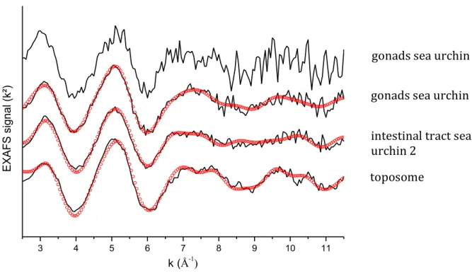
-172
4 M. The epsilon was calculated using the sequence of amino acids published by Noll et al.34 The toposome
173
was then frozen until further use. The protein purity is estimated to be 80% minimum. The toposome is
174
probably organized in the form of trimers. The two discernible fractions under the main band probably
175
correspond to the presence of the two isoforms described at 200 kDa and 180 kDa for the Paracentrotus
176
lividus toposome, which are present in the nutritive phagocytes of the gonads.35
177
178
Time Resolved Laser Induced Fluorescence spectroscopy (TRLFS)
179
Sample preparation, sea urchin: The test was dried, then crushed into powder. The powder was directly
180
analyzed with no further preparation. Concerning the gonads, they were dried and also directly analyzed
181
with no further preparation.
182
Sample preparation, toposome-uranium complex: The same procedure as described above was used.
183
However, the uranium was this time dissolved in a solution of Tris/HCl (10mM) NaCl (10mM), pH 5.5, to
184
reduce the ionic strength of the solution (Cl concentration). The solution was then kept at 4°C until analysis.
185
Data acquisition: A Nd-YAG laser (Model Surelite Quantel) operating at 355 nm (tripled) and delivering
186
about 10 mJ of energy in a 10 ns pulse with a repetition rate of 10 Hz, was used as the excitation source.
187
The laser output energy was monitored by a laser power meter (Scientech). The focused output beam was
188
directed onto the urchin part (gonad, shell) of the sea urchin (previously crushed) placed in a 1 mm
189
pathlength quartz cell of the spectrofluorometer (F900 Edinburgh). The detection was performed by an
190
intensified charge coupled device (Andor Technology) cooled by Peltier effect (-5°C) and positioned at the
191
polychromator exit for the emission spectra measurement and by a photomultiplier tube (PMT) to measure
192
fluorescence decay time. Logic circuits, synchronized with the laser shot beam, allowed the intensifier to be
193
activated with determined time delay (from 0.005 to 1000 μs) and during a determined aperture time (from
194
0.005 to 1000 μs). From a spectroscopic point of view, various gate delays and durations were used to ensure
195
the presence of only one complex by the measurement of a single fluorescence lifetime and spectrum.
196
Fluorescence lifetime measurements were performed by varying the temporal delay with fixed gate width.
197
198
X-ray Absorption Spectroscopy (XAS) Data Acquisition and Analysis
199
Sample preparation, sea urchins: sea urchins 2, 3 and 5 were analyzed by XAS: gonads and intestinal
200
tract for sea urchin 2 (EXAFS), only gonads for sea urchin 3 (EXAFS) and test for sea urchin 5 (X-ray
201
absorption near edge structure (XANES) (see Table S1). For sea urchin 2, gonads and intestinal tract were
202
freeze-dried for 24h. Solid pellets were then prepared by mixing the dry residue with polyethylene in order
203
to obtain homogenous solid pellets. For sea urchin 3, solid pellets were prepared with the gonads by mixing
204
fresh gonads with polyethylene. As polyethylene is only composed of light chemical elements, it does not
205
interfere with the EXAFS measurements. In both cases, the pellets were then kept at -20°C until the analysis
206
to avoid any deterioration of the biological system. For sea urchin 5, the test was mechanically ground and
207
pressed into solid pellets. The Liebigite reference sample (Ca2UO2(CO3)3) was obtained from the
208
mineralogy collection of the Museum National d'Histoire Naturelle (MNHN), Paris, France.
209
Sample preparation, U-toposome complex: uranium nitrate was directly dissolved in Tris/HCl (50mM)
210
NaCl (150mM), pH = 5.5. This pH value prevents any visible precipitation of uranium hydroxides. Absence
of hydrolysis was also verified using speciation codes. pH was adjusted to 5.5 with concentrated chlorhydric
212
acid. The solution was then mixed with the protein, to obtain a final concentration of uranium of 8.10-5 M,
213
and a concentration of protein in monomeric units estimated at 1.6. 10-4 M. An estimated excess of protein
214
ensures that no free uranyl would remain in the solution, which would interfere with the EXAFS analysis.
215
The solution was then kept at 4°C until analysis.
216
EXAFS data acquisition: experiments were performed on the MARS beamline of the SOLEIL
217
synchrotron facility. Energy calibration was performed at the yttrium K edge at 17038 eV and EXAFS
218
experiments at the U LIII edge. The MARS beamline is dedicated to the investigation of radioactive materials
219
in the hard X-ray range.36 The beamline optics consist essentially of a water-cooled double-crystal
220
monochromator (FMB Oxford), which is used to select the incident energy of the X-ray beam and for
221
horizontal focalization, and two large water-cooled reflecting mirrors (IRELEC/SESO) that are used for
222
high-energy rejection (harmonic part) and vertical collimation and focalization. All measurements were
223
achieved in fluorescence mode using a 13-element high purity germanium detector (ORTEC). The X-ray
224
absorption spectra for the test sample (from sea urchin 5) were measured at room temperature, whereas the
225
spectra for the gonad samples (from sea urchins 2 and 3) and for the intestinal tract sample (from sea urchin
226
3) were measured at -165 °C. To perform the latter measurements, the samples were inserted in a specifically
227
designed double containment cell (H. Hermange, SOLEIL) and inserted in the dedicated liquid nitrogen
228
cryostat of the beamline. The protein sample was measured at room temperature.
229
EXAFS data processing was performed using the ATHENA code.37-38 The E
0 energy was identified at
230
the maximum of the absorption edge. Fourier transform (FT) with k2 weighting was performed between 2.5
231
and 12 Å−1 for gonads and 10.5 Å−1 for the intestinal tract, with a Hanning window. The fits were performed
232
using the DEMETER code (version Demeter 0.9.25) and were fit in R space between 1 and 5 Å. EXAFS
233
data fitting: One global amplitude factor S0² and one energy threshold correction factor DE0 were used for
234
every path of the fits. The agreement factor r (%) and the quality factor (QF = reduced χ²) of the fits were
235
provided directly by DEMETER. Phases and amplitudes were calculated using the FEFF6 simulation code
236
integrated in DEMETER based on a partial structural model (in silico) of uranyl-acetate complex
237
(UO2(acetate)2.) This model was chosen because it exhibits both monodentate and bidentate carboxylate
238
ligation to the uranyl equatorial plane. The scattering paths used for the fitting procedure are: i) simple
239
scattering paths including U-Oax within the oxo bond, U-Oeq corresponding to the equatorial oxygen atoms
240
and U...C corresponding to the C atom of the bidentate carboxylate group; ii) multiple scattering paths
241
including the quadruple path U-Oax within the oxo bond, and the triple scattering U-O-C of the monodentate
242
carboxylate function. During the fitting procedure, the number of atoms of carbon in the monodentate and
243
bidentate functions was let free, in case only one coordination mode was present. The total number of
244
variables in the fit was equal to 12.
246
Transmission electron microscope (TEM) imaging
247
Gonads and intestinal tract of sea urchin 2 were analysed with TEM. Directly after dissection, fresh gonads
248
and fresh intestinal tract samples were fixed for 2 h at room temperature with 2.5% glutaraldehyde in
249
cacodylate buffer (0.1 M, pH 7.4) in artificial seawater, then washed with 0.1 M cacodylate buffer (pH 7.4)
250
and post-fixed with 1 % osmium tetroxide in cacodylate buffer containing 1% potassium ferrocyanide. The
251
samples were embedded in Epon resin after dehydration using an acetone/water solution and then acetone.
252
Ultrathin sections (70–80 nm) were cut using a diamond diatom mounted on an ultramicrotome (Ultracut S,
253
Leica) and placed on copper TEM grids coated with formvar film. Sections were observed with a JEOL
254
JEM 1400 TEM equipped with a CCD camera (Morada, Olympus SIS) at the Centre for Applied Microscopy
255
(CCMA, University of Nice Sophia Antipolis, Nice, France).
256
257
Scanning transmission X-ray microscope (STXM) imaging
258
Sample preparation: Gonad and intestinal tract cells of sea urchin 2 were analysed with STXM.
259
The STXM samples were prepared as described above for TEM analysis. Sections of 70-80 nm were placed
260
on a 100 nm thick Si3N4 membrane window (1 mm square) in a 10 mm frame obtained from Silson Ltd. A
261
second Si3N4 window was glued over the first to seal and confine the sample for radiological control
262
purposes.
263
Data acquisition: Data was recorded with the STXM on beamline 11.0.2 of the Advanced Light Source
264
(ALS) located at the Lawrence Berkeley National Laboratory in Berkeley, USA.39 The STXM methodology
265
employed in this study was similar to that described previously.40-41 The photon energy calibration of the
266
monochromator was performed at the neon K-edge (867.3 eV). The STXM measurements were performed
267
with a 25 nm zone plate in transmission mode and the ALS was operating in top-off mode with a beam
268
current of 500 mA. Images at a single energy were obtained by raster scanning the sample and collecting
269
X-rays as a function of sample position. Elemental maps of uranium were obtained by subtracting an image
270
taken before the absorption threshold from an image obtained at resonance utilizing the U N5 transition
271
(~738 eV), following image alignment.
272
Data treatment: Data treatment was performed with the aXis2000 code developed at McMaster
273
University.42
274
275
RESULTS AND DISCUSSION
276
Uranium uptake and distribution in the organs.
277
We have investigated the uptake of uranium by P. lividus in the closed aquarium system described in the
278
experimental section (1 spike per day for 10 days). Four similar experiments have been conducted with five
sea urchins that are detailed in Table S1. A control experiment with no spike was also performed at the same
280
time in a similar aquarium next to the one used for contamination. It showed that no detectable uranium was
281
naturally present in sea urchins (below the detection limit of ICP-OES, about 1x10⁻⁷ mg/L). Each P. lividus
282
specimen was sacrificed and dissected at the end of the 10 days. Gonads, digestive tube and test were
283
separated, and uranium content was measured by ICP-OES.
284
First, the total uranium balance was measured in the entire system with sea urchin 4 to ensure that the
285
major part of the uranium was distributed within the main components (sea water, gravel, sea urchin), and
286
not, for instance, adsorbed on the aquarium wall or filter. The concentration of uranium in each of the
287
different system components was measured. The results are presented in Figure S1. From the concentration
288
in seawater, the global Concentration Factor (CF) of sea urchin 4 for an exposure of 10 days was calculated
289
and is 0.37±0.02.
290
The bioaccumulation of uranium in each compartment of the sea urchin (test, gonad, intestinal tract) has
291
been assessed. Although competition between uranium and other cations was not explicitly taken into
292
account, the use of a natural medium implies that competition is implicitly included. It is presented in Figure
293
2 for sea urchins 1, 2 and 4 as the fraction of total uranium mass per dry weight of each compartment (see
294
Table S2). In the three specimens, the concentration in the digestive tube is almost 3 times and 10 times
295
higher than in the gonads and test, respectively. Differences can be observed between sea urchin 4 and the
296
two others. We explain these discrepancies by the seasonal variations in the gonads quantity and the seasonal
297
variations in the concentration of proteins inside the gonads. Indeed, the experiment on sea urchin 4 was
298
conducted out of the reproduction season (February-May), which was not the case for sea urchins 1 and 2.
299
Partial concentration factors for each compartment may also be calculated. The following CF were obtained
300
for sea urchin 1: CFgonads = 1.0±0.07, CFintestin = 2.8±0.1 and CFtest = 0.25±0.02. The overall very low value
301
obtained for the entire specimen is largely due to the low value of CFtest that is associated to the largest mass
302
of the specimen (the test). These results are in agreement with the values reported by Warnau et al. in 1996
303
as the same ordering was observed for most heavy metals: gonads, intestinal tract >> test. In their
304
communication, the concentration measured in the intestinal tract is the highest of the three compartments
305
for several heavy metals as iron, copper, tin and mercury. However, the gonads are more concentrated in
306
zinc than the intestinal tract, which means that the distribution is element specific and also confirms that
307
speciation is playing a key role in the accumulation mechanisms. In addition, Warnau et al. also highlighted
308
the fact that the concentrations of every metal but lead are always lower in the test than the gonads or the
309
intestinal tract, no matter which of the two latter compartments is the most concentrated. These data clearly
310
highlight the necessity of speciation investigation in each compartment separately, as the accumulation rate
311
of uranium is radically different between the three compartments.
312
313
Uranium speciation in the test
314
The test of sea urchin is mainly composed of monocrystalline calcite (calcium carbonate) rich in
315
magnesium.43 Uranium accumulation in test and spines is very low, as mentioned above, with an average
316
concentration of 11 ppm (CFtest = 0.25±0.02). Such a concentration lies just above the estimated EXAFS
317
detection limit under our experimental conditions. Therefore, an EXAFS spectrum could not be recorded
318
from the test with a reasonable signal to noise ratio and only the XANES part of the spectrum was
319
significant. Figure 3 compares the XANES spectra of the test after in vivo contamination of sea urchin 5
320
with data acquired from a Liebigite (Ca2UO2(CO3)3) solid state reference. Liebigite is taken here as the
321
model for the main species of uranium in seawater, the dicalcic uranyl tricarbonate species, Ca2UO2(CO3)3.
322
The enlarged insert of Figure 3 shows the derivatives of the spectra. A qualitative comparison of both
323
XANES spectra and their derivatives for the test samples and the Liebigite reference suggests that the
324
speciation of uranium in the test is similar to that in the Liebigite model, although it is not definite proof.
325
This could signify that a sorption mechanism occurs and is at the origin of the uranium accumulation in the
326
test. As adult spines do not grow once they reach their adult size, a mechanism involving sorption on the
327
calcite monocrystalline surface of the spines followed by slow diffusion of uranium inside the calcite
328
structure agrees well with the final very low concentration of uranium in the test, contrary to a mechanism
329
of incorporation of the uranium during the growth of the spines. To complement the XANES results, TRLFS
330
measurements were also performed on sea urchin 5. The wavelengths of the maximum of fluorescence
331
emissions obtained are 471, 488, 507, 528 and 552 nm (Figure 4).Previous studies on uranium compounds25
332
report the wavelengths obtained for different species, including uranium in seawater, several carbonated
333
calcium-uranium complexes, and sulphate and phosphate uranium complexes. Indeed, a slight
334
hypsochromic shift is noticed here, mainly for the higher wavelengths compared to free uranyl (reported
335
wavelengths: 470–488–509–534–559 nm), which is characteristic of Mx–UO2–(CO3)y complexes (with x :
336
1-2 and y : 2-3), with M =
Ca2+, Mg2+ and Sr2+.
337
Even though it is not possible to differentiate all the possible species, the results above are consistent with
338
the presence of the dicalcic uranyl tricarbonate species in the test at a relatively low level.
339
340
Uranium accumulation in the gonad and intestinal tract cells, imaging and spectroscopy
341
Transmission electron microscope (TEM) imaging was performed on the gonad cells of contaminated sea
342
urchin 2. Figure 5 shows a large field TEM image of the gonads. More specifically, two types of cells can
343
be observed; the reproductive cells (circular and darker), and the storage cells (various shapes, lighter). No
344
evidence of uranium precipitates can be noted on this micrograph in any of the cells although uranium is
345
present in the gonads at a concentration of around 50 ppm, as mentioned before. Precipitates of uranium
346
phosphate phases have been observed for different bacterial systems. For instance, Suzuki et al. reported
that nanoprecipitates of uranium were visible extra-cellularly in Deinococcus radioduransafter exposure.44
348
Uranium-phosphate crystals were also reported to be present inside cells of Stenotrophomonas Maltophilia
349
by Merroun et al.45It was also observed in living cells UMR-106, which are model osteoblastic cells.46 The
350
absence of any visible precipitate, although not a definite proof, suggests that uranyl is not incorporated as
351
an insoluble mineral phase. To further investigate this assumption, STXM elemental imaging was performed
352
on gonads cells of the same sea urchin specimen. Figure 6 shows the STXM image recorded at 738 eV, just
353
above the uranium NV edge. One can clearly distinguish the cell membrane and the different organelles
354
inside. The elemental map was then obtained via alignment and subtraction of the STXM image collected
355
at an energy preceeding the NV edge at 725 eV (Figure S5).The elemental map reveals a featureless map
356
with a shadow ring at the location of the cell wall although the contrast in the shadow ring is noticeable but
357
only 2% above background. Thus, the elemental map shows that uranium is not localized in specific hot
358
spots or precipitates to our degree of both spatial and spectral resolution but may be homogeneously
359
distributed around the cell membrane. As a consequence, both TEM and STXM images suggest that uranium
360
may be complexed within the cell by proteins, enzyme or metabolites even though this is clearly not a
361
definitive evidence because only a limited number of cells were observed and the signal of the shadow ring
362
in the uranium elemental map is very weak.
363
Considering the affinity of uranyl for hard donor oxygen groups like in the transferrin binding site
364
(aspartate, tyrosine, carbonate)7,47, complexation of the uranyl cation by carboxylate rich proteins, enzyme
365
or metabolites is a reasonable assumption. This could also explain the low contrast observed in the STXM
366
contrast image because it would be distributed over the entire cell membrane. Such ligation could involve
367
aspartic, glutamic or tyrosine residues, for instance. To further determine the speciation of uranium inside
368
the cells and possible complexation, contaminated gonads of sea urchin 2 and 3 were analysed by EXAFS
369
and XANES spectroscopy at the uranium LIII edge. The XANES spectrum of the gonads of sea urchin 2 is
370
provided in Figure 3 and exhibits the expected uranyl pattern. The EXAFS spectra obtained for the gonades
371
of sea urchins 2 and 3 are presented in Figure 7. The experimental spectrum for sea urchin 2 was adjusted
372
with a model composed of 3 scattering shells (Oyle, Oeq and C) as explained in the experimental section (the
373
spectrum corresponding to sea urchin 3 being superimposable, but at lower signal to noise ratio, is shown
374
but not fitted). The best fit metrical parameters are reported in Table 1, the Fourier transform (modulus and
375
imaginary parts) is provided in Figure S6. The first shell corresponds to the two axial oxygen atoms typical
376
of the uranyl oxocation. The second shell is composed of 5.1(3) O atoms at 2.37(1) Å (average) and the last
377
shell is composed of 3.3(9) C atoms at 2.94(3) Å. Such a coordination sphere is indeed typical of carboxylate
378
rich biomolecules, although a detailed path by path analysis is not possible here in the absence of a specific
379
structural model. Alternative attempts to fit our data with a Liebigite model lead to unrealistic metrical
380
parameters, thus supporting protein or metabolite complexation. Pible et al. in 2006 studied the interaction
between uranyl and a wide number of proteins.6 They calculated and reported the distances between uranium
382
and selected protein structures containing uranyl. Values for the U-Oeq distances fall into the range 2.31 -
383
2.61 Å for monodentate carboxylate groups and 2.41 - 2.84 Å for bidentate carboxylate groups. In the
384
specific case of transferrin, Hémadi et al. in 2009 and Wang et al. more recently reported the binding
385
mechanism of uranyl to transferrin by DFT calculations.8-9 In the model of Wang, the most probable
386
coordination sphere is composed of one bidentate carbonate with U-O at 2.46 Å, two tyrosines with U-O at
387
2.30 Å, and one monodentate aspartate at a distance of 2.44 Å. The values we obtained for the U-Oeq average
388
distance (reported in Table 1) seems to indicate the presence of monodentate carboxylate groups although
389
only average values are discussed here. Concerning the carbons atoms, the U...C average distances reported
390
in Table 1 in the range 2.9 - 3.0 Å also support the presence of bidentate carboxylate functions. No evidence
391
of monodentate complexation was found using the U...C interaction. However, this is not an absolute proof,
392
considering that this interaction is often very weak, due to the angle of the U-O-C bond (deviating from
393
focusing effect optimum angle, 180°) in monodentate configuration. On the other hand, the triple scattering
394
path U-C-O was found to be necessary to fit the experimental data. This path being a fingerprint of
395
monodentate ligation, it suggests the occurrence of at least one monodentate carboxylate function. We thus
396
propose here the following mode of coordination: two atoms of oxygen (for the oxo bonds), a mix of
397
monodentate and bidentate carboxylate functions with an unknown ratio, and possibly one water molecule
398
to complement the equatorial coordination sphere. The above structural data therefore suggest that uranyl
399
is coordinated to carboxylate residues of a protein or metabolite.
400
As for the gonad cells, the EXAFS (Figure 7) and XANES (Figure 3) spectra at the LIII edge of uranium
401
was recorded for the intestinal tract cells of sea urchin 2. Qualitatively, the spectra are very similar to what
402
is discussed above, suggesting that the same type of complexation occurs in this compartment as well.
403
Experimental and simulated EXAFS spectra are displayed in Figure 7, best fit structural parameters are also
404
reported in Table 1. The U-Oeq average distance (2.35(1) Å) is similar to those obtained for the gonad cells
405
(2.37(1) Å) and the same type of U...C path has been used. This suggests that the same biomolecule (or
406
same type) is involved in the uranium complexation in the intestinal tract.
407
In addition, TRFLS measurements were also performed (Figure 4) on the gonads of sea urchin 5. The
408
following fluorescence wavelengths were obtained: 501-521-542 nm. In contrast to what was obtained for
409
the test, a bathochromic effect with respect to free uranyl (488–509–534 nm) is observed, together with two
410
lifetimes of 140 µs and 1600 µs without modification of the spectra. This indicates that the carbonato calcite
411
form of uranyl is not the correct speciation within the gonads, as already supported by the EXAFS data
412
fitting. Bathochromic effects have been reported to occur in the presence of complexing ligands, such as
413
sulphate or phosphonate, the latter leading to the largest shift (main wavelengths: 496-519-545). In the
414
present case, the shift is even more important taken together with the broadening of the spectrum, indicating
that most of the uranyl is likely to be complexed with a strong complexing ligand with multiple interaction
416
sites such as macromolecules that establish several local environments (and account for the broadening
417
observed as well as the different lifetimes).
418
419
In conclusion, the best fit EXAFS data support the hypothesis of a complexation of the uranyl with
420
carboxylate rich proteins, enzymes or metabolites in gonads and intestinal tract cells. This assumption is
421
also in full agreement with both STXM and TEM imaging.
422
423
Uranium-toposome complexation
424
The toposome is the main protein in sea urchins and is present in both the gonads and intestinal tract. It
425
is described as a multimer of 180 kDa monomers, known as 22S fragment.48
426
This protein, also referred to as major yolk protein, was previously identified as a calcium binding
iron-427
less transferrin like protein.35 As a consequence, toposome is a potential candidate for uranyl complexation
428
in the cells. Different final masses are reported, depending on the number of monomers (from 1 to 6). As a
429
consequence, toposome is a potential candidate for uranyl complexation in the cells.
430
Following toposome extraction from gonad cells and purification, the EXAFS spectrum at the LIII edge of
431
uranium was recorded for the U-toposome complex in solution. The experimental XANES data, the
432
experimental EXAFS spectra and the best simulated EXAFS spectra are similar to both gonads and intestinal
433
tract spectra although some slight differences appear between 6 and 9 Å-1 (Figure 7). Nonetheless, the
434
similarities between the best fit structural parameters (reported in Table 1) obtained for the three systems
435
strongly suggest that the toposome protein is a good match for uranyl complexation in those two
436
compartments. The slight differences observed in the EXAFS spectra, notably in the wave between 6 and 9
437
Å-1 are not due to differences in the nature of the binding site (like carboxylate versus phosphate ligation for
438
instance) but most probably to differences in conformation as indicated by the similarities of the imaginary
439
parts of the moduli of the FT (Figure S6).
440
TRFLS measurements were also performed on a U-toposome solution (2:1 ratio, [UO2] = 1µM). The
441
spectrum represented in Figure 4 exhibits the same fluorescence wavelengths as observed before
(501-521-442
542nm). However, two other fluorescence wavelengths must be highlighted: 485nm and 465nm, the latter
443
being characteristic of carboxylic functions. This validates the EXAFS interpretation for the U-toposome
444
complex and suggests that the speciation of uranyl in the toposome complex and in gonads and intestinal
445
tract cells is similar. The absence of the 465 nm wavelength in the gonad TRLFS spectrum may be explained
446
because the gonads are not only composed of toposome, even though it is the main protein. Other proteins
447
or metabolites and thus other complexation conformations might be present. The spectrum obtained for
448
gonads is in consequence less specific.
450
The fact that the toposome is likely to complex uranyl inside the sea urchin is a result of great interest, as it
451
is a strong evidence of the metabolization of uranyl inside a living organism, from a dicalcic carbonato form
452
in seawater to a protein complex inside the sea urchin. This shows the importance of speciation as a key
453
parameter to understand and evaluate the potential impact on the environment and on humans that this
454
element, and by extension, other similar elements, can have, if released in the environment. There is still an
455
important need to continue these studies, as the speciation of multiple radioisotopes in biocycles remains
456
completely unknown.457
458
Associated content459
The Supporting Information is available free of charge on the ACS Publications website at …
460
The SI contains Table S1, which describes the different measurements on the 5 sea urchins. Results dealing
461
about the distribution of the uranium inside the sea urchin organs and inside the aquarium (Table S2 and
462
Figure S1). Elution profiles and SDS gel are presented Figure S2, S3 and S4. Complementary STXM images
463
and EXAFS data are also presented (Figure S5 and S6).
464
465
Author information466
Corresponding author:467
*C. Den Auwer, Phone: +0033 , E-mail : christophe.denauwer@univ-cotedazur.fr
468
The authors declare no competing financial interest.
469
470
ACKNOWLEDGMENTS:
471
This work was financed by the DNP (Direction of Nuclear Propulsion) of CEA DAM. We thank the
472
International Atomic Energy Agency, Environmental Laboratory of Monaco for providing the seawater. We
473
also acknowledge the LOV, UMR 7093 for providing the sea urchins and the algae. The XAS experiments
474
were performed at the MARS beamline of the SOLEIL synchrotron facility, Gif sur Yvette, France. This
475
research was supported in part (DES, DKS) by the Director, Office of Science, Office of Basic Energy
476
Sciences, Division of Chemical Sciences, Geosciences, and Biosciences Heavy Elements Chemistry
477
program of the U.S. Department of Energy under Contract Number DE-AC02-05CH11231 at Lawrence
478
Berkeley National Laboratory (LBNL). This research used resources of the Advanced Light Source, which
479
is a U.S. Department of Energy Office of Science User Facility supported under Contract No.
DE-AC02-480
05CH11231 at LBNL. Finally, we thank the Centre Commun de Microscopie Appliquée for the SEM
481
imaging.
482
Table 1: Best fit parameters for EXAFS data from the U LIII edge. Numbers in brackets are the estimated
484
uncertainties, numbers in italics have been fixed. σ2 is the Debye Waller factor of the considered scattering
485
path. S02 is the global amplitude factor, e0 is the energy threshold, Rfactor is the agreement factor of the fit in
486
% and Q is the quality factor (reduced CHI2) of the fit.
487
488
489
Sea
urchin 2
First coordination
shell
Second
coordination shell
Third
coordination shell
Fit
parameters
aGonads
2 Oax at 1.79 (1) Å, s2 = 0.003 Å2 5.1 (3) Oeq at 2.37 (1) Å, s2 = 0.008 Å2 3.3 (9) C at 2.94 (3) Å s2 = 0.007 Å2 S02 = 1.00 e0 = 1.56 eV Rfactor = 1.6 % Q= 2Intestinal
tract
2 Oax at 1.79 (1) Å, s2 = 0.007 Å2 5.8 (8) Oeq at 2.34 (1) Å, s2 = 0.010 Å2 3.8 (7) C at 2.96 (5) Å, s2 = 0.001 Å2 S02 = 1.00 e0 = 3.34 eV Rfactor = 2.2 % Q= 4Toposome
2 Oax at 1.78 (1) Å, s2 = 0.003 Å2 5.5 (9) Oeq at 2.35 (2) Å s2 = 0.011 Å2 3.4 (7) C at 2.89 (3) Å s2 = 0.001 Å2 S02 = 1.10 e0 = 3.29 eV Rfactor = 2.2 % Q= 12490
491
492
493
494
Figure 1: Picture of a sea urchin Paracentrotus lividus (one unit = one centimeter).
495
496
497
498
499
500
501
502
Figure 2: Uranium concentration (in mg/g, elemental U) for the three compartments of the sea urchins 1, 2
503
and 4.
504
505
Sea urchin 1 Sea urchin 2 Sea urchin 4
0,00 0,02 0,04 0,06 0,08 0,10 0,12 0,14
Q
ua
nt
ity
of
U
(mg
/g
)
Test
Gonads
Intestinal tract
506
Figure 3: XANES spectra at the U LIII edge of the test of sea urchin 5 (green), of the Liebigite reference sample (black), of the gonads (red) and the intestinal tract (blue) of sea urchin 2 (both shifted in ordinates for clarity). Insert: Enlargement of the corresponding first derivatives of the test and the Liebigite reference sample. 17200 0,0 0,5 1,0 1,5 2,0
no
rm(E)
Energy(eV)
17100 17150 17200 17250 17300 -0,1 0,0 0,1 0,2 D eri v(n orm(E)) Energy (eV)507
508
Figure 4: TRFLS spectra obtained for the contaminated test of sea urchin 5 (red), for the contaminated
509
gonads of sea urchin 5 (blue) and U-toposome complex (black)
510
511
512
513
425 450 475 500 525 550 575 600 0 50000 100000 150000 200000 In te nsi ty (g on ad s an d sh el l) Wavelength (nm) 0 500000 1000000 1500000 2000000 2500000 In te nsi ty (t op oso me )514
515
516
517
518
519
Figure 5: TEM imaging performed on the gonads of sea urchin 2.
520
521
522
523
Figure 7 : EXAFS spectra (k²) at the U LIII edge of the contaminated intestinal tract of sea urchin 2 (SU2)
524
and the U-toposome complex. The EXAFS spectra obtained for the contaminated gonads of sea urchin
525
3(SU3) is also presented. Experimental = straight line; fit = red dots.
526
527
528
gonads sea urchin 2
intestinal tract sea
urchin 2
toposome
gonads sea urchin 3
3 4 5 6 7 8 9 10 11 k (Å-1) EXAF S si gn al (k²)References:
529
530
[1] Pereira R., Barbosa S., Carvalho F.P., Uranium mining in Portugal: a review of the environmental
531
legacies of the largest mines and environmental and human health impacts. Environ. Geochem. Health 2014,
532
36 (2), 285-301.
533
[2] Wendel C.C., Fifield L.K., Oughton D.H., Lind O.C., Skipperud L., Bartnicki J., Tims S.G., Høibråten
534
S., Salbu B., Long-range tropospheric transport of uranium and plutonium weapons fallout from
535
Semipalatinsk nuclear test site to Norway. Environ. Int. 2013, 59, 92-102.
536
[3] Abe Y., Iizawa Y., Terada Y., Adachi K., Igarashi Y., Nakai I., Detection of uranium and chemical state
537
analysis of individual radioactive microparticles emitted from the Fukushima nuclear accident using
538
multiple synchrotron radiation X-ray analyses. Anal. Chem. 2014, 86 (17), 8521-8525.
539
[4] Bleise A., Danesi P.R., Burkart W., Properties, use and health effects of depleted uranium (DU): a
540
general overview. J. Environ. Radioactiv. 2003, 64 (2-3), 93–112.
541
[5] Michon J., Frelon S., Garnier C., Coppin F., Determinations of Uranium (VI) Binding Properties with
542
some Metalloproteins (Transferrin, Albumin, Metallotionein and Ferritin) by Fluorescence Quenching. J.
543
Fluoresc 2010, 20 (2), 581-590.
544
[6] Pible O., Guilbaud P., Pellequer J.-L., Vidaud C., Quéméneur E., Structural insights into protein–uranyl
545
interaction: towards an in-silico detection method. Biochimie 2006, 88 ,1631–1638.
546
[7] Vidaud C., Gourion-Arsiquaud A., Rollin-Genetet F., Torne-Celer C., Plantevin S., Pible O., Berthomieu
547
C., Quemeneur E. Structural Consequences of Binding of UO22+ to Apotransferrin: Can This Protein
548
Account for Entry of Uranium into Human Cells. Biochemistry 2007 46, 2215-2226.
549
[8] Hemadi M., Ha-Duong N.T., Plantevin S., Vidaud C., El Hage Chahine J.M., Can uranium follow the
550
iron-acquisition pathway? Interaction of uranyl-loaded transferrin with receptor 1. J. Biol. Inorg. Chem.
551
2010, 15, 497–504.
552
[9] Wang M., Ding W., Wang D., Binding mechanism of uranyl to transferrin implicated by density
553
functional theory study. RSC Adv. 2017, 7, 3667.
554
[10] Markich S.J., Uranium Speciation and Bioavailability in Aquatic Systems: An
555
Overview. TheScientificWorldJo 2002, (2), 707-729.
556
[11] data from U.S. Geological Survey, www.usgs.us
557
[12] Chen J.H., Lawrence Edwards R., Wasserburg G.J., 238U,234U and232Th in seawater. Earth Planet. Sc.
558
Lett. 1986, 80 (3–4), 241-251.
559
[13] Rona E., Gilpatrick L. O., Jeffrey L. M., Uranium determination in sea water. EOS T. Am. Geophys.
560
Un. 1956, 37 (6), 697-701.
561
[14] Ku T.L., Knauss K.G., Mathieu G.G., Uranium in open ocean: concentration and isotopic composition.
562
Deep-Sea Res. 1977, 24 (11), 1005-1010.
563
[15] Guegueniat, P., Germain, P., Metivier, H., Radionuclides in the oceans: inputs and inventories., 1996,
564
France: Les editions de physique.
565
[16] Soualili D., Dubois P., Gosselin P., Pernet P., Guillou M., Assessment of seawater pollution by heavy
566
metals in the neighbourhood of Algiers: use of the sea urchin, Paracentrotus lividus, as a bioindicator. ICES
567
J. Mar. Sci. 2008, 65, 132–139.
568
[17] Bernd S., The use of fish parasites as bioindicators of heavy metals in aquatic ecosystems: a review.
569
Aquat. Ecol. 2001, 35, 245–255.
570
[18] Mostafa H. M., Collins K. J., Heavy metal concentrations in sea urchin tissues from Egypt, Ireland and
571
United Kingdom. Chem. Ecol. 1995, 10 (1-2), 181-190.
572
[19] INTERNATIONAL ATOMIC ENERGY AGENCY, Sediment Distribution Coefficients and
573
Concentration Factors for Biota in the Marine Environment, Technical Reports Series No. 247, 1985.
574
[20] INTERNATIONAL ATOMIC ENERGY AGENCY, Sediment Distribution Coefficients and
575
Concentration Factors for Biota in the Marine Environment, Technical Reports Series No. 422, 2004.
576
[21] Jeffree R. A., Warnau M., Teyssié J-L., Markich S. J., Comparison of the bioaccumulation from
577
seawater and depuration of heavy metals and radionuclides in the spotted dogfish Scyliorhinus canicula