HAL Id: hal-03044376
https://hal.archives-ouvertes.fr/hal-03044376
Submitted on 7 Dec 2020
HAL is a multi-disciplinary open access
archive for the deposit and dissemination of
sci-entific research documents, whether they are
pub-lished or not. The documents may come from
teaching and research institutions in France or
abroad, or from public or private research centers.
L’archive ouverte pluridisciplinaire HAL, est
destinée au dépôt et à la diffusion de documents
scientifiques de niveau recherche, publiés ou non,
émanant des établissements d’enseignement et de
recherche français ou étrangers, des laboratoires
publics ou privés.
production in multiciliated cells
Diego Revinski, Laure-Emmanuelle Zaragosi, Camille Boutin, Sandra
Ruiz-Garcia, Marie Deprez, Virginie Thomé, Olivier Rosnet, Anne-Sophie
Gay, Olivier Mercey, Agnès Paquet, et al.
To cite this version:
Diego Revinski, Laure-Emmanuelle Zaragosi, Camille Boutin, Sandra Ruiz-Garcia, Marie Deprez, et
al.. CDC20B is required for deuterosome-mediated centriole production in multiciliated cells. Nature
Communications, Nature Publishing Group, 2018, 9, �10.1038/s41467-018-06768-z�. �hal-03044376�
CDC20B is required for deuterosome-mediated
centriole production in multiciliated cells
Diego R. Revinski
1
, Laure-Emmanuelle Zaragosi
2
, Camille Boutin
1
, Sandra Ruiz-Garcia
2
, Marie Deprez
2
,
Virginie Thomé
1
, Olivier Rosnet
1
, Anne-Sophie Gay
2
, Olivier Mercey
2
, Agnès Paquet
2
, Nicolas Pons
2
,
Gilles Ponzio
2
, Brice Marcet
2
, Laurent Kodjabachian
1
& Pascal Barbry
2
Multiciliated cells (MCCs) harbor dozens to hundreds of motile cilia, which generate
hydrodynamic forces important in animal physiology. In vertebrates, MCC differentiation
involves massive centriole production by poorly characterized structures called
deutero-somes. Here, single-cell RNA sequencing reveals that human deuterosome stage MCCs are
characterized by the expression of many cell cycle-related genes. We further investigated the
uncharacterized vertebrate-speci
fic cell division cycle 20B (CDC20B) gene, which hosts
microRNA-449abc. We show that CDC20B protein associates to deuterosomes and is
required for centriole release and subsequent cilia production in mouse and Xenopus MCCs.
CDC20B interacts with PLK1, a kinase known to coordinate centriole disengagement with the
protease Separase in mitotic cells. Strikingly, over-expression of Separase rescues centriole
disengagement and cilia production in CDC20B-de
ficient MCCs. This work reveals the
shaping of deuterosome-mediated centriole production in vertebrate MCCs, by adaptation of
canonical and recently evolved cell cycle-related molecules.
DOI: 10.1038/s41467-018-06768-z
OPEN
1Aix Marseille Univ, CNRS, IBDM, Marseille, France.2Université Côte d’Azur, CNRS, IPMC, Sophia-Antipolis, France. These authors contributed equally: Diego R. Revinski, Laure-Emmanuelle Zaragosi, Camille Boutin. Correspondence and requests for materials should be addressed to
B.M. (email:marcet@ipmc.cnrs.fr) or to L.K. (email:laurent.kodjabachian@univ-amu.fr) or to P.B. (email:barbry@ipmc.cnrs.fr)
123456789
M
ulticiliated cells (MCCs) are present throughout
metazoan evolution and serve functions ranging from
locomotion of marine larvae and
flatworms, to brain
homeostasis, mucociliary clearance of pathogens and
transpor-tation of oocytes in vertebrates
1–3. The formation of MCCs
requires the production of numerous motile cilia through a
complex process called multiciliogenesis
2,3. The transcriptional
control of multiciliogenesis has been decrypted to a large extent,
through studies in Xenopus and mouse
2. Seating at the top of the
cascade, the Geminin-related factors GemC1
4–7and Multicilin
8,9(MCIDAS in mammals) are both necessary and sufficient to
initiate MCC differentiation. GemC1 and Multicilin in complex
with E2F transcription factors have been reported to activate the
expression of Myb, FoxJ1, Rfx2, and Rfx3, which collectively
regulate the expression of a large body of effectors required for
the formation of multiple motile cilia
4,5,8–11. Recently, defective
multiciliogenesis caused by mutations in MCIDAS and Cyclin O
(CCNO) has been associated with congenital respiratory and
fertility syndromes in human
12,13.
Each cilium sits atop a modified centriole, called a basal body
(BB). After they exit from the cell cycle, maturing MCCs face the
challenge of producing dozens to hundreds of centrioles in a
limited time window. In vertebrate MCCs, bulk centriole
bio-genesis is mostly achieved through an acentriolar structure
named the deuterosome, although canonical amplification from
parental centrioles also occurs
1–3. The deuterosome was
first
described in early electron microscopy studies of various
mul-ticiliated
tissues
including
the
mammalian
lung
14and
oviduct
15,16, the avian trachea
17, and the Xenopus tadpole
epi-dermis and trachea
18. In mammalian MCCs, the deuterosome
was described as a spherical mass of
fibers organized into an
inner dense region and an outer, more delicate, corona
16. In
Xenopus, deuterosomes were initially named procentriole
orga-nizers and were reported as dense amorphous masses
18. Recent
studies have revealed that deuterosome-mediated centriole
synthesis mobilizes key components of the centriole-dependent
duplication pathway of the cell cycle, including CEP152, PLK4,
and SAS6
19–21. However, the deuterosome itself differs from the
centriole and may contain specific components. The
identifica-tion of one such component, called DEUP1 for Deuterosome
assembly protein 1, opened the possibility to investigate the
deuterosome at the molecular level
21. In mouse tracheal
epen-dymal cells, DEUP1 was detected in the core of the
deutero-some
21. DEUP1, also known as CCDC67, is a conserved
vertebrate paralogue of CEP63, itself known for its importance in
initiation of centriole duplication during the cell cycle
21,22.
Consistently, DEUP1 was shown to be essential for centriole
multiplication in mouse and Xenopus MCCs
21. Both CEP63 and
DEUP1 interact with CEP152, an essential event for centriole
duplication and multiplication in cycling cells and MCCs,
respectively
21,22. Once centriole multiplication is over,
neo-synthesized centrioles must disengage from deuterosomes and
parental centrioles, convert into BBs and migrate apically to dock
at the plasma membrane to initiate cilium elongation.
In this study, we aimed at better understanding deuterosome
biology. We found that the gene CDC20B was specifically
expressed in maturing MCCs during the phase of centriole
multiplication. We established the corresponding CDC20B
pro-tein as an essential regulator of centriole-deuterosome
disen-gagement. This work illustrates well the strong functional
relationships that exist between centriole release from
deutero-somes and centriole disengagement in mitotic cells. It also posits
CDC20B as a component of a
“multiciliary locus” that contains
several gene products, either proteins, such as MCIDAS, CCNO
or CDC20B itself, or microRNAs, such as miR-449abc, which are
all actively involved into vertebrate multiciliogenesis.
Results
MCC single-cell transcriptome at deuterosome stage. To
identify regulators of centriole multiplication, we analyzed the
transcriptome of human airway epithelial cells (HAECs) at the
differentiation stage corresponding to active centriole
multi-plication
23at the single-cell level (Fig.
1
a). Gene expression data
from 1663 cells were projected on a 2D space by t-distributed
Stochastic Neighbor Embedding (tSNE) (Fig.
1
b). We identified a
small group of 37 cells corresponding to maturing MCCs engaged
in deuterosome-mediated centriole amplification, as revealed by
the specific expression of MCIDAS
8, MYB
24, and DEUP1
21(Fig.
1
c, d and Supplementary Figure 1). This subpopulation was
characterized by the expression of known effectors of centriole
synthesis, such as PLK4, STIL, CEP152, SASS6, but also of cell
cycle regulators, such as CDK1, CCNB1, CDC20, SGOL2, and
NEK2 (Fig.
1
d, Supplementary Figure 1 and Supplementary
Table 1). We reasoned that uncharacterized cell cycle-related
genes that are specific to this subpopulation could encode
com-ponents of the deuterosome-dependent centriole amplification
pathway. A particularly interesting candidate in this category was
CDC20B (Fig.
1
d), which is related to the cell cycle regulators
CDC20 and FZR1
25(Supplementary Figure 2a). First, the
CDC20B gene is present in the vertebrate genomic locus that also
contains the key MCC regulators MCIDAS
8and CCNO
13.
Co-expression of CDC20B, MCIDAS, and CCNO throughout HAEC
differentiation was indeed observed in an independent RNA
sequencing study, performed on a bulk population of HAECs
(Supplementary Figure 2b). These results
fit well with the
observation that the promoter of human CDC20B was strongly
activated by the MCIDAS partners E2F1 and E2F4
(Supple-mentary Figure 2c), as also shown in Xenopus by others
9(Sup-plementary Figure 2d). Second, the CDC20B gene bears in its
second intron the miR-449 microRNAs, which were shown to
contribute to MCC differentiation
23,26–30. Finally, in Xenopus
epidermal MCCs, cdc20b transcripts were specifically detected
during the phase of centriole amplification (Supplementary
Figure 2e–m). This first set of data pointed out the specific and
conserved expression pattern of CDC20B in immature MCCs. In
the rest of this study, we analyzed the putative role of CDC20B in
deuterosome-mediated centriole multiplication.
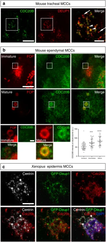
Composition and organization of vertebrate deuterosomes. We
first conducted a series of immunofluorescence analyses to gain a
better understanding of deuterosome organization in mouse
ependymal and Xenopus epidermal MCCs as models. In
whole-mounts of mouse ependymal walls, mature deuterosomes
revealed by DEUP1 staining appeared as circular structures
around a lumen (Fig.
2
a). We noticed that DEUP1 also stained
fibers emanating from the core into the corona. Nascent
cen-trioles revealed by the marker FOP were organized around the
DEUP1-positive core ring. STED super-resolution microscopy
helped to better appreciate the regular organization of individual
FOP-positive procentrioles (Fig.
2
b). Proximity labeling assays
have revealed that when ectopically expressed in centrosomes
CCDC67/DEUP1 is found close to Pericentrin (PCNT) and
γ-tubulin, two main components of the pericentriolar material
(PCM)
31. Interestingly, we found that PCNT was present in the
deuterosome corona (Fig.
2
a), and STED microscopy further
revealed that PCNT formed
fibers around growing procentrioles
(Fig.
2
b).
γ-tubulin staining was detected in the DEUP1-positive
deuterosome core, as well as in the corona (Fig.
2
a). STED
microscopy indicated that PCNT and
γ-tubulin stained distinct
interwoven
fibers in the deuterosome corona. Next, we stained
immature Xenopus epidermal MCCs with
γ-Tubulin and Centrin
to reveal centriole amplification platforms. These platforms
displayed irregular shapes and sizes (Fig.
2
c), in agreement with
early electron microscopy studies
18. Expression of low amounts of
GFP-Deup1 in MCCs induced by Multicilin confirmed that active
deuterosomes are embedded in
γ-Tubulin-positive masses
(Fig.
2
d). Overall, this analysis is consistent with early
ultra-structural studies, as the deuterosome core and corona can be
distinguished by the presence of DEUP1 and PCNT, respectively.
Moreover,
γ-tubulin is a conserved marker of centriole
amplifi-cation platforms in vertebrate MCCs. By analogy to the
organi-zation of the centrosome, we propose to coin the term
perideuterosomal material (PDM) to describe the corona, as this
region may prove important for deuterosome function.
Nasal turbinates 20 DEUP1+ MKl67+ 10 0 tSNE.2 tSNE.1
DEUP1 MCIDAS MYB FOXJ1 CDC20B
NEK2 CDC20 CCNB1 CDK1 PLK4 –10 –10 0 10 20 G1/S G2/M M M/G1 MKl67 FOXJ1 Pseudotime of differentiation MYB DEUP1 TP63 5 –5 4 3 2 1 0 0 S –20 –20 4 3 2 2.5 2.0 1.5 2.5 2.0 3.0 1.5 2.5 2.0 3.0 3.5 1.5 2 3 2 3 4 2 3 2 3 4 2 3 4 2 3 4 5
3D HAEC culture at centriole multiplication stage
Single cell suspension of whole-cell culture Single cell RNA-seq
a
b
c
d
Fig. 1 Single-cell RNA-seq analysis reveals MCC transcriptome at deuterosome stage. a Experimental design of the scRNA-seq experiment. b tSNE plot. Each point is a projection of a unique cell on a 2D space generated by the tSNE algorithm. Blue dots represent MKI67-positive proliferating cells, and red dots represent DEUP1-positive cells corresponding to maturing MCCs at deuterosome stage.c Cell cycle-related gene set expression in HAECs measured by scRNA-seq. Cells were ordered along a pseudotime axis, defined with the Monocle2 package. Phase-specific scores are displayed in the top heatmap. Expression of selected genes is displayed in the bottom heatmap.d tSNEs plots for a selection of genes specifically enriched in deuterosome stage cells. Note that CDC20B exhibits the most specific expression among deuterosome marker genes
CDC20B associates to vertebrate deuterosomes. We then
ana-lyzed the subcellular localization of CDC20B protein in
deu-terosome stage mouse and Xenopus MCCs. In immature mouse
tracheal MCCs, double immunofluorescence revealed the
asso-ciation of CDC20B to DEUP1-positive deuterosomes (Fig.
3
a).
We noticed that CDC20B tended to associate primarily to large
DEUP1 foci. As deuterosomes grow as they mature
21, this
sug-gests that CDC20B may penetrate into the deuterosomal
envir-onment at a late stage of the centriole multiplication process. The
same observation was made when comparing CDC20B staining
in the region of immature and mature deuterosomes of mouse
ependymal MCCs (Fig.
3
b). As double DEUP1/CDC20B staining
could not be performed on these cells, we analyzed CDC20B
distribution relative to FOP-positive procentrioles. In early
deu-terosome stage MCCs, CDC20B was expressed at low levels and
FOP staining was mostly concentrated in a large amorphous
cloud (Fig.
3
b). In such cells, no CDC20B staining was detected in
association to FOP-positive procentrioles growing around
deu-terosomes. In contrast, in mature deuterosome stage MCCs,
CDC20B was enriched in the innermost part of the PDM,
probably very close to the deuterosome core (Fig.
3
b). Further
evidence was provided with a custom-made polyclonal antibody
(Supplementary Figure 3b, c) used to analyze Cdc20b protein
distribution in Xenopus epidermal MCCs. Here also, Cdc20b was
found associated to Deup1-positive deuterosomes actively
engaged in centriole synthesis (Fig.
3
c). We
finally analyzed the
distribution of CDC20B in mature MCCs. As previously reported,
the CDC20B protein was detected near BBs
23, but also in cilia of
fully differentiated human airway MCCs (Supplementary
Figure 4a–c). This was confirmed by proximity ligation assays
that revealed a tight association of CDC20B with Centrin2 and
acetylated
α-Tubulin, in BBs and cilia, respectively
(Supplemen-tary Figure 4d–f). Fluorescent immunostaining also revealed the
presence of Cdc20b in the vicinity of BBs in Xenopus epidermal
MCCs (Supplementary Figure 4g–i). In contrast, no cilia staining
was observed in these cells. Altogether, our analyses revealed that
in three distinct types of MCCs in two distant vertebrate species,
CDC20B is tightly associated to mature deuterosomes. We next
investigated whether it may control their function.
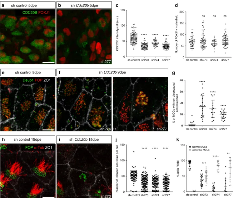
CDC20B is required for multiciliogenesis in vertebrates. For
that purpose, Cdc20b was knocked down in mouse ependymal
MCCs, through post-natal brain electroporation of three distinct
shRNAs. One of them, sh274, which targets the junction between
exons 3 and 4, and can therefore only interact with mature
Fig. 2 Composition and organization of vertebrate deuterosomes a, b Maturing mouse ependymal MCCs were immunostained as indicated, pictures were taken with confocal (a) or STED (b) microscope. a Individual deuterosomes (dashed boxes in top panels) are shown at higher magnification in bottom panels. DEUP1 stains the deuterosome core (ring) and a closefibrous area that defines the perideuterosomal region. The centriolar marker FOP reveals procentrioles arranged in a circle around the deuterosome. Pericentrin (PCNT) is enriched in the perideuterosomal region.γ-Tubulin (γ-TUB) stains the core as well as the periphery of the deuterosome.b STED pictures showing the organization of FOP, PCNT, and γ-TUB around deuterosomes. Individual centrioles identified by FOP staining are pointed out with arrowheads. The diagram was drawn from the adjacent FOP photograph to help reveal the regular concentric organization of nascent centrioles in a typical deuterosomalfigure. c Xenopus embryos were immunostained forγ-Tubulin (γ-Tub) and Centrin and high magnification pictures of immature epidermal MCCs were taken. In these cells, Centrin-positive procentrioles grow aroundγ-Tubulin-positive structures.d Xenopus embryos were injected with Multicilin-hGR and GFP-Deup1 mRNAs, treated with dexamethasone at gastrula st11 to induce Multicilin activity, and immunostained at neurula st18 forγ-Tubulin, GFP, and Centrin. Inc and d, zooms (right panels) were made on regions identified by dashed boxes. Scale bars: 5 µm (a, top), 500 nm (a, bottom), 500 nm (b), 10µm (c, d, large view), 1 µm (c, d, high magnification)Mouse ependymal MCCs DEUP1 PCNTγ-TUB γ-TUB PCNT FOP PCNT FOP DEUP1 Merge Merge PCNT FOP PCNT γ-TUB γ-TUB PCNT 1 3 2 4 5 6 1 3 2 4 5 6 γ-Tub γ-Tub γ-Tub γ-Tub GFP-Deup1 GFP-Deup1 Centrin Centrin Merge Merge Merge Centrin Xenopus epidermis MCCs
c
d
FOPa
b
FOP DEUP1 MergemRNA, was useful to rule out possible interference with the
production of miR-449 molecules from the Cdc20b pre-mRNA
(Supplementary Figure 5a). Five days after electroporation, all
three shRNAs significantly reduced the expression of CDC20B in
deuterosome stage MCCs (Fig.
4
c), but did not alter MCC
identity as revealed by FOXJ1 expression (Fig.
4
a, b, d). Centriole
production by deuterosomes was analyzed by FOP/DEUP1
double staining 9 days after electroporation. At this stage, control
MCCs had nearly all released their centrioles and disassembled
their deuterosomes (Fig.
4
e, g). In sharp contrast, Cdc20b shRNAs
caused a significant increase in the number of defective MCCs
that displayed centrioles still engaged on deuterosomes (Fig.
4
f,
g). Fifteen days after electroporation, a majority of
CDC20B-deficient MCCs still showed a severely reduced number of
released centrioles, and consequently lacked cilia (Fig.
4
h–k).
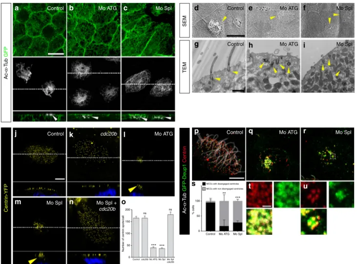
Cdc20b was also knocked down in Xenopus epidermal MCCs,
through injection of two independent morpholino antisense
oligonucleotides targeting either the ATG (Mo ATG), or the exon
1/intron 1 junction (Mo Spl) (Supplementary Figure 5b). The
efficiency of Mo ATG was verified through fluorescence
extinction of co-injected Cdc20b-Venus (Supplementary
Fig-ure 5c). RT-PCR confirmed that Mo Spl caused intron 1 retention
(Supplementary Figure 5d), which was expected to introduce a
premature stop codon, and to produce a Cdc20b protein lacking
96% of its amino acids, likely to undergo unfolded protein
response-mediated degradation. Thus, both morpholinos were
expected to generate severe loss of Cdc20b function. Consistent
with this interpretation, both morpholinos strongly reduced
Cdc20b immunostaining in deuterosome stage MCCs
(Supple-mentary Figure 5e). We verified that neither morpholinos caused
p53 transcript up-regulation (Supplementary Figure 5f), a
non-specific response to morpholinos that is sometimes detected in
zebrafish embryos
32. Importantly, whole-mount in situ
hybridi-zation indicated that miR-449 expression was not perturbed in
the presence of either morpholino (Supplementary Figure 5g).
We found that cdc20b knockdown did not interfere with
acquisition of the MCC fate (Supplementary Figure 6a–e), but
severely impaired multiciliogenesis, as revealed by
immunofluor-escence and electron microscopy (Fig.
5
a–i). This defect stemmed
from a marked reduction in the number of centrioles, and poor
docking at the plasma membrane (Fig.
5
g–o and Supplementary
Figure 6f–k). Importantly, centrioles and cilia were rescued in Mo
Spl MCCs by co-injection of cdc20b, venus-cdc20b or
cdc20b-venus mRNAs (Fig.
5
j–o and Supplementary Figure 6f–k). In
normal condition, Xenopus epidermal MCCs arise in the inner
mesenchymal layer and intercalate into the outer epithelial layer,
while the process of centriole amplification is underway
33. To rule
out secondary defects due to poor radial intercalation, we assessed
the consequences of cdc20b knockdown in MCCs induced in the
outer layer by Multicilin overexpression
8. Like in natural MCCs,
Cdc20b proved to be essential for the production of centrioles
and cilia in response to Multicilin activity (Supplementary
Figure 7a–g). We also noted that the apical actin network that
Mouse tracheal MCCs
CDC20B intensity at deuterosome (a.u.)
Mouse ependymal MCCs
ImmatureIntermediate Mature 0 50 100 150 Xenopus epidermis MCCs CDC20B DEUP1 Merge FOP CDC20B Merge Immature Immature Mature Mature FOP CDC20B Merge FOP CDC20B Merge FOP CDC20B GFP-Deup1 Cdc20b Merge Centrin Centrin Cdc20b Centrin GFP-Deup1 Cdc20bDAPI
a
b
c
GFP-Deup1 Cdc20b *** ****Fig. 3 CDC20B associates to vertebrate deuterosomes. a Double immunofluorescence was performed on mouse tracheal MCCs after 3 days of culture in air–liquid interface. Low magnification confocal panels show coincident CDC20B and DEUP1 staining in several individual MCCs. High magnification on a single MCC reveals the prominent association of CDC20B to large deuterosomes marked by DEUP1 (arrowheads). Note that some smaller deuterosomes do not contain CDC20B (arrows).b Mouse ependymal MCCs were immunostained as indicated, and high
magnification confocal pictures of cells with immature and mature deuterosomalfigures were taken. In these cells, centrioles revealed by FOP form a ring around deuterosomes. CDC20B staining forms a ring inside the ring of FOP-positive procentrioles indicating that CDC20B is tightly associated to deuterosomes. Note that the CDC20B signal associated to deuterosome increased with their maturation (high magnification pictures of >25 cells per category from two different animals were quantified in the graph; mean values and standard deviations are shown). Unpaired t test vs immature: p= 0.0005 (intermediate, ***); p < 0.0001 (mature, ****). In a andb, zooms were made on regions identified by dashed boxes. c Xenopus embryos were injected with GFP-Deup1 mRNA and immunostained at neurula st18 as indicated. Scale bars: 5µm (a, b, large view), 1.5 µm (a, high magnification), 500 nm (b, high magnification), 10 µm (c)
normally surrounds BBs was disrupted in absence of Cdc20b,
although this defect could be secondary to the absence of
centrioles (Supplementary Figure 7d–g). Centrioles in Cdc20b
morphant cells often formed clusters, suggesting that
disengage-ment from deuterosomes could have failed (Fig.
5
l,m). To better
assess this process, we injected GFP-Deup1 in Multicilin-induced
MCCs and stained centrioles with Centrin. In mature control
MCCs, deuterosomes were disassembled, centrioles were
con-verted into BBs, had docked and initiated cilium growth (Fig.
5
p,
s). In contrast, both morpholinos caused a marked increase in the
number of defective MCCs, which were devoid of cilia and
displayed centrioles still engaged on deuterosomes (Fig.
5
q–u).
Altogether our functional assays in mouse and Xenopus indicate
that CDC20B is required for centriole disengagement from
deuterosomes and subsequent ciliogenesis in MCCs. We next
investigated the molecular mechanism of action of CDC20B
underlying its role in centriole release.
Partners and effectors of CDC20B reveal its mechanism of
action. In mitotic cells, centriole disengagement is necessary to
license centriole duplication in the following cell cycle
34. This
process is known to depend on the coordinated activities of the
mitotic kinase PLK1 and the protease Separase
35. One proposed
mechanism involves the phosphorylation of PCNT by PLK1,
which induces its cleavage by Separase, thereby allowing centriole
disengagement through disassembly of the PCM
36,37. Separase is
known to be activated by the degradation of its inhibitor Securin,
which is triggered by the Anaphase Promoting Complex (APC/C)
upon binding to CDC20
25. PLK1, Separase (ESPL1), Securin
(PTTG1), CDC20, and PCNT were all found to be expressed in
human deuterosome stage MCCs (Fig.
1
d and Supplementary
Figure 1). We have shown above that PCNT is present in the
PDM and a recent study revealed the presence of CDC20 and
the APC/C component APC3 in mouse ependymal MCCs at the
stage of centriole disengagement
38. Based on this large body of
information, we hypothesized that centriole-deuterosome
disen-gagement involves the coordinated activities of PLK1 and
Separase, and that CDC20B would be involved in this scenario.
CDC20B encodes a protein of about 519 amino acids largely
distributed across the vertebrate phylum
23. In its C-terminal half,
CDC20B contains seven well conserved WD40 repeats, predicted
to form a
β-propeller, showing 49 and 37% identity to CDC20
and FZR1 repeats, respectively (Supplementary Figure 2a).
However, CDC20B lacks canonical APC/C binding domains
(Supplementary Figure 2a). Using mass spectrometry on
immu-noprecipitated protein complexes from transfected HEK cells, we
could identify multiple APC/C components interacting with
CDC20 but not with CDC20B (Supplementary Table 2). We
conclude that CDC20B is probably incapable of activating APC/
C. Interestingly, an unbiased interactome study reported
asso-ciation of CDC20B with PLK1
39. Using reciprocal
co-immunoprecipitation assays in HEK transfected cells, we
con-firmed that CDC20B and PLK1 could be found in the same
complex (Fig.
6
a and Supplementary Figure 8). This suggested
that CDC20B could cooperate with PLK1 to trigger centriole
disengagement. Consistent with this hypothesis, we found that
PLK1 was enriched in the PDM of mature deuterosomes in
mouse ependymal MCCs (Fig.
6
b), in agreement with a recent
report
38. Another interesting partner of CDC20B identified in a
second unbiased interactome study
40was SPAG5 (Astrin), which
was reported to control timely activation of Separase during the
cell cycle
41,42. Using the same strategy as above, we could detect
CDC20B and SPAG5 in the same complex (Fig.
6
c and
Supple-mentary Figure 8). As SPAG5 was found associated to DEUP1 in
a proximity labeling assay
31, we assessed its localization in
deuterosomes. Strikingly, SPAG5 was detectable in mature
deu-terosomes of mouse ependymal MCCs, with a clear enrichment in
the deuterosome core (Fig.
6
d). Finally, reciprocal
co-immunoprecipitations revealed that CDC20B and DEUP1 were
detected in the same complex when co-expressed in HEK cells
(Fig.
6
e and Supplementary Figure 8). Consistent with this result,
we observed that RFP-Cdc20b was recruited around spherical
Deup1-GFP structures positive for
γ-Tubulin and Centrin in
Xenopus epidermal MCCs (Supplementary Figure 7h–m). This
series of experiments suggested that CDC20B could participate in
the assembly of a protein complex in mature deuterosomes,
required to coordinate the activities of PLK1 and Separase for
centriole disengagement. As Separase is the last effector in this
scenario, we tested whether over-expressing human Separase in
Xenopus cdc20b morphant MCCs could rescue centriole
disen-gagement. In support to our hypothesis, over-expression of
wild-type, but not protease-dead Separase, efficiently rescued centriole
disengagement and cilia formation in cdc20b morphant MCCs
(Fig.
7
a–g and Supplementary Figure 7n–s). Separase could also
rescue multiciliogenesis in Multicilin-induced MCCs injected
with cdc20b Mos (Supplementary Figure 7t–z). We conclude that
CDC20B is involved in Separase-mediated release of mature
centrioles from deuterosomes in vertebrate MCCs (Fig.
7
h).
Discussion
In this study, we report the essential and conserved role of
CDC20B in vertebrate multiciliogenesis. Our data suggest that the
presence of CDC20B in the perideuterosomal region is necessary
to allow centriole disengagement. We note, however, that our
data, which are based on partial knockdowns, remain compatible
with an earlier function of CDC20B in promoting deuterosome
assembly and/or activity. A total genetic knockout of Cdc20b
should help to assess this possibility in mouse tracheal and
ependymal MCCs. By analogy to mitosis, we propose that
CDC20B is involved in Separase-dependent proteolysis at
deu-terosomes, allowing the release of mature centrioles and
sub-sequent ciliogenesis. This view is consistent with a recent report
showing that centriole disengagement in murine ependymal
MCCs involves the activities of PLK1, a partner of CDC20B, and
APC/C, the activator of Separase
38. The central question arising
from our work then becomes: how are CDC20B and Separase
activities integrated? The simple scenario of a CDC20-like
func-tion of CDC20B is very unlikely as it does not appear to bind
APC/C (Supplementary Table 2). CDC20 was detected in
cul-tured murine ependymal MCCs during the phase of centriole
disengagement
38, and FZR1 genetic ablation was reported to
cause reduced production of centrioles and cilia in the same
cells
43. APC/C is therefore likely activated in maturing MCCs by
its classical activators, CDC20 and/or FZR1, leading to Separase
activation through degradation of its inhibitor Securin. In that
context, we propose that additional factors linked directly or
indirectly to CDC20B may contribute to activation of Separase. It
was shown that SPAG5 inhibits or activates Separase depending
on its status of phosphorylation
41,42. As the phosphorylation
status of SPAG5 was shown to be controlled by PLK1
44, our data
suggest that the CDC20B/PLK1/SPAG5 complex could control
the timing of Separase activation locally in deuterosomes. It is
therefore possible that multiple modes of activation of Separase
may act in parallel to trigger the release of neo-synthesized
cen-trioles in maturing MCCs. Alternatively, different pathways may
be used in distinct species, or in distinct types of MCCs. An
important question for future studies regards the identity of PLK1
and Separase substrates involved in centriole disengagement.
Work on mitotic cells
36,37and our own analysis suggest that
candidate could be DEUP1 itself as it is clear that deuterosomes
are disassembled after the release of centrioles. In that respect, it
is interesting to note the presence of multiple PLK1 consensus
phosphorylation sites in human, mouse, and Xenopus DEUP1.
In this study, we have introduced the notion of
perideuter-osomal material, in analogy to the pericentriolar material. It is
striking that the two main components of the PCM, PCNT, and
γ-Tubulin, are also present in the PDM, which begs the question
whether additional PCM proteins may be present in the PDM.
The PDM may constitute a platform to sustain procentriole
growth, through the concentration and delivery of elementary
parts. It could also have a mechanical role to hold in place the
100 sh control *** **** ** Normal MCCs Abnormal MCCs 50 150 0 % cells / field sh273 sh274 sh277
% of MCCs with non disengaged
centrioles/field 0 30 40 10 20 sh control sh273 sh274 sh277 sh control sh273 sh274 sh277
Number of FOXJ1+ nuclei/field
0 50 100 150 200 0 50 100 150 sh control sh273 sh274 sh277
Number of released centrioles per cell
sh control 15dpe sh Cdc20b 15dpe
sh273
FOP α-Tub ZO1
sh274 sh277
Deup1 FOP ZO1
sh control 9dpe sh Cdc20b 9dpe
sh Cdc20b 5dpe sh control 5dpe CDC20B FOXJ1 sh277
b
a
c
d
e
f
g
j
h
i
k
**** **** **** CDC20B intensity/cell (a.u.) 0 50 100 150 sh control sh273 sh274 sh277 **** **** **** ns ns ns **** **** ****Fig. 4 CDC20B knockdown impairs multiciliogenesis in mouse ependymal MCCs. a, b Ependyma were stained for CDC20B (green) and FOXJ1 (nuclear MCC fate marker, red) 5 days post electroporation (5dpe) of control shRNA (a) or Cdc20b shRNA (b). sh277 is exemplified here, but all three Cdc20b shRNAs produced similar effects.c Graph showing the quantification of CDC20B protein levels in cells at the deuterosomal stage at 5dpe from two experiments. Mean values and standard error are shown. Unpaired t-test: ****p < 0.0001.d Dot plot showing the number of FOXJ1-positive nuclei observed for eachfield, with mean values and standard deviations from two experiments. Unpaired t-test: p = 0.3961 (sh273, ns), p = 0.1265 (sh274, ns), p = 0.3250 (sh277, ns). No significant variations were observed between conditions, indicating that MCC fate acquisition was not affected by Cdc20b knockdown.e, f Confocal pictures of 9dpe ependyma electroporated with control shRNA (e) or Cdc20b shRNAs (f) and stained for DEUP1 (deuterosome, green), FOP (centrioles, red) and ZO1 (cell junction, white). DEUP1-positive deuterosomes with non-disengaged FOP-positive centrioles were observed much more frequently in MCCs electroporated with Cdc20b shRNAs compared to control.g Dot plot showing the percentage of MCCs with non-disengaged centrioles perfield, with mean values and standard deviations. Two experiments were analyzed. Unpaired t-test: ****p < 0.0001. h, i Confocal pictures of 15dpe ependyma stained for FOP (centrioles, green),α-Tubulin (α-TUB, cilia, red), and ZO1 (cell junction, white) showing the morphology of normal MCCs in shRNA control condition (h), and examples of defects observed in MCCs treated with sh Cdc20b (i). j Dot plot showing the number of released centrioles per cell, with mean values and standard deviations.k Dot plot showing the percentage of normal and abnormal MCCs perfield of observation, with mean values and standard deviations. MCCs were scored abnormal when they did not display organized centriole patches associated to cilia. Three experiments were analyzed. Unpaired t-test: p= 0.0004 (sh273, ***), p = 0.0001 (sh274, ****), p = 0.0038 (sh277, **). Scale bars: 20 μm (a), 5μm (e, i)
growing procentrioles. Future work should evaluate
deuterosome-mediated centriole synthesis in absence of major PDM
components.
We found that beyond its association to deuterosomes during
the phase of centriole amplification, CDC20B was also associated
to BBs and cilia in fully differentiated mammalian MCCs. This
dual localization is consistent with failed ciliogenesis upon
CDC20B knockdown in mouse ependymal MCCs. However,
while we could detect Cdc20b near BBs of mature MCCs in
Xenopus, we found no evidence of its presence in cilia.
Further-more, cilia were rescued by Separase overexpression in Cdc20b
morphant MCCs. This suggests that Cdc20b is not required for
ciliogenesis in this species, although it could potentially
con-tribute to cilium structure and/or function. Thus, refined
tem-poral and spatial control of CDC20B inhibition will be needed to
study its function beyond centriole synthesis.
This and previous studies
23,26–28establish that the miR-449
cluster and its host gene CDC20B are commonly involved in
multiciliogenesis. Consistent with its early expression, it was
suggested that miR-449 controls cell cycle exit and entry into
differentiation of MCCs
23,27,30. This study reveals that CDC20B
itself is involved in the production of centrioles, the
first key step
SEM Control Mo ATG Mo Spl Ac-α -T ub GFP Control Mo ATG Mo Spl Mo Spl cdc20b cdc20b ns ns *** *** 0 50 100 150 200 Number of centr in spots/cell Mo ATG Mo ATG Centr in-YFP Mo Spl Mo Spl Mo Spl + cdc20b Control Control cdc20b TEM Control Mo ATG Mo Spl Ac-α -T ub GFP-Deup1 Centr in Control Mo ATG Mo Spl Control Mo ATG Mo Spl % cells 0 50 100 ** ***
a
b
c
j
k
m
n
l
p
q
r
s
MCCs with disengaged centriolest
u
MCCs with non disengaged centriolesd
e
f
g
h
i
o
Fig. 5 cdc20b knockdown impairs multiciliogenesis in Xenopus epidermal MCCs. a–c 8-cell embryos were injected in presumptive epidermis with GFP-CAAX mRNA and cdc20b morpholinos, as indicated. Embryos at tailbud st25 were processed forfluorescent staining against GFP (injection tracer, green) and Acetylated-α-Tubulin (Ac-α-Tub, cilia, white). White dotted lines indicate the position of orthogonal projections shown in bottom panels. Note that cdc20b morphant MCCs display cytoplasmicfilaments but do not grow cilia (white arrowheads). d–f Scanning electron microscopy (SEM) of control (d) and cdc20b morphant (e, f) embryos at tadpole st31. Yellow arrowheads point at normal (d) and defective MCCs (e, f). g–i Transmission electron microscopy (TEM) of control (g) and cdc20b morphant (h, i) embryos at tailbud st25. Yellow arrowheads point at normally docked basal bodies supporting cilia (g) and undocked centrioles unable to support cilia (h, i). j–n 8-cell embryos were injected in presumptive epidermis with centrin-YFP mRNA, cdc20b morpholinos, and cdc20b mRNA, as indicated. Centrin-YFPfluorescence was observed directly to reveal centrioles (yellow). Nuclei were revealed by DAPI staining in blue. White dotted lines indicate the position of orthogonal projections shown in bottom panels. Yellow arrowheads point at undocked centrioles.o Bar graph showing the mean number of BBs per MCC, and standard error mean, as counted by Centrin-YFP dots. One-way ANOVA and Bonferroni’s multiple comparisons test on two experiments, ***p < 0.0001. cdc20b knockdown significantly reduced the number of BBs per cell, and this defect could be corrected by cdc20b co-injection with Mo Spl.p–u Embryos were injected with Multicilin-hGR and GFP-Deup1 mRNAs, treated with dexamethasone at gastrula st11 to induce Multicilin activity, and immunostained at neurula st23 against Acetylated-α-tubulin (cilia, white), GFP (deuterosomes, green), and Centrin (centrioles, red).p Control cells showed individual centrioles, many of which had initiated ciliogenesis. Note that Deup1-positive deuterosomes were no longer visible at this stage. (q, r, t, u) cdc20b morphant MCCs showed procentrioles still engaged on deuterosomes and lacked cilia. In t and u, zooms were made on regions identified by dashed boxes in q and r. s Bar graph showing the mean percentage of cells that completed or not centriole disengagement with standard deviations. Three experiments were analyzed. Unpaired t-test: p= 0.0037 (Mo ATG, **), p = 0.0004 (Mo Spl, ***). Scale bars: 20µm (a, d), 1 µm (g, t), 5 µm (j, p)
of the multiciliogenesis process. From that perspective, the nested
organization of miR-449 and CDC20B in vertebrate genomes,
which allows their coordinated expression, appears crucial for
successful multiciliogenesis.
It is also noteworthy to point out the location of this gene in a
genomic locus where congenital mutations in MCIDAS and
CCNO were recently shown to cause a newly-recognized
MCC-specific disease, called reduced generation of multiple motile cilia
(RGMC). RGMC is characterized by severe chronic lung
infec-tions and increased risk of infertility
12,13. Its location in the same
genetic locus as MCIDAS and CCNO makes CDC20B a putative
candidate for RGMC. By extension, the deuterosome
stage-specific genes uncovered by scRNA-seq in this study also
repre-sent potential candidates for additional RGMC mutations.
myc-PLK1 – – + Input + + + GFP-CDC20B myc-PLK1 GFP-CDC20B myc-PLK1 myc-PLK1 CDC20B CDC20B CDC20B myc-DEUP1 myc-DEUP1 myc-DEUP1 GFP-CDC20B GFP-CDC20B myc-DEUP1 CDC20B – – + IP: GFP + 80 + + – – + Input + + + – – + IP: myc + + + CDC20B myc-SPAG5 myc-SPAG5 CDC20B GFP-CDC20B GFP-CDC20B myc-SPAG5 myc-SPAG5 – – + Input + + + – – + IP: GFP + + + – – + Input + + + – – + IP: myc + + + – – + Input + + + – – + IP: GFP + + + – – + Input + + + – – + IP: myc + + + SPAG5 FOP Mouse ependymal MCCs Mouse ependymal MCCs FOP SPAG5 FOP SPAG5 PLK1 DEUP1 PLK1 DEUP1 PLK1 DEUP1
a
b
c
d
e
80 65 80 140 80 65 140 80 80 65 80Fig. 6 CDC20B interacts with PLK1, SPAG5, and DEUP1. a, c, e Co-immunoprecipitations of PLK1, SPAG5, and DEUP1 with CDC20B were tested after transfections of different constructs in HEK cells, indicated at the top of each panel. Proteins (left legend) were revealed by immunoblotting. Representative blots from 3 independent experiments.b, d Maturing mouse ependyma were immunostained for the indicated proteins, and pictures were taken with a confocal microscope. PLK1 and SPAG5 are expressed in maturing MCCs. Inb, zoom was made on a different cell to reveal PLK1 enrichment in the perideuterosomal region. Ind, zoom was made on the dashed box to reveal SPAG5 enrichment in the deuterosome core. Scale bars in b, d: 5µm (large view), 1µm (high magnification)
Previous works have established the involvement of the
cen-triole duplication machinery active in S-phase of the cell cycle,
during centriole multiplication of vertebrate post-mitotic
MCCs
19–21. Our study further reveals a striking analogy
between centriole disengagement from deuterosomes in MCCs,
and centriole disengagement that occurs during the M/G1
tran-sition of the cell cycle (Fig.
7
g). Thus, it appears that centriole
production in MCCs recapitulates the key steps of the centriole
duplication cycle
34. However, the cell cycle machinery must adapt
to the acentriolar deuterosome to massively produce centrioles.
Such adaptation appears to involve physical and functional
interactions between canonical cell cycle molecules, such as
CEP152 and PLK1, and recently evolved cell cycle-related
deu-terosomal molecules, such as DEUP1
21and CDC20B. It remains
to examine whether additional deuterosomal cell cycle-related
molecules have emerged in the vertebrate phylum to sustain
massive centriole production.
In conclusion, this work illustrates how coordination
between ancestral and recently evolved cell cycle-related
mole-cules can give rise to a novel differentiation mechanism in
vertebrates.
Methods
Subjects/human samples. Inferior turbinates were from patients who underwent surgical intervention for nasal obstruction or septoplasty (provided by L. Castillo, Nice University Hospital, France). Experiments involving human tissues were performed according to the guidelines of the Declaration of Helsinki, after approval by the institutional review board“Comité de Protection des Personnes Sud Méd-iterranée V” (06/16/2015). All patients gave their written informed consent.
Single-cell RNA sequencing of human airway epithelial cells (HAECs). HAECs cultures were derived from nasal mucosa of inferior turbinates. After excision, nasal inferior turbinates were immediately immersed in Ca2+/Mg2+‐free HBSS supplemented with 25 mM HEPES, 200 U/mL penicillin, 200 µg/mL streptomycin, 50 µg/mL gentamicin sulfate, and 2.5 µg/mL amphotericin B (all reagents from Gibco). After repeated washes with cold supplemented HBSS, tissues were digested with 0.1% Protease XIV from Streptomyces griseus (Sigma) overnight at 4 °C. After incubation, fetal calf serum (FCS) was added to afinal concentration of 10%, and nasal epithelial cells were detached from the stroma by gentle agitation. Cell sus-pensions were further dissociated by trituration through a 21 G-needle and then centrifuged at 150×g for 5 min. The pellet was resuspended in supplemented HBSS containing 10% FCS and centrifuged again. The second cell pellet was then sus-pended in Dulbecco’s Modified Eagle’s Medium (DMEM, Gibco) containing 10% FCS and cells were plated (20 000 cells per cm2) on 75 cm2-flasks coated with rat
tail collagen I (Sigma-Aldrich). Cells were incubated in a humidified atmosphere of 5% CO2at 37 °C. Culture medium was replaced with Bronchial Epithelium Basal Medium (BEBM, Lonza) supplemented with BEGM SingleQuot Kit Supplements
b
c
d
e
f
h
Control Separase Mo ATG
Mo Spl
Mo ATG + separase
Mo Spl + separase
Centriole disengagement in mitotic cells Centriole release from deuterosome in post-mitotic MCCs
Pericentriolar material PLK1 Separase PLK1/CDC20B Separase Perideuterosomal material
a
Number of GFP+ normal MCCs/field
200 150 100 50 0 ns ns ns *** ***
Control Separase MoATG MoATG MoSpl
separase
MoSpl separase
g
Fig. 7 Separase overexpression rescues multiciliogenesis in absence of Cdc20b. a–f 8-cell Xenopus embryos were injected in the presumptive epidermis with GFP-gpi mRNA, cdc20b morpholinos, and human Separase mRNA, as indicated. Embryos werefixed at tailbud st25 and immunostained against GFP (injection tracer, green), Acetylated-α-Tubulin (cilia, white) and ɣ-Tubulin (BBs, red). White dotted lines indicate the position of orthogonal projections shown in bottom panels. Red arrowheads point undocked BBs. Left inset ine shows zoom on clustered centrioles. g Bar graph showing the mean number of properly ciliated MCCs among injected cells, perfield of observation, with standard error mean, from two independent experiments. Counting was performed on pictures taken at low magnification (×20), in order to score a large number of cells. Separase overexpression fully rescued multiciliogenesis in cdc20b morphant MCCs. One-way ANOVA and Bonferroni’s multiple comparisons on two experiments, ***p < 0.0001,nsp > 0.05. Scale bars: 5µm (a). h Model illustrating the analogy between centriole disengagement in mitotic cells and centriole release from deuterosomes in post-mitotic MCCs
(Lonza) on the day after and was then changed every other day. After 4 to 5 days of culture, after reaching about 70% confluence, cells were detached with trypsin-EDTA 0.05% (Gibco) for 5 min and seeded on Transwell® permeable supports (6.5 mm diameter; 0.4 µm pore size; Corning), in BEGM medium, with a density of 30,000 cells per Transwell®. Once the cells have reached confluence (typically after 5 days), they were induced to differentiate at the air–liquid interface by removing medium at the apical side of the Transwell®, and by replacing medium at the basal side with DMEM:BEBM (1:1) supplemented with BEGM SingleQuot Kit Supple-ments. Culture medium was changed every other day. Single-cell analysis was performed after 14 days of culture at the air–liquid interface, which corresponds to the maximum centriole multiplication stage. To obtain a single-cell suspension, cells were incubated with 0.1% protease type XIV from S. griseus in supplemented HBSS for 4 h at 4 °C. Cells were gently detached from Transwells® by pipetting and then transferred to a microtube. 50 units of DNase I (EN0523 ThermoFisher Scientific) per 250 µL were directly added and cells were further incubated at room temperature for 10 min. Cells were centrifuged (150×g for 5 min) and resuspended in 500 µL supplemented HBSS containing 10% FCS, centrifuged again (150×g for 5 min) and resuspended in 500 µL HBSS before being mechanically dissociated through a 26 G syringe (4 times). Finally, cell suspensions werefiltered through a Scienceware® Flowmi™ Cell Strainer (40 µm porosity), centrifuged (150×g for 5 min) and resuspended in 500 µL of cold HBSS. Cell concentration measurements were performed with Scepter™ 2.0 Cell Counter (Millipore) and Countess™ auto-mated cell counter (ThermoFisher Scientific). Cell viability was checked with Countess™ automated cell counter (ThermoFisher Scientific). All steps except the DNAse I incubation were performed on ice. For the cell capture by the 10× genomics device, the cell concentration was adjusted to 300 cells/µL in HBSS aiming to capture 1500 cells. We then followed the manufacturer’s protocol (Chromium™ Single Cell 3′ Reagent Kit, v2 Chemistry) to obtain single cell 3′ libraries for Illumina sequencing. Libraries were sequenced with a NextSeq 500/550 High Output v2 kit (75 cycles) that allows up to 91 cycles of paired-end sequencing: the forward read had a length of 26 bases that included the cell barcode and the UMI; the reverse read had a length of 57 bases that contained the cDNA insert. CellRanger Single-Cell Software Suite v1.3 was used to perform sample demulti-plexing, barcode processing and single-cell 3′ gene counting using default para-meters and human build hg19. Additional analyses were performed using R. Pseudotemporal ordering of single cells was performed with the last release of the Monocle package45. Cell cycle scores were calculated by summing the normalized intensities of genes belonging to phase-specific gene sets then centered and scaled by phase. Gene sets for each phase were curated from previously described sets of genes46(Table S2). Data was submitted to the GEO portal under series reference GSE103518. Data shown in Fig.1is representative of four independent experi-ments performed on distinct primary cultures.
RNA sequencing of HAECs. For Supplementary Fig. 2B, three independent HAEC cultures (HAEC1, HAEC2, HAEC3) were triggered to differentiate in air–liquid interface (ALI) cultures for 2 days (ALI day 2, undifferentiated), ALI day 14 (first cilia), or ALI day 28 (well ciliated). RNA was extracted with the miRNeasy mini kit (Qiagen) following manufacturer’s instructions. mRNA-seq was performed from 2 µg of RNA that wasfirst subjected to mRNA selection with Dynabeads® mRNA Purification Kit (Invitrogen). mRNA was fragmented 10 min at 95 °C in RNAseIII buffer (Invitrogen) then adapter-ligated, reverse transcribed and amplified (6 cycles) with the reagents from the NEBNext Small RNA Library Prep Set for SOLiD. Small RNA-seq was performed from 500 ng RNA with the NEBNext Small RNA Library Prep Set for SOLiD (12 PCR cycles) according to manufacturer’s instructions. Both types of amplified libraries were purified on Purelink PCR micro kit (Invitrogen), then subjected to additional PCR rounds (8 cycles for RNA-seq and 4 cycles for small RNA-seq) with primers from the 5500 W Conversion Pri-mers Kit (Life Technologies). After Agencourt® AMPure® XP beads purification (Beckman Coulter), libraries were size-selected from 150 nt to 250 nt (for RNA-seq) and 105 nt to 130 nt (for small RNA-RNA-seq) with the LabChip XT DNA 300 Assay Kit (Caliper Lifesciences), andfinally quantified with the Bioanalyzer High Sensitivity DNA Kit (Agilent). Libraries were sequenced on SOLiD 5500XL (Life Technologies) with single-end 50b reads. SOLiD data were analyzed with lifescope v2.5.1, using the small RNA pipeline for miRNA libraries and whole transcriptome pipeline for RNA-seq libraries with default parameters. Annotationfiles used for production of raw count tables correspond to Refseq Gene model v20130707 for mRNAs and miRBase v18 for small RNAs. Data generated from RNA sequencing were then analyzed with Bioconductor (http://www.bioconductor.org) package DESeq and size-factor normalization was applied to the count tables. Heatmaps were generated with GenePattern using the“Hierarchical Clustering” Module, applying median row centering and Euclidian distance.
Re-analysis ofXenopus E2F4 Chip-seq and RNA-seq. RNA-seq (samples GSM1434783 to GSM1434788) and ChIP-seq (samples GSM1434789 to GSM1434792) data were downloaded from GSE59309. Reads from RNA-seq were aligned to the Xenopus laevis genome release 7.1 using TopHat247with default parameters. Quantification of genes was then performed using HTSeq-count48 release 0.6.1 with“-m intersection-nonempty” option. Normalization and statistical analysis were performed using Bioconductor package DESeq249. Differential expression analysis was done between Multicilin-hGR alone versus Multicilin-hGR
in the presence of E2f4ΔCT. Reads from ChIP-seq were mapped to the X. laevis genome release 7.1 using Bowtie250. Peaks were called and annotated according to their positions on known exons with HOMER51. Peak enrichments of E2F4 binding site in the promoters of centriole genes and cell cycle genes9were esti-mated in presence or absence of Multicilin and a ratio of E2F4 binding (Multicilin vs no Multicilin) was calculated.
Promoter reporter studies. The human CDC20B promoter was cloned into the pGL3 Firefly Luciferase reporter vector (Promega) with SacI and NheI cloning sites. The promoter sequenced ranged from−1073 to +104 relative to the tran-scription start site. 37.5 ng of pGL3 plasmid were applied per well. pCMV6-Neg, pCMV6-E2F1 (NM_005225) and pCMV6-E2F4 (NM_001950) constructs were from Origene. 37.5 ng of each plasmid was applied per well. 25 ng per well of pRL-CMV (Promega) was applied in the transfection mix for transfection normalization (Renilla luciferase). HEK 293T cells were seeded at 20,000 cells per well on 96-well plates. The following day, cells were transfected with the indicated plasmids (100 ng of total DNA) with lipofectamine 3000 (Invitrogen). After 24 h, cells were pro-cessed with the DualGlo kit (Promega) and luciferase activity was recorded on a plate reader.
Proximity ligation assays. Fully differentiated HAECs were dissociated by incu-bation with 0.1% protease type XIV from S. griseus (Sigma-Aldrich) in HBSS (Hanks’ balanced salts) for 4 h at 4 °C. Cells were gently detached from the Transwells® by pipetting and then transferred to a microtube. Cells were then cytocentrifuged at 72×g for 8 min onto SuperFrostPlus slides using a Shandon Cytospin 3 cytocentrifuge. Slides werefixed for 10 min in methanol at −20 °C for Centrin2 and ZO1 assays, and for 10 min in 4% paraformaldehyde at room tem-perature and then permeabilized with 0.5% Triton X-100 in PBS for 10 min for acetylated-α-tubulin assays. Cells were blocked with 3% BSA in PBS for 30 min. The incubation with primary antibodies was carried out at room temperature for 2 h. Then, mouse and rabbit secondary antibodies from the Duolink® Red kit (Sigma-Aldrich) were applied and slides were processed according to manu-facturer’s instructions. Images were acquired using the Olympus Fv10i confocal imaging systems with ×60 oil immersion objective and Alexa 647 detection parameters.
Animals. All experiments were performed following the Directive 2010/63/EU of the European parliament and of the council of 22 September 2010 on the pro-tection of animals used for scientific purposes. Experiments on X. laevis and mouse were approved by the‘Direction départementale de la Protection des Populations, Pôle Alimentation, Santé Animale, Environnement, des Bouches du Rhône’ (agreement number F 13 055 21). Mouse experiments were approved by the French ethical committee no.14 (permission number: 62-12112012). Timed pregnant CD1 mice were used (Charles Rivers, Lyon, France).
Immunostaining on mouse ependyma. Dissected brains were subjected to 12 min fixation in 4% paraformaldehyde, 0.1% Triton X-100, blocked 1 h in PBS, 3% BSA, incubated overnight with primary antibodies diluted in PBS, 3% BSA, and incu-bated 1 h with secondary antibodies at room temperature. Ependyma were dis-sected further and mounted with Mowiol before imaging using an SP8 confocal microscope (Leica microsystems) equipped with a ×63 oil objective. The same protocol was used to prepare samples for super-resolution acquisition. Pictures were acquired with a TCS SP8 STED ×3 microscope equipped with an HC PL APO 93×/1.30 GLYC motCORRTMobjective (Leica microsystems). Pericentrin was
revealed using Alexa 514 (detection 535–564 nm, depletion 660 nm), γ-tubulin was revealed using Alexa 568 (detection 582–667 nm, depletion 775), and FOP was revealed using Alexa 488 (detection 498–531 nm, depletion 592 nm). Pictures were deconvoluted using Huygens software. Maximum intensity projection of 3 deconvoluted pictures is presented in Fig.4g. Primary antibodies: rabbit anti-CDC20B (1:500; Proteintech, 133376-1-AP), mouse IgG anti-PLK1 (1:500; Ther-moFisher, 33–1700), rabbit anti-Pericentrin (1:500, Abcam, ab4448), mouse IgG1 anti-FoxJ1 (1:1000; eBioscience, 14–9965), rabbit anti-Deup1 (1:1000; kindly pro-vided by Dr Xueliang Zhu), rabbit anti-Deup1 (1:250; Proteintech, 24579-1-AP), mIgG1 anti-γ-Tubulin (clone GTU88) (1:250; Abcam, Ab 11316), rabbit anti-ZO1 (1:600; ThermoFisher Scientific, 61–7300), rabbit anti-Spag5 (1:500; Proteintech, 14726-1-AP), mouse IgG1 anti-ZO1 (1:600; Invitrogen, 33-9100), mouse IgG2b anti-FGFR1OP (FOP) (1:2000; Abnova, H00011116-M01), mouse IgG1 anti-α-tubulin (1:500; Sigma-Aldrich, T9026). Secondary antibodies: Alexa Fluor 488 goat rabbit (1:800; ThermoFisher Scientific, A-11034), Alexa Fluor 647 goat anti-rabbit (1:800; ThermoFisher Scientific, A-21244), Alexa Fluor 514 goat anti-anti-rabbit (1:800; ThermoFisher Scientific, A-31558), Alexa Fluor 488 goat anti-mouse IgG2b (1:800; ThermoFisher Scientific, A-21141), Alexa Fluor 568 goat anti-mouse IgG2b (1:800; ThermoFisher Scientific, A-21144), Alexa Fluor 488 goat anti-mouse IgG2a (1:800; ThermoFisher Scientific, A-21131), Alexa Fluor 568 goat anti-mouse IgG1 (1:800; ThermoFisher Scientific, A-21134), Alexa Fluor 647 goat anti-mouse IgG1 (1:800; ThermoFisher Scientific, A-21240).
Mouse constructs. Expression constructs containing shRNA targeting specific sequences in the CDC20B coding sequence under the control of the U6 promoter
were obtained from Sigma-Aldrich (ref. TRCN0000088273 (sh273),
TRCN0000088274 (sh274), TRCN0000088277 (sh277)). PCX-mcs2-GFP vector (Control GFP) kindly provided by Xavier Morin (ENS, Paris, France), and U6 vector containing a validated shRNA targeting a specific sequence in the NeuroD1 coding sequence52(Control sh, ref. TRCN0000081777, Sigma-Aldrich) were used as controls for electroporation experiments.
Post-natal mouse brain electroporation. The detailed protocol for post-natal mouse brain electroporation established by Boutin and colleagues53was used with minor modifications. Briefly, P1 pups were anesthetized by hypothermia. A glass micropipette was inserted into the lateral ventricle, and 2μL of plasmid solution (concentration 3μg/μL) was injected by expiratory pressure using an aspirator tube assembly (Drummond). Successfully injected animals were subjected tofive 95 V electrical pulses (50 ms, separated by 950 ms intervals) using the CUY21 edit device (Nepagene, Chiba, Japan), and 10 mm tweezer electrodes (CUY650P10, Nepagene) coated with conductive gel (Signagel, Parker laboratories). Electroporated animals were reanimated in a 37 °C incubator before returning to the mother.
Statistical analyses of mouse experiments. Analysis of CDC20B signal intensity in deuterosomes (dot plot in Fig.3b). For each category, >25 cells from two different animals were analyzed. Deuterosome regions were delineated based on FOP staining and the intensity of CDC20Bfluorescent immunostaining was recorded using ImageJ software, and expressed as arbitrary units. Unpaired t test vs immature: p= 0.0005 (intermediate, ***); p < 0.0001 (Mature, ****).
Analysis of Cdc20b shRNAs efficiency (Fig.4c): For each cell at the deuterosomal stage, the intensity of CDC20Bfluorescent immunostaining was recorded using ImageJ software and expressed as arbitrary units. Data are mean ± sem. Two independent experiments were analyzed. A minimum of 35 cells per condition was analyzed. n= 3, 4, 5 and 5 animals for sh control, sh273, sh274, and sh277, respectively. Unpaired t test vs sh control: p < 0.0001 (sh273, sh274, and sh277 ****).
Analysis of the number of FOXJ1-positive cells at 5dpe (Fig.4d): Unpaired t test vs sh control: 0.3961 (sh273, ns), 0.1265 (sh274, ns), 0.3250 (sh277, ns).
Analysis of the number of cells with non-disengaged centrioles at 9dpe (Fig.4g): 15–20 fields were analyzed per condition. n = 4, 4, 3, and 4 animals for sh control, sh273, sh274, and sh277, respectively, from two independent experiments. Unpaired t test vs sh control: p < 0.0001 (sh273, sh274, sh277 ****).
Analysis of the number of centrioles per cell at 15dpe (Fig.4j): > 100 cells were analyzed per condition. n= 3, 3, 3, and 3 animals for sh control, sh273, sh274, and sh277, respectively, from two independent experiments. Unpaired t test vs sh control: p < 0.0001 (sh273, sh274, sh277 ****).
Analysis of ependymal cell categories at 15dpe (Fig.4k): Data are mean ± sem from three independent experiments. More than 500 cells were analyzed for each condition. n= 4, 4, 3, and 3 animals for sh control, sh273, sh274, and sh277, respectively. Unpaired t test vs sh control: p= 0.0004 (sh273, ***), 0.0001 (sh274, ****), 0.0038 (sh277, **).
Mouse tracheal epithelial cells (MTECs). MTECs cell cultures were established from the tracheas of 12 weeks-old mice. After dissection, tracheas were placed in cold DMEM:F-12 medium (1:1) supplemented with 15 mM HEPES, 100 U/mL penicillin, 100 µg/mL streptomycin, 50 µg/mL gentamicin sulfate, and 2.5 µg/mL amphotericin B. Each trachea was processed under a binocular microscope to remove as much conjunctive tissue as possible with small forceps and was opened longitudinally with small dissecting scissors. Tracheas were then placed in sup-plemented DMEM:F-12 containing 0.15% protease XIV from S. griseus. After overnight incubation at 4 °C, FCS was added to afinal concentration of 10%, and tracheal epithelial cells were detached by gentle agitation. Cells were centrifuged at 400 g for 10 min and resuspended in supplemented DMEM:F-12 containing 10% FCS. Cells were plated on regular cell culture plates and maintained in a humidified atmosphere of 5% CO2at 37 °C for 4 h to allow attachment of putative con-taminatingfibroblast. Medium containing cells in suspension was further cen-trifuged at 400×g for 5 min and cells were resuspended in supplemented DMEM:F-12 containing BEGM Singlequots kit supplements and 5% FCS. Cells were plated on rat tail collagen I-coated Transwell®. Typically, 5 tracheas resulted in 12 Transwells®. Medium was changed every other day. Air–liquid interface culture was conducted once transepithelial electrical resistance had reached a minimum of 1000 ohm/cm2(measured with EVOM2, World Precision Instruments).
Air–liquid interface culture was obtained by removing medium at the apical side of the Transwell®, and by replacing medium at the basal side with supplemented DMEM:F-12 containing 2% Ultroser-GTM(Pall Corporation). 10
µM DAPT (N-[N-(3,5-difluorophenacetyl)-L-alanyl]-S-phenylglycine t-butyl ester) (Sigma) was added one day after setting-up the air–liquid interface.
Immunostaining on HAECs and MTECs. Three days after setting-up the air–liquid interface, MTECs on Transwell membranes were pre-extracted with 0.5% Triton X-100 in PBS for 3 min, and thenfixed with 4% paraformaldehyde in PBS for 15 min at room temperature. HAECs were treated 21 days after setting-up the air–liquid interface. They were fixed directly on Transwells® with 100% cold methanol for 10 min at−20 °C (for CDC20B and Centrin2 co-staining,
Supplementary Figure 4a, b) or with 4% paraformaldehyde in PBS for 15 min at room temperature (for CDC20B single staining, Supplementary Figure 4c). All cells were then permeabilized with 0.5% Triton X-100 in PBS for 5 min and blocked with 3% BSA in PBS for 30 min. The incubation with primary and secondary antibodies was carried out at room temperature for 2 h and 1 h, respectively. Nuclei were stained with 4,6-diamidino-2-phenylindole (DAPI). Transwell® membranes were cut with a razor blade and mounted with ProLong Gold medium (Thermo-Fisher). Primary antibodies: rabbit anti-CDC20B (1:500; Proteintech, 133376-1-AP), rabbit anti-DEUP1 (1:500; Proteintech, 24579-1-133376-1-AP), anti-Centrin2 (Clone 20H5, 1:500; Millipore, 04-1624). Secondary antibodies: Alexa Fluor 488 goat rabbit (1:1000; ThermoFisher Scientific, A-11034), Alexa Fluor 647 goat anti-mouse (1:1000; ThermoFisher Scientific, A-21235). For co-staining of CDC20B and DEUP1, CDC20B primary antibody was directly coupled to CFTM633 with the
Mix-n-StainTMkit (Sigma-Aldrich) according to the manufacturer’s instruction.
Coupled primary antibody was applied after secondary antibodies had been extensively washed and after a 30 min blocking stage in 3% normal rabbit serum in PBS.
Western blot and immunofluorescence on transfected cells. Cos-1 or Hela cells cells were grown in DMEM supplemented with 10% heat inactivated FCS and transfected with Fugene HD (Roche Applied Science) according to manufacturer’s protocol. Transfected or control cells were washed in PBS and lysed in 50 mM Tris HCl pH 7.5, 150 mM NaCl, 1 mM EDTA, containing 1% NP-40 and 0.25% sodium deoxycholate (modified RIPA) plus a Complete Protease Inhibitor Cocktail (Roche Applied Science) on ice. Cell extracts separated on polyacrylamide gels were transfered onto Optitran membrane (Whatman) followed by incubation with rabbit anti-mouse CDC20B (1:500, Proteintech, 24579-1-AP) or homemade rabbit anti-Xenopus Cdc20b (1:300) antibody and horseradish peroxidase conjugated secondary antibody (Jackson Immunoresearch Laboratories, 711-035-152 and 715-035-150). Signal obtained from enhanced chemiluminescence (Western Lightning ECL Pro, Perkin Elmer) was detected with MyECL Imager (ThermoFisher Scientific).
For immunofluorescence staining, transfected cells were grown on glass coverslips andfixed for 6 min in methanol at −20 °C. Cells were washed in PBS, blocked in PBS, 3% BSA and stained with rabbit anti-Xenopus Cdc20b (1:300) or rabbit anti-CFTR (1:200, Santa-Cruz Biotechnology, 10747) as a negative control, in blocking buffer. After washings in PBS 0.1% Tween-20, cells were incubated with Alexafluor 488 donkey anti-rabbit antibody (ThermoFisher Scientific, R37118), washed, and DNA was stained with 250 ng/mL DAPI. Coverslip were then rinsed and mounted in Prolong Gold antifade reagent (ThermoFisher Scientific) and confocal images were acquired by capturing Z-series with 0.3μm step size on a Zeiss LSM 510 laser scanning confocal microscope.
Co-immunoprecipitation studies. Asynchronous HEK cells transfected with the plasmids described below, using lipofectamine 3000 according to manufacturer's instructions, were rinsed on ice with chilled Ca2+ and Mg2+ free Dulbecco’s PBS (DPBS, Invitrogen), harvested using a cell scraper and lysed on ice for 5 min in lysis buffer (0.025 M Tris, 0.15 M NaCl, 0.001 M EDTA, 1% NP-40, 5% glycerol; pH 7.4) supplemented with EDTA and Halt™ Protease and Phosphatase Inhibitor Cocktail (Pierce, ThermoFisher). Lysates were clarified (12,000×g, 4 °C, 10 min) and the protein concentrations were determined using the Bradford assay (Bio-Rad). Immunoprecipitations were performed with the Pierce co-immunoprecipitation kit (Pierce, ThermoFisher) according to the manufacturer’s instructions. For each immunoprecipitation, 1–1.5 mg of total lysate was precleared on a control column, then incubated on columns coupled with 20 µg of anti-GFP or anti-c-myc antibody (clone 9E10). Incubation was performed overnight at 4 °C. Columns were washed and eluted with 50 µL elution buffer. Samples were denatured at 70 °C for 10 min with Bolt™ LDS Sample Buffer and Bolt reducing agent, then separated on 4–12% gradient Bolt precast gels (ThermoFisher), transferred onto nitrocellulose (Milli-pore), and subjected to immunoblot analysis using either anti-CDC20B (Pro-teinTech, 133376-1-AP, 1/500) or anti-c-myc antibody (clone 9E10, 1/1000). In Fig.6, note that the high level of expression of myc-PLK1 (Fig.6a) and myc-SPAG5 (Fig.6b) drained out locally the ECL reagent at the peak of the protein. The resulting double bands correspond in fact to unique ones. Human SPAG5, sub-cloned into pCMV6-MT, was from OriGene. Human DEUP1 and PLK1 were cloned into pCS2-MT vector (Addgene). Human CDC20B was cloned into pEGFP-C1, pEGFP-N1 (Clontech) for the GFP fusion protein and pIRES-EYFP (Addgene) for the untagged protein.
In-gel digestion, NanoHPLC, and Q-exactive plus analysis. For mass spectro-metry analysis, protein spots were manually excised from the gel and destained with 100 µL of H2O/ACN (1/1). After 10 min vortexing, liquid was discarded, and the procedure was repeated 2 times. They were rinsed with acetonitrile and dried under vacuum. Extracts were reduced with 50μL of 10 mM dithiothreitol for 30 min at 56 °C, then alkylated with 15μL of 55 mM iodoacetamide for 15 min at room temperature in the dark. They were washed successively by: (i) 100 µL of H2O/ACN (1/1) (2 times) and (ii) 100 µL of acetonitrile. Gel pieces were rehy-drated in 60 µL of 50 mM NH4HCO3containing 10 ng/µL of trypsin (modified porcine trypsin, sequence grade, Promega) incubated for one hour at 4 °C. After the