Bacteria-Targeting Nanoparticles for Managing Infections
by P
Aleksandar Filip Radovic-Moreno B. S. Chemical Engineering The Pennsylvania State University, 2005
SUBMITTED TO THE HEALTH SCIENCES AND TECHNOLOGY PROGRAM IN PARTIAL FULFILLMENT OF THE REQUIREMENTS FOR THE DEGREE OF DOCTOR OF PHILOSOPHY IN CHEMICAL AND BIOMEDICAL ENGINEERING
AT THE
MASSACHUSETTS INSTITUTE OF TECHNOLOGY February 2013
0 2013 Massachusetts Institute of Technology. All rights reserved.
Signature of Author ...-... ... ... Health Sciences and Technology
November 9, 2012 C ertified by ...
Robert Langer, Sc. D. Institute Professor, MIT Th sis Co-Supervisor
Omid C. Fdegkhzad, M. D. Associate Professor of Anesthesia, Harvard Medical School Thesis Co-Supervisor
A ccepted by .... ... -.... ... Arup Chakraborty, Ph. D. Director, Institute for Medical Engineering and Sciences; Robert T. Haslam Professor of Chemical Engineering, Chemistry and Biological Engineering, MIT
Bacteria-Targeting Nanoparticles for Managing Infections
byAleksandar Filip Radovic-Moreno
Submitted to the Health Sciences and Technology Program on November 9, 2012 in Partial Fulfillment of the Requirements for the Degree of Doctor of Philosophy in
Chemical and Biomedical Engineering
ABSTRACT
Bacterial infections continue to be a significant concern particularly in healthcare settings and in the developing world. Current challenges include the increasing spread of drug resistant (DR) organisms, the side effects of antibiotic therapy, the negative consequences of clearing the commensal bacterial flora, and difficulties in developing prophylactic vaccines. This thesis was an investigation of the potential of a class of polymeric nanoparticles (NP) to contribute to the management of bacterial infections. More specifically, steps were taken towards using these NPs (1) to achieve greater spatiotemporal control over drug therapy by more targeted antibiotic delivery to bacteria, and (2) to develop a prophylactic vaccine formulation against the common bacterial sexually transmitted disease (STD) caused by Chlamydia trachomatis.
In the first part, we synthesized polymeric NPs containing poly(lactic-co-glycolic
acid)-block-poly(L-histidine)-block-poly(ethylene glycol) (PLGA-PLH-PEG). We show that
these NPs are able to bind to bacteria under model acidic infection conditions and are able to encapsulate and deliver vancomycin to inhibit the growth of Staphylococcus aureus bacteria in vitro. Further work showed that the PLGA-PLH-PEG-based NPs demonstrated the potential for competition for binding bacteria at a site of infection from soluble protein and model phagocytic and tissue-resident cells in a NP composition dependent manner. The NPs demonstrated low toxicity in vitro, were well tolerated by mice in vivo, and circulated in the blood on timescales comparable to control PLGA-PEG
NPs.
In the second part, we used PLGA-PLH-PEG-based NPs to design a prophylactic vaccine
against the obligate intracellular bacterium Chlamydia trachomatis, the most common cause of bacterial STD in the world. Currently, no vaccines against this pathogen are approved for use in humans. We first formulated NPs encapsulating the TLR7 agonist
R848 conjugated to poly(lactic acid) (R848-PLA) in PLGA-PLH-PEG-based NPs, then
incubated these R848-NPs with UV-inactivated C. trachomatis bacteria in acidity, forming a construct. Mice immunized with this vaccine via genital or intranasal routes demonstrated protection from genital infection post immunization in a primarily CD4* T cell-dependent manner.
These results may suggest avenues for future work in designing and developing more targeted drug therapies or vaccine formulations for managing bacterial infections using polymeric nanoparticles.
Thesis Co-Supervisor: Robert Langer Title: Institute Professor, MIT
Thesis Co-Supervisor: Omid C. Farokhzad
Acknowledgments
First and foremost, I would like to thank my parents and sister for their unending love, support, inspirational words, kindness, courage, and patience. Without them, nothing would have been possible.
I would like to express my deepest gratitude to my advisors, Professor Robert Langer and Professor Omid Farokhzad, among other things, for being inspirational figures and for creating and maintaining a thoroughly stimulating research environment. I would also like to thank my thesis committee members, Professor Paula Hammond and Professor Alexander Klibanov.
I have had the privilege to work with many extremely talented and bright colleagues and collaborators during my PhD. Most directly, I would like to acknowledge the expert guidance of Professor Timothy Lu, who contributed insights and ideas that were invaluable in the development of concepts contained in this thesis, particularly as they related to antibacterial therapies. For the work towards a prophylactic Chlamydia vaccine,
I am deeply indebted to Professor Uli von Andrian, Dr. Georg Stary, and Ms. Pamela Basto among others for collaborating to conceive the idea and executing the experiments.
I would also like to acknowledge the many contributions of my highly diligent and talented UROPs, with a special mention of Mr. Vlad Puscasu. Over the years I have greatly enjoyed extended discussions, collaborations, and friendships with (now Dr.) Ben Teply, Dr. Frank Gu, Dr. Frank Alexis, Dr. Andrew Wang, Dr. Etgar Levy-Nissenbaum, Eric Pridgen, Pedro Valencia, Dr. Liangfang Zhang, as well as members of the Langer and Farokhzad labs since 2006.
I would also like to thank everyone close to me for their support and for making the last several years so much more enjoyable. I cannot go further without a special thanks to Sukant Mittal, Jay Komarneni, Div Bolar, Nick Blasioli, Kevin Fowler, Alexandra de Paz, Pamela Basto, Rumi Chunara, Jessie Bright, Arun Bhagat, Jeff Yang, Brian Werner, Said Bogatyrev, Ben Caplan, Kathleen Lambert, Mara Macdonald, Yoni Goldwasser, Jack Milwid, David Nguyen, Manny Simons, and Jason Fuller, among many others.
Table of Contents
Abstract...2
Acknow ledgem ents... 5
Table of Contents... 6
List of Figures ... 8
List of Tables ... 11
Chapter 1: Introductory Rem arks and Overview ... 12
1.1 Introductory Rem arks ... 12
1.2 Thesis Overview ... 16
1.3 References ... 18
Chapter 2: Background: Nanoparticles for Treating Bacterial Infectious Diseases ..20
2.1 Introduction...20
2.2 Challenges to Effective Bacterial Clearance... 22
2.3 Nanoparticles Creating Opportunities for Improved Therapy ... 32
2.4 N anoparticle Platform s ... 43
2.5 Future Perspective... 52
2.6 Sum m ary ... 54
2.7 A cknow ledgm ents... 55
2.8 Reference Annotations... 56
2.9 References...56
Chapter 3: Bacteria-Targeting Nanoparticles for Antibiotic Delivery ... 69
3.1 Introduction...69
3.2 Results and Discussion ... 74
3.3 M aterials and M ethods... 89
3.4 References...97
Chapter 4: Mammalian Cell and Protein Interactions In Vitro and Preliminary Evaluation In Vivo...102
4.1 Introduction...102
4.2 Results and Discussion ... 105
4.3 M aterials and M ethods...124
4.4 References...131
5.1 Introduction...135
5.2 Results and Discussion ... 141
5.3 M aterials and M ethods...156
5.4 References...161
Chapter 6: Nanoparticles for Vaccination Against Chlamydia Trachomatis ... 167
6.1 Introduction...167
6.2 Results and Discussion ... 170
6.3 M aterials and M ethods...192
6.4 References...196
Chapter 7: Summary, Conclusions, and Suggestions for Future Work ... 200
7.1 Sum m ary and Conclusions ... 200
7.2 Sum m ary of Suggestions for Future W ork ... 209
List of Figures
Figure 2.1 Drug Resistance Mechanisms...23
Figure 2.2 Infection Microenvironments ... 27
Figure 2.3 Targeting Bacteria with Nanoparticles ... 33
Figure 2.4 Antibacterial Nanoparticle Platforms ... 44
Figure 3.1 Schematic representation of the designed nanoparticle (NP)-mediated drug targeting to bacterial cell w alls ... 75
Figure 3.2 Physicochemical characterization of NPs ... 77
Figure 3.3 NPs binding to representative bacteria...80
Figure 3.4 Fluorescent confocal microscopy of pH-dependent binding...81
Figure 3.5 Release of vancomycin from PLGA-PEG and PLGA-PLH-PEG NPs as a fun ction o f p H ... 83
Figure 3.6 NP-mediated vancomycin delivery to S. aureus ... 83
Figure 3.7 Competition Study using Sodium Polystyrene Sulfonate (PSS)...85
Figure 3.8 Henderson-Hasselbalch Analysis ... 87
Figure 3.9 Synthesis procedure for forming the PLGA-PLH-PEG copolymer ... 91
Figure 4.1 Mixed PLGA-PLH-PEG / PLGA-PEG NP Size and Zeta Potential C h aracterization ... 107
Figure 4.2 PLGA-PLH-PEG NP Uptake in the Presence of Bovine Serum Albumin (B S A ) ... 10 8 Figure 4.3 Mixed PLGA-PLH-PEG / PLGA-PEG NP Binding to S. aureus ... 110
Figure 4.5 pH-Titration of NP Uptake in LNCaP Cells...114 Figure 4.6 pH-dependent Uptake of PLGA-PEG NPs...114 Figure 4.7 Fluorescence Image of NP Uptake in LNCaP Cells...116 Figure 4.8 Inhibitor Study of Uptake of PLGA-PLH-PEG NPs in LNCaP cells at low pH ... 1 16 Figure 4.9 Mixed PLGA-PLH-PEG / PLGA-PEG NP Uptake in Mammalian Cells 117 Figure 4.10 Impact of LNCaP Cell Monolayer on Bacteria Targeting Ability ... 118 Figure 4.11 Mixed PLGA-PLH-PEG / PLGA-PEG NP Pharmacokinetics ... 119 Figure 4.12 Mixed PLGA-PLH-PEG / PLGA-PEG NP Biodistribution...122 Figure 4.13 Mixed PLGA-PLH-PEG / PLGA-PEG NP Biodistribution - Confocal Im ag es ... 12 3
Figure 5.1 Silver/Vanco Co-Delivery Nanoparticle Formulation and Characterization ... 14 2 Figure 5.2 UV-Vis Analysis of PLGA-PLH-PEG and silver-containing PLGA-PLH-PEG N P s ... 14 7
Figure 5.3 Free silver and vancomycin are synergistic against Staphylococcus aureus ... 14 8 Figure 5.4 Bacterial Growth Inhibition of Silver- and/or Vancomycin-containing N an op articles ... 150 Figure 5.5 Effect of Vancomycin-to-Silver Drug Loading Ratio on Bacterial Growth
In h ib ition ... 15 1
Figure 5.6 Growth Inhibition Study against Vancomycin-Resistant Enterococcus faecalis (V R E )...15 3
Figure 5.7 Nanoparticle Cytotoxicity Study ... 154
Figure 6.1 Schematic representation of the Chlamydia vaccine design ... 175
Figure 6.2 Zeta potential of Chlamydia trachomatis at different pH ... 176
Figure 6.3 Effect of Polymer Blending on Nanoparticle Size, Zeta Potential, and Stability ... 1 7 8 Figure 6.4 Nanoparticle-Chlamydia Vaccine Characterization...179
Figure 6.5 Flow Cytometry Analysis of NP-Chlamydia Vaccine ... 180
Figure 6.6 Flow Cytometry Analysis of NP-Chlamydia- Vaccine (II)...181
Figure 6.7 Induction of Chlamydia-specific T cells ... 182
Figure 6.8 Quantitative PCR Analysis of Chlamydia trachomatis burden in the uterus follow ing transcervical im m unization ... 184
Figure 6.9 Quantitative PCR Analysis of Chlamydia trachomatis burden in the uterus following intranasal or subcutaneous immunization ... 185
Figure 6.10 Inclusion Forming Unit (IFU) Analysis of Chlamydia trachomatis burden in the uterus follow ing im m unization ... 186
Figure 6.11 Analysis of Chlamydia-specific T cell levels in the uterus and lymphoid organs follow ing im m unization...186
Figure 6.12 Quantitative PCR analysis of Chlamydia trachomatis burden in the uterus following transcervical immunization in various immunodeficient mice ... 188
Figure 6.13 Protection is M HC-II Dependent...189
Figure 6.14 Protection requires functional RAG-2...190
List of Tables
Table 2.1 M ethods for Targeting Bacteria... 49
Table 2.2 Example of Nanoparticles Overcoming Challenges...51
Table 3.1 Minimum bactericidal concentration (MBC) at pH 6.0 and pH 7.4 in S. aureus ... 8 4 Table 4.1 XPS Analysis of PLGA-PLH-PEG and PLGA-PEG NPs...105
Table 4.2 Pharmacokinetic parameters - Two Compartmental Fit ... 120
Table 5.1 Selected drug combinations and their advantages ... 136
Table 5.2 X-ray Photoelectron Spectroscopy Analysis of Nanoparticles...144
Table 5.3 Fourier Transform Infrared Spectroscopy Analysis of Nanoparticles...145
Table 5.4 Differential Scanning Calorimetry Analysis of Nanoparticles...145 Table 5.5 Glass Transition Temperature Determined by Differential Scanning
Chapter 1
Introductory Remarks and Overview
1. 1. Introductory Remarks
Bacterial infections have been a scourge to mankind since the dawn of our species c.200,000 years ago. Exploring methods to improve treatment and prevention has been a continuing endeavor, albeit one characterized by a lack of clear vision or targeted methodology until perhaps the late 1 9 th century, when Robert Koch published a set of postulates which could be used to precisely determine if a microorganism was causing a disease. This seminal contribution was a capstone to centuries of observations including Anton van Leeuwenhoek's first visualization of a bacterium in the 1 7th century to Louis Pasteur's seminal studies in the 1 91h century disproving the theory of spontaneous
generation. These and many other examples together unambiguously made clear that bacteria can cause illness.1 Alongside these key advances in microbiology were the seminal contributions by Paul Ehrlich, who in the early 2 0 th century famously conceived of a "magic bullet" that could seek out and attack agents of disease with minimal collateral effects.2 The stage had now been set for the fortuitous discovery of penicillin by Alexander Fleming in 1928, which was later developed into a drug by scaled production techniques in the 1940s. The use of penicillin in humans was truly landmark because of its remarkably fast and potent activity combined with few side effects, even when ingested in gram quantities per day.3 This triggered a revolution known as the "golden era" of antibiotic discovery, a period of tremendous productivity from c.1940-1980, in which many of the major classes of antibiotics still in use today were
discovered.4
Particularly in these years, a host of drugs were developed, bringing unprecedented success against a wide variety of bacterial infections, leading to important insights into bacterial physiology, and most importantly, saving the lives of many.
However, the story does not end there. Bacteria are among the oldest living organisms on planet Earth and in the approximately 2 billion years of their existence, have evolved tools and strategies that make them highly adaptable to extreme environments or chemical attacks. Bacteria can be found at extremes of temperature and pressure, have survived cataclysmic events, withstood variations in atmospheric composition and surface temperature over the evolution of planet Earth, encountered chemical attacks from sources like the environment, competing microorganisms, and faced sophisticated assaults by the immune systems of multicellular organisms. In addition, bacteria have extremely short generation times - on the order of only a few minutes in some cases
-which allows them to rapidly iterate their genetic material across generations, and can readily transfer genes to each other using mobile genetic elements.5 These tools acquired over billions of years of harsh survival positioned them quite favorably to counter antibiotics. In fact, antibiotics provided a relatively straightforward target because of their high specificity. Bacteria were able to respond almost immediately, with the phenomenon of drug resistance (DR) being broadly recognized as a major challenge already in the 1940s. Perhaps even more remarkably, it appears that bacteria had already developed (cross) resistance to antibiotics - before we even developed them, as suggested by the
discovery of multi drug resistant bacteria in a deep cavern that had been isolated from all human intervention.6 DR is just one of several strategies used by bacteria. Bacteria are able to evade therapy by building, inducing, or finding microenvironmental niches, such
as biofilms, abscesses, or by intracellular localization.' 8 These niches provide a
protective barrier from many elements of humoral immunity and chemical intervention. Bacteria are also capable of entering into states of hibernation, in which very low metabolic activity reduces their susceptibility to antibiotics to almost zero, and can form extremely resilient endospores (for further discussion see Chapter 2).9
We cannot avoid bacteria - they are nearly omnipresent. Bacteria can be found in the air we breathe, on nearly all types of surfaces, in the food we eat, and perhaps most notably -in all of us. Bacteria outnumber us -in our own bodies approximately 10:1 (bacteria:human cells), living in commensal status on our skin, in our intestinal tracts, and on our mucosal surfaces.'0 For the most part this is an indifferent or mutualist interaction. However, even commensal organisms can cause deadly infections under inauspicious conditions, such as trauma, interruptions in the normal flora (such as by antibiotic therapy), or in cases of weakened immune systems due to age or comorbidities. Today we live in an increasingly interconnected world, where humans may come into contact with a wider diversity of bacteria more than ever before. This puts us not only at greater risk of infection, but allows for bacteria from different parts of the world to come together to "share" genetic information, perhaps leading to bacterial "superbugs" that are resistant to all known antibiotics.2 A historical perspective suggests that our species will likely be in a constant battle with bacteria. In fact, some believe that resistance is inevitable.2' " To remain one step ahead, it will be necessary to continuously develop novel tools and approaches based on an increasingly deeper understanding not only of bacterial (patho)physiology, but also how bacteria interact with humans in ways that reduce the effectiveness of therapeutic strategies. These insights will allow us to create more tailored
approaches that can not only adapt to bacterial adaptations, but also to anticipate and perhaps neutralize future moves. Our existing primary tools to treat infections, antibiotics, can be highly effective, but years of (mis)use are putting them at serious threat for obsolescence due to DR in many cases. Multi-drug resistance is on the rise, and agents of last resort are usually less effective, more toxic, and their increasing use is likely to lead to more widespread resistance (see Chapter 2 for thorough discussion). In addition, drug therapy of bacterial infections is affected by a variety of other factors, including microenvironmental conditions, biofilms, and drug pharmacokinetic challenges, all of which can significantly impact the outcome of therapy. These factors suggest that novel tools and approaches are needed to improve antibacterial drug therapy. 12
A complementary strategy to seeking new therapeutics is the search for novel methods of prevention. One part of a prevention strategy is proper sanitation, a concept pioneered by Ignaz Semmelweis and Joseph Lister in the 1 9th-2 0th centuries among many others, but
major effects can also be obtained by safe and effective prophylactic vaccination. Vaccines have been credited with the eradication of smallpox (a viral illness) and have also made significant impacts on a host of bacterial illnesses including those caused by
Bacillus anthracis, Streptococcus pneumoniae, Haemophilus influenzae type B, Clostridium tetani, and Mycobacterium tuberculosis, among others. These successes,
while modest in some cases (such as for TB), nevertheless have protected many from infection, saved lives, and reduced healthcare expenditures for decades. Despite these successes, many bacterial infections remain without a safe and effective prophylactic vaccine. It appears that traditional tools and approaches are insufficient to yield immunity
in several important examples, creating a clear and pressing need for the development of more advanced tools and platforms to yield safe and effective vaccines.
This thesis is a contribution to the ongoing work to improve the treatment and prevention of bacterial infections. In one succinct phrase, this thesis was motivated primarily by (1) a need to continue exploring new methods for enhancing the effectiveness of drug therapy, and (2) the need to identify new platforms for achieving safe and effective vaccination.
1. 2. Thesis Overview
My main interest in this thesis was to make a contribution towards developing technologies that might improve the management of bacterial infectious disease. Within this broad goal, I focused on two major activities: (1) treatment of bacterial infections, and (2) prevention of infection by prophylactic vaccination. To begin taking steps towards these aims, in collaboration with others I developed and tested a novel polymeric nanoparticle (NP) platform that could be used to encapsulate and deliver active agents. The precise method of using the NPs varies depending on the application - this is
discussed at length in the appropriate research chapters.
To begin, I discuss the rationale for NP-based approaches to treatment of bacterial infections in Chapter 2. This chapter is also a literature review, focusing on the different NP-based technologies that have been used to deliver drugs, particularly within the context of antibacterial therapy. I also include a discussion of why a polymeric NP platform is suitable for the work that follows. From there, I review some of the general principles that apply across NP platforms, including such concepts as passive targeting to inflammation by certain types of nanomaterials as well as methods to achieve binding to
bacteria, often termed "active" targeting. I emphasize the use of charge-charge interactions as a widely used basis for targeting bacteria with NPs, which also forms the basis for the work contained herein.
In Chapter 3, 1 delve into the synthesis and characterization of the polymeric NP platform that is used (albeit with some modification over the years) throughout this thesis. This chapter also documents the first steps we took to explore the applicability of our NP system for treating bacterial infections, beginning with the synthesis of the polymer, continuing with basic characterization, drug loading/release, confocal microscopy of interactions with bacteria, then culminating with in vitro studies of bacterial growth inhibition using Staphylococcus aureus as a model pathogen.
Chapter 4 is a continuation of the work done in Chapter 3, focused on complementary studies that can potentially better predict the outcome of using this NP platform to treat infections in vivo. We achieve this by first exploring how the NPs interact with model biological components present at sites of infection (other than bacteria) - namely proteins and host cells. Based on these studies, we devised and explored a method that might improve the specificity of NP binding to bacteria in more complex environments, such as those containing proteins and mammalian cells. The chapter concludes with an in vivo assessment of relevant NP properties that might inform future studies in this area.
Chapter 5 documents efforts to improve the potency of the antibacterial NP formulation by co-delivering drugs that work synergistically together. We describe the rationale for selecting silver(I) and vancomycin, describe the formation and characterization of these co-delivering NPs, then test them for their ability to inhibit bacterial growth.
Chapter 6 describes our efforts in applying our NP platform to yield a prophylactic vaccine against Chlamydia trachomatis. We begin the chapter with a short review of the pertinent literature, highlighting examples of how NPs can broadly be used as vaccines as well as explaining the continuing need for a vaccine against this disease. From there, we discuss the design and characterization of the vaccine formulation that was tested, leading to demonstrations of the ability to prevent infections in vaccinated mice.
Chapter 7 summarizes the highlights and conclusions of this thesis, as well as providing suggestions for future work in this area.
1. 3. References
1. Madigan, M. T.; Martinko, J. M.; Stahl, D. A.; Clark, D. P., Brock Biology of
Microorganisms. 13 ed.; Pearson Education, Inc: San Francisco, CA, 2012.
2. Wright, G. D., The antibiotic resistome: the nexus of chemical and genetic
diversity. Nat Rev Microbiol 2007, 5, 175-86.
3. Fleming, A., Nobel Lecture: Penicillin. In Nobelprize.org, 2012 ed.; 2012.
4. Overbye, K.; Barrett, J., Antibiotics: where did we go wrong. Drug Discovery Today 2005, 10, 45-52.
5. Levy, S. B.; Marshall, B., Antibacterial Resistance Worldwide: Causes,
Challenges and Responses. Nat Med 2004, 10, S122-S129.
6. Bhullar, K.; Waglechner, N.; Pawlowski, A.; Koteva, K.; Banks, E. D.; Johnston, M. D.; Barton, H. A.; Wright, G. D., Antibiotic Resistance Is Prevalent in an Isolated Cave Microbiome. Plos One 2012, 7.
7. Costerton, J. W.; Stewart, P. S.; Greenberg, E. P., Bacterial biofilms: a common cause of persistent infections. Science 1999, 284, 1318-22.
8. Donlan, R., Biofilms: Microbial life on surfaces. Emerging Infectious Diseases
2002, 8, 881-890.
9. Lewis, K., Persister cells, dormancy and infectious disease. Nat Rev Microbiol 2007, 5, 48-56.
10. Ackerman, J. How bacteria in our bodies protect our health Scientific American
11. D'Costa, V. M.; King, C. E.; Kalan, L.; Morar, M.; Sung, W. W.; Schwarz, C.; Froese, D.; Zazula, G.; Calmels, F.; Debruyne, R.; Golding, G. B.; Poinar, H. N.; Wright, G. D., Antibiotic resistance is ancient. Nature 477, 457-61.
12. Bush, K.; Courvalin, P.; Dantas, G.; Davies, J.; Eisenstein, B.; Huovinen, P.; Jacoby, G. A.; Kishony, R.; Kreiswirth, B. N.; Kutter, E.; Lerner, S. A.; Levy, S.; Lewis, K.; Lomovskaya, 0.; Miller, J. H.; Mobashery, S.; Piddock, L. J.; Projan, S.; Thomas, C. M.; Tomasz, A.; Tulkens, P. M.; Walsh, T. R.; Watson, J. D.; Witkowski, J.; Witte, W.; Wright, G.; Yeh, P.; Zgurskaya, H. I., Tackling antibiotic resistance. Nat Rev Microbiol 2011, 9, 894-896.
Chapter 2
Background: Nanoparticles for Treating Bacterial Infectious
Diseases
This chapter acknowledges contributions from: Radovic-Moreno A. F., Lu T. K., Langer R., Farokhzad 0. C. Review in preparation.
2. 1. Introduction
The clinical impact of drug resistant (DR) bacterial infections is unprecedented and growing. Currently, the majority of hospital-acquired infections involve microbes with resistance to at least one antibiotic and multidrug resistance is spreading.] It is estimated that the economic costs of treating resistant infections are as high as USD $30 billion annually.2 Drugs used to treat these resistant organisms are generally less effective, more
toxic, can have solubility problems, and are susceptible to resistance. Furthermore, the pipeline for new drugs is thin. There is a limited number of recently approved or investigational new drugs in clinical trials, with many of these belonging to existing drug classes and few providing obvious advantages over existing therapies.3
Complicating matters is the observation that drug resistance is only one of a host of strategies that bacteria use to evade therapy. Bacteria thrive in niches in host organisms that reduce the effectiveness of therapeutics and the immune system. Bacteria can cooperate with each other to form large, difficult to permeate colonies called biofilms that are extremely difficult to remove and very difficult to penetrate.4 Inside these colonies may reside persister cells - bacteria that have such low level metabolic activity as to be largely unaffected by the presence of antibiotics.5 In addition, bacteria have developed the ability to escape phagocytosis and can reside intracellularly, using the host cell
membrane as protection from both the immune system and from chemical attacks. Infection sites can also have a nefarious combination of conditions that can affect the outcome of therapy, with abscesses, localized acidity, and hyperviscous mucus barriers all potentially affecting the efficacy of therapeutics. These conditions can prolong duration of therapy, increase morbidity and mortality, or increase the likelihood of treatment failure.
In light of these urgent challenges, there is a pressing need to explore new strategies that might improve the treatment of infections. Currently, a variety of approaches are being evaluated including small molecule antibiotics,3, 7 bacteriophages,8 antimicrobial peptides,9 antivirulence or drug potentiators,"0 " 1 and nanoparticles (NP). Here, we focus on NP-based approaches. The potential of NPs stem from their small size, unique chemical, physical, electrical, or magnetic properties, ability to encapsulate and deliver drugs, and large surface area-to-volume ratio, among others. These properties can potentially be used to reduce the impact of delivery barriers, achieve improved efficacy, and reduce toxicity. Furthermore, now that the clinical evaluation of NPs for cancer therapy is well underway,12, 13 the relative safety and potential for efficacy of nanomedicine is becoming increasingly validated.
In this review, we highlight examples where NPs might enable improvements in the treatment of bacterial infections. We stress a deep understanding of the barriers to drug efficacy or delivery, showing how NP technology can potentially be engineered to help overcome these. Most examples will focus on bacterial infections, though applicable examples in treating fungi, protozoans, and cancer are included. Finally, we highlight a
sampling of the taxonomy of materials that have high potential, focusing on systemically deliverable formulations.
2. 2. Challenges to Effective Bacterial Clearance 2. 2. 1 Drug Resistance
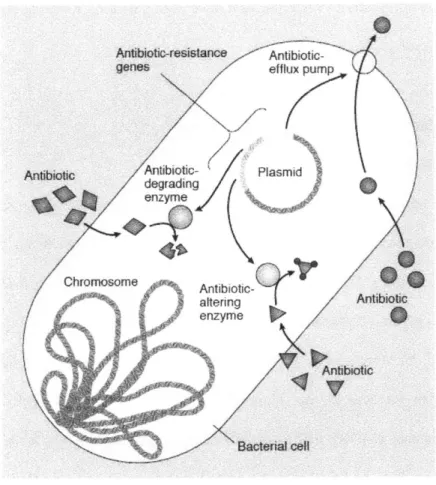
Bacterial drug resistance (DR) to antibiotics is one of the major challenges facing modem medicine. DR can be defined as the acquisition of gene(s) which act to reduce the effectiveness of a drug. This reduced drug activity can occur through several mechanisms, including reduced drug penetration into bacteria, increased drug efflux, drug modification or degradation, or drug target modification (Figure 1).5 DR can be observed in the laboratory as an increase in the minimum inhibitory concentration (MIC) of a drug. Multidrug resistance (MDR), that is, resistance to multiple antibiotics, is also on the rise, with as many as 16% of healthcare associated infections involving MDR pathogens in one report.1 5
Figure 2. 1. Drug Resistance Mechanisms. Common mechanisms of resistance to antibiotics include reduced drug penetration (not shown), increased drug efflux, antibiotic alteration by enzymes, antibiotic degradation, or drug target modification (not shown). Reprinted by permission from Macmillan Publishers Ltd: Nature Medicine Levy and
Marshall,2 copyright 2004.
Traditional antibiotics typically act on a narrow target and have a specific mechanism of action. While this specificity has obvious advantages, it also creates a selection environment favoring expansion of DR organisms. It is currently believed that subtherapeutic drug exposure is a mechanism driving DR.16 Theoretically, reasons why
antibiotics may fall below a therapeutic level in vivo include insufficient dose (caused by patient non-compliance, incorrect dosing, or dose-limiting toxicity), rapid elimination
from the site of infection, inactivation or loss of activity, or poor delivery to the infection site due to high barriers to diffusion or low tissue partition coefficients.
Nanomedicine is potentially well-suited to both improve traditional antibiotic formulations and contribute to overcoming the DR challenge. NPs have been shown to improve antibiotic drug efficacy or delivery,1 7', ' directly kill bacteria,19-21 or enable novel treatment paradigms such as targeted photothermal-mediated bacterial killing. ' In
addition, NPs can be used to reduce drug toxicity, potentially reduce clearance of beneficial bacteria, achieve drug concentrations high enough to overwhelm resistance mechanisms,24, 25 protect various antibiotics from degradative enzymes,2 6 or co-deliver multiple antibacterial agents. Further, because of the nature of NP-mediated killing or because of improvements in delivery, it may be intrinsically more difficult for bacteria to develop resistance to NP therapeutics. The sections below will further explore the potential of NPs to improve treatment of infections, which is likely to simultaneously
reduce the likelihood of DR emerging.
2. 2. 2. Infection Microenvironment
The microenvironment of an infection presents significant challenges for proper drug delivery and effective killing of bacteria. Infection sites are complex and dynamic entities, whose delivery challenges may vary as a function of the causative organism(s), the immune status of the patient, as well as the anatomic location. Infection sites can have intra- or extracellular bacteria, neutrophils, macrophages, lymphocytes, dendritic cells, host tissue cells, inflammatory mediators, bacterial toxins, and plasma proteins, among many others (Figure 2A). Other obstacles to delivery include aberrant tissue architecture
from chronic disease, hyperviscous mucus secretions, abscesses, acidity, and biofilms. Here we will highlight some examples of challenges to proper delivery together with nanomedicine approaches towards their resolution.
Chronic diseases can result in aberrations of the normal tissue architecture, which can impact the efficacy of therapy. For example, delivery of drugs to lung infections in patients with cystic fibrosis (CF), chronic obstructive pulmonary disease, or advanced
asthma is complicated by severe mucus plugging, areas of reduced ventilation, and significant tissue remodeling and fibrosis. In a step towards improving delivery to these regions, NPs have been developed which can penetrate mucus barriers. Tang et al
formulated NPs using a diblock copolymer of poly(sebacic acid)-block-poly(ethylene glycol) (PSA-PEG) designed to penetrate the hyperviscous mucus secretions of patients with cystic fibrosis.27 The authors densely coated the surface of the NPs with PEG to reduce interactions between the NPs and mucins, leading to more rapid and effective penetration rates. Using 173 nm PSA-PEG NPs, at a time scale of 1 second they demonstrated a 50-fold greater mean square displacement diffusion distance of the NPs than control latex NPs in mucus expectorated from CF patients. Further study showed that NP size is important, with NPs less than 200 nm in diameter moving more rapidly through low viscosity pores.28
Abscesses are collections of bacteria, white blood cells, and associated cell debris that are known to prevent effective antibiotic delivery. They are a significant clinical problem, with skin or subcutaneous abscesses alone accounting for ~2% of emergency room visits.29 Current clinical practice involves surgical drainage of the abscess with antibiotic therapy not practiced unless there are signs of systemic infection. Non-surgical methods
of therapy may help to reduce morbidity and tissue damage, leading to more rapid and satisfactory resolution. Nitric oxide-releasing NPs made from a hydrogel/glass composite have demonstrated activity against methicillin-resistant Staphylococcus aureus (MRSA) subcutaneous abscesses.30 These NPs were shown to not only inhibit MRSA growth and reduce abscess area in mice, but to also stimulate healing by promoting fibroblast migration and inducing collagen deposition.
Another factor that can reduce drug activity is the formation of localized acidity at a site of infection. Acidity has been documented across a range of different infections involving single and multiple organisms and at different anatomic locations. The mechanism of acidity is still incompletely understood but may involve a switch to anaerobic fermentation by bacteria under settings of low oxygen tension, leading to the production of organic acids.3 1 In addition, recruitment of acid-producing neutrophils and release of
products of inflammatory processes exacerbate the localized acidity, which can reach as low as pH -5.5. 3 Superficial skin infections, where the normal tissue pH is already
acidic, can be as low as pH 4.0.34 Localized acidity is significant in that the activity of several antibiotics is known to be affected by changes in pH. Selman Waksman, accepting the Nobel prize in 1952 for his discovery of streptomycin, noted the loss in bactericidal potency of this antibiotic in acidity. Loss of activity has been noted in amikacin, the fluoroquinolones ciprofloxacin and sparfloxacin,36 and vancomycin.37 Interestingly, certain
p-lactams
demonstrate increased potency in slight acidity.3 8 These observations suggest that there is a need to develop systems that may help to optimize antibiotic activity in different pH environments. In a step in this direction, Pornpattananangkul et al designed an acid-sensitive drug targeting system.34The authorsdeveloped cationic liposomes that remain stable at neutral pH due to surface-bound anionic gold NPs. As the local pH declines to below 5.0, the gold NPs dissociate, allowing the cationic liposomes to regain their ability to fuse with bacteria and deliver high doses of drugs.
0
)0Figure 2. 2. Infection Microenvironments. The microenvironment of an infection can have far-reaching implications in achieving proper drug delivery and bacterial killing. Schematic of a typical infection site. Nanoparticles (NP) circulate in the blood until they encounter a site of increased vascular permeability (dashed lines). NPs of appropriate size are able to extravasate and come into contact with the infectious process.
2. 2. 3. Biofilms
Discovering methods for clearing bacteria residing in biofilms is one of the most demanding challenges in bacterial infectious disease research today. Biofilms are a set of structurally diverse matrix-enclosed bacterial communities that adhere to surfaces and are remarkably resistant to antibiotic therapy. They form in a regulated developmental sequence, beginning with the adhesion of an active bacterium and production of an extracellular polymeric substance (EPS) matrix, often a polysaccharide, and have sophisticated architectures, growing flat or mushroom-shaped and with internal aqueous channels for diffusion of nutrients. The lack of efficacy of antibiotics against biofilms is
biofilms, (2) existence of bacteria in semi-starved states, leading to slower growth and subsequent reduced antibiotic susceptibility, and (3) existence of subpopulation of cells known as "persisters", which do not respond to antibiotics.5' 40 Biofilms are especially important in various chronic infections, including device-related infections, CF pneumonias, wounds, and periodontal disease.4 Nanoparticles have been hypothesized to be able to contribute to clearing biofilms through several mechanisms, including improved drug targeting, enhancing drug penetration into the biofilm, and reducing bacterial adhesion, the first step in biofilm formation.41 Efficacy against biofilms formed
by clinically significant pathogens including Pseudomonas aeruginosa and Staphylococcus aureus have been reported using nitric oxide-releasing silica NPs.42 In addition, magnetic silver ring-coated NPs ~30-40 nm in diameter have been developed that can penetrate deep into biofilms after application of an external magnetic field.43 Similarly, carboxyl-grafted superparamagnetic iron oxide NPs (SPIONs) were magnetically concentrated deep in biofilms, demonstrating ~8-fold higher percentage bacterial kill than gentamicin in a gentamicin-resistant strain of Staphylococci.4 4
Nanoparticles have also been used to prevent the formation of biofilms. A glycopeptide dendrimer was used to inhibit the formation of P. aeruginosa biofilms4 5 and silver
bromide NP/polymer composites, when used as a coating, demonstrated the ability to resist biofilm formation." Despite these promising advances, much more work is needed to investigate methods of completely eradicating bacteria in these colonies, including dormant persister cells.
Microorganisms have evolved the ability to evade the immune system by entering host cells, where intracellular conditions enable their continued survival. These organisms, broadly classified as "intracellular" can be very difficult to treat, have high mortality rates, and generally represent some of the most formidable challenges for designing therapeutics. Intracellular organisms inhibit the normal cellular digestive process in phagolysosomal compartments and reside there or escape into the cytoplasm.47 By residing intracellularly, bacteria are protected from attacks by antibodies, complement, and certain antibiotics.
A variety of intracellular organisms remain without a truly robust therapy, with rampant drug resistance, complex or lengthy regimens, lack of efficacy, and possible drug interactions in patients with comorbidities. Perhaps the most notorious is Mycobacterium
tuberculosis, the causative agent of tuberculosis (TB). TB is one of the most common and
dangerous diseases in the world and is highly multidrug resistant, with estimates by the World Health Organization suggesting that one third of the entire world population has latent TB. Other clinically significant intracellular pathogens include Listeria
monocytogenes, the causative organism of the highly fatal food-borne illness listeriosis, Salmonella typhi, the bacterium that causes typhoid fever, Legionella pneumophila, the
etiology of Legionnaires' disease, a dangerous infection of the respiratory tract, and
Chlamydia trachomatis among many others.8
The extent to which intracellular habitat affects antibiotic therapy depends on the drug and targeted cell type. Certain antibiotics, such clarithromycin, are actively transported into eukaryotic cells. Others, such as some
p-lactams,
vancomycin, or gentamicin, have relatively poor intracellular-to-extracellular ratios.6 Enhanced delivery precisely to thesubcellular site of bacterial habitat using NPs could potentially improve drug efficacy. Macrophages are a common target for intracellular bacteria due to this cell type's role in clearing pathogens. NPs can be engineered to target subcellular compartments in a wide variety of eukaryotic cells, including macrophages, using appropriate surface modifications. Consequently, several different NPs have been explored for their potential to treat intracellular infections.
Several NPs have been explored to treat TB. Poly(N-butylcyanoacrylate) and poly(isobutylcyanoacrylate) NPs encapsulating the often-used drugs isoniazid, rifampin, and streptomycin have been evaluated in terns of their uptake by human blood monocytes and their activity against TB.49 The NP-encapsulated drugs showed higher intracellular accumulation than free drugs and more potent antibacterial effect for isoniazid and streptomycin but not rifampin. In addition, PLGA particles have been used as an inhalable delivery system for rifampicin, isoniazid, and pyrazinamide, showing enhanced bioavailability and improved efficacy in a guinea pig model. In this study, only 5 doses of PLGA-formulated drugs led to complete clearance of bacteria - the equivalent of 46 daily doses of free drugs. This is particularly remarkable since complex dosing regimens lead to high patient non-compliance - a major contributing factor to the widespread multidrug resistant nature of TB.
Polyalkylcyanoacrylate (PACA) NPs are a class of materials that have been explored extensively for treating intracellular infections. Polyisohexylcyanoacrylate NPs with bound ampicillin were shown to have significantly improved efficacy as compared to free drug in a mouse model of listeriosis.5 1 The mechanism behind this improved activity in vivo was suggested to be improved activity of the NP-bound drug.5 2
Polyethylbutylcyanoacrylate NPs were developed which incorporated ciprofloxacin, a drug with a broader spectrum of action.5 3 Polyacrylate NPs have also been explored as
delivery systems for N-thiolated
p-lactams.
17 These modifiedp-lactams
are believed to act via a different mechanism of action than the conventional parent
P-lactams,
potentially making them suitable for use against drug resistant organisms. However, their low water solubility is a challenge for effective clinical translation. Polyacrylate NPs prepared with modified
p-lactam
monomers via emulsion polymerization in water demonstrated good drug encapsulation, small size (~40 nm), good stability, and low toxicity. Remarkably, the MIC of the NPs were 4-8x lower than the free drug monomer, suggesting significant enhancement of drug function by the NPs. Given the lack of antibacterial activity of empty NPs, it is believed that the lower MIC was due to either enhanced membrane permeability or higher local concentration of drug. Further, polyacrylate NPs formed containing acrylated penicillin G were evaluated in vivo by topical or intraperitoneal administration, demonstrating no obvious toxicity and promising activity, particularly in a model of an infected wound. Nevertheless, the potential for cytotoxicity of these and any other type of materials should be considered in further development, noting that PACA NPs may be cytotoxic at high concentrations.55Notable improvement in efficacy was found by encapsulating azithromycin in PLGA NPs. The PLGA NP formulation was shown to have an 8-fold lower MIC than free drug in treating S. typhi.8
These NPs may potentially be well-suited to target azithromycin to macrophages in the spleen or liver to treat typhoid fever. PLGA NPs have also been used to deliver the antibiotic combination of rifampin and azithromycin to Chlamydia-infected
cells, showing efficient targeting of the inclusion body and better efficacy than free drug.56
2. 3. Nanoparticles Creating Opportunities for Improved Therapy 2. 3. 1. Potential Advantages of Targeting Pathogenic Bacteria
One of the main advantages of using nanomaterials for treating bacterial infections is the potential to achieve more targeted effects. Possible advantages of targeting include improved drug efficacy, reduced side effects, reduced clearance of mutualist bacteria -which might impact a variety of diseases ranging from antibiotic-associated Clostridium
difficile diarrhea to immune diseases including asthma, eczema, and diabetes57 - reduced
potential for emergence of drug resistance, and ability to overcome drug resistance with drug concentrations not achievable using traditional antibiotic formulations due to toxicity. Nanomaterials have shown the ability to target bacteria through a number of different mechanisms, which generally fall under passive targeting or active targeting.
A
B
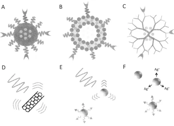
Figure 2. 3. Targeting Bacteria with Nanoparticles. Examples of different methods by which nanoparticles (NP) can target bacteria to clear infections. A) A combination of
surface moieties (purple triangles) and cationic surface charge disrupting the outer membrane (in green) of a Gram-negative bacterium. The large quantities of drug (in red) delivered in this manner can overwhelm drug efflux pumps (in blue). B) An approach where a targeting ligand (purple triangle) enables NP binding to the surface of a model protein (in orange), leading to more specific drug delivery and high local drug concentration. C) NPs targeting intracellular bacteria. A NP binds to a model membrane protein leading to internalization, where either fusion with bacteria-containing endosomes/phagolysosomes or endosome escape can lead to bacterial targeting. Note: NP not drawn to scale.
2. 3. 2 Passive Targeting
"Passive" targeting is the selective accumulation of nanomaterials at a site of disease by virtue of convection (primarily in the blood) and diffusion. This is contrast to "active" targeting, which includes specific interactions that occur between the nanomaterial and components of the targeted site that lead to accumulation, binding, or eukaryotic cell
internalization. Passive targeting of infections is initiated by the accumulation and release of bacterial components at the infection site, particularly pathogen-associated molecular patterns (PAMPs). These components activate immune cells, leading to generation of bradykinin, nitric oxide, prostaglandins, and other vascular mediators. Bacterial proteases, lipopolysaccharide, and lipoteichoic acids are also known to trigger production of bradykinin independent of immune cell action. This process can occur within minutes, lasting for hours to days following bacterial transmission and is characterized by vasodilation and increased vascular permeability locally in the vicinity of the infection. The vascular mediators trigger a widening of interendothelial gaps, allowing for extravasation of plasma components into the site of infection.58
Selective NP accumulation due to passive targeting can be modulated by physicochemical properties, including size, zeta potential, shape, rigidity, roughness,
surface hydrophilicity, density of poly(ethylene glycol) (PEG), and others (for a more thorough treatment, see reviews59'60). Significant effort has gone towards understanding the impact of these parameters on passive targeting. Much of this literature comes from studies of NP accumulation in tumors, which are similar to infections in that they exhibit local increases in vascular permeability. Important differences to consider include the aberrant vascular architecture, impaired lymphatic drainage, and reduced vascular density in tumors.61 Data that clearly and directly address the impact of NP properties on residence time at an infection are generally lacking. However, existing results suggest that NP infection residence time is significantly shorter than in tumors and is on the order of days vs. weeks.58 This suggests that a strategy that facilitates NP binding to the infection site may be preferable to one that relies on passive targeting alone.
Optimization of passive targeting is achieved by extending NP stability and circulation time in the blood, since NPs must circulate until they encounter the site of "leaky" vasculature. NP size is one of the principal factors governing passive targeting, with nanomaterials as small as 40 kDa and objects as large as bacteria (-1000 nm) being able to enter or leave the vascular space.58 However, NPs in the range 10-400 nm are
60
generally preferred for achieving extended circulation. Strongly cationic surfaces are a common feature of several antibacterial NPs. While certain examples have shown promise both in vitro and in vivo, in general, cationic charge would be expected to have high levels of non-specific eukaryotic cell uptake, negatively charged protein binding, and shorter circulation time. A mitigation strategy is to PEGylate the NP surface. However, even a single terminal cationic charge (primary amine) on PEG was shown to markedly affect the biodistribution of ~8-11 nm PEG-coated gadolinium oxide NPs, with the amine terminal group PEG-modified NPs showing much greater accumulation in the spleen and liver than similar negatively charged or neutral NPs.62 Consequently, efforts to design NPs with high surface charges should pay special attention to these considerations. The potential to target infections with nanoparticles is well-established.63 Technetium-99m-labeled liposomes of different mean sizes between 90 and 220 nm were shown to accumulate selectively at sites of S. aureus infection in rats.64 The biodistribution of these
liposomes was shown to be dependent on size, with differences occurring mainly in rates of splenic uptake. The % injected dose per gram of tissue (% ID/g) at the infection was not statistically different for liposomes 90, 120, 160, and 220 nm in size, with between
1.37-1.72% ID/g reaching the infection site. The targeting mechanism was believed to be passive targeting, with minimal contribution from monocyte uptake. Superparamagnetic
iron oxide nanoparticles (SPIONs) have been shown to rapidly and selectively accumulate in the lungs of mice infected with S. aureus.65 Accumulation could be observed as early as 24 hours postinfection - considerably before structural changes or edema could be appreciated using conventional T2* or T2-weighted imaging. SPIONs 18-30 nm have also been shown to accumulate in macrophages at sites of inflammation, including arthritic knees66 and soft tissue infections.67 The mechanism of selective accumulation in these examples is believed to be a combination of passive targeting followed by selective phagocyte uptake at the infection site.
2. 3. 3 Active Targeting
Active targeting involves the engineering of the nanomaterial to specifically interact with an infection site. Three general strategies have been explored to achieve active targeting: (1) cationic surface charge, which interacts with the negative surface charge of bacteria, (2) specific binding to the bacterial surface using targeting ligands, such as antimicrobial peptides or peptidomimetics, lectins, cell wall-targeting antibiotics, inflammation targeting, antibodies, or aptamers, and (3) targeting internalization inside of phagocytic cells for reaching intracellular organisms.
2. 3. 3. 1. Cationic Materials
Engineering a cationic surface charge to bind to the negative surface charge of bacteria is the most widely used mechanism to achieve active targeting. Components of the bacterial cell wall that contribute to a negative charge include (lipo)teichoic acids, peptidoglycan, negatively charged phospholipids and the lipid A/acidic phospholipids of the Gram-negative outer membrane. A wide variety of bacteria are Gram-negatively charged under physiologic conditions, making this approach suitable for different types of infections or
for infections that are polymicrobial. In addition, multivalent effects and the variety of chemical structures that can be engineered to produce a cationic charge make this an attractive method for targeting bacteria. Cationic bacteria-targeting materials generally fall under two major categories: (1) synthetic antimicrobial polycations and (2) natural peptide or peptidomimetic structures. The majority of interest in developing these materials has been to yield surfaces with contact-killing properties. Such surfaces are particularly well-suited to prevent bacterial colonization and have been explored principally as coatings for medical devices or other objects one might find in healthcare settings.
Synthetic antimicrobial polycations include materials such as quaternary ammonium6 or phosphonium69 compounds, or alkyl pyridiniums.70 Many more synthetic antimicrobial polymers have been explored for their antimicrobial effects (for review, see Kenawy et a171). These polymers could potentially be grafted onto the surfaces of nanomaterials and used for their bacteria-targeting and/or contact-killing properties, or be engineered to self-assemble into nanoscale structures. In general, accumulated studies have shown that high charge density, particularly zeta potential above +40 mV, and chain mobility are important for achieving a bactericidal surface.72 However, a challenge with these types of
materials is their lack of specificity for bacterial membranes. High charge density is known to correlate with toxicity to human cells, and many of the reported polymers are not biodegradable. In addition, the antimicrobial efficacy of these materials is typically reported at concentrations in the mg/mL range, which is too high for systemic application in many cases. Furthermore, there is risk with the mode of action of some of these polymers - bacteriolysis can lead to endotoxin release and fatal anaphylactic shock.
Despite these challenges, there have been notable successes. Cationic amphiphilic biodegradable polycarbonates that self-assembled into -40-200 nm NPs with low polydispersity were developed, demonstrating low micromolar MICs against Bacillus subtilis, MRSA, and Enterococcus faecalis, among others.2 0 These NPs did not show evidence of damaging red blood cell membranes even at concentrations much greater than their MIC, and were well tolerated in mice. The selective targeting appears to be a result of the highly cationic surface charge (+47 to +65 mV) interacting with the more negative microbial membranes. The biodegradable nature of these NPs, their excellent efficacy, and their broad spectrum activity make these synthetic structures very promising
antimicrobial NPs.
Synthetic NPs that use cationic charge-based targeting have shown excellent potential in vivo. A NP composed of a linear structure of TAT peptide (sequence YGRKKRRQRRR),
hexarginine (R6), triglycine (G3), and cholesterol (C) (combined = CG3R6TAT) was designed to target drug-resistant infections in the central nervous system." The hydrophobic cholesterol region triggered self-assembly of NPs 177 nm in diameter with a zeta potential of +55 mV. In vitro studies demonstrated low MIC across a range of pathogens, including MRSA and vancomycin resistant Enterococcus. Further, the NPs were shown to have an MIC six times lower than the soluble G3R6TAT peptide,
suggesting that the high positive charge density of the NP was important for the high potency of the antimicrobial effects. The NPs appeared to have selectivity for bacterial membranes, demonstrating relatively low rates of hemolysis and good tolerability in vivo. PLGA NPs have been given a cationic surface charge by incorporating Eudragit RL100 into the NP formulation step.7 3 The cationic NPs were shown to bind avidly to both S.
aureus and P. aeruginosa, with potential application in the targeted and sustained
delivery of ciprofloxacin to the eye.
2. 3. 3. 2. Antimicrobial Peptides or Peptidomimetics
Antimicrobial peptide (AMP) or peptidomimetic structures can be used to target bacteria. Currently, more than 1000 AMPs have been described, with a large diversity of structures and subclassifications.9
In general, AMPs are peptides of -10-50 residues composed of both cationic and hydrophobic regions with secondary structures such as a-helices or
p-sheet-like tubes. Above certain critical concentrations, AMPs lead to increases in membrane permeability, resulting in loss of membrane function and ultimately bacterial death. It is believed that the cationic regions of AMPs mediate the initial attraction step to negatively charged regions of the bacterial membrane. Following this initial interaction, hydrophobic regions adhere to the hydrophobic portion of the lipid membrane, leading to the formation of pores.7 4
Advantages of antimicrobial peptides as either targeting moieties or as drugs to be delivered include a binding mechanism of action that cannot easily be invalidated by microbial evolution, wide variety of structures and functionalities, relatively small size, and selectivity for bacterial membranes. However, certain AMPs only function under a defined set of conditions. Consequently, special care should be taken to ensure that AMPs will function under local pathologic conditions. In addition, the orientation of the AMPs on the NP surface may also play a role in their proper function. Finally, one should note that the site of interaction of many AMPs, the cytosolic membrane, may lie underneath one or more outer layers, making it difficult for the AMP to reach cytosolic membrane. Despite this, there is evidence to suggest NP-conjugated
AMPs may still be able to reach the cytoplasmic membrane, even in Gram-negative organisms that have an outer membrane. 10 nm gold NPs conjugated to the AMP Sushi 1 (Si) were shown to be able to target the Gram-negative E. coli, with NP penetration across all layers, though the majority (-77%) associated with the outer membrane.75 Further, the potential to target E. coli with -20 nm quantum dots conjugated to Si was demonstrated using fluorescence microscopy. These concerns are likely to be less significant in Gram-positive organisms, where the lack of an outer membrane allows for easier access to the cytoplasmic membrane. In fact, molecules of molecular weight up to 90 kDa are known to be able to diffuse across the peptidoglycan layer of the Gram-positive organism S. aureus.7 1 Potential challenges to AMP use include toxicity at high concentrations, lack of efficacy, enzymatic degradation, or negative effects on NP pharmacokinetics, particularly for highly cationic charged AMPs. Some of these challenges may be mitigated using peptidomimetics.7 6
2. 3. 3. 3. Lectins
The sugars of the bacterial membrane represent a potential binding site for targeted NPs. A gliadin NP containing acetohydroxamic acid was targeted to Helicobacter pylori using
the lectins Ulex Europeaus Agglutinin I (UEA I) or Conconavalin A (Con A).77 The lectin-targeted NPs demonstrated greater growth inhibition of H. pylori in vitro than both untargeted NPs and free drug. The lectin-targeted NPs also reduced the binding of H. pylori to samples of the human gastric mucosa ex vivo. A potential mechanism for the enhanced efficacy of the targeted NPs was a higher local concentration of drug in the vicinity of the bacteria.