HAL Id: tel-02185968
https://tel.archives-ouvertes.fr/tel-02185968
Submitted on 17 Jul 2019HAL is a multi-disciplinary open access archive for the deposit and dissemination of sci-entific research documents, whether they are pub-lished or not. The documents may come from teaching and research institutions in France or abroad, or from public or private research centers.
L’archive ouverte pluridisciplinaire HAL, est destinée au dépôt et à la diffusion de documents scientifiques de niveau recherche, publiés ou non, émanant des établissements d’enseignement et de recherche français ou étrangers, des laboratoires publics ou privés.
Role of Alix in endocytosis at presynaptic boutons
Kwangil Chi
To cite this version:
Kwangil Chi. Role of Alix in endocytosis at presynaptic boutons. Neurons and Cognition [q-bio.NC]. Université Grenoble Alpes, 2019. English. �NNT : 2019GREAV001�. �tel-02185968�
THÈSE
Pour obtenir le grade de
DOCTEUR DE LA COMMUNAUTE UNIVERSITE
GRENOBLE ALPES
Spécialité : Neurosciences, Neurobiologie
Arrêté ministériel : 25 mai 2016
Présentée par
Kwangil / CHI
Thèse dirigée par Rémy SADOUL, PR, Université Grenoble Alpes
préparée au sein du Grenoble Institut des Neurosciences – INSERM U1216
dans l'École Doctorale de Chimie et Sciences du Vivant
Rôle d'Alix dans l'endocytose
au niveau des boutons
pré-synaptiques
Thèse soutenue publiquement le 15 janvier 2019, devant le jury composé de :
M, Winfried, WEISSENHORN PR, Université Grenoble Alpes, Président M, Michael, COUSIN
PR, The University of Edinburgh, Rapporteur M, Jean, GRUENBERG
PR, Université de Genève, Rapporteur Mme, Lydia, DANGLOT
CR1, Université Paris Diderot, Examinateur Mme, Mireille, ALBRIEUX
MCF, Université Grenoble Alpes, Examinateur M, Rémy, SADOUL
The Role of Alix in
Endocytosis at Presynaptic
Boutons
Kwangil CHI
A Thesis submitted for the degree of Doctor of
Philosophy
ACKNOWLEDGEMENTS
I would like to thank the members of my jury, Dr. Mireille Albrieux, Dr. Michael Cousin, Dr. Lydia Danglot, Dr. Jean Gruenberg and Dr. Winfried Weissenhorn, for your time. I would like to express my deepest appreciation to the reviewers, Dr. Michael Cousin and Dr. Jean Gruenberg, for the critical review of my dissertation.
I would also like to extend my deepest gratitude to Rémy and Christine for their guidance through the process. I really honestly couldn’t thank you two enough. Thank you Rémy for entrusting me with this project, and being patient with me. Thank you also for the inspiration that you are and for encouraging me on throughout the study. I know there were difficult times, but even then you were always so determined and strong, and I thank you so much for that. There were times when I regretted my decision to continue my studies, but I have never once regretted my choice to stay with you. It has really been an amazing few years. Christine. Thank you for teaching me so much and for always being there to listen to me. This project would have been much more difficult without you. Thank you also for the practical advices, and helping me with some experiments. I will miss the chats we had in the neuron culture room.
I would like to extend my gratitude also to the former members of Team Sadoul. The restaurant Jimin and I went to with your wedding gift was fabulous. We thoroughly enjoyed the meal. As a team you made me feel welcome, and I really felt I belong here. I can’t thank you enough for that.
Thank you Bea for the practical advices you have given me. Thank you also for the stories of your adventures in the mountains. They were very nice to listen to and quite refreshing. Thank you Fiona for all the critical questions you’ve asked me which helped me to think a lot more about the little details of the project. I would like to thank you for the Scottish dance event you invite us (Marine, Jimin and me) which was quite an experience. Sometimes I can hear the instructor voice saying, “saut tam tam”. I remember you came at once when you heard that I had an accident (one of many that I had in Grenoble – I think this one is the neck-injury one) to take me to the emergencies. Thank you so much for that. Thank you Karine and Charlotte for being patient with me while teaching me how to do primary hippocampal cultures. The little tips you gave helped me become quite fast at it. Thank you both for listening to my concerns about the project and about the future. It helped me develop a more tangible plan for my future. Thank you for your reassuring words in response to my concerns. Thank you Sandrine for the promise you made me make. It was a good driving force! We are nearly there. Yves. I would came to you with a few questions (and I’m sure some of them were ‘yes’ or ‘no’ questions) and you always had a very extensive answer to each question. But each time, your answers helped me take a different approach to my problems. Thank you so much for helping me in times of need. Thank you Anne for your advice on experiments. Thank you Fred for your encouragement. I enjoyed the badminton session with you and Eric. I would like a rematch one day! Thank you Vincent for your words of encouragement. I still remember the exact words and those words have helped me along the way. Marine, my mini-boss. Thank you for the French lessons. The first phrase, “il pleut comme vache qui…”. I still haven’t got to use it yet. The second phrase, “les doigt dans le nez”. A little more useful, but I still haven’t used it yet. On a more
serious note, thank you. Among many things, thank you for running around the town with me to help me find a flat. I think it was raining like the phrase that day. Thank you for the potato book, Chi cat and the little events you did for us like finding the Easter eggs.
I would like to thank all my friends at GIN (Adrien, Camille, Raphael, Remi, Rebecca, Max, Charlotte and Celine), for the friendly environment you all have made. Special thanks to members of team 12 (Alain, Fabien, Jose, Sylvie, Mireille, Muriel and Eve). It was really nice to have you around as my neighbours for the second half of my study. Thank you Fabien and Jose for the work you’ve put into this project. Thank you Julien for going bouldering with me on occasions and making me feel as if I’m actually good at climbing. You have been a good writing buddy also. I shouldn’t forget Arnaud the barista. Thanks mate for a lot of things. Thanks for the temptations with your dealabs deals. I have two online bank accounts now! Thank you also for making the time to have those ‘before going home’ coffee breaks. But seriously, I can’t express how really nice it was to have you around. We talked about all sorts of things and I’ve learnt so much from you. I will really miss all the coffee breaks…. and the coffee breaks… and the coffee breaks. Thank you Eric and Jacques for always going that extra mile to help me with the analysis. Merci Yasmina de ne jamais parler en anglais avec moi. J'ai dû améliorer mon français en partie grace à toi. Merci de m'apprendre à manipuler le microscope.
I would like to thank my Grenoble family, Abror, Gizem and Jamie for always encouraging me. The fun and laughs we shared really helped me get through this.
I would like to thank my family for their prayers and support. A few words in Korean for my parents. 양가 부모님께 감사의 글을 올립니다. 힘든 과정을 거치는 동안 항상 믿어주시고 기도로써 힘이
되어주셔서 감사합니다. 특히 지친 몸과 마음으로 집에 갈 때마다 바쁘신 중에도 시간을 내셔서 따뜻하게 맞아 주시고 부족함 없이 편히 쉬고 재충전 할 수 있도록 사랑으로 배려를 해주심에 진심으로 감사드립니다. Thank you
Jonny and HJ for your encouragement! I guess I have you two to thank for my being here in France. Thanks KJ for the laughs. I still remember your ‘Gandalf spins’ down the ski slopes. Classic.
I would like to dedicate this thesis to my grandfather. Thank you for your prayers and support while you were here with us. I have tried to live out your life lessons and ended here. I hope I have made you proud.
Last, but not least. I would like to express my gratitude to my wife, 지미니, who was by my side throughout the journey. Saying thank you throughout this dissertation won’t be enough to express how thankful I am for supporting me all the way through. This would not have been possible without you.
Table of Contents
List of Abbreviations ... 1 Preface ... 3Introduction ... 4
1. Synapses ... 5 1.1 Presynaptic Boutons ... 6 1.1.1 Active Zones ... 61.1.2 Synaptic Vesicles and Synaptic Vesicle Pools... 9
1.2 Postsynaptic Terminals ... 13
1.3 Synaptic Transmission ... 13
1.3.1 Kiss-and-Run ... 13
1.3.2 Exocytosis ... 16
1.3.3 Endocytosis ... 18
1.3.3.1 Membrane Deformation in Endocytosis ... 20
1.3.4 Endocytosis in Presynaptic Boutons ... 23
1.3.4.1 Clathrin-Mediated Endocytosis ... 23
1.3.4.2 Clathrin-Independent Endocytosis ... 28
1.3.4.2.1 CIE: Ultrafast Endocytosis ... 28
1.3.4.2.2 CIE: Activity-Dependent Bulk Endocytosis ... 29
1.3.4.3 Common Molecular Actors in Synaptic Vesicle Recycling ... 34
1.3.5 Exo-Endocytosis Coupling ... 38
1.3.5.1 Cytomatrix of the Active Zone ... 38
1.3.5.2 Exocytosis and Synaptic Vesicle Proteins ... 38
1.3.5.3 Membrane Lipids ... 39
1.4 Synaptic Plasticity ... 41
1.4.1 Short-Term Plasticity ... 41
1.4.2 Long-Term Plasticity ... 44
2. The Protein Alix ... 47
2.1 Structure and Interactions ... 48
2.2 Functions ... 50
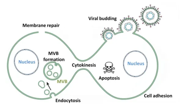
2.2.2 Endosomal-Lysosomal Pathways ... 53 2.2.3 Exosome Biogenesis ... 54 2.2.4 Endocytosis ... 55 2.2.5 Membrane Repair ... 57 2.2.6 Viral Budding ... 57 2.2.7 Cell Adhesion ... 58 2.2.8 Cell Death ... 58 2.2.9 Cytokinesis ... 59 2.2.10 Brain Development ... 60
3. Aim of the Thesis ... 61
Results ... 62
1. Context of Study ... 63
2. Article: Alix is Required for Activity-Dependent Bulk Endocytosis at Brain Synapses ... 64
Additional Results ... 103
1. Synaptophysin-pHluorin Response to Electrical Stimulation Suggests No Impairment of ‘Normal’ SV Endocytosis in Alix KO Neurons ... 104
2. Synaptophysin-pHluorin Response Indicates Reduced SV Exocytosis in the Presence of EGTA and BAPTA ... 105
3. Fluorescence Increase in Presynaptic Boutons During Intense Stimulation Do Not Represent Injury-Induced Varicosities ... 107
Discussion ... 108
1. Synaptic Density is Not Affected by the Lack of Alix... 109
2. Changes in Synaptic Morphology in Alix KO Neurons... 110
3. Changes in Synaptic Function in Alix KO Neurons ... 112
4. Recruitment of Alix and Its Partners to Presynaptic Boutons ... 114
5. ADBE Impairment in Alix Neurons ... 121
6. Possible Mechanisms Underlying the Role of Alix in ADBE ... 123
6.1 Possible Roles of Endophilin ... 123
6.2 Possible Involvement of Actin ... 123
6.3 Possible Mechanisms of Action Underlying ADBE Involving Alix ... 125
7. General Conclusion ... 127
1
List of Abbreviations
ADBE – Activity-Dependent Bulk Endocytosis Adp1 – Auxin binding protein 1
ADP / ATP – Adenosine DiPhosphate/Adenosine TriPhosphate AIP1 – Apoptosis Interacting Protein 1
ALG-2 – Apoptosis Linked Gene 2 Alix – ALG-2 Interacting protein X
AMPA(R) – α-Amino-3-hydroxy-5-Methyl-4-isoxazolePropionic Acid (Receptors) AP180 – Assembly Protein 180
AP2 – Adaptor Protein 2
ARF6 – ADP-Ribosylation Factor 6 Arp2/3 – Actin-related protein 2/3
BAR - Bin-Amphyphysin-Rvs (N-BAR: N-terminal amphipathic helix-BAR / F-BAR: Fes/CIP4 homology-BAR) BAPTA – 1,2-Bis(o-AminoPhenoxy)ethane-N,N,N’,N’-Tetraacetic Acid
CaMK-II – Calcium/CalModulin-dependent protein Kinase II cAMP – cyclic Adenosine MonoPhosphate
CAZ – Cytomatrix of the Active Zone CDC42 – Cell Division Control protein 42 CDK5 – Cyclin-Dependent Kinase 5 CEP55 – CEntrosomal Protein 55 CGN – Cerebellar Granule Neurons
CHMP – CHarged Multivesicular body Protein CME – Clathrin-Mediated Endocytosis CIE – Clathrin-Independent Endocytosis CIN85 – Cbl-Interacting Protein of 85kDa CME – Clathrin-Mediated Endocytosis CTxB – Cholera Toxin chain B
EGF(R) – Epidermal Growth Factor (Receptor)
EGTA – Ethylene Glycol-bis(β-aminoethyl ether)-N,N,N’,N’-Tetraacetic Acid EIAV – Equine Infectious Anemia Virus
ELKS/CAST – protein rich in the amino acids E,L, K and S/Cytomatrix at the Active zone-Associated STructural protein
EPS15 – EPidermal growth factor receptor Substrate 15 EPSC – Excitatory PostSynaptic Current
EPSP – Excitatory PostSynaptic Potential
ESCRT – Endosomal Sorting Complexes Required for Transport F-actin – Filamentous-actin
FCHO1/2 – Fer/Cip4 Homology domain-Only 1/2 GABAA(R) – γ-AminoButyric Acid A (Receptor)
G-actin – Globular-actin
GIT – G protein coupled receptor kinase 2 InTeracting protein GPI – GlycosylPhosphatidylInositol
2 GSK3 – Glycogen Synthase Kinase 3
GTP – Guanosine TriPhosphate
HDPTP – His Domain PhosphoTyrosine Phosphatase HIV – Human Immunodeficiency Virus
ILV – IntraLumenal Vesicles LBPA – LysoBisPhosphatidic Acid LTD – Long-Term Depression LTP – Long-Term Potentiation MEF – Mouse Embryonic Fibroblast
MHCI – major histocompatibility complex class I MVB – MultiVesicular Bodies
N-WASP – Neuronal Wiskott-Aldrich Syndrome Protein NMDA(R) – N-Methyl-D-aspartic Acid (Receptor) PAR1 – Protease Activated Receptor 1
PIP – PhosphatidylInositol Phosphates
PIPKIγ – PhosphatidylInositol Phosphate Kinase type Iγ PI3K – PhosphatidylInositol-3-Kinase
PI(4,5)P2 – PhosphatidylInositol 4,5-bisPhosphate
PI(3,4,5,)P3 – PhosphatidylInositol 3,4,5-trisPhosphate
PKA – Protein Kinase A
PPD – Paired Pulse Depression PPF – Paired Pulse Facilitation PPR– Paired Pulse Ratio
PRD / PRR – Proline-Rich Domain / Proline-Rich Region PSD – PostSynaptic Density
PTD – Post Tetanic Depression PTP – Post Tetanic Facilitation RIM – Rab3-Interacting Molecules RIM-BP – RIM-Binding Protein RRP –Readily Releasable Pool SH3 – Src Homology 3
SNAP – SyNaptosomal-Associated Protein
SNARE – Soluble NSF (N-ethylmaleimide-sensitive factor) Attachment protein Receptor STED – STimulated Emission Depletion
SV – Synaptic Vesicle
TNF(R) – Tumour Necrosis Factor (Receptor) Tsg-101 – Tumour susceptibility gene-101 VAMP – Vesicle-Associated Membrane Protein VGLUT – Vesicular GLUtamate Transporter Vps4 – Vacuolar Protein Sorting 4
3
Preface
Synaptic vesicle recycling refers to the regeneration of synaptic vesicles from the presynaptic membrane after fusion. The term was coined by J.E. Heuser and T.S. Reese in 1973 through their work on frog neuromuscular junctions (Heuser and Reese, 1973). This electron microscopic study revealed the formation of cisternae derived from the presynaptic membrane after stimulation. The cisternae then divided to form fusion-competent synaptic vesicles. Since then, a number of different modes of synaptic vesicle recycling and the molecules involved have been discovered, that can be categorised as clathrin-mediated endocytosis or clathrin-independent endocytosis. Clathrin-mediated endocytosis is the best understood and it was considered the main route of synaptic vesicle regeneration. The significance of clathrin-independent endocytosis has only recently been brought to light and it is less well understood due to its recent discovery. Therefore, it is vital to identify the molecules involved and their mechanism of action in this process. The present study aims to contribute to the growing knowledge on synaptic vesicle recycling by putting forward Alix (ALG-2 Interacting Protein X) and its interacting proteins (ALG-2 and endophilin) as molecular actors in clathrin-independent endocytosis in synapses.
4
5
1. Synapses
Neuronal communication is central to the functioning of the central nervous system and underpins its physiological roles such as learning and storing memory. The 100 billion nerve cells that make up the human brain are called neurons, and these are excitable cells that are responsible for transmitting chemical and electrical signals. Neurons are compartmentalised into somato-dendritic compartments, that receive and integrate electrical signals, and axons, where action potentials are generated. Action potentials are temporary shifts in the neuron’s membrane potential. Neurons transmit signals between each other through chemical or electrical synapses that are made up of a presynaptic terminal (or bouton) and a postsynaptic terminal. Electrical synapses allow flow of ions and metabolites between neurons through specialised structures called gap junctions. Chemical synapses allow information relay via the release of neurotransmitters from the presynaptic bouton that is detected by receptors on the postsynaptic terminal, and they can be excitatory or inhibitory (Pereda, 2014). In chemical excitatory synapses of the central nervous system, postsynaptic terminals are defined by the presence of postsynaptic densities (PSDs) on the plasma membranes of dendrites whereas presynaptic terminals are often present as boutons in axons. Briefly, synaptic transmission occurs when action potentials depolarise the presynaptic membrane, causing the release of neurotransmitters via synaptic vesicle (SV) fusion to the presynaptic membrane. Neurotransmitters will then diffuse across the synaptic cleft and bind receptors on the postsynaptic membrane to cause depolarisation. Like the PSDs of postsynaptic terminals, there are some defining features of presynaptic boutons and postsynaptic terminals. Together, the presynaptic boutons and postsynaptic terminals form chemical synapses through which neurons communicate.
6
1.1. Presynaptic Boutons
Presynaptic boutons are specialised parts of the axon that are characterised by the presence of active zones and SVs. This is where the SV fusion occurs to allow the release of neurotransmitters.
1.1.1. Active Zones
The active zones of presynaptic boutons are electron-dense meshwork of a range of multi-domain proteins which are called the cytomatrix of the active zone, or CAZ, and these are the sites of SV exocytosis (Gundelfinger and Fejtova, 2012). There are five constituent proteins of CAZ that are conserved across organisms (such as flies, rodents and worms) which include ELKS/CAST1
, liprin, Munc13, Rab3-interacting molecules (RIM) and RIM-binding proteins (RIM-BP) (Sudhof, 2012; Michel et al., 2015). CAZ in vertebrates also comprise bassoon and piccolo. These proteins contain protein-protein interaction domains such as C2, PDZ2
and Src homology 3 (SH3) domains that allow the tight association of the proteins for regulation of SV fusion to the presynaptic membrane. The protein composition and the relative organisation of CAZ are summarised in figure 1 and table 1. The active zone is encompassed by a periactive zone, which is where SV endocytosis occurs (Sudhof, 2012). Periactive zones also house cell-adhesion molecules and presynaptic receptors that modulate neurotransmitter release (e.g. endocannabinoid CB1 receptors). How the different components of the CAZ allow the fusion of SVs for the release of neurotransmitters will be discussed later.
1
ELKS/CAST: protein rich in the amino acids E,L, K and S/cytomatrix at the active zone-associated structural protein
2 named after postsynaptic density protein (PSD-95), discs-large tumor suppressor (Dlg), and tight junction
7
Figure 1: Molecular organisation of a synapse
A) Electron micrograph of a brain slice showing a synapse: a synaptic vesicle-filled presynaptic terminal and a
postsynaptic terminal. B) A model of the molecular organisation of a synapse.
Key : Adhesion molecules Active zone proteins
Endocytic proteins Exocytic proteins
Ion channels and glutamate receptors Postsynaptic density proteins
Adapted from (Chua et al., 2010)
B
A
8 Category Protein Function Interactions
Active zone proteins
ELKS/CAST SV exocytosis Liprin, Rab6
Liprin Active zone assembly
SV exocytosis CASK, ELKS, ERC, GIT1, RIM
Munc13 SV priming Munc18, RIM, syntaxin
Munc18 SV priming Munc13, Syntaxin, VAMP2
RIM Ca
2+
-channel recruitment SV docking and priming
Ca2+-channel, ELKS, Munc13, Rab3 (SV), Rab27, RIM-BP RIM-BP Ca2+-channel recruitment Ca2+-channel, RIM
CASK SV recruitment Liprin, β-Neurexin
Bassoon SV clustering
SV exocytosis ERC (RIM)
Piccolo SV clustering Ca2+, stonin
Exocytic machinery
Ca2+-channel Ca2+ entry RIM, RIM-BP, synaptotagmin PI(3,4)P2 Synaptotagmin recruitment Synaptotagmin
SNAP25 SV fusion Syntaxin, VAMP2
Synaptobrevin SV fusion Munc18, SNAP25
Synaptotagmin SV fusion (Ca2+ sensing) Β-Neurexin, Munc13, PI(3,4)P2
Syntaxin SV fusion Munc18, SNAP25
Endocytic machinery
(CME)
Amphiphysin Dynamin recruitment Dynamin, endophilin, phospholipase D2, synaptojanin Accessory proteins
(AP2, AP180, auxilin, Eps15, epsin, stonin 2)
Cargo recognition and clustering Membrane deformation
Cargo, clathrin, PI(3,4)P2, AP180,
AP2, epsin, synaptojanin
Clathrin Membrane deformation Amphiphysin, auxilin, AP180, AP2,
Dynamin Membrane scission Amphiphysin, PI(3,4)P2 , synaptojanin, syndapin Endophilin Membrane deformation
Synaptojanin recruitment
Amphiphysin, dynamin, synaptojanin Synaptojanin Clathrin coat disassembly Endophilin, PI(3,4)P2
Adhesion molecules
Neurexin
SV recruitment AMPAR and NMDAR
organisation
CASK, synaptotagmin, neuroligin (AMPAR, NMDAR)
EphB, Ephrin-B AMPAR and NMDAR
trafficking and function AMPAR, NMDAR, PSD-95 N-cadherin F-actin organisation Β-catenin
NLG PSD formation
NMDAR clustering PSD-95
Scaffold proteins
Homer PSD structure maintenance Shank
PSD-95 PSD structure maintenance Stargazin (AMPAR), GKAP, NLG, NMDAR
SAPAP PSD structure maintenance PSD-95, Shank Shank Links PSD with F-actin Cortactin, GKAP, homer Glutamate
receptors
AMPAR Na+ entry Stargazin
NMDAR Na+ and Ca2+ entry CaMKII, PSD-95
9
1.1.2. Synaptic Vesicles and Synaptic Vesicle Pools
SVs are storehouses of neurotransmitters that are uniform in size with diameters of around 30 to 50 nm (Poudel and Bai, 2014). It is suggested that these uniform SVs also contain approximately the same amount of neurotransmitters which is the basis of reliable quantal release of neurotransmitters. They are compact structures with over 60% of their total mass coming from proteins alone. It is estimated that up to 200 protein molecules are present in a single SV. The protein composition of a typical SV is summarised in table 2 (Südhof, 1999; Wilhelm et al., 2014). Different synapses have varying numbers of SVs, for example, rodent hippocampal synapses typically have around 200 SVs whereas the calyx of Held around 300 SVs (Gan and Watanabe, 2018). Such a difference reflects the physiological requirements of these synapses. Compared to the relatively small hippocampal synapses, the calyx of Held, found in the auditory nervous system, is a large synapse which is often subject to very high firing rates3
that can reach up to around 1 kHz, and therefore requires more SVs (Baydyuk, Xu and Wu, 2016; Gan and Watanabe, 2018). Despite the uniformity in size, SVs show heterogeneity in terms of function and organisation (Crawford and Kavalali, 2015) and these differences helped categorise SVs into different groups known as SV pools. These pools are readily releasable pool, recycling pool, total recycling pool and resting pool (Alabi and Tsien, 2012; Fowler and Staras, 2015) and are summarised in figure 2.
SVs of the readily releasable pool are the first to be released upon stimulation. In hippocampal synapses, the readily releasable pool accounts for around 10 SVs (Gan and Watanabe, 2018). These are typically the SVs that are docked to the active zone, ready for release. It is generally considered that the readily releasable pool depletion can be achieved by various stimulation conditions such as a few seconds of hypertonic shock or tens of action potentials at mild frequencies (Rizzoli and Betz, 2005; Fowler and Staras, 2015). The replenishment of the readily releasable pool takes less than 30 seconds to complete in hippocampal synapses (Goda and Stevens, 1998).
3
10
Figure 2: synaptic vesicle pools
A) The four different synaptic vesicle pools present in a presynaptic bouton. The readily releasable pool (RRP)
represents the docked synaptic vesicles that are primed for release. The recycling pool accounts for around 5~20% of all synaptic vesicles, that will release immediately after the RRP. Together, the RRP and the recycling pool are called the total recycling pool. The resting pool accounts for around 15~85% of all synaptic vesicles, that are reluctant to release. The superpool represents the synaptic vesicles that are transported along axons to different presynaptic boutons, likely for the sharing of synaptic vesicles where necessary. B) the kinetics of release of the various synaptic vesicle pools. Goldfish bipolar cells were stimulated in the presence of FM1-43, a styryl dye that fluoresces upon integration into membranes. Three kinetic components of exocytosis were reported that were suggested to derive from different synaptic vesicle pools (indicated on the graph). A) and B) are adapted from (Rizzoli and Betz, 2005)
11
Table 2: Seven major synaptic vesicle proteins and their corresponding copy number
Adapted from (Mutch et al., 2011)
The release of the recycling pool then follows. The recycling pool accounts for around 16 SVs in hippocampal synapses (Gan and Watanabe, 2018) or 5~20% of all vesicles generally (Rizzoli and Betz, 2005). This pool replenishes the readily releasable pool during mild stimulation conditions and maintains steady release (Alabi and Tsien, 2012). Together the readily releasable pool and recycling pool are known as the total recycling pool which represents the SVs that undergo SV recycling during stimulation conditions within physiological boundaries.
The remaining SVs are grouped in the resting pool that is reluctant to release. The size of resting pool in hippocampal synapses are estimated to be around 200 SVs (Gan and Watanabe, 2018), or around 15% to 85% generally (Fowler and Staras, 2015). It was shown that the resting pool could be released in an extreme condition when the total recycling pool has been depleted as a study on the temperature sensitive-mutant of Drosophila, shibire, showed (Kuromi and Kidokoro, 1998). The shibire gene encodes dynamin in flies and the
shibire mutation was found to cause mutation in the GTPase domain of this protein (Van Der
Bliek and Meyerowrtz, 1991). In shibire-mutation carrying Drosophila, SV endocytosis is inhibited at ‘non-permissive temperature’ leading to the depletion of the total recycling pool at synapses (Kuromi and Kidokoro, 1998). In this condition, release of the resting pool could be observed indicating that the resting pool could be released once the total recycling pool has been depleted. The physiological role of the resting pool, however, still remains vague, but it appears that SVs are exchanged with the total recycling pool (Fowler and Staras, 2015).
Category Protein Description Average Copy Number /Vesicle
Synaptic vesicle proteins
Proton ATPase Pumps protons into SVs 1.27
SV2 Function uncertain 5.04
Synaptogyrin-1 Implicated in SV recycling and synaptic plasticity
6.80
Synaptophysin Implicated in synapse formation, SV docking, SV endocytosis
13.06
Synaptotagmin-1 SV fusion (Ca2+ sensing) 7.16
VAMP2 SV fusion 10.53
12
Certain conditions or factors seem to promote the transfer of SVs from the resting pool to the total recycling pool such as cyclin-dependent kinase 5 (CDK5) activity (Kim and Ryan, 2010) or the activation of N-methyl-D-aspartic acid receptor (NMDAR; Ratnayaka et al., 2012). The transfer of SVs from the total recycling pool to the resting pool has been suggested with an antidepressant (Jung et al., 2017) or during calcineurin activity (Marra et
al., 2012). These studies suggest SVs are not fixed to a specific pool, but that they could shift
from a pool to another.
In addition to these pools, a new pool, termed the superpool has been identified. The superpool is transported along the axon, residing transiently in presynaptic boutons, and allows sharing of SVs across boutons (Fowler and Staras, 2015). In a study on rat hippocampal neurons, it was shown that newly recycled SV vesicles from one bouton can be transferred to a neighbouring bouton to be released in a subsequent stimulation (Darcy et
al., 2006). This indicates that the superpool is a mix of SVs from the total recycling pool. The
transport of superpools between presynaptic boutons may be active, along the cytoskeleton, or passive, by free diffusion (Westphal et al., 2008). It is suggested that certain factors regulate the superpool and that the function of this pool might be to provide an extra supply of SVs where necessary (Staras and Branco, 2010). This further supports the idea that various SV pools are intermixable.
In terms of the organisation within presynaptic boutons, the total recycling pools are generally found closer to sites of SV fusion and the resting pools are found distant from such sites (Fowler and Staras, 2015). It has been suggested that various molecular markers help SV pool organisation by differentially associating with the different SV pools. For example, vesicle-associated membrane protein 7 (VAMP7) appeared to be enriched in SVs of the resting pool, and these SVs were distinct from those enriched in VAMP2 and vesicular glutamate transporter-1 (VGLUT-1) that represented the recycling pool (Hua et al., 2011). Synapsin is a protein that has been implicated as a molecular glue that binds the resting pool together, forming boundaries and limiting lateral leakage of SVs into axons (Orenbuch et al., 2012). Altogether, the dynamic nature of SVs and their highly-regulated organisation allow distinct pools of SVs that can intermix in adaptation to changes at synapses.
13
1.2. Postsynaptic Terminals
Postsynaptic terminals are specialised structures on dendrites and the molecular organisations can differ depending on the nature of the synapse, either excitatory or inhibitory (Sheng and Kim, 2011). For the purpose of this thesis, only excitatory synapses will be discussed. PSDs are electron-dense regions of the dendritic spines harbouring a range of proteins including calcium/calmodulin-dependent protein kinase II (CaMK-II), glutamate receptors, PSD-95, Shank, and components of the actin cytoskeleton. Other proteins of the postsynaptic terminal and their relative organisation are shown in figure 1 and table 1.
Postsynaptic terminals and presynaptic boutons are held together by a number of adhesion molecules that are in the synaptic cleft including integrins, neurexins and ephrin receptors (Harris and Weinberg, 2012). In addition to the adhesion function, some of these proteins have roles in synaptogenesis and synaptic plasticity.
1.3. Synaptic Transmission
Action potentials arriving at the presynaptic terminal (or bouton) cause depolarisation. This triggers the opening of voltage-gated calcium channels which leads to the fusion of neurotransmitter-filled synaptic vesicles (SVs) to the presynaptic membrane in full-collapse fusion, namely exocytosis. Then compensatory endocytosis will reform SVs by pinching off a part of the presynaptic membrane and replenish the SV pool. This process is termed SV recycling (Heuser and Reese, 1973) and it is at the core of neurotransmission to allow maintenance of efficient neuronal communication in the dynamic environment of synapses.
1.3.1. Kiss-and-Run
Since the first discovery of SV recycling, it has been theorised that some SVs might not undergo full-collapse fusion, but rather retain their SV identity through transient fusion called ‘kiss-and-run’ – a term which was first coined by Fesce and his colleagues in 1994 (Ceccarelli et al., 1973; Heuser and Reese, 1973; Fesce et al., 1994). In this mode of SV
14
recycling, SVs fuse transiently with the presynaptic membrane, allowing a partial discharge of neurotransmitters through the fusion pore before the retrieval of the SV through fusion pore closure. Initial speculations were generally based on morphological observations such as the predominance of vesicle openings in active zones even during the recovery phase (endocytic phase; Fesce et al., 1994). A number of evidences in support of the existence of kiss-and-run have since been brought forward. These include capacitance4
flickers that correspond to rapid opening and closing of fusion pores with diameters of >2.3nm (He et al., 2006), partial release of styryl dyes (Aravanis, Pyle and Tsien, 2003), and the retention of 15nm quantum dots during cycles of neurotransmission (Zhang, Li and Tsien, 2009). Such studies do not neglect the existence of other modes of endocytosis. No mechanisms have been shown regarding kiss-and-run at synapses, but suggestions have been made which include a special organisation of the active zone to allow a limited number of full-collapse fusions, and a possible existence of a ring at the neck to restrict full-collapse fusion (Saheki and De Camilli, 2012). A number of studies provide evidence against the presence of kiss-and-run. It was demonstrated that prolonged stimulation of cultured hippocampal neurons at 10Hz (a condition which does not deplete the recycling pool of SVs) leads to an efficient release of FM4-64, suggesting full-collapse fusion events (Fernández-Alfonso and Ryan, 2004). Additionally, it was found that SV proteins that are exocytosed are not always the same as those which are subsequently endocytosed (Fernández-Alfonso et al., 2006; Wienisch and Klingauf, 2006). This implies that SVs undergo full-collapse fusion before a new SV is reformed through compensatory endocytosis, arguing against the existence of kiss-and-run.
4
Membranes act as an insulator separating two electrolytic media (the cytoplasm and extracellular environment), resulting in membrane capacitance. The addition of membrane by exocytosis will lead to increased capacitance whereas the deduction of membrane by endocytosis will lead to decreased capacitance.
16 Figure 3: Synaptic vesicle exocytosis (↑)
A) Intermediary steps in SNARE-mediated membrane vesicle fusion. Adapted from (Jahn and Fasshauer, 2012). B) Four steps of the SNARE/SM protein cycle. The top diagram shows VAMP (v-SNARE) in its native, unfolded state, syntaxin-1 SNARE) in its closed conformation and bound to Munc18-1 (SM protein), and SNAP-25 (t-SNARE). Synatxin-1 and SNAP-25 can form heterodimers. The diagram on the left shows membranes primed for fusion. The priming process involves the formation of trans-SNARE complexes: the coiled coil interaction between v-and t-SNAREs. The bottom diagram shows the formation of a fusion pore induced by the formation of full trans-SNARE complexes. The trans-SNARE complexes convert into cis-SNARE complexes. The cis-SNARE complexes are disassembled by the action of ATPase NSF and its adaptors α/β/γ-SNAPs, preparing the SNARE/SM proteins for more synaptic vesicle exocytosis. C) Active zone proteins during synaptic vesicle exocytosis. Four components of the CAZ (α-Liprins, Munc13s, RIMs and RIM-BPs) are depicted in this diagram. The interaction between the various active zone proteins are mediated by the C2-/SH3-/zinc finger domains. The MUN domain of Munc13 cooperates with Munc18 to allow the correct formation of trans-SNARE complex (Lai et al., 2017). B) and C) are adapted from (Südhof and Rizo, 2011)
1.3.2. Exocytosis
As mentioned earlier, neurotransmitter release or SV fusion occurs at active zones. Synapses of the central nervous system are smaller than other synapses, with only one active zone per synapse in the case of hippocampal synapses (Gan and Watanabe, 2018). SV fusion is a very rapid process, taking only around 200μsec, due to the tight connection between the molecular actors involved (Adler et al., 1991).
The fusion and merging of two bilayers into one requires a substantial amount of energy as there are factors that have to be overcome: the electrostatic repulsion by negative charges on the surfaces of the bilayers, and the dehydration barrier. As storage compartments of neurotransmitters, SVs are an important part of neurotransmission and harbour a vast range of proteins and lipids including the ‘soluble NSF (N-ethylmaleimide-sensitive factor) attachment protein receptor’ (SNARE) complex proteins (Takamori et al., 2006). SNAREs are capable of providing the energy to overcome the unfavourable fusion process. They can be grouped into two types5: v-SNAREs that are on SVs and t-SNARES that
5 t-SNARE: target SNARE; v-SNARE: vesicular SNARE; q-SNARE: SNAREs with Gln/Q residue; r-SNARE: SNAREs
17
are on the presynaptic membrane; or alternatively R-SNAREs or Q-SNAREs, respectively (Hong, 2005). Synaptobrevin or VAMP-2 is a v-SNARE, and syntaxin and synaptosomal-associated protein 25 (SNAP-25) are t-SNAREs (Südhof and Rizo, 2011). Other proteins such as the calcium-sensing protein, synaptotagmin, are also involved. Initially SVs will be tethered and primed for exocytosis at the active zone, through a process which is carried out by certain Rab family of small GTPases and their effector proteins. At this point the v-SNAREs and t-SNAREs will interact to form a trans-SNARE complex bringing the SV membrane in tight apposition with the presynaptic membrane. Complexin plays a role in facilitating the formation of this complex, but prevents further zippering (Li et al., 2011). This leaves SVs in a primed state until action potentials induce the entry of calcium through voltage-gated calcium channels in the active zones. Synaptotagmins are SV proteins that harbour two C2 domains (C2A and C2B) that are capable of binding calcium, membrane and SNARE complexes (Yoshihara and Littleton, 2002; Jahn and Fasshauer, 2012). Upon binding calcium, synaptotagmins allow the fusion of SVs to the presynaptic membrane by interacting with the SNARE complex to relive the inhibitory effect of complexin (Schaub et al., 2006), and initialise and stabilise fusion pores (Yoshihara and Littleton, 2002). It was proposed that the C2B domain of synaptotagmin-1, which interacts with the trans-SNARE complex as well as the plasma membrane, is essential in calcium-dependent SV fusion by deforming the plasma membrane (Wang et al., 2016). The capacities of the C2B domain to deform the membrane and to displace the inhibitory complexin allow the two apposed membranes to come closer together, which leads to the formation of fusion pores. The various steps in the SV fusion process, the SNARE/SM (Sec1/Munc18-like protein) protein cycle and the involvement of the active zone proteins in SV exocytosis are summarised in figure 3.
Components of the CAZ also play a role in SV exocytosis. It was demonstrated that RIMs allow the clustering of calcium channels to the active zone either by direct interaction or through its interaction with RIM-BP (Kaeser et al., 2011). The depletion of RIMs led to reduced calcium influx at presynaptic boutons and a concomitant decrease in neurotransmitter release, suggesting that calcium channel recruitment to active zone mediated by RIMs has a central role in SV exocytosis (Han et al., 2011; Kaeser et al., 2011). Furthermore, an electron tomography study showed that knockout of an isoform of RIM, RIM1α, caused defects in SV tethering to the active zone and reduced the readily releasable
18
pool size (Fernández-Busnadiego et al., 2013). This suggested that RIM is necessary for tethering SVs to the active zone.
Munc13 of the CAZ has also been implicated in SV exocytosis. Depletion of an isoform, Munc13-1, in mice led to a significant decrease in the size of readily releasable pool which was accompanied by a blockage of neurotransmitter release at excitatory glutamatergic synapses (Augustin et al., 1999). Furthermore, a double-knockout of 1 and Munc13-2 in mice resulted in a complete blockage of SV exocytosis in both excitatory glutamatergic synapses and inhibitory, γ-aminobutyratergic (GABAergic), synapses (Varoqueaux et al., 2002). Munc13, through its MUN domain, plays a role in SV exocytosis by accelerating the transition of the closed Munc18-syntaxin complex into the trans-SNARE complex (Ma et al., 2011). A recent study revealed another role of the MUN domain of Munc13 in the regulation SV exocytosis by ensuring proper assembly of the trans-SNARE complex by cooperating with Munc18 (Lai et al., 2017). Such proper assembly of the trans-SNARE complex allows the interaction of the complex with complexin-1 and synaptotagmin-1 to favour calcium-triggered vesicle fusion. In addition, it was shown that Munc13 and RIM are able to form tripartite complexes along with a Ras-related protein, Rab3 (a GTP-binding protein), that facilitate SV priming (Dulubova et al., 2005).
1.3.3. Endocytosis
Endocytosis refers to a set of highly regulated cellular processes that allow the uptake of extracellular material by capturing a part of the plasma membrane. The role of endocytosis can vary from the internalisation of receptors for signalling, to the regulation of the plasma membrane composition. The ingested materials are contained in membrane-bound endocytic vesicles and can either be recycled back to the plasma membrane or degraded in specialised compartments. Such pathways can be hijacked by pathogens to spread disease, or exploited by therapeutic interventions to ameliorate diseases or disorders (Pelkmans and Helenius, 2003). In general, endocytosis is mediated by various molecular actors that act to curve the plasma membrane inward to form invaginations which will be cut into vesicles by scission (Doherty and McMahon, 2009). Endocytosis can be classified into different categories according to the molecular actors involved, as summarised in figure 4.
19
The well-understood clathrin-mediated endocytosis (CME) involves the coat protein, clathrin, and its adaptor proteins to internalise essential nutrients (e.g. iron) and receptors (e.g. fibroblast growth factor receptor and G-protein coupled receptors; Le Roy and Wrana, 2005; Mukherjee, Tessema and Wandinger-Ness, 2006). Clathrin-independent endocytosis, on the other hand, as the name suggests, does not involve clathrin and is not so well understood. It encompasses a range of endocytic mechanisms such as ADP-ribosylation factor 6 (ARF6)-mediated and cell division control protein 42 (CDC42)-(ARF6)-mediated pathways (Sabharanjak et
al., 2002; Kirkham and Parton, 2005; Kirkham et al., 2005; Mayor et al., 2014).
Figure 4: Different types of endocytosis
A range of different endocytic modes can be found in cells. The various modes utilise different proteins to take up different cargoes and varying amounts of the plasma membrane. The nomenclature depends on the main proteins involved or the cargo taken up. For example, clathrin-mediated endocytosis and flotillin-dependent endocytosis utilise clathrin and flotillin, respectively. CLIC/GEEC stands for clathrin-independent carriers/ glycosylphosphatidylinositol-anchored protein enriched endocytic compartments. For more details of each pathway, see (Doherty and McMahon, 2009). Many forms of clathrin-independent endocytosis exist and are not as well-understood as the clathrin-mediated endocytosis. Adapted from (Doherty and McMahon, 2009).
20
1.3.3.1. Membrane Deformation in Endocytosis
Deforming or re-shaping a flat membrane to form vesicles requires energy due to the propensity of the membrane to remain flat (Hurley et al., 2010). In endocytosis positive membrane curvature is induced which means that the membrane bends inwards, towards the cytoplasm. Negative membrane curvature, which is the outward bending of the membrane, away from the cytoplasm, is induced in processes such as viral budding and exosomes generation. Positive membrane curvature can occur through a number of different mechanisms as summarised in figure 5.
Among the different way to induce positive membrane curvature, protein shape-induced membrane bending is of particular interest to the present study. Bin-Amphyphysin-Rvs (BAR) domain proteins such as endophilin and amphiphysin comprise curved and positively charged BAR domains that are able to bind the negatively charged polar heads of lipids (Peter et al., 2004). The BAR domain superfamily comprises a number of families including the F-BAR (Fes/CIP4 homology-BAR), the N-BAR (N-terminal amphipathic helix-BAR) and others that are reviewed in Frost et al., 2009. A majority of BAR domain proteins have protein-to-protein interaction domains like the SH3 domain (Stanishneva-Konovalova et al., 2016). BAR domain proteins may function by inducing membrane curvature. In support, certain BAR domain proteins such as endophilin and amphiphysin harbour amphipathic α-helices that insert into the lipid bilayers that may facilitate membrane curvature (Frost et al., 2009). Also supporting the idea, a study on large synthetic lipid vesicles called giant unilamellar vesicles showed that amphiphysin acts as curvature sensing modules at low protein density, but acts as curvature-inducing modules at high density (Sorre et al., 2012). An ensuing study reported a similar finding with endophilins suggesting that at high protein density, endophilin also acts to form tubules on giant unilamellar vesicles (Shi and Baumgart, 2015).
21
Figure 5: Positive membrane curvature inducing mechanisms.
A) Lipid-induced membrane curvature arising from the presence of lipids with varied polar head groups to acyl
chains ratios. B) Membrane curvature induced by the insertion of hydrophobic or amphipathic proteins. C) Protein assembly-induced membrane curvature, where adaptor proteins anchored to the membrane are pulled by coat proteins such as clathrin. D) High concentrations of proteins with curved domains that are positively charged (e.g. BAR domains) alone are enough to generate membrane curvatures and invaginations. E) Crowding of surface density proteins can induce membrane curvature. Adapted from (Kirchhausen, 2012).
22 Figure 6: Synaptic vesicle recycling
During synaptic activity, synaptic vesicles fuse with the presynaptic membrane at the active zone. Approximate timings are indicated in brackets below. It was suggested that vesicles may fuse transiently, partially releasing neurotransmitters, before being retrieved through kiss-and-run. Ultrafast endocytosis occurs immediately after fusion, at sites adjacent to active zones and takes up large volumes of membrane (equivalent to approximately four synaptic vesicles). Clathrin-mediated endocytosis, which is the main form of endocytosis during mild activity, takes up single synaptic vesicles in a relatively slow process involving clathrin (green dotted lines) and its adaptors. Bulk endocytosis (or activity-dependent bulk endocytosis), the main form of endocytosis during intense or high frequency activity, takes up bulks of membrane from which synaptic vesicles can be regenerated in a clathrin-dependent manner.
23
1.3.4. Endocytosis in Presynaptic Boutons
At synapses, compensatory endocytosis ensures efficient neurotransmission by avoiding swelling of the presynaptic membrane and replenishing the limited pool of synaptic vesicles. Initially it was suggested that newly formed vesicles fuse with endosome-like structures before new SVs are generated (Heuser and Reese, 1973). An ensuing study added to this classical model of clathrin-mediated endocytic recycling and proposed that CME can derive single SVs directly from the presynaptic membrane (Takei et al., 1996). The discovery of other modes of endocytosis at presynaptic boutons then followed. Action potential firing rates in hippocampal neurons tend to remain low (up to around 10Hz), but in times of high activity can reach up to 100 Hz (Gan and Watanabe, 2018). SV fusion is a rapid process (around 200μsec) and CME is a relatively slow process with a time constant of around 7 to 10 seconds (Adler et al., 1991; Clayton and Cousin, 2009b; Gan and Watanabe, 2018). Considering these facts, it is clear that other modes of endocytosis exist in synapses. A few different forms of CIE have been identified in synapses that led to a deeper understanding of the complex nature of SV recycling. The various modes of SV recycling or endocytosis in presynaptic boutons are summarised in figure 6.
1.3.4.1. Clathrin-Mediated Endocytosis
The initiation of CME is mediated by a pioneer module which comprises the adaptor proteins Fer/Cip4 homology domain-only 1/2 (FCHO1/2), epidermal growth factor receptor substrate 15 (EPS15) and intersectin (Henne et al., 2010; Kaksonen and Roux, 2018). FCHO1/2 interact with phosphatidylinositol 4,5-bisphosphate (PI(4,5)P2) and mediate the
recruitment of other endocytic proteins including adaptor protein 2 (AP2), stonin 2 and assembly protein 180 (AP180). Such adaptor proteins recognise the cytosolic domains of various cargo molecules such as VGLUT-1, synaptotagmin and VAMP2 (Diril et al., 2006; Voglmaier et al., 2006; Koo et al., 2011) allowing eventual recapture of these SV proteins through CME. Clathrin light and heavy chains are then recruited in the form of triskelia that polymerise into the characteristic lattice, deforming and invaginating the membrane, forming clathrin-coated pits (Brodsky et al., 2001). This is depicted in figure 7. The adaptor proteins then recruit BAR domain proteins like amphiphysin and endophilins that are
24
capable of recognising and attaching to curvatures in the membrane. Although BAR domain proteins are also capable of deforming the membrane, such a function has not been described in CMEs, rather, these BAR domain proteins recruit other proteins like the GTPase dynamin and polyphosphoinositide phosphatase synaptojanin-1 (Di Paolo et al., 2002; Schuske et al., 2003; Verstreken et al., 2003). The GTPase dynamin performs the pinching (fission) process by forming a spiral around the necks of clathrin coated pits that constrict by the hydrolysis of GTP (Cocucci et al., 2014). Synaptojanin-1, along with Hsc70 and auxilin, then disassemble (uncoat) the clathrin coat from the nascent vesicle by PI(4,5)P2 to induce
dissociation of clathrin adaptor proteins (Milosevic, 2018). Newly formed vesicles either fuse with endosomes-like intermediates or act as fusion-competent SVs as mentioned above. The sequence of events and the sequential recruitment of the various proteins involved in CME at synapses are summarised in figure 8.
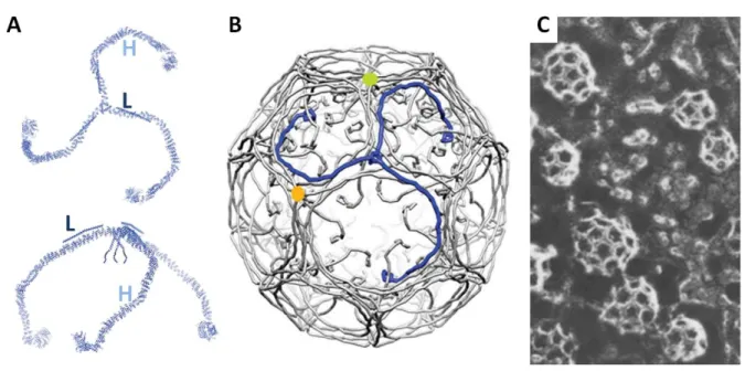
Figure 7: Clathrin triskelia and coat
A) The diagram depicts the clathrin triskelion viewed from different angles. H for heavy chain and L for light chain. B) Model of clathrin assembly that forms the clathrin coat. C) Basket-like structures that represent clathrin-coated vesicles observed in frog neuromuscular junction by freeze-fracture electron microscopy. A) and B) are adapted from (Xing et al., 2010), and C) from (Heuser, 1980).
25
CME at synapses has been observed at various synapses over the last four decades and the significance of this mode of SV endocytosis has been evidenced through studies that interfere with the components of CME. For example, clathrin knockdown or dominant-negative inhibition of AP180 leads to a near-complete blockage of endocytosis at hippocampal synapses suggesting that CME accounts for the majority of membrane reuptake (Granseth et al., 2006). In addition, knockdown of AP2 (Kim and Ryan, 2009) or knockout of auxilin (Yim et al., 2010) resulted in significant impairment of CME at hippocampal synapses. Other studies in support of the presence and significance of CME at synapses are well described in a number of reviews (Saheki and De Camilli, 2012; L.-G. Wu et
al., 2014; Milosevic, 2018).
The recent controversies that surround CME at hippocampal synapses should also be discussed, as these observations seem to refute what has been well-accepted for the last four decades. It was discerned that most studies that put CME forward as an important mode of SV endocytosis were performed at room temperature (Watanabe et al., 2014; Soykan et al., 2017). Watanabe et al., 2014, found that at physiological temperature, actin-dependent CIE is the dominant mode of endocytosis, taking up bulks of membrane from which clathrin and its adaptor proteins regenerate SVs (Watanabe et al., 2014). Reducing the temperature significantly impairs actin polymerisation, and thus blocks the actin-dependent ultrafast endocytosis (a form of CIE; see ‘CIE: ultrafast endocytosis’ later) at synapses. There was an increase in the number of clathrin-coated pits on the presynaptic membrane, indicating a shift from ultrafast endocytosis to CME when the experiment is performed at room temperature. This suggested that CME is only dominant at room temperature, and that CIE is the dominant mode of endocytosis at physiological temperature. The dispensable nature of CME at synapses was also suggested in a study by Kononenko et al., 2014, which shows that reducing the levels of clathrin or AP2 has very little impact on SV endocytosis during mild frequency stimulation and has no impact during high frequency stimulation (Kononenko et al., 2014). This suggests that SV endocytosis occurs only partially as CME and dominantly as CIE at a range of stimulation frequencies. The study also indicates that the CIE involved retrieves membrane through a dynamin and endophilin-dependent mechanism to form endosomes-like structures, from which a clathrin-dependent mechanism regenerates SVs. More recently Soykan et al., 2017, demonstrated that ultrafast endocytosis is likely to
26
be the dominant mode of endocytosis in response to a few initial action potentials, however, longer stimulations will favour a slower mode of endocytosis that is generally independent of clathrin and AP2 (Soykan et al., 2017). The study goes on to explain that the CIE observed is heavily dependent on actin dynamics through formins, regulators of linear actin filaments. Such studies indicate a model in which CIE is the dominant mode of endocytosis under all stimulation conditions, forming endosomes-like structures, from which, clathrin-dependent reformation of SVs occur. It should be mentioned that the two studies discussed were performed in hippocampal neurons which has relevance to this thesis work.
27 Figure 8: Clathrin-mediated endocytosis
The steps involved in clathrin-mediated endocytosis are given at the top and the sequential recruitment of the molecular actors involved is given at the bottom of the diagram. Clathrin-mediated endocytosis begins with ligand-receptor interaction (cargo of mediated endocytosis which recruits FCHo (nucleator of clathrin-mediated endocytosis; Henne et al., 2010). FCHo in turn recruits the structural proteins intersectin and Eps15. The recruitment of clathrin adaptors such as AP2 then follows. AP2 acts as a hub of interaction between the various molecular actors of clathrin-mediated endocytosis. Adapted from (Saheki and De Camilli, 2012).
28
1.3.4.2. Clathrin-Independent Endocytosis
A few forms of CIE occur in synapses which include the activity-dependent bulk endocytosis (ADBE) and ultrafast endocytosis (Milosevic, 2018). They differ in the size of membrane taken up, the rate at which they occur and the molecular actors that mediate them.
1.3.4.2.1. CIE: Ultrafast Endocytosis
This mode of endocytosis had only recently been described, and thus our knowledge is limited. Two recent studies used optogenetics in conjunction with ‘flash-and-freeze’ electron microscopy to demonstrate the existence of ultrafast endocytosis in two different model systems (mouse hippocampal synapses and Caenorhabditis elegans neuromuscular junctions) (Watanabe et al., 2013b; Watanabe et al., 2013a). This method involves light stimulation to induce a single or a pair of action potentials that cause neurotransmitter release. Then by the use of high-pressure freezer, cells are frozen at various defined time points before being observed under an electron microscope. These studies report that one action potential is enough to induce the fusion of one or two SVs around 30ms after stimulation. Such fusion events were quickly followed by compensatory endocytosis which occurred 50 to 100ms after stimulation, at sites immediately adjacent to active zones. Invaginations arising from such events generally lacked the classic coat, indicating that they are not clathrin-dependent, and the surface areas of these invaginations were estimated to be equivalent to four SVs. The studies also show that calcium influx alone is not enough to trigger ultrafast endocytosis, and that ultrafast endocytosis does not occur without exocytosis. Furthermore, it was suggested that actin dynamics is necessary to initialise membrane deformation, and dynamin function necessary for vesicle fission. SV regeneration then occurs from endosomes in a clathrin-dependent manner (Watanabe et al., 2014). Finally the fact that ultrafast endocytosis occurs at a distinct site (within around 200nm of fusion sites) suggests a special membrane organisation in this region. More recently, it was demonstrated that endophilin-A and synaptojanin-1 are involved in ultrafast endocytosis by using synaptojanin-1 knockout or endophilin-A1/2 double knockout mice (Watanabe et al., 2018). The study showed that the 5-phosphatase of synaptojanin-1 is required for the neck
29
formation in ultrafast endocytosis. Finally the fact that ultrafast endocytosis occurs at a distinct site (within around 200nm of fusion sites) suggests a special membrane organisation in this region (Watanabe et al., 2013b; Watanabe et al., 2013a).
1.3.4.2.2. CIE: Activity-Dependent Bulk Endocytosis
ADBE takes over as the main form of endocytosis when level of synaptic activity increases, when an exhaustive amount of fusion outbalances the rate of CME (Clayton and Cousin, 2009b; Saheki and De Camilli, 2012). First identified in frog neuromuscular junctions (Miller and Heuser, 1984), it is characterised by a large uptake of the presynaptic membrane to form endosomes-like structures called bulk endosomes in response to intense stimulation. The molecular aspect of ADBE is still under intense scrutiny, but recent works have shed light on some key molecules that may be involved in ADBE. Hypothetical molecular underpinnings of ADBE are depicted in figure 9.
A study by Wu et al., 2009, suggests that calmodulin activation is necessary for ADBE (Wu et al., 2009). Calcineurin is a calcium and calmodulin-binding phosphatase that has been implicated in ADBE. A study using an antagonist of calcineurin (cyclosporin A) suggested that calcineurin is crucial in allowing SV regeneration through ADBE (Cheung and Cousin, 2013). Calcineurin, when it binds calcium, dephosphorylates dynamin 1 to allow the interaction between dynamin 1 and syndapin 1, and this interaction is reported to be important in ADBE (Clayton et al., 2009b). The role of dynamin 1 in ADBE, however, was challenged by a dynamin 1/3 double knockout study that indicated that ADBE proceeds unaffected (Y. Wu et
al., 2014). This study, nonetheless, provides a possible explanation for such a discrepancy by
suggesting that dynamin 2, which is still present in very low levels, may replace the role of dynamin 1. More evidence in support of the role of dynamin 1 in ADBE exists. In fact, an earlier evidence for the involvement of dynamin 1 in ADBE was provided by a study on
Drosophila shibire mutants (Van Der Bliek and Meyerowrtz, 1991). The multiple forms of
dynamin encoded by the shibire gene in these mutants are rendered temperature-sensitive causing them to be inactive at low temperatures (Chen et al., 1991; Van Der Bliek and Meyerowrtz, 1991). At low non-permissive temperatures, it was reported that tubular
30
invaginations remained attached to the plasma membrane, suggesting that the activity of dynamin is required for the scission of these invaginations to form endosomes-like structures (Van Der Bliek and Meyerowrtz, 1991). Moreover, it was shown that rephosphorylation of dynamin 1 is crucial to maintain ADBE in rodent neurons, as preventing rephosphorylation of dynamin 1 by cyclin-dependent kinase 5 (CDK5) and glycogen synthase kinase 3 (GSK3) blocked the continuation of ADBE (Evans and Cousin, 2007; Clayton et al., 2010). Finally, it was demonstrated that disrupting the interaction between dynamin 1 and syndapin 1 leads to the blockage of ADBE (Clayton et al., 2009b). Taken together, it appears that dynamin is a key component of ADBE although the presence of the different isoforms may allow for functional redundancy.
The role of syndapin 1 in ADBE is unclear, but its molecular interactions provide hints. Syndapin 1 has binding sites for both N-WASP and dynamin (Qualmann et al., 1999). N-WASP has a proline-rich domain (PRD), allowing its interaction with Src homology 3 (SH3) domains of various proteins, and it is able to interact also with actin and actin-related protein 2/3 (Arp2/3) complex via another region (Takenawa and Miki, 2001). In migrating cells, the recruitment of Arp2/3 allows for fast changes in the actin cytoskeleton, allowing the formation of filopodia. Such studies led to the hypothesis that syndapin could be the bridge between the actin cytoskeleton and dynamin in ADBE (Clayton and Cousin, 2009b).
The molecular underpinnings of ADBE was initially inferred through studies on a similar process such as macropinocytosis (Royle and Lagnado, 2003). Suppressing the dynamics of actin and the activity of phosphatidylinositol-3-kinase (PI3K) led to the impairment of macropinocytosis in non-neuronal cells, indicating the involvement of actin dynamics in this process (Nichols and Lippincott-Schwartz, 2001; Royle and Lagnado, 2003). It was then postulated that a similar mechanism may underlie ADBE in synapses. Evidence in support of this were then provided using frog neuromuscular junctions (Richards, Rizzoli and Betz, 2004) and retinal bipolar cells (Holt et al., 2003), demonstrating that actin dynamics and PI3K activity are crucial for ADBE.
The possible connection between ADBE and PI3K implies that membrane lipids and lipid metabolism play a role in ADBE. Phosphatidylinositol phosphate kinase type Iγ (PIPKIγ), an enzyme that synthesises PI(4,5)P2, has been shown to be dephosphorylated by calcineurin under stimulation conditions that would induce ADBE (Sang et al., 2005). The study
31
demonstrates that the dephosphorylated form of PIPKIγ interacts with talin, which leads to the activation of PIPKIγ and to increase in the level of PI(4,5)P2 at synapses. This could mean a possible role of PI(4,5)P2 in ADBE at synapses. Further speculations regarding the role of membrane lipids can be found in a review on ADBE by Clayton and Cousin, 2009 (Clayton and Cousin, 2009b). All in all, much of the molecular mechanisms underlying ADBE has been inferred from other studies and thus requires further investigation that will provide deeper insight.
It is already known that certain cargoes are taken up via CIE in non-neuronal cells. For example, β-integrins, glycosylphosphatidylinositol (GPI)-anchored proteins, interleukin-2 receptor alpha chain and major histocompatibility complex class I (MHCI) are all ingested via the ARF6-mediated endocytosis (Maldonado-Báez, Williamson and Donaldson, 2013). It seems quite plausible to postulate that a similar phenomenon may be true for CIE at synpases. A recent study on mouse cerebellar and hippocampal neurons showed that VAMP4 is a specific cargo of the ADBE (Nicholson-Fish et al., 2015). In fact, the knockdown of VAMP4 leads to the blockage of ADBE suggesting that VAMP4 is not simply a cargo, but is a crucial element of the process. Interestingly, inhibiting ADBE does not affect the uptake of various SV proteins including synaptophysin and synaptotagmin-1. Clathrin heavy chain knockdown and pharmacological inhibition of CME led to reduced uptake of these SV proteins at high frequency stimulations suggesting that these SV proteins are taken up via CME. In contrast, an earlier study suggested that synaptotagmin-1 uptake during similar stimulation condition was unaffected by the impairment of CME using shRNA against clathrin heavy chains or pharmacological inhibition (Kononenko et al., 2014). With the current understanding of SV recycling, it is difficult to explain the contradiction. A recent proteomic study revealed the ADBE core proteome which included cell adhesion and cytoskeletal molecules as well as trafficking proteins (Kokotos et al., 2018). The study looks particularly into the Rab proteins that were purified with bulk endosomes and discover that the overexpression of Rab11 increases the number of synapses capable of ADBE and increases the rate of CME. Adaptor proteins AP1 and AP3, which were previously shown to be necessary for the regeneration of SVs from bulk endosomes (Cheung and Cousin, 2012), were also among the proteins purified with bulk endosomes (Kokotos et al., 2018).