Publisher’s version / Version de l'éditeur:
European Cells and Materials, 26, SUPPL 6, 2013
READ THESE TERMS AND CONDITIONS CAREFULLY BEFORE USING THIS WEBSITE. https://nrc-publications.canada.ca/eng/copyright
Vous avez des questions? Nous pouvons vous aider. Pour communiquer directement avec un auteur, consultez la première page de la revue dans laquelle son article a été publié afin de trouver ses coordonnées. Si vous n’arrivez pas à les repérer, communiquez avec nous à [email protected].
Questions? Contact the NRC Publications Archive team at
[email protected]. If you wish to email the authors directly, please see the first page of the publication for their contact information.
NRC Publications Archive
Archives des publications du CNRC
This publication could be one of several versions: author’s original, accepted manuscript or the publisher’s version. / La version de cette publication peut être l’une des suivantes : la version prépublication de l’auteur, la version acceptée du manuscrit ou la version de l’éditeur.
Access and use of this website and the material on it are subject to the Terms and Conditions set forth at
Microfluidic lab on-a-chip devices for high throughput rare cell sorting
Fatanat-Didar, T.; Li, K.; Veres, T.; Tabrizian, M.
https://publications-cnrc.canada.ca/fra/droits
L’accès à ce site Web et l’utilisation de son contenu sont assujettis aux conditions présentées dans le site
LISEZ CES CONDITIONS ATTENTIVEMENT AVANT D’UTILISER CE SITE WEB.
NRC Publications Record / Notice d'Archives des publications de CNRC:
https://nrc-publications.canada.ca/eng/view/object/?id=9cb1033f-92ad-45f5-99aa-e41f37ba3d54 https://publications-cnrc.canada.ca/fra/voir/objet/?id=9cb1033f-92ad-45f5-99aa-e41f37ba3d54
European Cells and Materials Vol. 26. Suppl. 6, 2013 (page 139) ISSN 1473-2262
http://www.ecmjournal.org
Microfluidic Lab on-a-Chip devices for high throughput rare cell sorting
T Fatanat-Didar
1, K Li
3, T Veres
3, M Tabrizian
1,21
Biomedical Engineering Dept. and
2Faculty of Dentistry, McGill University, Montreal, Canada
2Heath Science Division, National Research Council of Canada, Boucherville, Canada
INTRODUCTION: Biomedical devicesdeveloped for detection, sorting and in vitro culture of cells are important tools in both clinical diagnostics and fundamental research. Recently, with the advances in miniaturization, Lab-on-Chip (LOC) devices have started to play an important role in detection and enrichment of rare cells1. Combined with label-free methods that do not alter the properties of target cell, LOCs provide a suitable option for cell separation. We developed two main label-free approaches exploring the difference in size or adhesion properties of cell to sort them. The first device was a multilayered, fully thermoplastic-based microfluidic chip that was designed and fabricated for high throughput size-based separation of rare cells2. The second microchip was employing a well–designed biointerface that could separate the cells based on their affinity towards the microfabricated surface3.
METHODS: The first microfluidic device was composed of a bottom layer, commercially polycarbonate porous membrane layer, top layer and a pneumatic air control layer, fabricated on thermoplastic elastomers, all by hot embossing. Top layer had three access holes that could be used as inlets or outlets depending on the peristaltic micro-pumping configuration. These layers were connected through the membrane in a circular channel area where liquid exchange could occur. The microfluidic device was then connected to a homemade 12-channels pneumatic control manifold using silastic laboratory tubing.
For the second platform microcontact printing was used to print aminopropyltriethoxysilane (APTES) onto the glass surface to provide region specific patterns of amine groups which were later activated through EDC-NHS chemistry. Microfluidic patterning was then used to create highly resistant protein patterns. This achieved by forming the PDMS-Glass microfluidic platform and flowing biomolecules of choice into the device to covalently attach them to the patterned amine groups on the surface.
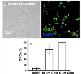
RESULTS: Separation of rare oligodendrocyte progenitor cells (OPCs) from rat brain primary cultures was used as the proof on concept. Using multilayered microfluidic chip with a 5 µm pores membrane and micropumping system, a separation
efficiency of 99% was achieved. This microchip was able to operate at flow rates up to 100 µl/min, capable of separating OPCs from a confluent 75 cm2 cell culture flask in less than 10 min.
Fig. 1. (a) Image of initial cell mixture (b) OPCs population after separation using 5 μm pore size (c) % of OPCs in the initial cell mixture and after
separation using chips with 10 μm and 5 μm
membrane pore sizes.
Similar separation efficiency of OPCs, were obtained with the second platform. In addition, target cell attachment and single cell spreading could be precisely controlled. Isolated OPCs could proliferate and differentiate into mature oligodendrocytes on the separation platform.
DISCUSSION & CONCLUSIONS: The traditional macroscale techniques for OPCs separation require pre-processing of cells and/or multiple time consuming steps with low efficiency leading very often to alteration of their properties. The proposed label-free methodologies could separate OPCs, either based on their smaller size compared to other glial cells based on their affinity towards the developed biointerface, with a separation efficiency greater than 95%.
REFERENCES: 1K. Sun Min et al. (2008). Lab
on a Chip 8:1015-23. 2T. Fatanat Didar et al. (2013) Biomaterials 34(22):5588-93. 3 T. Fatanat Didar et al. (2011) Anal Chem 84(2):1012-8.
ACKNOWLEDGEMENTS: Genome Quebec, Nano Quebec, NSERC, Fac. Medicine, Drs G. Almazan, D. Junker, M. Mekhail and M. Dipaula.