BORDEAUX VIVA WINTER SCHOOL - XXXIII LIAC MEETING
29 November to 1 December | 29 Novembro a 1 Dezembro
DOI: 10.19277/bbr.14.2.169
Biomédica e BiofarmacêuticaProgram
29 Novembrer | 29 de Novembro
Reception of participant Winter School Meeting
Winter School meeting (organizers: A. Bikfalvi & J. Badaut) Presentation of students - What should be achieved in this winter school
Opening meeting : Thierry Couffinhal (VIVA action, Director) & Michel Spina (LIAC President)
CLINICAL AND EPIDEMIOLOGIC ASPECT OF VASCULAR AGING (chairmen: C. Tzourio & L. Monteiro)
Early vascular aging - P.M. NILSSON, Malmö - Sweden
Neurovascular epidemiology of aging - C. TZOURIO, Bordeaux
Cardiovascular epidemiology of aging - P. BOUTOUYRIE, Paris
Forecasted trends in disability and life expectancy - S. AHMADI-ABHARI, Liverpool - UK
Neurovascular genetic epidemiology - S. DEBETTE, Bordeaux
Vascular and thrombosis genetic epidemiology - D. TREGOUET, Paris
SELECTED ORAL PRESENTATION (Chairmen: S. Debette & J. Badaut)
• Mitochondrial function regulates vascular aging in mice - K. FOOTE, Cambridge - UK
• Structural imaging of the vascular wall - S. ALMAGRO, Reims
• Numerical assessment and comparison of pulse wave velocity methods aiming at measuring aortic stiffness - H. OBEID, Paris
• Long-term trajectories of cardiometabolic risk factors in prodromal dementia: the Three-City Study - M. WAGNER, Bordeaux
EVENING PHILOSOPHICAL CONFERENCE ANTONIO-MARIO TAMBURRO (chairman: M. Spina)
When does the vascular system age and when is there a disease? Conceptual and theoretical issues - M. LEMOINE, Tours
30 Novembrer | 30 de Novembro
PATHOPHYSIOLOGIC FEATURES OF VASCULAR AGING (Chairmen: J.F. Arnal & M. Formato)
From physiological aging to pathological aging - J.B. MICHEL, Paris
Physiological models to study the human microcirculation - effects of hyperoxia - L. MONTEIRO-RODRIGUES, Lisbon - Portugal
Murine models for investigating vascular ageing - G. FAURY, Grenoble
New insights into vascular biology by single cell RNA sequencing - M. VANLANDEWYCK, Stockholm - Sweden
Thrombosis in aging - C. JAMES, Bordeaux
Retinal vascular imaging - C. HELMER, Bordeaux
Lunch of Winter School Meeting
Winter School meeting (organizers: A. Bikfalvi & J. Badaut) Meet the experts - Round table with experts
MATRIX STRUCTURAL AND MECHANICAL STUDIES (Chairmen: I. Brunet & P. Dufourcq)
Model of pseudoxanthoma elasticum – Vascular calcification and aging, from gene to bedside - L. MARTIN, G. LEFTHERIOTIS, Angers
Vascular aging: structure and dynamics of constitutive macromolecules and sub-fragments - V. SAMOUILLAN, Toulouse
Tubular scaffolds for applications of vascular tissue engineering - V. LA CARRUBBA, Palermo - Italy
Bioprosthetic heart valve aging deterioration and age-related benefits - M. SPINA, Padova - Italy
Elastin modification during vascular aging and pathophysiological consequences - L. DUCA, Reims
SELECTED ORAL PRESENTATION (Chairmen: M. Laffargue & D. Henrion)
• MicroRNA expressed in progenitor cells circulating in peripheral blood as prospective biomarkers of agerelated macular degeneration (AMD) in its dry
and wet form – preliminary data - M. KAWA, Poland
• Molecular, conformational and thermal characterization of ventricular Remodeling in a pig model of tachycardia-induced cardiomyopathy - V.
LLORENTE CORTES, Barcelona - Spain
Program (cont.)
• Vascular calcification during chronic kidney disease: role of the RAGE/Cathepsin S/elastin peptides axis - A. WAHART, Reims
VIVA EVENING CONFERENCE (Chairman: T. Couffinhal)
A short history of the vessel - A. BIKFALVI, Bordeaux
1 December | 1 de Dezembro
CELL BIOLOGY AND SIGNALING (Chairwomen: C. Duplàa & V. Llorrente Cortes)
SELECTED ORAL PRESENTATION
• Targeting Connexin40 expression or function reduces angiogenesis in the developing mouse retina - F. ALONSO, Bordeaux
• Imatinib inhibits hypoxia-induced MMP-9 overexpression and activation through impairment of LRP1 and Pyk2 phosphorylation in human coronary
vascular smooth muscle cells - A. BENITEZ, Barcelona - Spain
• NRP1 supresses TGFβ-dependent ERK1/2 activation in endothelial cells - A. LAMPROPOULOU, London - UK
• The elastin receptor complex interacts with CD36 through NEU1 and regulates oxidized LDL uptake in macrophages: potential implication in the
proatherogenic effects of elastin-derived peptides - P. MAURICE, Reims
• Ubiad1 promotes tumor angiogenesis by inhibiting dephosphorylation of vegfr2 in a redox dependent manner - R. SEWDUTH, Belgium
Cell senescence and telomere - E. GILSON, Nice
Estrogens as vascular anti-ageing hormones? Experimental and clinical evidences and uncertainties - J.F. ARNAL, Toulouse
Arteriolar and microcirculation aging - D. HENRION, Angers
Oxydation and inflammation in Atherosclerosis - A. LEPEDDA, Italy
Winter School Meeting (organizers: A. Bikfalvi & J. Badaut) Highlights and major topics identified by the students, group proposals (3 per group)
for fictive/future projects and presentation
OPEN SESSION (Chairwomen: M.A. Renault & J. Lods)
• Endothelial cell dysfunction is a new potential therapeutic target for the treatment of critical limb ischemia - C. CARADU, Pessac
• Podosomes Mediate Basement Membrane Collagen-IV Proteolysis During Sprouting Angiogenesis - E. GENOT, Bordeaux
•How plant food bioactives may have benefits for vascular health? The case of curcumin, L.E. MONFOULET, Lille
Table des matières
CLINICAL AND EPIDEMIOLOGIC ASPECT OF VASCULAR AGING
Early vascular aging
Neurovascular epidemiology of aging
Cardiovascular epidemiology of aging
Forecasted trends in disability and life expectancy
Neurovascular genetic epidemiology
Vascular and thrombosis genetic epidemiology
EVENING PHILOSOPHICAL CONFERENCE ANTONIO-MARIO TAMBURRO
When does the vascular system age and when is there a disease? Conceptual and theoretical issues
PATHOPHYSIOLOGIC FEATURES OF VASCULAR AGING
From physiological aging to pathological aging
Physiological models to study the human microcirculation – effects of hypeoxia
Murine models for investigating vascular ageing
New insights into vascular biology by single cell RNA sequencing
Thrombosis in aging
Retinal vascular imaging
MATRIX STRUCTURAL AND MECHANICAL STUDIES
Model of pseudoxanthoma elsticum – Vascular calcification and aging, from gene to bedside
Vascular aging: structure and dynamics of constitutive macromolecules and sub-fragments
Tubular scaffolds for applications of vascular tissue engineering
Bioprosthetic heart aging détérioration and age-related benefits
Elastin modification during vascular aging and pathophysiological consequences
VIVA EVENING CONFERENCE
A short history of the vessel
CELL BIOLOGY AND SIGNALING
Cell senescence and telomere
Estrogens as vascular anti-ageing hormones? Expérimental and clinical evidences and uncertainties
Arteriolar and microcirculation agin
Oxidation and inflammation in atheriosclerosis
SELECTED ORAL PRESENTATION
Mitochondrial function regulates vascular aging in mice
Structural imaging of the vascular wall
Numerical assessment and comparison of pulse wave velocity methods aiming at measuring aortic stiffness
Long-term trajectorie of cardiometabolic risk factors in prodromal dementia: the Three-City
MicroRNA expressed in progenitor cells circulating in peripheral blood as prospective biomarkers of age related degeneration (AMD) in its dry and wet form – preliminary data
Molecular, conformational and thermal characterization pf ventricular remodeling in a pig model of tachycardia-induced cardiomyopathy
Valvular calcification: implication if the Semicarbazide-Sensitive Amine Oxidaxe (SSAO)?
Vascular calcification during chronic kidney disease: role of the RAGE/Capthepsin S/elastin peptides axis
Targeting Connexin40 expression or function reduces angiogenesis in the developing mouse retina
Imatinib inhibits hypoxia-induced MMP- overexpression and activation through impairment of LRP1 and Pyk2 phosphorylation in human cprpnary vascular smooth musle cells
NRP1 supresses TGFB-dependent ERK1/2 activation in endothelial cells
The elastin receptor complex interacts with CD36 through NEU1 and regulates oxidized LDL uptake in macrophages : potential implication in the proatherogenic effects of
elastin-derived peptides
Ubiad1 promotes tumor angiogenesis by inhibiting dephosphorylation of vegfr2 in a redox dependent manner
OPEN SESSION
Endothelial cell dysfunction is a new potential therapeutic target for the treatment of critical limb ischemia
Podosomes mediate basement membrane collagen-IV proteolysis during sprouting angiogenesis
How plant foot bioactives may have benefits for vascular health? The case of curcumin
Early Vascular Ageing (EVA) in translation: from cell senescence to
clinical implications
Peter M Nilsson, MD, PhD
* 11 Professor of Clinical Cardiovascular Research, Lund University, Department of Clinical Sciences, Skane University Hospital, S-205 02 Malmo, Sweden
* Intervenant
Already Thomas Sydenham (1624-1689) told that “A man is as old as his arteries”.
The ageing process of the vasculature has increasingly been studied over the last 25 years. New data on the stiffening of arteries (arteriosclerosis) has been added to long-term studies on atherosclerosis. It is believed that arteriosclerosis starts early in life, based on fetal programming of the elastin content of the arterial media, as well as of the vasculature and capillaries. This will impact on the morphological changes of large elastic arteries and central hemodynamic regulation. Later on the process of atherosclerosis, as being more proximal to clinical cardiovascular events, will play an important role while the two processes will be more and more mixed and jointly increase the risk. In the arterial wall the increasing age-dependent stiffness is characterized by a relative decrease of the elastin content of the arterial media and a relative increase of the collagen content as well as more pronounced collagen linkages. In addition there is a mechanism called mechanotransduction that will impact on the structure of the extra-cellular matrix in relation to increased tension of the arterial wall, i.e. following increased blood pressure. This will also influence vascular smooth muscle cells (VSMC) to undergo changes as the VSMC contractile phenotype is associated with an increased arterial stiffness. This can take place both in the arterial media and the adventitia [1]. In addition, some specific processes related to the adventitia may further increase stiffness. When the vascularization of the arterial wall via vasa vasorum is impaired, as following the administration of anti-angiogenic drugs for cancer therapy, this has been shown to increase arterial stiffness.
Furthermore, impaired autonomous nervous function is related to both increased arterial stiffness and impaired glucose metabolism. Finally, the existence of perivascular adipose tissue (PVAT) is a source of local perivascular inflammation via secretion of vasoactive cytokines. This will also influence arterial stiffness by reducing endothelial function to vasodilate.
Based on epidemiological evidence from the Malmo Diet Cancer Cohort it has been shown that increased carotid-femoral pulse wave velocity (c-f PWV) is independently associated with markers of impaired glucose metabolism [2], even adjusted for hemodynamic factors and risk factors for atherosclerosis (LDL cholesterol, smoking, renal function). This shows the close link between arterial stiffness, impaired glucose metabolism and overt type 2 diabetes – a different risk factor cluster than the more traditional cluster associated with atherosclerosis (hypertension, smoking, hyperlipidaemia).
We have also found that c-f PWV is associated with a family history of cardiometabolic disease [3], impaired cognition [4], and is predictive of type 2 diabetes [5] and all-cause mortality, even adjusted for known risk factors/markers and family history of cardiometabolic disease (abstract, ARTERY 2017, Pisa, Italy).
New drugs are being developed for vascular protection and the reduction of arterial stiffness. One promising experimental drug, currently in human Phase-1 studies, is the selective Angiotensin-II (AT2) agonist compound 21 (C21). In one animal study it was shown that C21 could reduce PWV in L-NAME induced hypertension without interfering with blood pressure [6].
In summary, early vascular ageing (EVA) has inspired research to better understand the morphological changes of the arterial wall (especially the media and adventitia) and its age-related hemodynamic changes [7]. Increased c-f PWV is a predictor not only of cardiovascular events, independent of traditional risk factors, but also of type 2 diabetes and total mortality. This shows that arterial stiffness is a marker of biological ageing in general and should be the focus for prevention and testing of new drugs for vascular protection such as C21, besides the efforts to reduce the burden of atherosclerosis, plaque formation and risk of cardiovascular events.
References
1. Lacolley P, Regnault V, Segers P, Laurent S. Vascular Smooth Muscle Cells and Arterial Stiffening: Relevance in Development, Aging, and Disease. Physiol Rev. 2017;97:1555-1617.
2. Gottsäter M, Östling G, Persson M, Engström G, Melander O, Nilsson PM. Non-hemodynamic predictors of arterial stiffness after 17 years of follow-up: the Malmö Diet and Cancer study. J Hypertens. 2015;33:957-65. 3. Fatehali AA, Gottsäter M, Nilsson PM. Family history of cardiometabolic diseases and its association with arterial stiffness in the Malmö Diet Cancer cohort. J Hypertens. 2017;35:2262-2267.
4. Nilsson ED, Elmståhl S, Minthon L, Nilsson PM, Pihlsgård M, Tufvesson E, Nägga K. Nonlinear association between pulse wave velocity and cognitive function: a population-based study. J Hypertens. 2014;32:2152-7. 5. Muhammad IF, Borné Y, Östling G, Kennbäck C, Gottsäter M, Persson M, Nilsson PM, Engström G. Arterial Stiffness and Incidence of Diabetes: A Population-Based Cohort Study. Diabetes Care. 2017 Sep 29. pii: dc171071. 6. Paulis L, Becker ST, Lucht K, Schwengel K, Slavic S, Kaschina E, Thöne-Reineke C, Dahlöf B, Baulmann J, Unger T, Steckelings UM. Direct angiotensin II type 2 receptor stimulation in Nω-nitro- L-arginine-methyl ester-induced hypertension: the effect on pulse wave velocity and aortic remodeling. Hypertension. 2012;59:485-92. 7. Nilsson PM, Boutouyrie P, Cunha P, Kotsis V, Narkiewicz K, Parati G, Rietzschel E, Scuteri A, Laurent S. Early vascular ageing in translation: from laboratory investigations to clinical applications in cardiovascular prevention. J
Hypertens. 2013;31:1517-26.
Forecasted trends for dementia, disability, and lifeexpectancy in England and Wales to 2040: a probabilistic Markov modelling study
Sara Ahmadi-Abhari*1Maria Guzman Castillo2, Piotr Bandosz2,3, Martin J Shipley1, Archana
Singh-Manoux1,4, Mika Kivimäki1,5, Simon Capewell2, Martin O’Flaherty2, Eric J Brunner1
1 Department Epidemiology & Public Health, University College London, United Kingdom 2 Department of Public Health and Policy, University of Liverpool, United Kingdom 3 Department of Prevention and Medical Education, Medical University of Gdansk, Poland 4 INSERM, U1018, Centre for Research in Epidemiology & Public Health, Hôpital Paul Brousse, France
5 Clinicum, Faculty of Medicine, University of Helsinki, Finland
* Intervenant
Background: As life-expectancy rises, dementia and disability are expected to impose increasing societal and healthcare burden. Forecasts for
prevalence of dementia and disability, and life-expectancy are vital in defining future needs and can be improved with a dynamic modelling approach that integrates calendartrends in incidence of dementia and disability with those for mortality and cardiovascular disease.
Methods: A probabilistic Markov-model labelled IMPACT-BAM (Figure 1) was developed and validated to track the progression of the
population of England and Wales aged 35+ through states of cardiovascular disease, cognitive impairment, dementia, and disability through to death and forecast burden of disease and life-expectancy for the future. Data from the English Longitudinal Study of Ageing (ELSA), a representative panel study with 6 waves of data across 2002-2013, was used to inform the baseline prevalence estimates and transition probabilities in the model. Calendar trends in incidence of cardiovascular disease, dementia, and mortality rates were obtained from ELSA and data from the UK Office for National Statistics and projected to the future. Disability was defined as inability to independently perform one or more basic activities of daily living.
Results: Average Life-expectancy is estimated at 21.8 year in 2017 and projected to increase to 26.4 years by 2040 (Figure 2). The proportion of
disabled to disability-free life expectancy declined over time suggesting compression of morbidity. Using IMPACT-BAM we estimated approximately 792000 (95% Uncertainty Interval 760000–823000) people are living with dementia and 3,590,000 (95% UI 3540000- 3669000) with disability in England and Wales in 2017. Despite the decline in incidence of dementia and cardiovascular disease, the numbers of people living with dementia and disability are projected to increase to 872000, 1092000, and 1205000 for dementia and 3771000, 4383000, and 4902000 for disability in 2020, 2030, and 2040 respectively. Population ageing is a main contributor as the age-standardized prevalence is declining.
Conclusion: Despite the decline in age-specific incidence of dementia and cardiovascular disease, the number of people living with dementia
and disability in England and Wales is likely to increase by 52% for dementia and 37% for disability from 2017 to 2040. This increase is mainly driven by increasing lifeexpectancy.
Our projections have important implications for planning to meet future health and social care needs.
Figure 1: IMPACT-Better Ageing Model
(IMPACT-BAM)
Figure 2: Average expectancy and disabled
life-expectancy at age 65 in England and Wales estimated from IMPACT-BAM
Figure 3: Projected number of people living with dementia
and disability in England and Wales using IMPACT-BAM, 2015-2040. Dashed lines represent 95% uncertainty intervals.
Cardiovascular epidemiology of aging
Pierre Boutouyrie
* 11
Pharmacologie, Cardiologie - Hôpital Européen Georges Pompidou, Inserm U970, Equipe 7, Paris
*
Intervenant
This presentation will concentrate on the arterial consequences of arterial aging, especially
arterial stiffness, arterial remodeling and wave reflections. We will present the
pathophysiological aspects, the different techniques available and the principal results of
epidemiological surveys. We will then briefly present some directions for treating early vascular
aging.
Neurovascular genetic epidemiology
Stéphanie Debette *1
1 Inserm U1219, Bordeaux
* Intervenant
With increasing longevity, cerebrovascular disease has become the most common age-related source of disability and dependence, as well as a
major determinant of dementia, representing a huge societal burden. Compared to cardiovascular disease, unraveling the genomic underpinnings
of cerebrovascular disease has proven more complex due to its much more heterogeneous nature. Recent multiethnic collaborative efforts have
enabled important progress in revealing genetic determinants of stroke and MRI-defined covert vascular brain injury. These findings are
primarily based on common variants but preliminary results on rare and low-frequency variants are beginning to emerge. Both the “lumping
approach” combining all types of strokes with large numbers and strategies focusing on highly specific subtypes of cerebrovascular disease has
proven efficient, with complementary findings. Analysis of shared genetic variation with other vascular phenotypes has also provided interesting
mechanistic insight. Experimentally follow-up of some of these findings is generating novel hypotheses on the biological pathways underlying
stroke.
Vascular and thrombosis genetic epidemiology
David-Alexandre TREGOUET *1
1Inserm UMR_S 1166, Paris
*
Intervenant
During the last 30 years, the evolution of the genetic epidemiology discipline paralleled the development of high-throughput technologies. In the
80's, familial linkage analysis and candidate gene association approaches were the method of choice for identifying genetic markers associated to
human complex diseases. In the last 10 years, high density arrays, followed now by next generation sequencing techniques, allowed the
screening for (epi-)genetic and transcriptomic marks that influence disease susceptibility. Henceforth, it is time to interrogate other kind of
molecular phenotypes, incl metabolites, proteins, through new high-throughput techniques , and to integrate their analysis with
that of (epi-)genetic data in order to disentangle in a more comprehensive way the complex architecture underlying common human diseases.
This opens the era of the genomic epidemiology discipline or what could be referred to as epidemiolomics.
When does the vascular system age and when is there a
disease? Conceptual and theoretical issues
Maël LEMOINE
*11
University of Tours - Philosophy of biological and medical sciences
*
Intervenant
This talk takes a very general perspective in order to question the distinction between aging and disease, in the specific case of the endothelial
dysfunction. To what extent is it possible to hold the claim that the difference does not matter insofar as we treat everything we can all the same,
whether it is aging or a disease? I will show that it is difficult, but possible to hold it in the face of commonly held but unscientific claims such as
“atherosclerosis is aging rather than a disease because it eventually strikes everyone”. Then, I will examine the hypothesis that a difference can
be made locally between what resorts to aging of the arteries and what resorts to a disease of the arteries, and whether it can
matter. Eventually, I will assess the interest of general biological theories of aging to justify the distinction. In the conclusion, I will try to gather
an answer to the question: does it matter to have a distinction between aging and disease of the arteries?
Physiological models to study the human microcirculation - effects of
hyperoxya
Luis Monteiro-Rodrigues
*1
1
Universidade Lusófona de Humanidades e Tecnologias - Lisbon, Lisboa, Portugal
* Intervenant
Models, animal or human, are crucial to look further into the mechanisms involved in a pathological or in a
regulatory process to study vascular function and disease, also considering that currently, Vascular Dysfunction
represents the 3rd major cause of cerebral-vascular disease. In the last years we explored various approaches to
measure microcirculation in vivo, as near as possible from the normal physiological state, and identified several
determinants. More recent physiological models are helping to improve the experimental conditions and contribute
to accelerate knowledge in these areas.
Hyperoxia is a well known strategy in medicine, also used as a stressor to text microcirculatory regulation
mechanisms. We have explored several models, human as animal using hyperoxia. More recently, focusing the
improvement of neovascularization process following ischemia, we combined hyperoxia and the hindlimb ischemia,
using C57/BL6 mice. In the present communication we intend to share some of our latest experience, and data, in
these areas in order to better understand microcirculation physiology.
EVENING PHILOSOPHICAL CONFERENCE ANTONIOMARIO TAMBURRO
New insights into vascular biology by single cell RNA
Sequencing
Michael VANLANDEWIJCK
*11
Department of Immunology, Genetics and Pathology, Vascular Biology; Research group Christer Betsholtz,
Uppsala University, Sweden
*
Intervenant
Cerebrovascular disease is the third most common cause of death in developed countries, yet our understanding of the cells that make the
cerebral vasculature is limited. Using large-scale analysis of vascular single cell transcriptomes, we provide molecular definitions to the
principal blood vascular and vessel-associated cell types in the adult mouse brain. We uncover the transcriptional basis for the gradual
phenotypic change (zonation) that occur along the arterio-venous (A-V) axis in the two major vascular cell types – endothelial and mural cells -
and reveal unexpected differences between them: a seamless endothelial continuum, as opposed to a punctuated mural continuum. The latter
reflects two distinct mural cell groups comprising arterial/arteriolar smooth muscle cells, and pericytes/venous smooth muscle cells, respectively.
Brain pericytes are highly homogenous, but a comparison between brain and lung reveals significant pericyte organotypicity. For example, brain
pericytes express a unique repertoire of molecular transporters, suggesting their active role at the blood-brain barrier. We also define a
population of perivascular fibroblast-like cells that form a previously unrecognized adventitial layer in the brain vasculature. These cells are
distinct from mural cells, occur on all vessel types except capillaries and provide distinct anatomical landmarks at the arteriolar-capillary and
capillary-venular borders.
Summary Thrombosis in Aging
Chloé James
*11
Inserm U1034, Biology of Cardiovascular Diseases, Pessac – Bordeaux
*
Intervenant
Age is a well-known risk factor for thrombosis, both in the arterial (acute myocardial infarction, stroke) and the venous (deep vein thrombosis,
pulmonary embolism) system. Because ageing in humans in most cases occurs in parallel to other thrombotic risk factors (such as hypertension,
obesity, cancer, diabetes), the “true” effects of ageing per se may not be easily dissected.
Recently, epidemiologic studies have suggested that aging “per se” is a thrombotic risk factor.
Based on the literature, we will expose how ageing can favour thrombosis, and especially focus on the role of the endothelium, platelets, and
coagulation.
Model of pseudoxanthoma elasticum
Ludovic Martin
*1
1
CHU Angers
*Intervenant
Pseudoxanthoma elasticum (PXE) is a prototype of heritable ectopic mineralization. This rare, monogenic (autosomal
recessive) condition presents with intriguing manifestations in the skin, retina and cardiovascular systems. Notably,
all these systems are rich in elastic tissue and PXE pathological changes are closely related to elastic fibers. The
causative gene of PXE is ABCC6, which encodes an ATP-binding transporter primarily expressed in the liver. PXE
looks a metabolic condition with central defect in the liver and remote mineralization in the above mentioned
systems. The reality might be more complicated... This talk will briefly overview the clinical presentation of PXE,
review the specific pathomechanisms operative in ectopic mineralization, including the central role of inorganic
pyrophosphate, and discuss whether or not PXE might be seen as an example of premature aging.
Murine models for investigating vascular ageing
Gilles Faury
*11
Laboratory HP2, Université Grenoble Alpes, France
* Intervenant
In human beings, investigating the normal or pathological ageing processes is a difficult task because of the long time course of all the
implicated mechanisms, in the range of decades. The access to the elderly is also uneasy because of the laws regulating investigations in patients.
In order to overcome these difficulties, numerous murine models have been designed or obtained, by genetic engineering or natural/classical
selection. Regarding the investigations of normal ageing, some models allow for easier longitudinal studies because of a selected shorter
lifespan, such as senescence-accelerated mice (SAM mice), whereas other models aim at the characterization of the involvement of specific
target/candidate mechanisms by gene modification of invalidation, such as Klotho-/- mice. Regarding pathological ageing, or age-related
pathologies, a series of models are used, mostly aiming at targeting specific actors (by invalidation, over-expression, ...), such as the mice
knock-out for the Apolipoprotein E (ApoE-/-, for studies on atherosclerosis) mice or Spontaneously Hypertensive Rats rats (SHR, for studies of the
mechanisms or consequences of hypertension). Some other murine models, not overtly related to the ageing, have also brought some insight into
the possible modulations of or alternatives to the normal ageing processes, such as the mice heterozygous for the elastin gene (Eln+/-). However,
most of these models present some undesirable side-effects which could lead to biases in the possibilities to generalize to the ageing processes
that are present in "normal" animals.
The presentation will review some of the main uses of and results from several murine models widely used in the studies of (cardio-) vascular
ageing.
Vascular calcification and aging, from gene to bedside
Georges Leftheriotis
*1
1
CHU Angers
*
Intervenant
Arterial calcification is a widely demonstrated independent risk factor associated to the cardiovascular morbi-mortality. Calcification of the
intimal or medial (i.e. so called mediacalcosis) layers of the arterial wall are frequently observed in acquired metabolic diseases such as chronic
kidney disease or diabetes.
Since the last decade, our knowledge of the biological mechanisms underlying this degenerative process has considerably widened with the
study of several rare inherited calcifying diseases. For example, the study of PXE, generalized calcification in infancy (GACI) or Arterial
Calcification due to Deficiency of CD73 (ACDC) have pointed the importance of the ectonucleotidic regulation of the calcifying process. The
complex role of the cell transdifferentiation has been recently evidenced in inflammatory conditions whereas atrogenic induction or protection of
calcification are still under the scope of epidemiological studies. Our lecture will focus on the different facets and current concepts of the
underlying complex biomineralizing mechanisms and their therapeutic perspectives.
Recommanded readings before the lecture are accessible here:
http://jasn.asnjournals.org/content/15/12/2959.full.pdf
http://ac.els-cdn.com/S147148921630008X/1-s2.0-S147148921630008X-main.pdf?_tid=5de16b18-
e692-11e5-9c9f-00000aab0f01&acdnat=1457595382_5827a2f8e420f132701fcee7fc6a31b7
Vascular aging: structure and dynamics of constitutive
macromolecules and sub-fragments
V. Samouillan*1, J. Dandurand1, C. Lacabanne1, M. Spina2, V. Llorente-Cortes3, B.
Bochicchio
4, A. Pepe
41CIRIMAT, Université Paul Sabatier, Physique des Polymères, Toulouse
2Dipartimento di Scienze Biomediche Sperimentali, Università di Padova, Viale Colombo, Padova, Italy. 3Institute of Biomedical Research of Barcelona - Spanish National Research Council, Barcelona, Spain.
4Dipartimento di Scienze, Universita degli Studi della Basi licata,Potenza, Italy *Intervenant
The main constitutive macromololecules of the cardiovascular tissues, essential to the cohesion and resiliency, are also active components which can evolve with ageing.
In the first part of this talk we will show how vibrational, calorimetric and dielectric techniques can be adapted to characterize elastin and collagens. In peculiar we will see that the vibrational spectra of elastin and type I collagen allow to discriminate between their secondary structures. Using Differential Scanning Calorimetry (DSC), the thermal fingerprints of elastin and collagens evidence the strong correlation between chain architecture and mesophase organization (Fig 1).
Dielectric techniques reveal the distinct dynamics of these proteins, correlated to their hydrogen bonds network1. This set of data collected on pure proteins is used to help the understanding of vascular aging trough different examples.
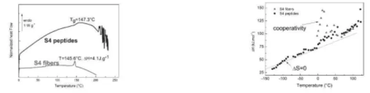
One application deals with the use of DSC to compare the thermal signature of elastin and elastin fractions from human thoracic aortas during aging. It is shown that in the thoracic aortas from children group, proteins induce a more flexible structure in contrast with that observed in the thoracic aortas from adults group, due to the total cross-linking of elastin. In this case, there is a segregation between proteins and elastin. No significant differences can be found between the glass transition temperature of elastin in adults and old persons groups, meaning that the mechanical properties of elastin are conserved during ageing. Nevertheless, an irreversible denaturation phenomena of sheet or helical structured proteins is detected with aging, and corroborated by biochemical analyses which report an important increase of polar amino acids in the elastin-proteins fraction of elderly patients2. In this case the protein fraction would form an homogenous phase with elastin by ionic bonds. A second application deals with the combined use of FTIR, DSC and dielectric techniques to characterize a sub fragment of elastin involved in pathological aging. Under certain pathological conditions elastin is attacked by the metalloproteinase MMP12 releasing short polypeptides able to give rise to amyloid-like fibers. Amyloid fibrils are highly ordered parallel cross -sheet structures usually associated with Alzheimer’s disease, Parkinson’s disease, type II diabetes. Peculiar signatures of the amyloid fibers can be detected both at the localized and delocalized levels in the condensed state: DSC analysis evidences that S4 peptides are amorphous whereas S4 fibers present a long range order (Fig. 2). Their self-assembly into amyloid fibers lead to macroscopic dipole moments explaining the enhancement of these dipolar properties3 (Fig.3).
In the last part, we will show how the dielectric techniques, coupled with thermal and vibrational analyses, allow to characterize vascular tissues themselves, both at the nanometric and mesoscopic levels, in the physiologic and pathologic states; this is promising to track vascular aging and also to optimize the conception of substitutive biomaterials.
References
1. Samouillan V, Lamure A., Lacabanne C. Dielectric relaxations of collagen and elastin in the dehydrated state. Chem Phys 2000;255:259–271.
2. Samouillan V, Dandurand J, Lacabanne C, Stella A, Gargiulo M, Degani A, Gandaglia A, Spina M. Analysis of the molecular mobility of collagen and elastin in safe, atheromatous and aneurysmal aortas. Pathol Biol (Paris) Elsevier Masson SAS; 2012;60:58–65.
3. Samouillan V, Dandurand J, Causse N, Lacabanne C, Bochicchio B, Pepe A. Influence of the Architecture on the Molecular Mobility of Synthetic Fragments Inspired from Human Tropoelastin. IEEE Trans Dielectr Electr Insul 2015;22:1427–1433.
Figure 1: DSC thermograms of freeze dried collagen and elastin
Tubular scaffolds for applications of vascular tissue
Engineering
Vicente La Carrubba
*11
DICAM, Università di Palermo, Viale delle Scienze building 8, 90128 Palermo
* Intervenant
Polymeric tubular scaffolds have been prepared through phase-separation based methods, namely Diffusion Induced Phase Separation (DIPS)
and particulate leaching.
According to the DIPS protocol the scaffold, which present a vessel-like shape, can be obtained obtained by performing a phase separation
process (induced by a decrease of solvent power) around a nylon fibre covered by a viscous polymeric solution. The fibre was first immersed in
a PLLA/dioxane solution (dipcoating bath) at a fixed temperature. Then the fibre was slowly drawn at a constant rate from the solution and put
in a second bath (DIPS bath), containing distilled water or dioxane/water at different ratios, at the same temperature, for a well-defined time
interval This procedure was repeated once or twice, obtaining scaffolds characterized by a single or double layer respectively, with diameters of
2 and 4 mm.
The scaffolds with double layer are more suitable for vascular tissue engineering applications, due to the more elevated and uniform porosity and
the larger mechanical strength.
According to the particulate leaching protocol, the scaffold can be obtained by immersing a fibre in a PLLA/dioxane solution containing NaCl
sieved particles with a well defined size, slowly drawing the fibre at a constant rate from the solution and immersing in a second bath (DIPS
bath). After drying for 24 hours, the salt particles are extracted via leaching by using sistilled water.
Also in this case single and double layer scaffolds have been prepared with diameters of 2 and 4 mm at various extraction rates. Scaffolds
obtained at the extraction rate of 5 cm/min via salt leaching are more uniform with respect to the scaffolds produced via DIPS.
Cell cultures (RPMI 2650, nasal mucosa) on scaffolds produced via particulate leaching exhibited better results than the scaffolds obtained via
DIPS, with a satisfactory level of adhesion and proliferation and the development of an architecture comparable to the original texture of nasal
mucosa in vivo, also confirming the non-cytotoxicity of the used materials.
These preliminary results may address the use of particulate leaching towards vascular tissue engineering applications.
Bioprosthetic heart valve aging deterioration and agerelated
Benefits
Michel Spina
*11
Department of Biomedical Sciences, University of Padova, Italy
* Intervenant
Following population aging and due to congenital and acquired diseases the annual number of patients requiring heart valve replacement
worldwide has been estimated to triple from approximately 290,000 in 2003, to over 850,000 by 2050 [1]. Prosthetic heart valves are broadly
divided in mechanical heart valves (MHV) and bioprosthetic heart valves (BHV) while ≈55% are MHVs and ≈45% BHVs [2] not taking into
account most recent transcatheter heart valves. MHVs are made of non-biological material while BHVs are either of allogenic (syn. homografts)
or xenogenic origin. The last ones are treated with glutaraldehyde to prevent immune rejection. Mechanical devices have a superior durability
but the recipients need permanent lifelong anticoagulant therapy. In turn biological valves exhibit excellent hemodynamic behaviour with a low
risk of thromboembolic complications but show limited longevity following structural valve degeneration (SVD) particularly in patients <35
years of age. The definition of SVD has been associated with the intrinsic permanent changes of prosthetic valve leading to degeneration and/or
haemodynamic dysfunction that necessitates reoperation [3]. According to an overall evaluation, 49-63% of all valve replacements with
glutaraldehyde-treated BHVs [3] have been reported to be free from SVD 20 years after intervention. Otherwise the risk of structural failure is
inversely related to the age of patients: it is higher in individuals <35 years of age in which up to 100% of BHVs fail within 5 years postsurgery
whereas after 15 years 60-70% of older patients (>75 years) are still free of SVD [4].
In the vast majority of cases SVD is associated with leaflet calcium deposits even if in 19-25% of BHV implants reoperations occur in the
absence of leaflet mineralization [5, 6] (excluding nonintrinsic abnormalities, thrombosis and infections). Therefore, considering the elevated
levels of oxidized amino acids always associated with bioprosthesis dysfunction, a potentially important role has been prospected also for
oxidized amino acid formation [7]. Although not fully understood the mechanism of early failure appears to be complex and multifactorial [6].
The process starts by host IgM/IgG interaction (α-Gal epitope) and macrophage deposition followed by collagen breakdown and
finally calcification [8]. Otherwise calcification is believed to be driven by the chemical interaction between phospholipids, residual and
oxidized aldehyde groups on one side and circulating calcium ions [4]. A procedure for the suppression of both tissue calcium binding sites and
oxidation effects has been developed [9], while to avoid immune injury the use of BHV made of α-Gal free tissue (Gal knockout
pigs) has been reported [10]. Moreover a novel procedure aimed to overcome comprehensively immune injury, calcification and oxidative
modifications has also been recently proposed [11]. At the present only one of these procedures has been implemented in a clinical trial [9, 12].
[1] Yacoub MH and Takkenberg JJM. Will Heart Valve Tissue Engineering Change the World? Nat Clin Pract Cardiovasc Med. 2005; 2: 60-61.
[2] Siddiqui RF, Abraham JR, Butany J. Bioprosthetic heart valves: modes of failure. Histopathology 2009; 55: 135-144.
[3] Capodanno D, Petronio AS, Prendergast B, Eltchaninoff H, Vahanian A et al..
Standardized definitions of structural deterioration or valve failure in assessing long-term durability of transcatheter and surgical aortic
bioprosthetic valves: a consensus statement from the European Association of Percutaneous Cardiovascular Interventions (EAPCI) endorsed by
the European Society of Cardiology (ESC) and the European Association for Cardio-Thoracic Surgery (EACTS). Eur J Cardio-Thoracic Surg
2017; 52: 408-417.
[4] Schoen FJ, Levy RJ. Tissue heart valves: current challenges and future research perspectives. J Biomed Mater Res 1999; 47: 439-465.
[5] Schoen FJ, Kujovich JL, Webb CL, Levy RJ. Chemically determined mineral content of explanted porcine aortic valve bioprostheses:
correlation with radiographic assessment of calcification and clinical data. Circulation. 1987;76:1061–1066.
[6] Barbarash O, Rutkovskaya N, Hryachkova O, Gruzdeva O, Ucasova E, et al. Impact of recipient-related factors on structural dysfunction of
xenoaortic bioprosthetic heart valves. Pat Pref Adher 2015; 9: 389-399.
[7] Lee S, Levy RJ, Christian AJ, Hazen SL, Frick NE, et al. Calcification and oxidative modifications are associated with progressive
bioprosthetic heart valve dysfunction. J Am Heart Ass 2017; 6:e005648. DOI:10.1161/JAHA.117.005648.
[8] Konakci K, Bohle B, Blumer R et al. Alpha-Gal on bioprostheses: xenograft immune response in cardiac surgery. Eur J Clin Invest 2005; 35;
17–23.
[9] Dove J, Dobler D, Davidson J, Wright G. Capping bioprosthetic tissue to reduce calcification. Patent No US2014/0200659 A1.
[10] McGregor C, Byrne G, Rahmani B, Chisari E, Kyriakopoulou K, Burriesci G. Physical equivalencyof wild type and galactose α 1,3
galactose free porcine pericardium; a new source material for bioproisthetic heart valves. Acta Biomater 2016; 41: 2014-209.
[11] Naso F, Stefanelli U, Buratto E, Lazzari G, Perora A, Galli C, Gandaglia A. Alpha-Gal inactivated heart valve bioprostheses exhibit an
anticalcification propensity similar to knockout tissues. Tissue Eng A 2017. 23: 1181-1195.
[12] Puskas JD, Bavaria JE, Svensson LG, Blackstone EH, Griffith B, Gammie JS et al. The COMMENCE trial: 2-year outcomes with an aortic
bioprosthesis with RESILIA tissue. Eur J Cardiothorac Surg 2017; 52: 432–439.
Elastin modification during vascular aging and
pathophysiological consequences
Laurent DUCA
*11
UMR CNRS / URCA 7369, Reims
*Intervenant
Cardiovascular diseases (CVD) are the leading cause of death worldwide and represent a major problem of public health. Over the years, life
expectancy has considerably increased throughout the world and the prevalence of CVD is inevitably rising with the growing aging of the
population. The normal process of aging is associated with progressive deterioration in structure and function of the vasculature, commonly
called vascular aging. At the vascular level, extracellular matrix (ECM) aging leads to molecular alterations of long half-life proteins, such as
elastin and collagen, and have critical impacts on vascular diseases. Elastin, is an insoluble and hydrophobic protein mainly produced by
smooth muscle cells in the media and fibroblasts in the adventitia. As elastogenesis is restricted to fetal and infancy, mature elastin fibers remain
for lifespan. Indeed, its strong reticulation makes elastin a highly stable molecule with longevity comparable with human lifespan and any
proteolytic damage that does occur with age and disease is essentially irreparable. Under pathological conditions, vascular and inflammatory
cells produces elastases able to degrade elastin, leading to the production of elastin derived peptides (EDPs) that actively participate through the
Elastin Receptor Complex (ERC) to the development of diseases such as diabetes, atherosclerosis, thrombosis and vascular calcification.
Consequently, the proactive role of the ECM suggests that the development of a prognostic value and an innovative therapy based on matrix
biology has a strong potential. Finally, some modifications affecting elastin such as glycation or carbamylation could occur and possibly affect
the elastin network. The works presented here will summarize and discuss these points.
History and conceptual developments in vascular biology and angiogenesis research
Andréas Bikfalvi
*11
Inserm U1219, University of Bordeaux, Talence
*
Intervenant
Vascular biology is an important scientific domain that has gradually penetrated many medical and scientific fields. Scientists are most often
focused on present problems in their daily scientific work and lack awareness regarding the evolution of their domain throughout history and of
how philosophical issues are related to their research field. In this article, I provide a personal view with an attempt to conceptualize vascular
development research that articulates lessons taken from history, philosophy, biology and medicine. I discuss selected aspects related to the
history and the philosophy of sciences that can be extracted from the study of vascular development and how conceptual progress in this
research field has been made. I will analyze paradigm shifts, cross-fertilization of different fields, technological advances and its impact on
angiogenesis and discuss issues related to evolutionary biology, proximity of different molecular systems and scientific methodologies. Finally, I
discuss briefly my views where the field is heading in the future.
Estrogens as vascular anti-ageing hormones?
Experimental and clinical evidences and uncertainties
Jean-François Arnal
*11
Inserm U1048 - CHU Rangueil, Toulouse
*Intervenant
Aging is associated with structural and functional changes in the vasculature, including endothelial dysfunction, arterial stiffening and
remodeling, impaired angiogenesis, and defective vascular repair, and with increased prevalence of atherosclerosis.
Cardiovascular risk is similar for older men and women, but lower in women during their fertile years.
In women, vascular aging seems to be accelerated during the menopause transition, particularly around the late perimenopausal period. This age-
and sex related difference points to estrogen as a protective factor because menopause is marked by the loss of endogenous estrogen production.
Experimental studies have attributed part of the protective effects of estrogen to its modulatory action on vascular endothelium. Indeed, estrogen
promotes endothelial-derived NO production through increased activity of endothelial NO synthase. However, inhibition of endothelial NO
production does not really impair the atheroprotective actions of estrogens. In fact, numerous other cell targets can mediate the actions of
estrogens, from smooth muscle cells to various immune cells… Modulations of numerous other factors, such as prostacyclin and hromboxaneA2
release, were also reported and could play a role.
In contrat to the previous epidemiological and observational evidences, the only interventional trial (Women Health Initiative) failed to report
any cardiovascular benefit from estrogen replacement therapy, and revealed even an increase in CV events in the older postmenopausal women
(i.e. women treated 20 years or more after menopause). This raised the “Timing Hypothesis” states that estrogen mediated vascular benefits
occur only before the detrimental effects of aging are established in the vasculature, and could offer a possible « explanation» for these
discrepancies. Nevertheless,a gap remains in current knowledge of cardiovascular aging mechanisms in women.
The talk will attempt to summarize clinical and experimental data on the effects of aging, estrogens, and hormone replacement therapy on
vascular function of females.
VIVA EVENING CONFERENCE
Arteriolar and microcirculation aging
Daniel HENRION
*1
1
CNRS UMR 6015 – Inserm U1083, University of Angers
*Intervenant
Resistance arteries are the small blood vessels located upstream from capillaries. They are crucial for the delivery of blood to vital tissues at
relevant flow and pressure. Disorders in resistance arteries structure and function raise capillary pressure and cause downstream organ damage
such as that seen in diabetes, hypertension or kidney disease, whose frequency increases with age. Resistance arteries have a basal tone allowing
a tight control of local blood flow. This basal tone results of the interaction between myogenic tone due to the activation by pressure of vascular
smooth muscle contractility and flowmediated dilatation (FMD) due to the activation of endothelial cells by shear stress. A reduced FMD is
the hallmark of endothelium dysfunction. FMD is altered in ageing and in both cardio-vascular and metabolic disorders. Long-term decreases or
increases in blood flow induce inward or outward remodelling of resistance arteries respectively. Although flow-mediated outward remodelling
is essential in post-ischemic revascularization, it is altered in ageing and this reduced responsiveness is aggravated by the main risk factors.
These latter increase oxidative stress and inflammation at the tissue level leading to alter FMD and then to inward remodelling which in turn
accelerates oxidative stress and inflammation. Breaking this vicious circle is a major challenge in most countries with a rise in longevity
and a high level of exposure to the risk factor associated to the so-called Western way of life. Recent studies have identified the estrogens
receptors has key modulators of both FMD and flow-mediated outward remodelling independently of their effect on reproduction, thus opening
a new field of investigation in the search for more accurate therapeutic tools reducing vascular ageing.
Oxidation and Inflammation in Atherosclerosis
Antonio Junior Lepedda
*11
Department of Biomedical Sciences - University of Sassari, Sassari, Italy
*Intervenant
Atherosclerosis is a chronic inflammatory condition, characterized by the accumulation of lipids and fibrous elements in the intimal layer of
medium and large arteries, that could turn into an acute clinical event due to plaque ulceration and thrombosis. Indeed, plaque rupture is the
predominant underlying process in the pathogenesis of acute coronary syndromes and peripheral vascular disease. Although a great deal of work
has been done on it, the mechanisms underlying plaque formation and progression to advanced lesions are not yet completely known.
It is generally held that plaque instability is caused by a substantial increase in inflammatory and proteolytic activities. Furthermore, some lines
of evidence suggest that unstable plaques are also characterized by pronounced oxidative environment. In situ oxidative events may determine
lipid/protein metabolic fate, bioactivity, and antigenic properties. In this respect, oxidized LDL is readily internalized by macrophages through
the so-called “scavenger receptor” pathway. These early modifications could initiate and/or contribute to atherogenesis, mainly when an
imbalance between oxidant and antioxidant agents takes place. Although several studies report that atherosclerotic plaques contain high
concentrations of some amino acid oxidation products, caused mainly by carbonylation, ROS, and RNS oxidation or thiolation, limited
information are available regarding the relationship between the accumulation of markers of oxidized proteins and atherosclerosis severity.
Furthermore, different oxidation-specific epitopes can be detected in blood and may reflect atherosclerosis manifestations. At present, the
mechanisms underlying the formation of these by-products and the relevance for disease progression are under investigation.
Although oxidative stress has been long associated with the genesis and progression of the atherosclerotic plaque, scanty data on its in situ
effects on protein sulfhydryl groups oxidation are available. In this respect, reduced protein sulfhydryls, which are in most cases key functional
groups, represent a target for several post-translational oxidative modifications. In the last years, we investigated the entity of these oxidations,
focusing on low molecular weight thiols adduction, in advanced human carotid plaques, also in relation with plaque vulnerability. By this way,
we evidenced deep differences between stable and unstable atherosclerotic plaques as well as between extractable plaque proteins and plasma
proteins. Furthermore, by analysing both circulating and plaque-filtered serum albumin (HSA), we evidenced that the pro-oxidant environment
present in atherosclerotic tissue could modify filtered proteins also by protein-SH group oxidation, probably contributing to plaque progression.
In this regard, our studies suggest, for the first time, that once filtered, albumin represents a harmful source of homocysteine and
cysteinylglycine inside the plaque environment. In this respect, the contribution of GSH to the intra-plaque protein-bound LMW thiols
equilibrium seems to be particularly relevant.
Numerical assessment and comparison of pulse wave
velocity methods aiming at measuring aortic stiffness
Structural imaging of the vascular wall
Azzam Alwannl, Amandine Wahart2, Sébastien Blaise3, Edith Grall-Maës4, Béatrice Romier5, Pierre Beauseroy4, Laurent Duca5, Michael Sheratt6, Laurent Debelle5, ManuelDauchez7,8, Rémi Cogranne9, and Sébastien Almagro*10
1Laboratoire de Modélisation et Sûreté des Systèmes (LM2S), Institut Charles Delaunay, UMR CNRS 6279 — lm2s —
pôle ROSAS, Université de Technologie de Troyes., France
2Laboratoire Signalisation et Récepteurs Matriciels (SiRMa), Unité Matrice Extracellulaire et Dynamique Cellulaire
(MEDyC), UMR CNRS 7369, — CNRS : UMR7369 — Université de Reims-Champagne-Ardenne., France
3Laboratoire Signalisation et Récepteurs Matriciels - MEDyC UMR URCA/CNRS n 7369 - Equipe 2 Veillissement
matriciel et remodellage vasculaire (MEDyC - Equipe 2) — Université de Reims - Champagne Ardenne — Université de Reims Champagne Ardenne UFR Sciences Exactes et Naturelles Campus du Moulin de la Housse BP 1039 51687 Reims
cedex 2, France, France
4Laboratoire Modélisation et Sûireté des Systemes (LM2S) — Université de Technologie de Troyes, CNRS : UMR6281 —
UTT - 12, rue Marie Curie - BP 2060 - 10010 TROYES CEDEX, France
5Unité Matrice Extracellulaire et Dynamique Cellulaire (MEDyC), — CNRS : UMR7369 — Université de
Reims-Champagne-Ardenne., France
6Division of Cell Matrix Biology and Regenerative Medicine, School of Biological Sciences, Faculty of Biology Medicine
and Health„ — The University of Manchester, Room 1.529, Stopford Building, Oxford Road, Manchester M13 9PT, Royaume-Uni
7Plateau de Modélisation Moléculaire Multiéchelle (P3M), Reims — Université de Reims - Champagne Ardenne, CNRS :
UMR7369 — Moulin de la Housse, 51687 Reims Cedex 2, France
8UMR CNRS/URCA 7369 (MEDyC), Reims (1) — Université de Reims - Champagne Ardenne, CNRS : UMR7369 —
Moulin de la Housse, 51687 Reims Cedex 2, France
9Laboratoire de Modélisation et Sûreté des Systèmes (LM2S), Institut Charles Delaunay, UMR CNRS 6279 — CNRS :
UMR6279 — , pôle ROSAS, Université de Technologie de Troyes., France
10Laboratoire Signalisation et Recepteurs Matriciels (SiRMa), Unite Matrice Extracellulaire et Dynamique Cellulaire
(MEDyC), . — CNRS : UMR7369 — Université de Reims-Champagne-Ardenne, France *Intervenant
Résumé
The extracellular matrix is a complex assembly produced by our cells. This structure is crucial for the proper functioning of tissue because it provides support and mechanical anchoring. Elastin is a fibrous protein of the extracellular matrix that is synthetized before teen age. Elastin bestows its elastic properties to the arterial wall and therefore contributes to its mechanical resistance. Elastin is intrinsically auto-fluorescent and thus observable on tissue slice. As a consequence, elastin alterations such as those occurring during normal aging or chronic age-related vascular pathologies can be imaged.
The aim of our work is to image elastin inside the murine aortic wall and hereafter, to extract morphological and quantitative parameters from these images. Statistical analysis of these data could characterize the status of the system and could allow us to infer about the evolution of the vascular integrity. This preliminary work was started using murine aortic slices observed by fluorescence microscopy. Further, large aortic segments of several millimeters were imaged by X-Ray computed micro-tomography at the submicron scale.
Our very first results suggest that it is possible with a minimal set of morphological descriptors to characterize the matrix condition of an aortic section, and thus, to potentially detect the occurrence of a pathology, even at an early stage, before its clinical onset. Importantly, our first experiments with a synchrotron light source indicate that it is possible to image "large segment" of very small artery at high resolution allowing the global analysis of the ultrastructure of the matrix within the arterial wall. Using this high-resolution methodology, we observed a "weft" inside the aortic matrix structures. We think that this feature may provide mechanical resilience to the artery and that its integrity could be the sign of its good health.
Grants :
The authors thank D.R.R.T. of Champagne-Ardenne for financial support. This work was carried out with the support of the Diamond Light Source. We thank Diamond Light Source for access to beamline 113 (proposal number MT12776-1) that contributed to the results presented here.
Long-term trajectories of cardiometabolic risk factors in prodromal
dementia: the Three-City Study
Maude Wagner
1,2, Catherine Helmer
2, Christophe Tzourio
3, Claudine Berr
4, Cécile
Proust-Lima
1*, and Cécilia Samieri
2*1
Univ. Bordeaux, Inserm U1219, Bordeaux Population Health Research Center team Biostatistics
2Univ. Bordeaux, Inserm U1219, Bordeaux Population Health Research Center team LEHA
3Univ. Bordeaux, Inserm U1219, Bordeaux Population Health Research Center, Team HEALTHY
4
Univ. Montpellier, Inserm U1061, Neuropsychiatry: Epidemiological and Clinical Research
*These authors contributed equally to this work
Objective: To describe the trajectories of major cardiometabolic risk factors (body mass index [BMI], systolic and diastolic blood pressure
[SBP, DBP], fasting blood lipids [HDL and LDL cholesterol, triglycerides] and glycemia) in the 14 years preceding dementia diagnosis, and to
compare them to the trajectories of matched subjects free of dementia, using latent process mixed models.
Design: Case-control study nested in a prospective cohort.
Setting: The Three-City Study, a population-based cohort of older persons (≥65 years) enrolled between 1999 and 2001 in three French cities
(Bordeaux, Dijon, and Montpellier) who underwent up to six repeated home visits with neuropsychological testing and measurements of
anthropometric parameters and blood pressure, and up to three repeated blood draws for assessment of biological parameters.
Participants: 785 incident cases of dementia and with evaluation of cardiometabolic risk factors at baseline individually matched (for gender,
age, education, and study center) to 3,140 controls at the time of dementia cases diagnosis.
Results: In both cases and controls, BMI, DBP and HDL declined in the 14 years preceding the matching visit, whereas SBP and glycemia
increased and LDL and triglycerides remained stable over time.
Compared to controls, dementia cases had a faster decline in BMI and a lower increase in SBP (P<.001and P<0.049 respectively for
group-by-time interactions) leading to significantly lower BMI and SBP values among cases respectively 2 and 4 years before diagnosis. The other
cardiometabolic risk factors (DBP, lipids and glycemia) did not evolve differently according to disease status (all P for group-bytime
interactions>0.20); however cases presented lower DBP and higher fasting glycemia levels over the entire period.
Conclusion: Higher BMI, blood pressure and blood lipids were not found as strong risk factors for dementia in the older age range; on the
contrary BMI and blood pressure decreased months to several years before dementia diagnosis (possibly as a consequence of changes in the
prodromal phase of the disease). Blood glucose was the single cardiometabolic risk factor with constantly higher values over the 14 years before
diagnosis among incident dementia cases, suggesting that elevated glycemia is among the strongest cardiometabolic risk factor for dementia in
older persons.
MicroRNA expressed in progenitor cells circulating in peripheral blood as prospective
biomarkers of agerelated macular degeneration (AMD) in its dry and wet
form – preliminary data
A. Machalińska
1
, M.P. Kawa*
2
, A. Sobuś
2
, Z. Litwińska
2
, A. Kowalska-Budek
1
, K. Babiak
2
,
A. Grabowicz
2
, B. Machaliński
2
1. First Department of Ophthalmology,
2. Department of General Pathology,
Pomeranian Medical University, Szczecin, Poland
*Intervenant
Abstract:
Objectives: Age-related macular degeneration (AMD), a worldwide public health problem, has been recognized as a common cause of visual
dysfunction in elderly people. MicroRNAs (miRNAs) present in sera are characteristically altered in many pathological conditions and have
been used as biomarkers for specific diseases, including AMD. Several miRNAs can be detected in bodily fluids including serum, urine and
saliva, where they are released in form of exosome vesicles. However, different physical conditions, including blood oxygen tension, alter the
exosome release and affect the global miRNA profile in fluids modifying their bioactivity as a consequence. In contrast, being resistant to
exonucleolytic activities, intracellular miRNAs are more stable and have longer half-life than extracellular miRNA, therefore may play a role in
development and progression of diseases. Due to the high abundance, stable and accessible characteristics, intracellular miRNAs could serve as
putative biomarker molecule in clinically relevant samples. Thus, in our study, we analyzed selected miRNAs from peripheral blood
mononuclear cells (PBMCs) in patients with dry and wet form of AMD and healthy volunteers (HV), and we investigated the intracellular
miRNAs expression as potential biomarker for early diagnosis of dry or wet form of AMD.
Material and Methods: PBMCs were collected from 94 AMD patients (46 wet form and 48 dry form) and 46 HV as a source of miRNAs.
Self-validated individual quantitative RT-PCR study was then performed to evaluate the expression levels of selected miRNAs. The Mann-Whitney
U test was used for statistical analysis.
Results: miRNA expression analysis revealed a decreased expression of selected angiogenesis-related miRNAs in both forms of AMD,
including miR-223-3p, miR-17-5p (both greatly down-regulated in dry AMD), and miR-150-5p (stronger down-regulation in wet AMD). In
contrast, two angiogenic miRNAs: miR126-5p and miR126-3p were upregulated, especially in dry AMD.
Conclusion: Our results indicated that altered expression of selected miRNAs may play a role in the AMD development and progression.
Further studies are required to determine causal relationships and elucidate underlying mechanisms. This work was supported by grants:
STRATEGMED1/234261/2NCBR/2014 and UMO-2013/09/B/NZ7/04031
Keywords: MicroRNA, age-related macular degeneration (AMD), dry AMD, wet AMD, angiogenesis, intracellular miRNAs, disease
biomarkers
Molecular, conformational and thermal
characterization of ventricular Remodeling in a pig
model of tachycardia-induced cardiomyopathy
Valerie Samouillan', Aleyda Benitez-Amarot", Esther Jorge", Jany Dandurand
1,
Laura Nasarre
3, David De Gonzalo-Calvo
3, Olga Bornachea
2'
3, Colette Lacabanne
l, Jose
M Guerra', and Vicenta Llorente-CortesI"
Centre interuniversitaire de recherche et dingenierie des materiaux (CIRIMAT) — Institut National Polytechnique [Toulouse], Universite Paul Sabatier - Toulouse 3, Centre National de la Recherche Scientifique : UMR5085 — Laboratoire de Physique des Poly 118,route de Narbonne 31062 TOULOUSE
CEDEX 4, France
2lnstitute of Biomedical Research of Barcelona-Spanish National Research Council (IIBB-CSIC) —
Rosse116, 161 6' i
7
a planta, 08036 Barcelona, Espagne3Biomedical Research Institute Sant Pau (IIB Sant Pau) — Hospital de la Santa Creu i Sant Pau. Sant
Antoni Ma Claret, 167 Pavello n° 16, Sant Frederic, 08025 Barcelona, Espagne
4Universitat Autonoma de Barcelona (UAB) — Placa Mica, s/n, 08193 Bellaterra, Barcelona, Espagne
*Auteur correspondant:
valerie.sarnouillan@univ-tlse3.frt
Intervenant
t
Auteur correspondant:
cllorente@csic-iccc.orgResume
Aims: Non-ischemic dilated cardiomyopathy (NIDCM) is characterized by ventricular dilatation associated to pathological remodeling. Our aim was to compare molecular, conformational and physical characteristics of healthy and NIDCM myocardium to identify crucial changes in the evolution to pathological cardiac remodeling.
Methods and Results: Our study includes healthy pigs (control, N=7) and tachycardia-induced NIDCM pigs (DCM,
N=10). Samples from right (RV) and left ventricle (LV) were frozen at -80°C. Real-time PCR showed a strong increase in collagen I and collagen III in both RV (2.06 and 1.38-fold) and LV (2.43 and 1.48-fold) dilated ventricles. Fourier
Transform Infrared Spectroscopy (FTIR) 1st [A(1171cm-1)/A(1338cm-1)] and 2nd [A(1304cm1)/A(1338cm-1)] indicators reflect myofiber/collagen ratio while [A(1043cm-1)/A(2800cm-1- 3000cm-1)] 3rd indicator reflect carbohydrate/lipid ratio. 1st indicator was reduced in both dilated ventricles (to 43% in RV and to 37% in LV), while 2nd indicator was only reduced in dilated RV (to 37%). 3rd indicator was upregulated in both dilated ventricles, but to a higher extent in RV (2.60-fold vs 1.61-fold, P=0.049). Histological Masson's trichromic staining showed unorganized and agglomerated collagen in dilated ventricles, and a higher percentage of fibrosis in RV compared to LV (P=0.016). Both FITR and thin
layer chromatography revealed the cancellation of the differences in cholesteryl ester and triglyceride content between RV and LV (inherent to control pigs) in DCM group. Differential Scanning Calorimetry (DSC) evidenced a strong decrease of protein denaturation temperature (a shift of 50°C in the freeze-dried state) in both dilated ventricles, associated with a significant depression of the freezable water melting point.
Conclusion: Our results point to reduced myofiber/collagen ratio, increased carbohydrate/lipid ratio and lower protein
thermal stability as key alterations contributing to the deposition of unorganized and agglomerated collagen in dilated ventricles. The exacerbation of these alterations in RV underlies deeper remodeling of RV vs LV in the evolution to dilated cardiomyopathy.