ORIGINAL ARTICLE
Ultra-deep sequencing confirms immunohistochemistry
as a highly sensitive and specific method for detecting
BRAF
V600E
mutations in colorectal carcinoma
Matthias Rössle&Michèle Sigg&Jan H. Rüschoff&
Peter J. Wild&Holger Moch&Achim Weber&
Markus P. Rechsteiner
Received: 19 August 2013 / Revised: 19 September 2013 / Accepted: 20 September 2013 / Published online: 2 October 2013 # Springer-Verlag Berlin Heidelberg 2013
Abstract The activating BRAFV600 mutation is a well-established negative prognostic biomarker in metastatic colo-rectal carcinoma (CRC). A recently developed monoclonal mouse antibody (clone VE1) has been shown to detect reliably BRAFV600E
mutated protein by immunohistochemistry (IHC). In this study, we aimed to compare the detection ofBRAFV600E mutations by IHC, Sanger sequencing (SaS), and ultra-deep sequencing (UDS) in CRC. VE1-IHC was established in a cohort of 68KRAS wild-type CRCs. The VE1-IHC was only positive in the three patients with a knownBRAFV600E muta-tion as assessed by SaS and UDS. The test cohort consisted of 265 non-selected, consecutive CRC samples. Thirty-nine out of 265 cases (14.7 %) were positive by VE1-IHC. SaS of 20 randomly selected IHC negative tumors showedBRAF wild-type (20/20). Twenty-four IHC-positive cases were confirmed by SaS (24/39; 61.5 %) and 15 IHC-positive cases (15/39; 38.5 %) showed aBRAF wild-type by SaS. UDS detected a BRAFV600E
mutation in 13 of these 15 discordant cases. In one tumor, the mutation frequency was below our threshold for UDS positivity, while in another case, UDS could not be performed due to low DNA amount. Statistical analysis showed sensitivities of 100 % and 63 % and specificities of 95 and 100 % for VE1-IHC and SaS, respectively, compared to
combined results of SaS and UDS. Our data suggests that there is high concordance between UDS and IHC using the anti-BRAFV600E(VE1) antibody. Thus, VE1 immunohistochemis-try is a highly sensitive and specific method in detecting BRAFV600E
mutations in colorectal carcinoma.
Keywords BRAF mutation . Immunohistochemistry . Sanger sequencing . Ultra-deep sequencing . Colorectal carcinoma
Introduction
Colorectal carcinoma (CRC) is the third most malignant neo-plasm in males and second most in females worldwide [1]. In a metastasized stage, the 5-year survival rate decreases to less than 10 % [2]. A combined systemic therapy including cetuximab, a monoclonal anti-EGFR antibody, increases the overall and progression-free survival in tumors bearing a wild-type of KRAS (‘Kirsten rat sarcoma viral oncogene homolog’) and BRAF (‘v-raf murine sarcoma viral oncogene homolog B1’) genes [3,4]. In contrast, mutated BRAF is a marker of worse prognosis in CRC [5–7]. The most common genetic change in the BRAF gene of CRC is a c.1799 T > A point mutation in Exon 15, causing an amino acid exchange from valine to glutamic acid (p.V600E), which leads to a 10-fold increased kinase activity [8–10].
Usually, analysis of the BRAFV600Estatus is performed using different polymerase chain reaction (PCR)-based detection methods like Sanger sequencing, pyrosequencing, and qPCR. Besides the average costs of approximately $300 per analysis [11, 12], these techniques are time consuming and require a high-quality laboratory infrastructure with well-trained staff. Currently, Sanger sequencing is probably the most often used technique because of its reliability and high specificity.
Matthias Rössle and Michèle Sigg contributed equally to this study. Electronic supplementary material The online version of this article (doi:10.1007/s00428-013-1492-3) contains supplementary material, which is available to authorized users.
M. Rössle (*)
:
M. Sigg:
J. H. Rüschoff:
P. J. Wild:
H. Moch:
A. Weber:
M. P. RechsteinerInstitute of Surgical Pathology, University Hospital Zurich, Schmelzbergstrasse 12, 8091 Zurich, Switzerland e-mail: [email protected]
However, its sensitivity usually does not allow detecting muta-tions below a frequency of 15–20 % tumor cells [13,14].
Among other more sensitive techniques, next-generation sequencing technology overcomes this disadvantage and re-veals low-frequency mutations in heterogeneous tumor popu-lations. Recently, we demonstrated the reliability of this tech-nique as a standard procedure in diagnostics of CRC using genomic DNA isolated from formalin-fixed paraffin-embedded tissues (FFPE) [15].
In contrast, immunohistochemistry is a tissue-based, cost-effective technique, which is easy to perform and routinely available in most pathology laboratories [16]. Capper et al. [17] developed a monoclonal mouse antibody (clone VE1), which recognizes the BRAFV600E protein. Several studies have shown that this antibody reliably detects BRAFV600E mutations in malignant neoplasms by immunohistochemistry in routinely processed FFPE tissues [18–26].
In our study, we aimed to evaluate the reliability of this new antibody in detecting BRAFV600Emutations compared to con-ventional Sanger sequencing in a cohort of consecutive CRCs. Furthermore, we aimed to assess the sensitivity of the anti-body below the detection limit of Sanger sequencing by including ultra-deep sequencing (UDS).
Materials and methods Patients
For this study, tumor tissue, and tissue microarray (TMA) slides from two cohorts were analyzed. We have previously described a cohort of 68 KRAS wild-type CRCs [15]. Among these 68 KRAS wild-type CRCs, we identified three tumors with BRAFV600Emutations as assessed by Sanger sequencing (SaS) and UDS. All cases were diagnosed at the Institute of Surgical Pathology, University Hospital Zurich (Switzerland) between 2006 and 2011. The median age was 61 ranging from 35 to 83. Nineteen patients were females and 49 males.
The test cohort consisted of non-selected, consecutive CRC samples of 265 patients, diagnosed at the Institute of Surgical Pathology, University Hospital Zurich between 2001 and 2011. The age ranged from 21 to 95 years (median 72 years). Both sexes were equally distributed (133 females, 132 males). The study was approved by the Cantonal Ethics Committee of Zurich (KEK-ZH-NR: 2010-0093/0).
Tissue microarray construction
Original hematoxylin and eosin stained sections of all CRC samples were retrieved from the archives of the Institute of Surgical Pathology, University Hospital Zurich and reviewed by two histopathologists (antibody establishment cohort: J.R., H.M.; test cohort: M.S., A.W.). The TMAs were constructed
with core replicas as described elsewhere [27]. In brief, two tissue cores (diameter 0.6 mm) of a representative tumor area of each patient were taken from a“donor” block and arranged in a new“recipient” block using a custom-built instrument. Each recipient block also included 14 colon cancer cell lines with known BRAF mutational status (six cell lines with a BRAFV600E
mutation, eight cell lines with BRAF wild-type), which served as controls. Recipient blocks of the test cohort additionally include normal colorectal tissue of CRC patients as additional control tissue.
Immunohistochemistry
Immunohistochemistry (IHC) was conducted on 2-μm thick sections of both TMAs with Ventana Benchmark XT automat-ed staining system (Ventana Mautomat-edical Systems, Tucson, AZ, USA) and according to the manufacturer's instructions. Briefly, after antigen retrieval with cell conditioner 1 (Ventana Medical Systems) for 64 min and pre-primary peroxidase inhibition, the TMA slides were incubated with BRAFV600Eantibodies (clone VE1, dilution 1:200; Spring Bioscience, Pleasanton, USA) for 32 min at 37 °C. After that, incubation with OptiView DAB IHC Detection Kit (Ventana Medical Systems) followed. Slides were counterstained with hematoxylin and Bluing reagent for 4 min each. The reaction quality was controlled using cell lines on the TMAs with known BRAFV600Emutational status. The immunostained slides were independently evaluated by two pathologists (M.S. and M.Rö.), both blinded to the BRAF mutation status. TMA spots lacking of tumor tissue were excluded from the analysis. According to the unequivocal cytoplasmic staining of a majority of the tumor cells, TMA spots were ranked into three staining categories: score 0 for negative, score 1 for weakly/moderately positive, and score 2 for strongly positive. Of each case, the strongest immunoreac-tivity of the two spots was counted.
DNA extraction
Three tissue cylinders (diameter 0.6 mm) were punched from formalin-fixed, paraffin-embedded (FFPE) tissue block of each CRC patient. After extraction of genomic DNA using DNeasy Blood & Tissue Kit 250 (Qiagen, Hilden, Germany), DNA-quantification was done with NanoDrop.
Sanger sequencing
Sanger sequencing evaluating the mutational status of BRAF exon 15 was done for all positive and for 20 IHC-negative cases of the test cohort. DNA was PCR amplified (AmpliTaq Gold, Roche, Switzerland) using the primers de-scribed in Table 1. The cycling conditions were 5 min at 95 °C, 40 cycles each with 1 min at 95 °C, 1 min at 53 °C, 1 min at 72 °C and a final elongation step of 10 min at 72 °C.
PCR products were purified using the Qiagen MiniElute PCR Purification Kit (Qiagen) and sequenced with a Genetic Analyzer 3130xl (Applied Biosystems) using the same primers mentioned above. A change of the electropherogram was classified as mutation, if the peak of the aberrant base (i) was clearly above background, (ii) was present in forward and reverse direction, and (iii) its height exceeded more than 10 % of the reference base peak height. Examples for Sanger se-quencing declared as wild-type are shown in Supplementary Figure S1. As a comparison of a “true” mutation, co40 is depicted as positive control in Supplementary Figure S1. Ultra-deep sequencing (UDS)
Additional UDS was performed to assess the BRAF status in all cases of the test cohort, which showed discordant results after IHC and Sanger sequencing. The UDS procedure is described elsewhere [15]. Shortly, an independent PCR with fusion primers including multiplex identifiers (MIDs) (Table1) was performed from the same DNA sample used for Sanger sequencing. Amplicon processing was done as described by the Amplicon Library Preparation and emPCR (Lib-A) Method GS Junior Titanium Series manual from Roche. 500,000 enriched beads were loaded on a 454 Junior Sequencer (Roche). Demultiplexing and variant calling was done with the Amplicon Variant Analyzer v2.7 (AVA) soft-ware from Roche. Selection criteria (at least 50 reads contain-ing the mutation and at least 0.38 % mutation frequency) for a true positive variant are described in Rechsteiner et al. [15]. Statistics
The calculation of the IHC VE1 sensitivity, specificity, and positive (PPV) and negative predictive value (NPV) including
their 95 % confidence intervals (CI) by comparison with the combined results of both sequencing methods as “gold stan-dard” was performed using Microsoft Excel 2010 (Microsoft Corporation, Redmond, USA). Measurement of interobserver agreement was performed using SPSS 20.0 (IBM Corporation, Armonk, USA).
Results
BRAFV600E(VE1) antibody establishment
Immunohistochemistry of cell line controls showed moderate positivity in one and strong positivity in five of the six known BRAFV600E
mutated cell lines, while all eight cell lines with known BRAF wild-type were immunohistochemically completely negative. A representative staining of the cell lines is shown in Supplementary Figure S2.
The BRAFV600E(VE1) antibody was further established in 68 KRAS wild-type CRCs with three (4.4 %) BRAFV600E mutations previously identified by SaS and UDS. All three BRAFV600E
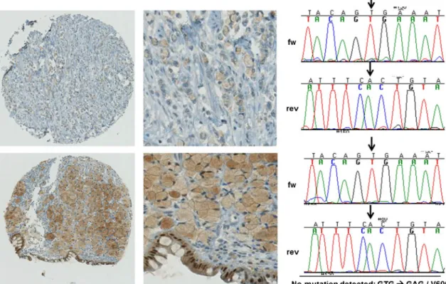
mutated CRC were immunohistochemically pos-itive and showed strong staining reactivity with the VE1 antibody. Some CRCs showed an uncharacteristic weak to strong staining in the intracellular mucus of signet ring-like cells. In the upper row of Fig.1, one CRC with such a staining pattern is depicted with its Sanger sequence on the right panel without visible mutation. Another CRC with strong staining is presented in the lower row of Fig.1with its correspond-ing sequence. Due to the fact that no BRAF mutation could be detected in both CRCs by sequencing, all similar stainings in the intracellular mucus of signet ring-like cells were classified as artifacts and scored negative. All other cases were unequivocally negative, resulting in a sensitivity
Table 1 Primers used for Sanger sequencing and ultra-deep sequencing of BRAF exon 15
Sequencing Primer
Sanger sequencing
BRAF Ex.15 F 5'-TCATAATGCTTGCTCTGATAGGA-3' BRAF Ex.15 R 5'-GGCCAAAAATTTAATCAGTGGA-3' Ultra-deep sequencinga
BRAF 15 F_MID1 5'-CGTATCGCCTCCCTCGCGCCATCAGACGAGTGCGTTCATAATGCTTGCTCTGATAGGA-3' BRAF 15 R_MID1 5'-CTATGCGCCTTGCCAGCCCGCTCAGACGAGTGCGTGGCCAAAAATTTAATCAGTGGA-3' BRAF 15 F_MID2 5'-CGTATCGCCTCCCTCGCGCCATCAGACGCTCGACATCATAATGCTTGCTCTGATAGGA-3' BRAF 15 R_MID2 5'-CTATGCGCCTTGCCAGCCCGCTCAGACGCTCGACAGGCCAAAAATTTAATCAGTGGA-3' BRAF 15 F_MID3 5'-CGTATCGCCTCCCTCGCGCCATCAGAGACGCACTCTCATAATGCTTGCTCTGATAGGA-3' BRAF 15 R_MID3 5'-CTATGCGCCTTGCCAGCCCGCTCAGAGACGCACTCGGCCAAAAATTTAATCAGTGGA-3' BRAF 15 F_MID4 5'-CGTATCGCCTCCCTCGCGCCATCAGAGCACTGTAGTCATAATGCTTGCTCTGATAGGA-3’ BRAF 15 R_MID4 5'-CTATGCGCCTTGCCAGCCCGCTCAGAGCACTGTAGGGCCAAAAATTTAATCAGTGGA-3’ Ex exon, F forward, R reverse
a
and specificity of 100 % each. There was a perfect interobserver agreement with a kappa value of 1.000.
VE1 expression in colorectal carcinoma of the test cohort Both observers agreed on the interpretation of the VE1 im-munohistochemical staining in all but five cases (three cases: score 0 vs. score 1; two cases: score 1 vs. score 2) resulting in a kappa value of 0.921 (p <0.01). After discussion, a consensus was obtained. One case was not evaluable because of loss of
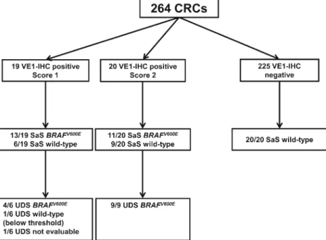
tumor material during staining process. Of the remaining 264 cases, 225 (85.2 %) were immunohistochemically negative, 19 (7.2 %) showed an score 1, and 20 (7.6 %) an IHC-score 2 (Fig.2).
BRAF mutational analysis by Sanger sequencing in colorectal carcinoma of the test cohort
These 39 positive cases as well as 20 randomly selected IHC-negative tumors were submitted to BRAFV600E mutation
Fig. 1 Unspecific staining of tissue by VE1-IHC. Moderate (score 1, upper row) and strong (score 2, lower row) staining of intracellular mucus in signet ring-like cells (original magnifications: left panel×10, middle
panel×40) and nuclei of normal surface epithelium. The right panel shows the corresponding wild-type Sanger sequences. Arrows indicate the loca-tion of the T/A (forward strand) mutaloca-tion leading to BRAFV600E
Fig. 2 VE1 immunohistochemistry of different colorectal carcinoma of the test cohort with score 0 (a ×40 original magnification), score 1 (b ×40), and score 2 (c ×40) for VE1-immunohistochemistry
analysis. SaS of the 20 randomly chosen IHC-negative cases confirmed the immunohistochemical results and showed no mutation in exon 15 of the BRAF gene. Of the 19 IHC-score 1 cases, SaS showed a BRAFV600Emutation in 13 cases and a BRAF wild-type situation in six tumors. Among the 20 IHC-score 2 cases, SaS resulted in 11 BRAFV600Emutated and in nine wild-type cases (Fig.3).
BRAF mutational analysis by Ultra-deep sequencing in colorectal carcinoma of the test cohort
UDS was performed in 14 IHC-positive cases, which were BRAF wild-type by SaS. The DNA amount was too low for analysis in one case. Additionally, one case with IHC-score 1 in one TMA core (site 1) and IHC-score 2 on the second TMA core (site 2) was also included in the UDS analysis. Interestingly, DNA extraction from site 1 and subsequent SaS detected a BRAF mutation in the core with IHC-score 1 and none in the DNA extracted from site 2. Therefore, UDS was performed with the DNA derived from site 2 to investi-gate whether a low frequency mutation was missed by SaS. Moreover, a positive control was included in the UDS analy-sis, in which SaS detected a BRAFV600Emutation.
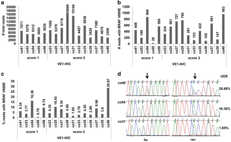
In Fig.4a, the total number of reads from the UDS analysis, including mutated and wild-type BRAF sequences, is shown per patient (mean 7,077 reads). In Fig.4b, the fraction of reads with BRAFV600Emutations is depicted (mean 449 reads). In 14 cases, a BRAFV600E mutation was detected by UDS with mutation frequencies ranging from 1.85–16.36 % (Fig. 4c). Importantly, a mutation with a frequency of 1.85 % was de-tected in the DNA extracted from site 2 of the abovementioned
case underscoring the reliability of the VE1 staining. In the remaining case, a mutation frequency of 0.79 % with only 29 reads containing the mutation was identified which, however, was below our UDS threshold to be included as a true variant (Fig.4c). The positive control (co40) was successfully detected at high frequency (29 %).
Comparision of results of IHC, SaS, and UDS in colorectal carcinoma of the test cohort
As a comparison of VE1-IHC, SaS, and UDS, these 15 cases and the positive control are shown in the Supplementary Figure S1. The mutated base peak height from SaS was comparable to the percentage of the mutation found by UDS. However, all cases except for the positive control were classified as wild-type when using SaS according to our mutation detection threshold level explained in the
“Materials and methods” section. In contrast, SaS and UDS
results did not correlate in all cases with the VE1-IHC stain-ing. This is mainly due to the heterogeneity of the tissue (tumor/stromal/normal cells), which is shown for one case (co25; Fig.5). In this case, the TMA punches gave an IHC-score 2 which matches the TMA extraction cores of the whole tissue cut in Fig.5. In contrast, the punches taken for DNA extraction included one punch at a location with low tumor content which might have resulted in the wild-type classifica-tion by SaS and in the low percentage of mutated reads by UDS (10.43 %).
Together, SaS and UDS detected BRAFV600Emutations in 37 (14 %) and BRAF wild-type in 22 tumors (Fig.3). The case with VE1-IHC positivity and a BRAF mutation below the
Fig. 3 Flow chart of immunohistochemistry (IHC), Sanger sequencing (SaS), and ultra-deep sequencing (UDS) on test cohort
detection limit of UDS (29 reads) was counted as false posi-tive and included in the wild-type cases. The case with nega-tive SaS and missing subsequent UDS was not counted for further statistical analysis.
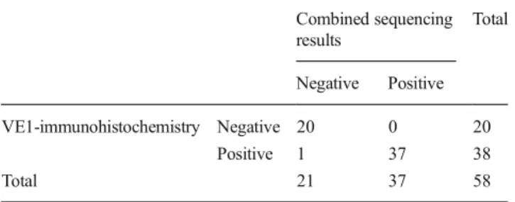
Statistical analysis showed a sensitivity of 100 % (95 % CI 90.75–100.00 %) and a specificity of 95.24 % (95 % CI 76.18–99.88 %) for VE1-IHC compared with combined re-sults of both SaS and UDS as“gold standard” (see Table2). The PPV was 97.44 % (95 % CI 86.52–99.94 %) and the NPV was 100.0 % (95 % CI 83.16–100.0 %) for VE1-IHC.
For SaS compared with combined results of both sequencing methods (see Table3) a sensitivity of 63.16 % (95 % CI 45.99– 78.19 %), a specificity of 100.0 % (95 % CI 83.16–100.00 %),
a PPV of 100.0 % (95 % CI 85.75–100.00 %), and a NPV of 62.86 % (95 % CI 40.7–75.35 %) was calculated.
Discussion
Our results show a very high reliability of the VE1 immuno-histochemistry detecting BRAFV600E mutations in colorectal carcinoma with a sensitivity of 100 % and a specificity of 95.24 %. Similar results were obtained in previous studies using the VE1 antibody on different tumors like malignant melanoma [24,28], Langerhans cell histiocytosis [26], hairy cell leukemia [18], papillary thyroid carcinoma [20, 23],
Fig. 4 Ultra-deep sequencing (UDS) of selected colorectal carcinoma cases negative for BRAFV600Eby Sanger sequencing but positive by VE1-IHC. co40: positive control with a BRAFV600E
mutation detected by Sanger sequencing. a Total read count of wild-type and BRAFV600E mutated
sequences. b Reads with BRAFV600Emutations. c Percentage of BRAFV600E mutated reads compared to total reads. d Sanger sequences of co40, co54, and co31 with BRAFV600Emutations of 28.68, 16.36, and 1.85 % mutation frequencies, respectively, assessed by UDS
Fig. 5 Tissue heterogeneity of VE1-IHC stained colon carcinoma tissue. Left panel: whole tissue section. Right panel: big cores taken for TMA construction and smaller cores punched for DNA extraction and sequencing
epithelial ovarian tumors [19,25], and pulmonary adenocar-cinoma [22].
The almost perfect interobserver agreement shows that the interpretation of immunohistochemical staining results is quite clear and simple in most cases. Using the well-defined criteria (“diffuse and homogeneous granular cytoplasmic staining of tumor cells”) for positivity, only few pitfalls, which have been partly described previously [20,21,29,30], occur. In colo-rectal tissues, sometimes, a nuclear staining of normal epithe-lial cells as well as an unspecific positivity of the intracellular mucus of goblet cells and signet ring-like tumor cells can be observed. Additionally, tumor-associated macrophages that sometimes show a cytoplasmic granular positivity may be difficult to differentiate from true-positive tumor cells.
According to the COSMIC database and literature, BRAF mutations are found in about 4–16 % of CRC [3,13, 31], which fits very well with the frequency in our test cohort bearing in mind that BRAFV600E changes counts for more than 80 % of all BRAF mutations in CRC [9]. As we performed Sanger sequencing only in 20 out of 225 VE1-IHC-negative cases, we cannot exclude that some of the remaining IHC-negative cases might have low frequency or other BRAF mutations, as well, than BRAFV600E. However, the aim of this study was rather to assess the detection limit of the VE1 antibody than the detection limit of our UDS ap-proach. The lower rate of BRAFV600Emutations in our anti-body establishment cohort (4.4 %) can be explained by a selection bias, while in contrast our test cohort consisted of non-selected, consecutive colorectal carcinoma specimen.
Our study shows some limitations of Sanger sequencing (SaS) in detecting BRAF mutations. It is well known that for
successful SaS, a minimal tumor cell amount of 10–20 % is necessary. In summary, four (all VE1-IHC-score 2) out of 15 Sanger-negative cases had a tumor cell amount on TMA cores below 30 %. Three ones (all VE1-IHC-score 1), including that one on which UDS could not be performed and that one with UDS negativity due to low number of positive reads, showed a heterogeneous VE1-IHC staining pattern. In a recent study on mismatch repair protein-deficient colorectal carcinomas, Capper et al. also reported one case with a high amount of non-tumorous cells, which showed positive VE1-IHC and negative Sanger sequencing and pyrosequencing [32]. However, in eight cases with homogeneous VE1-IHC posi-tivity (three with score 1, five with score 2) and a tumor cell percentage >60 %, SaS failed repeatedly to detect the BRAF mutation. Thus, despite the fact that we used tumor punches instead of whole sections for DNA extraction, which usually results in a higher tumor cell load, SaS failed in 38 % of our cases to detect the BRAFV600Emutation. One critical aspect of punching the tumor area for DNA extraction is to allocate and to hit the right area for punching. Such a case is shown in Fig.5, where one punch at a location with low tumor content was included. This punch might have resulted in the wild-type classification by SaS and in the low percentage of mutated reads by UDS. Another case reflecting the same problem was investigated by comparing sequencing data obtained from DNA extracted from two different sites of a tumor which both were positive for VE1-IHC. SaS detected a BRAFV600E mu-tation only in one of these two sites. Subsequent UDS of the Sanger-negative material revealed the same mutation at low frequency. It may be speculated if VE1-IHC, which can also highlight single tumor cells, was more sensitive detecting the BRAF mutation than SaS or if VE1-IHC was false positive in these tumor cells. The fact that UDS detected the mutation at low frequency rather suggests a higher sensitivity of VE1-IHC. Whether, in this case, the low mutation frequency was due to the contamination of normal cells or due to intratumoral heterogeneity remains elusive. Assuming the latter variant, these results might reflect true intratumoral heterogeneity of BRAF mutation, which has been reported as rare events (1–3 %) in primary CRC [33,34].
Another reason why, in our study, 38 % of the BRAF mutations were missed by SaS might be the stringent selection criteria we applied for classifying a mutation as true by SaS. Retrospectively, one might adjust the detection threshold as in most cases of the 15 previously classified wild-type cases a mutated base peak was visible in SaS.
Our results agree with previous studies comparing SaS with other sensitive detection methods like pyrosequencing or deep sequencing. Guerra et al. reported a similar result comparing BRAF mutational status of macrodissected papil-lary thyroid carcinoma by SaS and pyrosequencing [35]. Thus, VE1-IHC seems to be sensitive enough to overcome the problem of tissue and tumor heterogeneity. Taken together,
Table 2 Comparing results of VE1-immunohistochemistry with com-bined results of Sanger and ultra-deep sequencing for BRAFV600E mutations Combined sequencing results Total Negative Positive VE1-immunohistochemistry Negative 20 0 20 Positive 1 37 38 Total 21 37 58
Table 3 Comparing results of Sanger sequencing alone with combined results of Sanger and ultra-deep sequencing for BRAFV600Emutations
Combined sequencing results Total Negative Positive
Sanger sequencing Negative 20 14 34
Positive 0 24 24
we demonstrate the high sensitivity and specificity of detecting BRAFV600Emutations in colorectal carcinoma by a new mutation-specific antibody. Our IHC results agree with two other recently published studies investigating the VE1 antibody on CRC specimens [29,36]. Both groups reported a high concordance between VE1-IHC and pyrosequencing [29] or multiplex allelic-specific PCR-based assay [36] as reference methods. In contrast, a study by Adackapara et al. [30] showed a much lower sensitivity (35 %) for VE1-IHC on CRC specimen compared to pyrosequencing results. They used a manual technique with overnight incubation for VE1-IHC. Therefore, methodological differences could explain the difference to ours and other studies.
Our study represents the first comparison between UDS and VE1-IHC in CRC and we demonstrate a very high con-cordance between UDS and VE1-IHC. In the light of the relatively high costs and demanding infrastructure of ultra-deep sequencing as well as the lower sensitivity of SaS, the VE1-IHC, which can be performed routinely in probably most pathological institutes, seems to be a reliable, simple, and cheap primary tool to analyze BRAFV600Estatus for diagnos-tic, predictive, and prognostic purposes. Only VE1-negative or equivocal cases may be further investigated by more sen-sitive methods like ultra-deep sequencing.
Acknowledgements We thank Dr. Adriana von Teichman, Marion Bawohl, and Sonja Brun-Schmid for their excellent technical assistance. We are very grateful to Prof. Dieter Zimmermann for his critical proof-reading of the manuscript.
Conflict of interests The authors declare that they have no conflicts of interest.
References
1. Jemal A, Bray F, Center MM, Ferlay J, Ward E, Forman D (2011) Global cancer statistics. CA: A Cancer Journal for Clinicians 61(2): 69–90. doi:10.3322/caac.20107
2. Ries L, Young J, Keel G, Eisner M, Lin Y, Horner M-J (2007) SEER survival monograph: cancer survival among adults: U.S. SEER Pro-gram, 1988–2001, Patient and Tumor Characteristics. National Can-cer Institute, SEER Program, Bethesda, MD
3. Van Cutsem E, Kohne CH, Lang I et al (2011) Cetuximab plus irinotecan, fluorouracil, and leucovorin as first-line treatment for metastatic colorectal cancer: updated analysis of overall survival according to tumor KRAS and BRAF mutation status. J Clin Oncol 29(15):2011–2019. doi:10.1200/JCO.2010.33.5091
4. Di Nicolantonio F, Martini M, Molinari F et al (2008) Wild-type BRAF is required for response to panitumumab or cetuximab in metastatic colorectal cancer. J Clin Oncol 26(35):5705–5712. doi:10.1200/JCO.2008.18.0786
5. Bokemeyer C, Cutsem EV, Rougier P et al (2012) Addition of cetuximab to chemotherapy as first-line treatment for KRAS wild-type metastatic colorectal cancer: pooled analysis of the CRYSTAL and OPUS randomised clinical trials. Eur J Cancer 48(10):1466– 1475. doi:10.1016/j.ejca.2012.02.057
6. Safaee Ardekani G, Jafarnejad SM, Tan L, Saeedi A, Li G (2012) The prognostic value of BRAF mutation in colorectal cancer and mela-noma: a systematic review and meta-analysis. PLoS ONE 7(10): e47054. doi:10.1371/journal.pone.0047054
7. Roth AD, Tejpar S, Delorenzi M et al (2010) Prognostic role of KRAS and BRAF in Stage II and III resected colon cancer: results of the translational study on the PETACC-3, EORTC 40993, SAKK 60–00 trial. Journal of Clinical Oncology 28(3):466–474. doi:10. 1200/jco.2009.23.3452
8. Fransén K, Klintenäs M, Österström A, Dimberg J, Monstein H-J, Söderkvist P (2004) Mutation analysis of the BRAF, ARAF and RAF-1 genes in human colorectal adenocarcinomas. Carcinogenesis 25(4): 527–533. doi:10.1093/carcin/bgh049
9. Davies H, Bignell GR, Cox C et al (2002) Mutations of the BRAF gene in human cancer. Nature 417(6892):949–954. doi:10.1038/ nature00766
10. Rajagopalan H, Bardelli A, Lengauer C, Kinzler KW, Vogelstein B, Velculescu VE (2002) Tumorigenesis: RAF/RAS oncogenes and mismatch-repair status. Nature 418(6901):934–934. doi:10.1038/ 418934a
11. Behl AS, Goddard KA, Flottemesch TJ, Veenstra D, Meenan RT, Lin JS, Maciosek MV (2012) Cost-Effectiveness Analysis of Screening for KRAS and BRAF Mutations in Metastatic Colorectal Cancer. J Natl Cancer Inst 104(23):1785–1795. doi:10.1093/jnci/djs433
12. Blank PR, Moch H, Szucs TD, Schwenkglenks M (2011) KRAS and BRAF mutation analysis in metastatic colorectal cancer: a cost-effectiveness analysis from a Swiss perspective. Clinical Cancer Research 17(19):6338–6346. doi:10.1158/1078-0432.CCR-10-2267
13. Lamy A, Blanchard F, Le Pessot F et al (2011) Metastatic colorectal cancer KRAS genotyping in routine practice: results and pitfalls. Mod Pathol 24(8):1090–1100. doi:10.1038/modpathol.2011.60
14. Anderson S, Bloom KJ, Vallera DU et al (2012) Multisite analytic performance studies of a real-time polymerase chain reaction assay for the detection of BRAF V600E mutations in formalin-fixed, paraffin-embedded tissue specimens of malignant melanoma. Ar-chives of Pathology & Laboratory Medicine 136(11):1385–1391. doi:10.5858/arpa.2011-0505-OA
15. Rechsteiner M, von Teichman A, Rüschoff JH et al (2013) KRAS, BRAF, and TP53 deep sequencing for colorectal carcinoma patient diagnostics. The Journal of Molecular Diagnostics 15(3):299–311. doi:10.1016/j.jmoldx.2013.02.001
16. Raab SS (2000) The cost-effectiveness of immunohistochemistry. Archives of Pathology & Laboratory Medicine 124(8):1185–1191. doi:10.1043/0003-9985(2000)124<1185:tceoi>2.0.co;2
17. Capper D, Preusser M, Habel A et al (2011) Assessment of BRAF V600E mutation status by immunohistochemistry with a mutation-specific monoclonal antibody. Acta Neuropathol 122(1):11–19. doi:
10.1007/s00401-011-0841-z
18. Andrulis M, Penzel R, Weichert W, von Deimling A, Capper D (2012) Application of a BRAF V600E mutation-specific antibody for the diagnosis of hairy cell leukemia. The American Journal of Surgical Pathology 36(12):1796–1800. doi:10.1097/PAS. 0b013e3182549b50
19. Bösmüller H, Fischer A, Pham DL et al (2013) Detection of the BRAFV600E mutation in serous ovarian tumors: a comparative analysis of immunohistochemistry with a mutation-specific mono-clonal antibody and allele-specific PCR. Human Pathology 44(3): 329–335. doi:10.1016/j.humpath.2012.07.010
20. Bullock M, O'Neill C, Chou A et al (2012) Utilization of a MAB for BRAFV600Edetection in papillary thyroid carcinoma. Endocrine-Related Cancer 19(6):779–784. doi:10.1530/erc-12-0239
21. Capper D, Berghoff A, Magerle M et al (2012) Immunohistochem-ical testing of BRAF V600E status in 1,120 tumor tissue samples of patients with brain metastases. Acta Neuropathol 123(2):223–233. doi:10.1007/s00401-011-0887-y
22. Ilie M, Long E, Hofman V et al (2012) Diagnostic value of immu-nohistochemistry for the detection of the BRAFV600E mutation in primary lung adenocarcinoma Caucasian patients. Annals of Oncol-ogy 24(3):742–748. doi:10.1093/annonc/mds534
23. Koperek O, Kornauth C, Capper D et al (2012) Immunohistochem-ical detection of the BRAF V600E-mutated protein in papillary thyroid carcinoma. The American Journal of Surgical Pathology 36(6):844–850. doi:10.1097/PAS.0b013e318246b527
24. Long GV, Wilmott JS, Capper D et al (2013) Immunohistochemistry is highly sensitive and specific for the detection of V600E BRAF mutation in melanoma. The American Journal of Surgical Pathology 37(1):61–65. doi:10.1097/PAS.0b013e31826485c0
25. Preusser M, Capper D, Berghoff AS et al (2013) Expression of BRAF V600E mutant protein in epithelial ovarian tumors. Applied immu-nohistochemistry & molecular morphology 21(2):159–164 26. Sahm F, Capper D, Preusser M et al (2012) BRAFV600E mutant
protein is expressed in cells of variable maturation in Langerhans cell histiocytosis. Blood 120(12):e28–e34. doi: 10.1182/blood-2012-06-429597
27. Kononen J, Bubendorf L, Kallioniemi A et al (1998) Tissue micro-arrays for high-throughput molecular profiling of tumor specimens. Nat Med 4(7):844–847. doi:10.1038/nm0798-844
28. Colomba E, Hélias-Rodzewicz Z, Von Deimling A et al (2013) Detection of BRAF p.V600E Mutations in Melanomas: Comparison of Four Methods Argues for Sequential Use of Immunohistochemis-try and Pyrosequencing. The Journal of Molecular Diagnostics 15(1): 94–100. doi:10.1016/j.jmoldx.2012.09.001
29. Affolter K, Samowitz W, Tripp S, Bronner MP (2013) BRAF V600E mutation detection by immunohistochemistry in colorectal carcino-ma. Genes, Chromosomes and Cancer 52(8):748–752. doi:10.1002/ gcc.22070
30. Adackapara CA, Sholl LM, Barletta JA, Hornick JL (2013) Im-munohistochemistry using the BRAF V600E mutation-specific monoclonal antibody VE1 is not a useful surrogate for genotyping in colorectal adenocarcinoma. Histopathology 63(2):187–193. doi:10.1111/his.12154
31. De Roock W, Claes B, Bernasconi D et al (2010) Effects of KRAS, BRAF, NRAS, and PIK3CA mutations on the efficacy of cetuximab plus chemotherapy in chemotherapy-refractory metastatic colorectal cancer: a retrospective consortium analysis. The Lancet Oncology 11(8):753–762. doi:10.1016/S1470-2045(10)70130-3
32. Capper D, Voigt A, Bozukova G et al (2013) BRAF V600E-specific immunohistochemistry for the exclusion of Lynch syndrome in MSI-H colorectal cancer. Int J Cancer 133(7):1624–1630. doi:10.1002/ijc. 28183
33. Baldus SE, Schaefer K-L, Engers R, Hartleb D, Stoecklein NH, Gabbert HE (2010) Prevalence and heterogeneity of KRAS, BRAF, and PIK3CA mutations in primary colorectal adenocarcinomas and their corresponding metastases. Clinical Cancer Research 16(3):790– 799. doi:10.1158/1078-0432.ccr-09-2446
34. Richman SD, Chambers P, Seymour MT, Daly C, Grant S, Hemmings G, Quirke P (2011) Intra-tumoral heterogeneity of KRAS and BRAF mutation status in patients with advanced colorectal cancer (aCRC) and cost-effectiveness of multiple sample testing. Analytical Cellular Pathology 34(1):61–66. doi:10.3233/acp-2011-0005
35. Guerra A, Fugazzola L, Marotta V et al (2012) A high percentage of BRAFV600E
alleles in papillary thyroid carcinoma predicts a poorer outcome. Journal of Clinical Endocrinology & Metabolism 97(7): 2333–2340. doi:10.1210/jc.2011-3106
36. Sinicrope FA, Smyrk TC, Tougeron D et al (2013) Mutation-specific antibody detects mutant BRAFV600Eprotein expression in human colon carcinomas. Cancer 119(15):2765–2770. doi:10.1002/cncr.28133