ORIGINAL ARTICLE
Correlation of lung abnormalities on high-resolution CT
with clinical graft-versus-host disease after allogeneic versus
autologous bone marrow transplantation in children
Laura Merlini&Irene Maria Olivia Borzani&
Mehrak Anooshiravani&Isabelle Rochat&
Ayse Hulya Ozsahin&Sylviane Hanquinet
Received: 12 September 2007 / Revised: 10 November 2007 / Accepted: 1 June 2008 / Published online: 4 September 2008
# Springer-Verlag 2008 Abstract
Background Late-onset noninfectious pulmonary compli-cations (LONIPCs) are life-threatening complicompli-cations of bone marrow transplantation (BMT). Several pathological patterns are described in the literature with different prognoses, and with different relationships to graft-versus-host disease (GVHD). The role of high-resolution CT (HRCT) is not yet well established.
Objective To illustrate different patterns of LONIPCs on HRCT in allogeneic versus autologous BMT in order to investigate the correlation with chronic GVHD (cGVHD). Materials and methods A total of 67 HRCT scans were performed in 24 patients with noninfectious pulmonary disease at least 3 months after BMT (16 allogeneic, 8 autologous). Abnormality patterns and extension on HRCT images were correlated with the clinical outcome and with the severity of cGVHD.
Results Of 24 patients, 9 showed LONIPCs (1 autologous, 8 allogeneic). There was a significant association between abnormalities on HRCT and severe cGVHD (P=0.038), with no specific pattern. Prognosis seemed to be related to the severity of cGVHD and not to the extent of abnormal-ities on HRCT.
Conclusion The significant association between abnormal-ities on HRCT and severe GVHD suggests that LONIPCs can be a pulmonary manifestation of the disease. HRCT is a useful tool when combined with clinical data.
Keywords HRCT . Graft-versus-host disease . Late-onset noninfectious pulmonary complications . Bone marrow transplantation . Children
Introduction
Pulmonary complications after bone marrow transplantation (BMT) occur in 25% of children [1], and are responsible for decreased survival time and increased mortality. There has been a decrease in infectious complications due to the widespread use of infection prophylaxis, and noninfectious complications are now the major pulmonary cause of morbidity and mortality in patients after BMT [2]. In particular, late-onset noninfectious pulmonary complica-tions (LONIPCs) occurring beyond 3 months after BMT have now become recognized as life-threatening complica-tions. The presence of graft-versus-host disease (GVHD) seems to be a contributing factor to the advent of non-infectious pulmonary complications [3]. Furthermore, in children, the occurrence of severe GVHD increases the relative risk of developing pulmonary complications by a factor of two [1]. Diagnosis is crucial since the only L. Merlini (*)
:
I. M. O. Borzani:
M. Anooshiravani:
S. Hanquinet
Paediatric Radiology Unit,
University of Geneva Children’s Hospital, Rue Willy-Donzé 6,
Geneva 1205, Switzerland e-mail: [email protected] I. Rochat
Paediatric Pneumology Unit,
University of Geneva Children’s Hospital, Geneva, Switzerland
A. H. Ozsahin
Paediatric Oncology Unit,
University of Geneva Children’s Hospital, Geneva, Switzerland
available treatment for many noninfectious conditions is increased immunosuppression [4].
While pulmonary histological confirmation is not always available and bronchoalveolar lavage (BAL) cytology is generally performed in BMT patients to differentiate infectious from noninfectious processes, high-resolution CT (HRCT) is usually a complementary tool, although its specific role in the diagnosis of LONIPCs is not well defined. The pathological spectrum of LONIPCs is still undefined. Palmas et al. [4] proposed that the classification include bronchiolitis obliterans (BO), BO with organizing pneumonia (BOOP), diffuse alveolar damage (DAD), lymphocytic interstitial pneumonia, and nonclassifiable interstitial pneumonia. Afessa et al. [5] categorized LONIPCs as BO, BOOP, and idiopathic pneumonia syndrome. Data in the literature on prognostic significance and the relationship between different pathological spectra and chronic GVHD (cGVHD) are scarce and contradictory,
especially in children. Some authors [6,7] consider BO as
the end-stage damage of pulmonary GVHD, preceded by BOOP and idiopathic pneumonia syndrome; others
consid-er subclassifications of LONIPCs as separate entities with different clinical outcomes and different relationships to GVHD.
The aim of this longitudinal study was to compare HRCT findings between paediatric allogeneic BMT patients present-ing with respiratory complications and oncology patients with similar clinical symptoms but receiving autologous BMT. We sought to determine whether specific HRCT patterns corre-lated with the presence of extrapulmonary GVHD in allogeneic BMT patients with different clinical outcomes, and could therefore be helpful for diagnostic purposes and therapeutic interventions.
Materials and methods Patients
All patients from a paediatric tertiary care teaching hospital presenting with noninfectious respiratory complications at least 3 months after allogeneic and autologous BMT were
Table 1 Clinical details for 24 children at least 3 months after allogeneic or autologous BMT.
Characteristic Autologous BMT Allogeneic BMT Total
No. of children 8 (33%) 16 (67%) 24 (100%) Boys 3 11 14 Girls 5 5 10 Age at BMT (years) Mean 10 9.2 9.3 Range 3.1–15.3 0.3–17 0.3–17 Diagnosis Neuroblastoma 2 – Ewing sarcoma 2 – Rhabdomyosarcoma 1 –
Primitive neuroectodermal tumour 1 –
Acute lymphoblastic leukaemia 1 6
Medulloblastoma 1 –
Acute myelogenous leukaemia – 3
Aplastic anaemia – 2
Chronic myeloid leukaemia – 1
Juvenile myelogenous leukaemia – 1
Fanconi anaemia – 1
Severe combined immune deficiency – 1
Drepanocytosis – 1 BMT ×2 1 (13%) 4 (25%) 5 (21%) cGVHD 9 (56%) Grade I/II – 5 (31%) Grade III/IV – 4 (25%) Donor type Related – 6 (38%) Nonrelated – 10 (63%)
Years between diagnosis and BMT (range) 1.5 (0.5–3) 1.7 (0.2–7) 1.6 (0.2–7) No. of HRCT scans 24 (35.8%) 43 (64.2%) 67 (100%) No. of deaths during the study 3 (37.5%) 5 (31.3%) 8 (33%)
included in the study. To exclude infection, BAL, trans-bronchial lung biopsy or open lung biopsy was performed; standard culture and staining methods for bacterial, viral, and protozoan pathogens were employed.
Patients were identified by reviewing all the medical charts of patients who had undergone a BMT between January 2001 and January 2007. Clinical characteristics were obtained from medical charts and all laboratory results from electronic patient files. The following data are reported for each patient group: age, BMT type (allogeneic or autologous), donor type (related or nonrelated), condi-tioning regimen, methotrexate dose, use of cyclosporine A, stage of the underlying disease (first remission or advanced disease at the time of transplantation), number of BMTs per patient, clinical outcome, and the presence of GVHD in allogeneic BMT. In allogeneic BMT patients, clinical manifestations of GVHD were scored as previously described by Przepiorka et al. [8]
Respiratory complications were defined as symptoms such as cough, dyspnoea, hypoxaemia, haemoptysis or unexplained fever with respiratory symptoms, or persistent infiltrates on chest radiographs.
HRCT
HRCT (Somatom Sensation 40/64, Siemens, Erlangen, Germany; MX 8000, Philips, Best, The Netherlands) was performed at end inspiration using 1-mm collimation at 5-or 10-mm intervals, 90–120 mAs and 100–120 kVp. Two paediatric radiologists analysed the HRCT scans and reached final decisions by consensus with regard to the spatial extent of the lesions (considered as limited when no more than one lung segment was involved), and pattern of abnormality (ground-glass and air-space opacities, bronchi-al dilatation, bronchibronchi-al wbronchi-all thickening, tree-in-bud opaci-ties, reticular opaciopaci-ties, large or small nodules, mosaic attenuation, perfusion). Criteria for these findings were those defined in the Fleischner Society’s glossary of terms [9].
Once a patient was identified as having LONIPCs, we attempted to further classify the disease on the basis of clinical and radiological findings, routine pulmonary function tests when possible, and pathological findings when available, according to Afessa et al. [5], as BO, BOOP, interstitial pneumonia and DAD. In the literature, patients have been classified as having BO if they show a
decline in FEV1(forced expired volume in 1 s) of less than
80% of the predicted value and FEV1/FVC (forced volume
capacity) of less than 70% [10]. However, in young chronically ill children, pulmonary function tests are often not applicable and seldom performed [11]. Signs of BO on HRCT are low- or high-attenuation areas, bronchial
dilatation, bronchial thickening, vascular attenuation and Table
2 Clinical characteristics and radiological patterns in patients with LONIPCs. GVHD Age (years) Sex BMT type Donor Diagnosis HRCT signs Classification of LONIPCs Outcome Interstitial thickening Nodular opacities Consolidations
Ground- glass opacities Pneumothorax and bullae III/IV 0.8 M Allogeneic Nonrelated Severe combined immune deficiency + − ++ − DAD Died (day +21) 6.3 F Allogeneic Related Sickle cell disease + + + − + – Died (day +175) 9 F Allogeneic Nonrelated Acute myelogenous leukaemia + + −− + – Died (day +29) 9.6 M Allogeneic Related Acute lymphoblastic leukaemia + −− −− – Died (day +210) I/II 8.4 M Allogeneic Nonrelated Chronic myeloid leukaemia + − ++ − – Alive 1 1.7 F Allogeneic Nonrelated Acute lymphoblastic leukaemia + − ++ + – Alive None 8 M Allogeneic Nonrelated Acute lymphoblastic leukaemia + + + + − BOOP Alive 8.7 M Allogeneic Nonrelated Juvenile myelogenous leukaemia + − ++ − BOOP Alive Autologous 10.8 M Autologous – Primitive neuroectodermal tumour + −− −− – Alive
expiratory air trapping [12]. Bullae or pneumothorax are considered consequences of obstructive lung disease [13]. Histologically, BO is recognized by intraluminal fibrosis [14]. Idiopathic pneumonia syndrome is diagnosed if a patient shows bilateral diffuse parenchymal interstitial/ alveolar infiltrates on chest radiographs and/or CT images
with associated hypoxaemia (PaO2 <70 mmHg, A-aDO2
>20 mmHg) [15].
The diagnosis of DAD requires fulfilment of the following criteria: diffuse bilateral pulmonary infiltrates, progressively haemorrhagic BAL fluid from at least three separate lobes and haemosiderin-laden macrophages on microscopic examination of the BAL fluid, no improve-ment with correction of underlying coagulopathy, and/or fluid overload and no evidence of cardiogenic pulmonary
oedema [4, 16]. BOOP is diagnosed on the basis of the
chest radiographic appearance of patchy air-space opacity, foci of air-space consolidation and ground-glass opacity on
HRCT images [17–19]. Pathologically, BOOP is
character-Fig. 2 Allogeneic BMT patients with severe cGVHD and poor outcome. HRCT images shows limited alteration with bullae (a, b arrows), or extensive abnormalities and pneumothorax (c arrowheads)
Fig. 1 A 10-year-old child with autologous BMT and good outcome. Interstitial thickening (arrow) was correlated with the area of therapeutic radiation
ized by granulation tissue within the bronchiolar lumen that proliferates and involves adjacent alveoli [20].
Statistics
SPSS 11.0 statistical software was used for all calculations. Fisher’s exact test was used to compare categorical
variables. Statistical significance was set as P≤0.05.
Categorical variables compared were: diagnosis of LONIPCs in allogeneic and autologous BMT recipients versus presence of GVHD stratified according to grade (I/II or III/IV), number of BMTs, total body irradiation (TBI, only in patients after allogeneic BMT) and overall survival of patients with LONIPCs and GVHD.
Results Study patients
The review of the medical charts identified 27 patients (14 boys) who had undergone BMT between January 2001 and January 2007. Three patients died in the days following the BMT and were excluded from the study. The clinical details for 24 children surviving at least 3 months after allogeneic
or autologous BMT are summarized in Table1.
Of the 24 patients, 16 underwent allogeneic BMT (6 had related donor BMT and 10 had nonrelated donor BMT) and 4 of these 16 underwent BMT twice. The mean age of the allogeneic BMT patients at the time of transplantation was 9.2 years (range 4.5 months to 17 years) and the mean time between diagnosis and BMT was 1.7 years (range 0.2– 7 years). Over the same time period, eight patients underwent
autologous BMT and one had two BMTs. Their mean age was 10 years (range 3.1–15.3 years) and the mean time between
diagnosis and BMT was 1.5 years (range 0.5–3 years).
The conditioning regimen included chemotherapy and TBI at a total dose of 12 Gy in eight patients (all allogeneic BMT), chemotherapy and cranial irradiation in one patient (allogeneic BMT) and chemotherapy alone in 15 patients (7 allogeneic and 8 autologous BMT). Autologous BMT patients did not receive TBI, but some had radiotherapy for their underlying pathology before the transplantation. Mean methotrexate dose was not significantly different between allogeneic and autologous BMT patients
(1,363 mg/m2 and 2,047 mg/m2, respectively; P=NS).
Cyclosporine A was uniformly started at a dose of 3 mg/kg
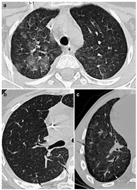
Fig. 4 Allogeneic BMT patients with no clinical cGVHD and good outcome. Extensive involvement of the lung parenchyma with consolidation (a), ground-glass opacity (b arrow) and nodules (b arrowhead) and pleural effusion (a, b). The final diagnosis was BOOP Fig. 3 An 8-month-old patient with allogeneic BMT, severe GVHD
and poor outcome. HRCT image shows diffuse extensive involvement of the lung parenchyma with ground-glass opacity, consolidation and interstitial thickening. The diagnosis after BAL was DAD
per day intravenously on day 1, and then was adjusted according to blood levels. The overall characteristics of the two groups of patients were similar.
Of the 16 patients, 12 presented with respiratory symptoms or chest radiographic anomalies at least 3 months after allogeneic BMT and underwent 43 HRCT scans, and 8 of the 16 patients (50%) had HRCT evidence of LONIPCs. All of the eight autologous BMT patients had HRCT scans (24 HRCT scans in total) on follow-up of the underlying diseases. One of the eight (12.5%) had HRCT evidence of LONIPCs. Thus the overall percentage of LONIPCs in our patients was of 37.5%. In the group of children who had two BMTs (4 allogeneic and 1 autolo-gous), 3 developed LONIPCs, while of the 19 patients who underwent only one BMT, 5 had LONIPCs.
Of the 16 patients, 7 did not develop GVHD; 2 had LONIPCs. Five patients had mild GVHD (stage I/II), of whom two had LONIPCs. Four had severe GVHD (stage III/ IV) and all of them had LONIPCs. All patients with moderate-to-severe GVHD had at least skin and gastrointestinal involvement. All four patients with severe GVHD died. HRCT
A total of 67 HRCT scans were performed in 20 patients (1– 8 studies per patient). The patterns of abnormalities on HRCT
in the nine patients with LONIPCs are summarized in Table2.
The pattern and the extension of CT findings were variable. Interstitial thickening was shown in all nine patients. The only patient with autologous BMT and abnormalities on
Fig. 5 Allogeneic BMT patients with mild cGVHD and good outcome. HRCT images show both extensive lung in-volvement with ground-glass opacity (a) and interstitial thickening (b black arrows), or centrilobular nodules and micronodules (c white arrow)
HRCT had marked interstitial thickening (Fig. 1) in strict spatial correlation with therapeutic radiation.
On the basis of HRCT it was impossible to subclassify patients with LONIPCs (BO, interstitial pneumonia, DAD, BOOP). None of the patients fulfilled the clinical and CT definitions for BO. Three of four patients with GVHD had
bullae (Fig. 2) and unilateral pneumothorax (Fig. 2).
However, patients with GVHD did not show a specific
pattern of abnormalities on HRCT (Table2). In the patient
with DAD, the pattern on HRCT, severe GVHD and poor clinical outcome are in accordance with reports in the
literature [4,16]. In this patient the lung abnormalities were
extensive (Fig. 3). Two patients were classified as having
BOOP on the basis of clinical and CT findings; lung
parenchyma involvement was extensive (Fig.4). In the two
patients who had allogeneic BMT and mild GVHD, the
clinical course was satisfactory (Fig.5).
Statistics
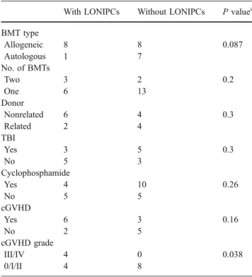
There was no statistically significant difference in the incidence of LONIPCs between patients after one or multiple BMTs (P=0.2), nor between patients receiving TBI or chemotherapy only as the conditioning regimen (P= 0.26). There was no statistically significant association between the presence of GVHD and a diagnosis of
LONIPCs on HRCT (P=0.16). However, there was a significant association between abnormalities on HRCT
and severe GVHD (P=0.038; Table 3). All patients with
severe GVHD died. The association between severe GVHD
(stage III/IV) and death was significant (P=0.002; Table4).
Discussion
The current longitudinal study is the first in the paediatric literature to investigate the incidence of LONIPCs on HRCT in allogeneic versus autologous BMT in order to ascertain whether a correlation exists with GVHD [21]. The incidence of LONIPCs was higher than that reported previously (range 3–26%) [22–24]. This can be partially explained by the relative homogeneity of the patients in our series in contrast with other studies that included adults and different conditioning regimens. LONIPCs are found almost exclusively in patients with allogeneic rather than
autologous BMT [1,13,25]. In fact, the only patient (one
of eight) with abnormalities on HRCT after autologous BMT had interstitial thickening following local radiation, thus probably not directly related to the BMT.
The patients in the two groups were relatively
homoge-neous (Table 3). The only significant association was with
the presence of severe GVHD (P=0.038), confirming the belief that the lung can be a target of GVHD [3]. The spatial extension of abnormalities on HRCT was not correlated with either GVHD or with clinical outcome. In fact, the two patients with extensive ground-glass opacities and consolidations were the only two without GVHD and had a very good outcome. These two children had been diagnosed with BOOP on the basis of HRCT and clinical findings. Our study supports reports in the literature that BOOP is not a manifestation of GVHD [26] and usually has a good outcome. On the contrary, most patients with abnormalities on HRCT associated with severe GVHD and
a poor outcome showed limited spatial extension (Fig. 2).
There was no correlation between a specific pattern and the presence or severity of GVHD, or with the clinical outcome in those with pulmonary pathology, although bullae and pneumothorax tended to show an association with severe
GVHD (Table 2).
We investigated the possibility of subclassifying LONIPCs into BO, BOOP and interstitial pneumonia Table 3 Comparison between patients with LONIPCs and without
LONIPCs.
With LONIPCs Without LONIPCs P valuea BMT type Allogeneic 8 8 0.087 Autologous 1 7 No. of BMTs Two 3 2 0.2 One 6 13 Donor Nonrelated 6 4 0.3 Related 2 4 TBI Yes 3 5 0.3 No 5 3 Cyclophosphamide Yes 4 10 0.26 No 5 5 cGVHD Yes 6 3 0.16 No 2 5 cGVHD grade III/IV 4 0 0.038 0/I/II 4 8 a
Fisher’s exact test
Table 4 Outcome in relation to cGVHD grade.
cGVHD grade Dead Alive P valuea
III/IV 4 0 0.002
0/I/II 1 11
a
according to the classification of Afessa et al. [5], which could be important in predicting the clinical outcome. This subclassification was not possible on the basis of HRCT findings alone. However, HRCT findings combined with clinical data and BAL results may be of real value in narrowing the differential diagnosis. For instance, in our series no patient fulfilled all the criteria for the diagnosis of BO, probably because they were too young or their clinical condition was too poor to perform accurate pulmonary function tests [11]. The finding of bullae or pneumothorax on HRCT is regarded as indirect evidence of BO or interstitial pneumonia. All of the patients with bullae and pneumothorax had GVHD and the clinical outcome was unfavourable. Also, in the only patient with DAD, the correct diagnosis resulted from the combination of clinical data, BAL and HRCT findings. The association of DAD with severe GVHD and poor clinical outcome is well
described in the literature [4,16] and was also found in our
patient. All patients with severe GVHD died, verifying the significant association between severe GVHD (stage III/IV) and death (P=0.02).
The clinical symptoms of LONIPCs may be insidious at the beginning of the disease, and routine radiological examination may also be normal. Furthermore, in LONIPCs, response to immunosuppressive therapy is limited, the best outcome being preservation of remaining lung function rather than significant improvement [14]. Since enhanced immunosuppression increases the risk of infection, the utility of such therapy is questionable when pulmonary dysfunction is chronic. Thus it is of the utmost importance to perform HRCT early in order to suggest the diagnosis of LONIPCs. Further prospective studies are required to determine the role of HRCT in patients with GVHD before the development of LONIPC-related respi-ratory symptoms.
Conclusion
The significant association between HRCT abnormalities and severe cGVHD (grades III/IV) suggests that LONIPCs can be a pulmonary manifestation of the disease. The prognosis seems to be related to the severity of GVHD and not to the pattern or extension of HRCT abnormalities. The accurate diagnosis of LONIPC subclasses is not possible on the basis of HRCT alone. However, HRCT combined with clinical data and BAL assists in narrowing the differential diagnosis and establishing the prognosis.
Acknowledgements The authors are grateful to Prof. Gian Paolo Cornalba, Department of Radiology, Università degli Studi di Milano, for his support and to Prof. Savvas Andronikou, Department of Radiology, Stellenbosch University Medical School, for his editorial assistance.
References
1. Eikenberry M, Bartakova H, Defor T et al (2005) Natural history of pulmonary complications in children after bone marrow transplantation. Biol Blood Marrow Transplant 11:56–64 2. Griese M, Rampf U, Hofmann D et al (2000) Pulmonary
complications after bone marrow transplantation in children: twenty-four years of experience in a single pediatric center. Pediatr Pulmonol 30:393–401
3. Yanik G, Cooke KR (2006) The lung as a target organ of graft-versus-host disease. Semin Hematol 43:42–52
4. Palmas A, Tefferi A, Myers JL (1998) Late-onset noninfectious pulmonary complications after allogeneic bone marrow transplan-tation. Br J Haematol 100:680–687
5. Afessa B, Litzow MR, Tefferi A (2001) Bronchiolitis obliterans and other late onset non-infectious pulmonary complications in hematopoietic stem cell transplantation. Bone Marrow Transplant 28:425–434
6. Yousem SA (1997) The histological spectrum of chronic necrotizing forms of pulmonary aspergillosis. Hum Pathol 28:650–656
7. Trisolini R, Bandini G, Stanzani M et al (2001) Morphologic changes leading to bronchiolitis obliterans in a patient with delayed non-infectious lung disease after allogeneic bone marrow transplantation. Bone Marrow Transplant 28:1167–1170 8. Przepiorka D, Weisdorf D, Martin P et al (1995) 1994 Consensus
Conference on Acute GVHD Grading. Bone Marrow Transplant 15:825–828
9. Austin JH, Muller NL, Friedman PJ et al (1996) Glossary of terms for CT of the lungs: recommendations of the Nomenclature Committee of the Fleischner Society. Radiology 200:327–331 10. Clark JG, Crawford SW, Madtes DK et al (1989) Obstructive lung
disease after allogeneic marrow transplantation: clinical presentation and course. Ann Intern Med 111:368–376
11. Sargent MA, Cairns RA, Murdoch MJ et al (1995) Obstructive lung disease in children after allogeneic bone marrow transplantation: evaluation with high-resolution CT. AJR 164:693–696
12. Worthy SA, Flint JD, Muller NL (1997) Pulmonary complications after bone marrow transplantation: high-resolution CT and pathologic findings. Radiographics 17:1359–1371
13. Gower WA, Collaco JM, Mogayzel PJ Jr (2007) Pulmonary dysfunction in pediatric hematopoietic stem cell transplant patients: non-infectious and long-term complications. Pediatr Blood Cancer 49:225–233
14. Watkins TR, Chien JW, Crawford SW (2005) Graft versus host-associated pulmonary disease and other idiopathic pulmonary complications after hematopoietic stem cell transplant. Semin Respir Crit Care Med 26:482–489
15. Clark JG, Hansen JA, Hertz MI et al (1993) NHLBI workshop summary. Idiopathic pneumonia syndrome after bone marrow transplantation. Am Rev Respir Dis 147:1601–1606
16. Ben-Abraham R, Paret G, Cohen R et al (2003) Diffuse alveolar hemorrhage following allogeneic bone marrow transplantation in children. Chest 124:660–664
17. Dodd JD, Müller NL (2005) Bronchiolitis obliterans organizing pneumonia after bone marrow transplantation: high-resolution computed tomography findings in 4 patients. J Comput Assist Tomogr 29:540–543
18. Flowers JR, Clunie G, Burke M et al (1992) Bronchiolitis obliterans organizing pneumonia: the clinical and radiological features of seven cases and a review of the literature. Clin Radiol 45:371–377
19. Akira M, Yamamoto S, Sakatani M (1998) Bronchiolitis obliterans organizing pneumonia manifesting as multiple large nodules or masses. AJR 170:291–295
20. Levine DS, Navarro OM, Chaudry G et al (2007) Imaging the complications of bone marrow transplantation in children. Radio-graphics 27:307–324
21. Kotloff RM, Ahya VN, Crawford SW (2004) Pulmonary complications of solid organ and hematopoietic stem cell transplantation. Am J Respir Crit Care Med 170:22–48
22. Yoshihara S, Yanik G, Cooke KR et al (2007) Bronchiolitis obliterans syndrome (BOS), bronchiolitis obliterans organizing pneumonia (BOOP), and other late-onset noninfectious pulmonary complications following allogeneic hematopoietic stem cell transplantation. Biol Blood Marrow Transplant 13:749–759 23. Watkins TR, Chien JW, Crawford SW (2005) Graft versus
host-associated pulmonary disease and other idiopathic pulmonary
complications after hematopoietic stem cell transplant. Semin Respir Crit Care Med 26:482–489
24. Patriarca F, Skert C, Sperotto A et al (2004) Incidence, outcome, and risk factors of late-onset noninfectious pulmonary complications after unrelated donor stem cell transplantation. Bone Marrow Transplant 33:751–758
25. Worthy SA, Flint JD, Muller NL (1997) Pulmonary complications after bone marrow transplantation: high-resolution CT and pathologic findings. Radiographics 17:1359–1371
26. Mathew P, Bozeman P, Krance RA et al (1994) Bronchiolitis obliterans organizing pneumonia (BOOP) in children after allogeneic bone marrow transplantation. Bone Marrow Transplant 13:221–223