Abstract This is a prospective study of spinal magnetic resonance imag-ing (MRI), electrophysiological record-ings, and neurological examinations of 100 patients admitted for surgery for adolescent idiopathic scoliosis (AIS), which was conducted to as-sess the prevalence of structural and functional abnormalities within the spinal cord in patients with clinically normal neurologic condition. In all patients the clinical diagnosis and in-tact neurological condition was as-certained by a spinal orthopedic sur-geon. Full-length spinal axis MRI studies (T1/T2 sequences) and so-mato-sensory evoked potentials of the tibial nerves (tSSEPs) were pre-operatively assessed by independent evaluators blinded to the patients’ medical histories. Structural spinal cord abnormalities were found in three of 100 AIS patients on MR imaging. In one patient a Chiari mal-formation type 1 with an accompa-nying syringomyelia was diagnosed, which required a suboccipital de-compression. In the other two pa-tients small thoracic syringomyelias were diagnosed. Abnormalities of spinal cord function were detected in 68% of the 100 patients: tSSEP la-tencies corrected for body height were increased in 56% of the pa-tients; pathological differences
be-tween tSSEPs on the left and right sides were present in 17% (12% in combination with a prolongation of the latency). The findings of this study indicate that MRI and electro-physiological examinations are es-sential to assess spinal cord abnor-malities that are clinically not de-tectable in AIS patients. Even in pa-tients with intact neurologic condi-tion and clinically typical right-curved thoracic scoliosis, the possibility of intraspinal pathologies should be ruled out by MRI. It is especially im-portant to detect structural patholo-gies like syringomyelia and Chiari malformation before proceeding with scoliosis surgery, as these conditions are associated with a higher neuro-logical risk during scoliosis surgery. The electrophysiological recordings made in the present study, with the high number of pathological tSSEPs, are indicative of functional abnor-malities with a subclinical involve-ment of the recorded neuronal path-ways. The relevance of the latter findings is not yet clear, but pre-op-erative tSSEP examinations offer the possibility of assessing alterations in spinal cord function that are unde-tectable by clinical examination. Keywords Idiopathic scoliosis · MRI · Tibial SSEP · Syringomyelia Oliver N. Hausmann Thomas Böni Christian W. A. Pfirrmann Armin Curt Kan Min
Preoperative radiological
and electrophysiological evaluation
in 100 adolescent idiopathic
scoliosis patients
Received: 18 September 2002 Revised: 8 February 2003 Accepted: 12 April 2003 Published online: 2 August 2003 © Springer-Verlag 2003
O. N. Hausmann · T. Böni · K. Min (✉) Department of Orthopedic Surgery, University Hospital Balgrist, University of Zürich, Forchstrasse 340, 8008 Zurich, Switzerland Fax: +41-1-3861609, e-mail: Min.Kan@balgrist.ch C. W. A. Pfirrmann Department of Radiology, University Hospital Balgrist, University of Zürich, Switzerland A. Curt
ParaCare, Swiss Paraplegic Center, University Hospital Balgrist, University of Zürich, Switzerland
Introduction
Adolescent idiopathic scoliosis (AIS) is typically seen in girls, and a right-sided thoracic curve is most common. However, AIS may present as the first symptom of an in-traspinal pathology. These pathologies, which include sy-ringomyelia, Chiari malformations, tethered cord, and tramedullary tumors, are risk factors for neurological in-jury during spine correction [6]. Surgical correction with an existing intradural pathology may be deleterious; thefore, these pathologies have to be investigated and, if re-quired, treated before the spine correction.
The aim of this study was to assess the value and clin-ical impact of a preoperative electrophysiologclin-ical exami-nation in combiexami-nation with a magnetic resonance imaging (MRI) evaluation to rule out any potential underlying in-tradural pathology. In the literature the indication for a routine MRI in AIS is still controversial [2, 6, 11, 13, 17, 18].
Materials and methods Patients
The study enrolled 100 adolescent patients with AIS admitted for sur-gical correction (80 female and 20 male; average age 15.3±2.2 SD years; enrollment from July 1997 until February 2002). Inclusion criteria were restricted to patients with an unsuspicious medical history (no pain, numbness or weakness of the lower extremities) of idiopathic scoliosis and with no abnormal findings on physical and neurological examination. Patients with neuromuscular or de-generative scoliosis or with any accompanying neurological disor-der were excluded from this study.
Preoperative clinical examinations
Prior to surgical correction, all patients underwent a comprehen-sive evaluation, which consisted of a patient history, and physical and neuro-orthopedic examinations by the orthopedic spinal sur-geon. Specific attention was given to assessing any disturbances of motor strength in the upper and lower extremities, exploration of sensation of the extremities and trunk, including cold and warm sensation, tendon reflexes and pathological pyramidal signs (levels of cutaneous abdominal reflexes, Babinski phenomenon, or in-creased muscle tone).
The curve pattern and size of the scoliosis was determined on standing plain antero-posterior and lateral radiographs of the whole spine, measuring the Cobb angle, as well as on supine bend-ing films. The Risser sign was used to denote the degree of skele-tal maturity.
Electrophysiological examinations
The pre-operative electrophysiological examinations by tibial so-mato-sensory evoked potential (tSSEP) recordings were performed by specially trained staff at a neurophysiology lab. The neurologist was blinded to the individual medical histories of the patients. Pre-operative SSEP evaluation is obtained routinely in all cases of complex spinal surgery that require intraoperative monitoring. Pre-operative SSEPs are used as baseline recordings for the intraoper-ative monitoring as well as for postoperintraoper-ative comparison.
Preoperative electrophysiological evaluation was performed 1–3 days before surgery. Tibial SSEPs were elicted using electrical stimulation by a conventional EMG machine (Dantec Keypoint; software 2.0) using surface electrodes. The tibial nerves were stim-ulated at the ankle (proximally placed cathodes, the anode placed 2 cm distally) with a square-wave stimulation for 0.2 ms at a fre-quency of 3 Hz. The stimulus intensity (up to a maximum of 40 mA) was adjusted to produce a clear muscle contraction. For recording, scalp electrodes were applied at Cz’ and Fz using the International 10/20 electrode system. The electrode impedance was maintained below 5 kOhm. The amplifier was set at 5µV/division, and the fre-quency bandpass was set at 50–2000 Hz. Two sets of 500 replica-tions were averaged to improve the signal to noise ratio for latency and amplitude measurements. All measurements were carried out at ambient temperature.
As the characteristic P40 latency of the early tSSEP is signifi-cantly influenced by body height, in all patients the value of P40 la-tency was corrected for body height (y=0.199×body height+3.9037) [8]. Therefore, body height corrected latencies of ±2SD (1SD= 0.57 ms) of the P40 were accepted as normal. Beside the latency of the early SSEP component, the interside difference of the P40 latency was calculated, as this value has been proven to be a sensitive marker to identify unilateral abnormalities of the tSSEP. Interside differ-ences of the P40 latency >2 ms were regarded as pathological [8].
MRI protocol
MR imaging of the whole spine was performed on a 1.5-T scanner (Siemens Symphony, Siemens Medical Systems, Erlangen, Ger-many), using a dedicated receive-only spine coil. The cervical, tho-racic and lumbar spine were imaged separately. Imaging protocol included a sagittal T2-weighted fast spin echo (FSE) sequence (TR 5000 ms/TE 130 ms) and a coronal T1-weighted spin echo (SE) se-quence (TR 700 ms/TE 12 ms) with the following parameters: matrix 512×225, field of view (FOV) 225×300 mm; slice thickness 4 mm, interslice gap 0.8, number of excitations (NEX) 4, echo train length (ETL) 15, as well as T2-weighted axial FSE scans (TR 5000 ms/TE 72 ms, matrix 210×256, FOV 150×150 mm; interslice gap 0.8 mm, NEX 2; ETL 7). For the cervical spine, axial T2-weighted gradient echo sequences were used: (TR 666 ms/TE 22 ms, flip angle 20°, matrix 210×256, FOV 200×200 mm, interslice gap 0.8 mm, NEX 2). All sequences were acquired without fat saturation. All examina-tions were evaluated preoperatively by experienced spinal radiolo-gists. Abnormalities of the spinal cord, the presence or absence of a syringomyelia and malformations such as the Chiari malforma-tion or diastematomyelia were noted.
The hindbrain herniation (Chiari malformation type 1) was de-fined as an elongation of the cerebral tonsils by more than 5 mm into the cervical spinal canal, a cervicomedullary kinking, and a re-duction of subarachnoid space anterior to the brain stem and pos-teroinferior to the cerebellum [15].
Results
In all 100 consecutive patients with AIS, full clinical and radiological workups were done. In 7/100 patients, preop-erative electrophysiological data were not available due to administrative problems with patients from abroad.
Preoperative findings
There were 34 single thoracic curves, 2 of them were left convex. There were 23 double thoracic curves, 12 double
major curves, 5 double thoracic curves, and 4 triple major curves, in which all the main thoracic curves were right sided. Further, there were 3 single lumbar, 8 lumbar major (all left-sided), and 11 single thoracolumbar curves. The mean Cobb angle was 56°±12° (range 43°–96°). The skele-tal maturity was distributed as follows: Risser 0 n=17; Risser 1 n=3; Risser 2 n=10; Risser 3 n=13; Risser 4 n=48; Risser 5 n=9.
Electrophysiology
Preoperative electrophysiological evaluation with SSEP of the tibial nerve was obtained in 93 of the 100 patients. Functional spinal cord abnormalities were detected in 68% of the 100 patients. Body height corrected P40 latencies were prolonged (body height corrected latency ±2SD) in 56% of patients. In 17% (n=16) the right–left interside difference was pathologically increased (>2 ms). How-ever, five of these patients (5.4%) had normal P40 laten-cies. Figure 1 plots the absolute (dark rhomboids) and the calculated body height corrected P40 latencies (line shows normal mean ±2SD latency).
MRI findings
In three of the 100 MR examinations (3%), relevant struc-tural intraspinal abnormalities were found.
Case 1
A 14-year-old girl presented with a progressive right-sided thoracic scoliosis. Patient’s history and clinical ex-amination findings were unremarkable. Menarche was 6 months prior to surgery. Radiological examination revealed a right convex long thoracic scoliosis from T4 to L4 of 57° with its apex at T9/10. MRI of the spine demonstrated a syringomyelia extending from T5 to L1 with a maximal diameter of 6 mm. The body height corrected tSSEP P40 latencies (right 36 ms, left 37 ms) were pathologically in-creased (body height 151 cm, expected normal P40 la-tency±2SD: 33.9±1.14 ms). The interside difference of 1 ms was within normal limits. Neurosurgical treatment of the syringomyelia was not considered necessary prior to scoliosis surgery. To reduce the risk of a stretching of the spinal cord by distraction of the spine, a ventral release by discectomy from T6/7 to T11/12 was performed prior to the dorsal corrective spinal fusion from T5 to L2. For the posterior instrumented fusion, titanium implants were used to allow follow-up of the syringomyelia with MR imaging. The perioperative course and postoperative fol-low-up were unremarkable.
Case 2
A 12-year-old pre-menarchal girl presented with a pro-gressive left-sided thoracic scoliosis from T5 to T12 of 50° with its apex at T8. The patient’s history and clinical examination findings were unremarkable. Preoperative Fig. 1 P40 latencies of
preop-erative somato-sensory evoked potentials of the tibial nerves (tSSEPs) are plotted against body height to rule out func-tional spinal cord abnormalities The dark rhomboids represent the absolute P40 latencies; the line with standard deviations represents the calculated body height corrected latencies (±2SD bars). Body height cor-rected P40 latencies were pro-longed (body height corrected latency ±2SD) in 56% of pa-tients
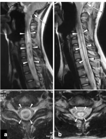
MRI demonstrated a Chiari malformation type 1 with a syringomyelia (Fig. 2a). Decompression of foramen mag-num, C1 laminectomy and partial resection of the cerebral tonsils combined with duraplasty and untethering of the filum terminale were performed by the neurosurgeon. Post-operative MRI showed a remarkable reduction of the size of the syringomyelia (Fig. 2b). Two months later, the sco-liosis was corrected by an anterior instrumentation from T6 to T12 from the left. The anterior instrumentation pre-vented lengthening of the spine during correction and the use of titanium implants enabled MRI follow-up of the sy-ringomyelia. The pre-operative electrophysiological record-ings of this patient (absolute and body height corrected values, as well as interside difference) were normal.
Case 3
A 17-year-old woman presented with a progressive dou-ble thoracic scoliosis with a main right convex thoracic scoliosis from T6 to L1 of 55° with its apex at T8 and a left convex upper thoracic scoliosis from T2 to T5 of 35°. Her history and clinical examination findings were unre-markable. The MRI revealed a small syringomyelia in the lower thoracic cord with extension from T8 to T10 with a maximal diameter of 4 mm (Fig. 3). Surgical treatment of the syringomyelia was not considered necessary due to its small size. The body height corrected latencies and mor-phology of the preoperative tSSEP were normal. Distrac-tion of the spinal cord was avoided during T2–T12 cor-rection from dorsal with titanium implants. Surgery, intra-operative monitoring, and the postintra-operative course were uneventful.
Fig. 2 a Preoperative sagittal and axial T2-weighted magnetic res-onance imaging (MRI) demonstrates a Chiari malformation type 1 with a large cervical syringomyelia (arrowheads) extending from C2 to T1. b Following the foramen magnum decompression (par-tial resection of the cerebral tonsils and the arch of C1 and a du-raplasty), the postoperative sagittal and axial T2-weighted MR im-ages show a remarkable reduction in the size of the syringomyelia (arrowheads)
Fig. 3 Sagittal and axial T2-weighted MR images show a small syringomyelia in the lower thoracic cord with exten-sion from T8 to T10 with a maximal diameter of 4 mm (arrow)
Surgery
The spine correction consisted of a posterior spinal fusion in 69 patients combined with same-day anterior release in 21 of them, and anterior fusion in 31 cases. The decision regarding isolated posterior or anterior approach depended on the type/magnitude of the curve and the degree of skele-tal maturity. All surgeries were performed by the senior author (K.M.). Intraoperative monitoring was routinely performed for all operations with SSEPs, and in a major-ity of the cases also with motor evoked potentials. There were no intraoperative or postoperative neurological com-plications.
Discussion
Although the present study included only patients with severe AIS requiring surgery (Cobb angle >43°), we did not find a higher incidence of structural spinal intradural pathologies compared with other reports in which cases with less severe scoliosis were also included [3, 5, 7, 10, 11, 16, 19]. We found structural intradural pathology in 3 of 100 AIS patients (3%) who presented without any ob-vious neurological deficits. This is more frequent than the findings of Do et al., who recently reported an intradural pathology of 0.6% in their large series of 327 AIS patients (average Cobb angle 57° of the major curve) [2]. Of the three patients with intradural pathology in our series, two had typical right-sided thoracic curves, and one had a left-sided thoracic curve – this latter patient being one of only two patients in the entire series with a left-sided thoracic curve. This gives a rate of 2% MRI abnormalities in typi-cal right-sided thoracic curves and 50% in left-sided tho-racic curves.
AIS is typically seen in girls, and a right-sided thoracic curve is most common. However, scoliosis per se can be the earliest manifestation of an occult intraspinal pathol-ogy, as such pathologies are known to be associated with the development of progressive scoliotic curves. A prospective MRI evaluation of idiopathic left thoracic scoliosis (mean Cobb angle 20°) found 2 out of 29 examined patients (7%) had an underlying syringomyelia [11].
The policy at our institution is to obtain preoperative MR images in all cases of AIS, independently of the side of the curvature. In our opinion the consequences of a missed spinal cord pathology that deteriorates due to the scoliosis correction would be in no way comparable to the costs of routine preoperative MRI examinations.
Patients with syringomyelia may present with scoliosis before manifesting any neurological abnormalities [1, 3, 9]. Further, intramedullary tumors can present with scoli-osis without neurological deficits [7]. However, the mech-anism by which a syringomyelia or a Chiari malformation type 1 (CM-1) causes the scoliosis is poorly understood [11, 12]. It has been suggested that an asymmetrically
ex-panding syringomyelia causes pressure and injures the lower motor neurons in the gray matter of the anterior horn in-nervating the trunk muscles. This imbalance of the paraver-tebral muscles may presumably cause the scoliosis [3]. Another explanation could be that patients with CM-1 have an equilibrium dysfunction disturbing the postural reflex system and causing scoliosis [11, 12].
Untreated syringomyelia poses higher neurological risk during surgical scoliosis correction, possibly due to tenu-ous blood supply making the spinal cord more vulnerable for distraction and compression [16]. Intradural patholo-gies should be known preoperatively, so that titanium im-plants can be used to enable further MRI follow-up exam-inations. Forty percent of scoliosis patients with a known syringomyelia present with neurological symptoms, usu-ally with subtle findings such as asymmetry of the reflexes; less commonly, however, they present with motor distur-bance, spasticity, muscle mass asymmetry, or temperature insensitivity [3]. An absent superficial abdominal reflex in an idiopathic scoliosis patient is suggestive for an un-derlying syringomyelia. Zadeh et al. reported that in all ten children they studied with IS and syringomyelia, the superficial abdominal reflex was consistently absent on the same side as the convexity of the curve [19]. In a study by Ono et al., hyperreflexia appeared in all patients with Cobb angle >20° who had syringomyelia with CM-1 [14]. However, we could not confirm this finding.
Known intradural pathologies should be treated before scoliosis correction. The treatment of syringomyelia is tailored to the individual, but generally consists of a du-raplasty with or without syringosubarachnoid shunt or sy-ringoperitoneal shunt. Cases of CM-1 are treated with a foramen magnum decompression („suboccipital craniec-tomy“) combined possibly with a duraplasty. The partial resection of the cerebellar tonsils (as in case 2) is neces-sary only in exceptional cases. Improvement of the scoli-osis after surgery of the syringomyelia has been reported in children [14].
The results of the present study, with pathologically prolonged tibial SSEP latencies of 56%, confirm our pre-vious results. In an earlier study we reported that preoper-ative body height corrected tSSEP latencies were pro-longed in 61% of neurologically normal AIS patients with no intradural pathology on MR imaging. The impairment of tSSEPs was not related to the extent of spine deformity as assessed by the Cobb angle [8]. The finding of pro-longed latencies in tSSEPs and unremarkable MRI find-ings may support the theory that occult structural changes of the spinal cord, undetectable on MRI, may be present in idiopathic scoliosis. We do not expect a prolongation of the tSSEP latencies due to the young age of this patient group, as the myelination is achieved during the first 2 years of life [15]. This corroborates the findings of Cheng and col-leagues, who could not find any correlation between tSSEP latencies and either age or body weight in a cohort of young (age 11–22, mean 12.5 years) and healthy subjects
[4]. In our series, the SSEP abnormalities were not spe-cific for morphological abnormality in spinal cord detectable with MRI. Of three patients with MRI abnormality, only one patient showed abnormal SSEPs.
Conclusions
The study reveals a clear indication for a routine preoper-ative MR imaging of the whole spine even in adolescent patients suffering from scoliosis suspected to be of idio-pathic etiology. Normal clinical neurological and neuro-physiological assessments, independent of the side of the
curve of scoliosis, are not reliable to exclude structural pathologies within the spinal cord. The high percentage of altered tSSEP recordings in AIS patients indicates a sub-clinical alteration of spinal cord function that is not re-lated to structural abnormalities detectable with MRI. The possibility of structural and functional alterations of spinal cord in AIS demands serious assessment, as unknown in-tradural findings such as Chiari, syringomyelia, or a teth-ered cord may cause complications arising from the cor-rection of the scoliosis. Preoperative SSEP assessment is mandatory as a baseline examination for intraoperative monitoring and to allow comparisons with the postopera-tive condition.
1. Baker AS, Dove J (1983) Progressive scoliosis as the first presenting sign of syringomyelia. J Bone Joint Surg Br 65: 472–473
2. Do T, Fras C, Burke S, Widmann R, Rawlins, Boachie-Adjei O (2001) Clin-ical value of routine preoperative mag-netic resonance imaging in adolescent idiopathic scoliosis. J Bone Joint Surg Am 83:577–579
3. Charry O, Koop S, Winter R, Lonstein J, Denis F, Bailey W (1994) Syringo-myelia and scoliosis: a review of twenty-five pediatric patients. J Pediatr Orthop 14:309–317
4. Cheng JC, Guo X, Sher AH (1998) Posterior tibial nerve somatosensory cortical evoked potentials in adolescent idiopathic scoliosis. Spine 23:332–337 5. Emery E, Redondo A, Rey A (1997)
Syringomyelia and Arnold Chiari in scoliosis initially classified as idio-pathic: experience with 25 patients. Eur Spine J 6:158–162
6. Freund M, Hähnel S, Thompson M, Sartor K (2001) Treatment planning in severe scoliosis: the role of MRI. Neu-roradiology 43:481–484
7. Gupta R, Sharma R, Vashisht S, Ghandi D, Jayaswal AK, Dave PK, Berry M (1999) Magnetic resonance evaluation of idiopathic scoliosis: a prospective study: Australas Radiol 43: 461–465
8. Hausmann O, Min K, Böni T, Erni T, Dietz V, Curt A (2003) SSEP in sur-gery of idiopathic scoliosis: the influ-ence of spine deformity and surgical approach. Eur Spine J 12:117–123 9. Hugus JJ, McGee-Collett M, Besser M,
Gurr KR, Taylor TK (1990) Scoliosis in syringomyelia: a new perspective. J Bone Joint Surg Br 72:1098 10. Majocco B, Deeney VF, Coulon R,
Parks PF (1997) Adolescent idiopathic scoliosis and the presence of spinal cord abnormalities. Spine 22:2537– 2541
11. Mejia EA, Hennrikus WA, Schwend RM, Emans JB (1996) A prospective evaluation of idiopathic left thoracic scoliosis with magnetic resonance imaging. J Pediatr Orthop 16:354–358 12. Muhonen MG, Menezes AH, Sawin
PD, Weinstein SL (1992) Scoliosis in pediatric Chiari malformations without myelodysplasia. J Neurosurg 77:69–77 13. O’Brien MF, Lenke LG, Bridwell KH, Blanke K, Baldus C (1994) Preopera-tive spinal canal investigation in large adolescent idiopathic scoliosis curves (70°–140°): is it warranted? Spine 19: 1606–1610
14. Ono A, Ueyama K, Okada A, Echi-goya N, Yokoyama T, Harata S (2002) Adult scoliosis in syringomyelia asso-ciated with Chiari I malformation. Spine 27:E23–E28
15. Osborn A (1994) Diagnostic neuro-radiology. Mosby, St. Louis
16. Phillips WA, Hensinger RN, Kling TF (1990) Management of scoliosis due to syringomyelia in childhood and adoles-cence. J Pediatr Orthop 10:351–354 17. Shen WJ, McDowell GS, Burke SW,
Levine DB, Chutorian AM (1996) Routine preoperative MRI and SEP studies in adolescent idiopathic scolio-sis. J Pediatr Orthop 16:350–353 18. Winter RB, Lonstein JE, Heithoff KB,
Kirkham JA (1997) Magnetic reso-nance imaging evaluation of the ado-lescent patient with idiopathic scoliosis before instrumentation and fusion. Spine 22:855–858
19. Zadeh HG, Sakka SA, Powell MP, Mehta MH (1995) Absent superficial abdominal reflexes in children with scoliosis. J Bone Joint Surg Br 77:762– 767