ORIGINAL PAPER
Resection of pediatric intracerebral tumors with the aid
of intraoperative real-time 3-D ultrasound
Nils H. Ulrich&Jan-Karl Burkhardt&Carlo Serra&
René-Ludwig Bernays&Oliver Bozinov
Received: 8 August 2011 / Accepted: 30 August 2011 / Published online: 17 September 2011 # Springer-Verlag 2011
Abstract
Purpose Intraoperative ultrasound (IOUS) has become a useful tool employed daily in neurosurgical procedures. In pediatric patients, IOUS offers a radiation-free and safe imaging method. This study aimed to evaluate the use of a new real-time 3-D IOUS technique (RT-3-D IOUS) in our pediatric patient cohort.
Material and methods Over 24 months, RT-3-D IOUS was performed in 22 pediatric patients (8 girls and 14 boys) with various brain tumors. These lesions were localized by a standard navigation system followed by analyses before, intermittently during, and after neurosurgical resection using the iU22 ultrasound system (Philips, Bothell, USA) connected to the RT-3-D probe (X7-2).
Results In all 22 patients, real-time 3-D ultrasound images of the lesions could be obtained during neurosurgical resection. Based on this imaging method, rapid orientation in the surgical field and the approach for the resection could be planned for all patients. In 18 patients (82%), RT-3-D IOUS revealed a gross total resection with a favorable neurological outcome.
Conclusion RT-3-D IOUS provides the surgeon with advanced orientation at the tumor site via immediate live two-plane imaging. However, navigation systems have yet
to be combined with RT-3-D IOUS. This combination would further improve intraoperative localization.
Keywords Neuronavigation . Pediatric neurosurgery . Intraoperative ultrasound . Image-guided surgery . Real-time 3-D ultrasound
Introduction
Image-guided systems, including neuronavigation, intra-operative MRI (iMRI), or intraintra-operative ultrasound (IOUS) have become essential neurosurgery intraoperative tools in recent decades. Especially in pediatric neurosurgery, the outcome of surgery often relies on the extension of tumor removal. In such situations, image-guided systems seem to be useful [1, 3, 4]. However, the integration of neuro-navigation based on preoperative data is limited, due to intraoperative changes, such as brain shift. Such tools as iMRI and intraoperative CT (iCT) seem to address this limitation [2,8, 14]. Unfortunately, iMRI and iCT require long image acquisition times, immense economic invest-ments, special surgical equipment (iMRI), and special workspaces. In addition, the ionizing radiation associated with iCT and low image definitions are hardly acceptable for many pediatric patients. Further improvements in imaging modalities, such as advanced IOUS, seem to be a promising alternative.
IOUS is an affordable, simple, and time-saving intraoperative tool that might be a valuable neuro-imaging technique of choice to shorten operation time, avoid radiation, and improve resection control. So far, only studies with real-time 2-D imaging or reconstructed (time-delayed) 3-D imaging have been conducted [10,
17,25,26].
N. H. Ulrich (*)
:
J.-K. Burkhardt:
C. Serra:
R.-L. Bernays:
O. Bozinov
Department of Neurosurgery, University Hospital Zurich, Frauenklinikstr.10,
8091 Zurich, Switzerland e-mail: nils.ulrich@usz.ch J.-K. Burkhardt
Department of Neurological Surgery, New York-Presbyterian Hospital, Weill Cornell Medical College,
In this study, we present our initial experience using a real-time 3-D IOUS (RT-3-D IOUS) system for pediatric cranial surgery. The aim of this study was to analyze the feasibility of RT-3-D IOUS to assist and improve the surgeon's orientation in pediatric neurosurgical cases.
Patients and methods
Patients
Twenty-two pediatric patients (8 females, 14 males; age range, 1–17 years) presenting supra- (n=12) or infratento-rial (n=10) brain lesions consecutively underwent surgery performed between June 2009 and May 2011 at the Department of Neurosurgery in Zurich by the senior author (O.B., Table1).
The lesion was diagnosed using pathohistological crite-ria. The identified lesions included five pilocytic astrocy-tomas (WHO grade I), five medulloblasastrocy-tomas (WHO grade VI), two atypical territorial/rhabdoidal tumors (WHO grade VI), two glioblastomas (WHO grade IV), and eight other lesions. All patients had comprehensive physical and
neurological examinations upon admission and discharge, including pre- and early postoperative MRI.
Intraoperative ultrasound technique
The US probe X7-2 was used to acquire real-time 3-D images in combination with the iU22 ultrasound system (Philips, Bothell, USA). The transducer and cord were placed in a sterile transparent plastic sheath, and the scan head was secured by a rubber band. The X7-2 transducer operated at 2–7 MHz. Sterile saline was used for irrigation between the sheathed transducer and the dura for adequate transmission of the acoustic beam. The X7-2 was mainly designed for cardiologists and gynecologists. The xMA-TRIX array technology utilizes 2,400 fully sampled elements for 360-degree focusing and steering. The array probe enables live xPlane imaging to acquire two full-resolution planes simultaneously from the region of interest. The system's multidirectional beam steering pro-vides unlimited planes in all directions, and live volume imaging allows the acquisition and rendering of full-volume data at true real-time frame rates with unparalleled isovoxel resolution.
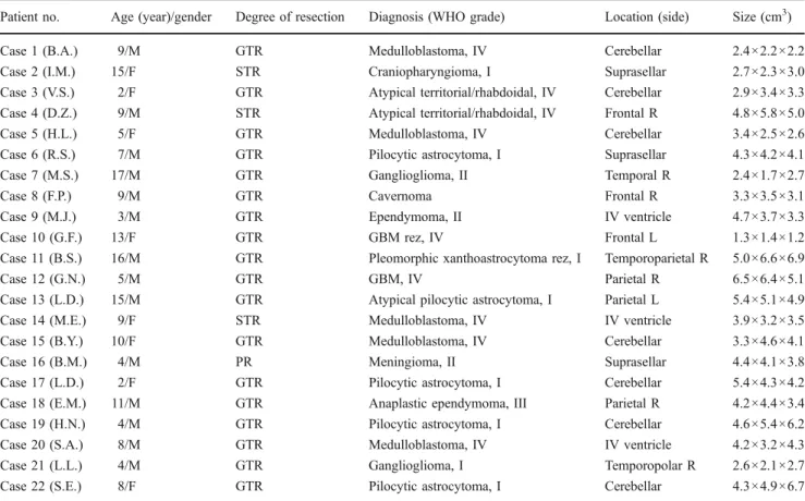
Table 1 Pediatric patient population
Patient no. Age (year)/gender Degree of resection Diagnosis (WHO grade) Location (side) Size (cm3)
Case 1 (B.A.) 9/M GTR Medulloblastoma, IV Cerebellar 2.4×2.2×2.2
Case 2 (I.M.) 15/F STR Craniopharyngioma, I Suprasellar 2.7×2.3×3.0
Case 3 (V.S.) 2/F GTR Atypical territorial/rhabdoidal, IV Cerebellar 2.9×3.4×3.3
Case 4 (D.Z.) 9/M STR Atypical territorial/rhabdoidal, IV Frontal R 4.8×5.8×5.0
Case 5 (H.L.) 5/F GTR Medulloblastoma, IV Cerebellar 3.4×2.5×2.6
Case 6 (R.S.) 7/M GTR Pilocytic astrocytoma, I Suprasellar 4.3×4.2×4.1
Case 7 (M.S.) 17/M GTR Ganglioglioma, II Temporal R 2.4×1.7×2.7
Case 8 (F.P.) 9/M GTR Cavernoma Frontal R 3.3×3.5×3.1
Case 9 (M.J.) 3/M GTR Ependymoma, II IV ventricle 4.7×3.7×3.3
Case 10 (G.F.) 13/F GTR GBM rez, IV Frontal L 1.3×1.4×1.2
Case 11 (B.S.) 16/M GTR Pleomorphic xanthoastrocytoma rez, I Temporoparietal R 5.0×6.6×6.9
Case 12 (G.N.) 5/M GTR GBM, IV Parietal R 6.5×6.4×5.1
Case 13 (L.D.) 15/M GTR Atypical pilocytic astrocytoma, I Parietal L 5.4×5.1×4.9
Case 14 (M.E.) 9/F STR Medulloblastoma, IV IV ventricle 3.9×3.2×3.5
Case 15 (B.Y.) 10/F GTR Medulloblastoma, IV Cerebellar 3.3×4.6×4.1
Case 16 (B.M.) 4/M PR Meningioma, II Suprasellar 4.4×4.1×3.8
Case 17 (L.D.) 2/F GTR Pilocytic astrocytoma, I Cerebellar 5.4×4.3×4.2
Case 18 (E.M.) 11/M GTR Anaplastic ependymoma, III Parietal R 4.2×4.4×3.4
Case 19 (H.N.) 4/M GTR Pilocytic astrocytoma, I Cerebellar 4.6×5.4×6.2
Case 20 (S.A.) 8/M GTR Medulloblastoma, IV IV ventricle 4.2×3.2×4.3
Case 21 (L.L.) 4/M GTR Ganglioglioma, I Temporopolar R 2.6×2.1×2.7
Case 22 (S.E.) 8/F GTR Pilocytic astrocytoma, I Cerebellar 4.3×4.9×6.7
Intraoperative ultrasound scanning
For optimal US imaging, all patients were positioned during surgery with a nearly vertical craniotomy channel such that saline remained in the resection cavity after dura opening. The initial US acquisition was performed extra-durally following craniotomy. After dura opening, when used intermittently or for a final scan, the US probe was moved over the field of interest with continuous irrigation of saline. The localization of the tumor and the usual surrounding anatomical landmarks (falx, tentorium, ven-tricles, brainstem, or skull base) were verified in each case. Colored Doppler mode was used (when needed) to verify vascular structures.
An acquisition procedure was not necessary, as we worked in a completely live 3-D mode. The“X-Plane” mode enables the user to visualize two US images in two different planes (in the degree of choice; Fig. 1). For instance, if the surgeon holds the US probe in an axial position, then a choice of 90° or 270° will illustrate the corresponding coronal (from the
lateral approach) or sagittal (from the anterior approach) image of interest simultaneously. Furthermore, during the “Live 3-D” mode, all US information from the array probe cone is displayed (Fig. 3). The width can be adjusted according to the needs of the surgeon. The images were displayed online and immediately responded (live) to all the movements of the probe.
Tumor visualization and resection control
After data collection was completed, tumor removal was performed. While debulking the lesion, intraoperative scanning was performed to estimate tumor remnants. After surgery was completed, an ultrasound scan was performed as a final resection control. In cases of suspicious areas that may harbor possible lesion remnants, microscopic reeval-uation was performed, and those areas were excised if possible.
The senior surgeon (O.B.) estimated the resection grade based on macroscopic findings and final US scan. These
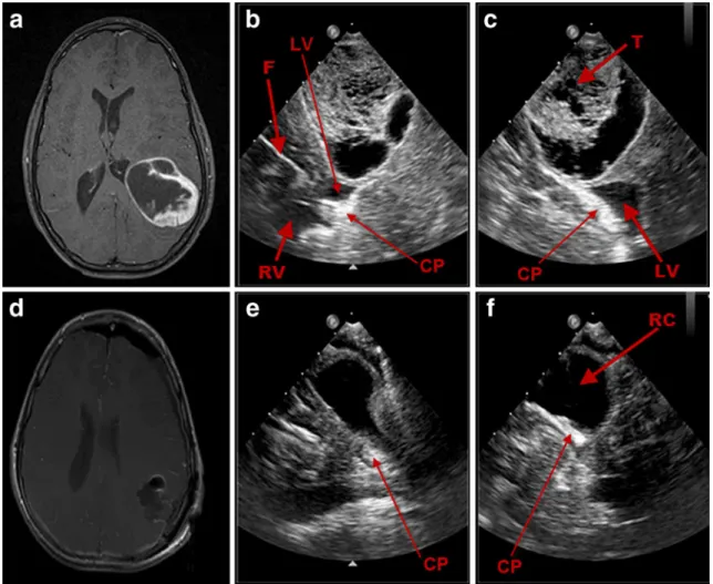
Fig. 1 Illustrative case no. 1 (case 13, L.D.): preoperative axial T1 MRI with contrast (a) of a 15-year-old male patient with a left parietal atypical pilocytic astrocytoma and the corresponding RT-3-D IOUS
images in“X-Plane” mode as shown in the coronal (b) and sagittal (c)
planes before tumor removal. RT-3-D IOUS of the coronal (e) and sagittal (f) planes and postoperative axial T1 MRI with contrast after 24 h (d) confirm total removal of the lesion. F falx, RV right ventricle, T tumor, LV left ventricle, RC resection cavity, CP choroid plexus
data were used at the end of the surgery to differentiate between a complete and incomplete resection. The extent of resection was confirmed by postoperative MRI within 24– 72 h by a pediatric neuroradiologist.
Results
In our consecutive case series of 22 pediatric patients, we were able to localize all lesions using the RT-3-D IOUS system without any complications. In all cases, rapid orientation of the surgeon was achieved via RT-3-D IOUS mode location of the lesion along with its leading anatomical landmarks (falx, tentorium, ventricles, brain-stem, or skull base). RT-3-D IOUS was useful in confirming lesion location and differentiating between solid tumors and cystic components (Figs. 2 and 3 in case 19, H.N., illustrative case no. 2). The average scanning time per
procedure was less than 10 min with an average scanning time of approximately 1–3 min per scan.
In all cases, the chosen craniotomy was sufficient for the US probe. No enlargement of the craniotomy for the US probe was needed. There were no complications, such as bleeding through manual scanning. After dural closure, the images were more difficult to interpret, due to remnant air bubbles located in the subdural space.
Gross total resection (GTR) was achieved in 18 out of 22 cases (81.8%; e.g., illustrative case 1, Fig.1). In two cases (cases 2 and 16), tumor infiltration involved the hypothal-amus, and radical resection was avoided. In another case (case 4), GTR was avoided, due to multiple occurrences of the tumor as well as infiltration of the eloquent areas. In only one case (case 14, M.E.), complete resection was determined by the surgeon but not confirmed by the neuroradiologist (Fig. 4, illustrative case no. 3). A residual tumor (1.0×0.5×0.5 cm) was detected and resected in a
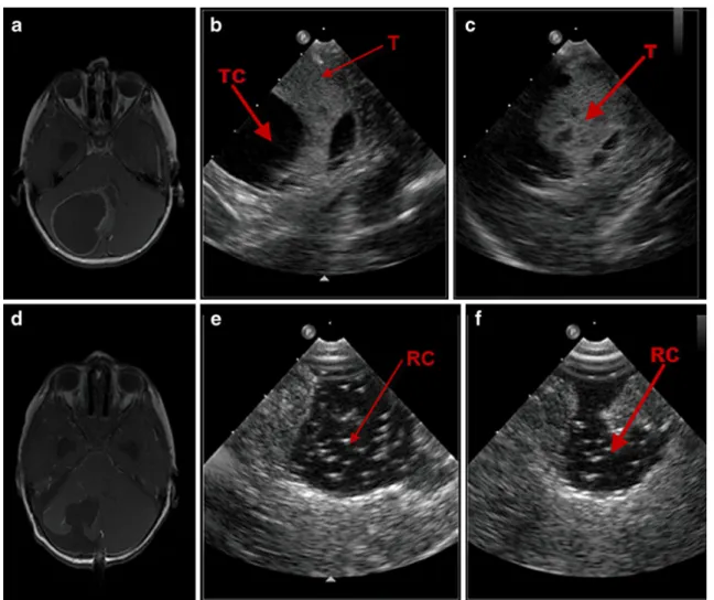
Fig. 2 Illustrative case no. 2 (case 19, H.N.): preoperative axial T1 MRI with contrast (a) of a 4-year-old male patient with a pilocytic astrocytoma in the right cerebellum and the corresponding
intra-operative RT-3-D IOUS images (b, c) in“X-Plane” mode as shown in
the coronal (b) and sagittal (c) planes before tumor removal. RT-3-D IOUS of the coronal (e) and sagittal (f) planes and postoperative axial T1 MRI with contrast after 24 h (d) confirm total removal of the lesion. TC tumor cyst, T tumor, RC resection cavity
second surgery 1 week after the primary surgery. Excluding the three intended subtotal resections, the GTR rate was 94.7% (1 out of 19). With regard to the localization of the
lesions, almost the same GTR resection rate of 90% (one out of ten) was detected for all posterior fossa tumors. There was no permanent neurological deficit in any case
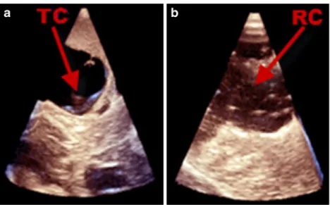
Fig. 3 Illustrative case no. 2 (case 19, H.N.): preoperative “Live 3-D” mode (a) of case 19 displaying the cystic component surrounding the tumor before tumor removal. In the
postre-section“Live 3-D” mode, b
tumor border differentiation is difficult. TC tumor cyst, RC resection cavity
Fig. 4 Illustrative case no. 3 (case 14, M.E.): preoperative axial T1 MRI with contrast (a) of a 9-year-old female with a medulloblastoma
in the fourth ventricle and the corresponding“X-Plane” 3-D IOUS
images in“X-Plane” mode as shown in coronal (b) and sagittal (c)
planes before tumor removal. Total removal was intended by the
surgeon but was not confirmed by the neuroradiologist. On the postoperative axial T1 MRI with contrast, a small residual was seen (left to the asterisk) corresponding to the IOUS in coronal (e) and sagittal (f) planes. RT residual tumor
due to GTR. Only temporary gait disturbance in most of the posterior fossa cases and limited instances of hormonal dysfunction in suprasellar tumor surgery have been noted.
Discussion
Intraoperative imaging and IOUS
Intraoperative imaging methods, such as iMRI and IOUS, that compensate for brain shift and mass reduction have been introduced in adult surgery and also, recently, in pediatric care [18,19,24]. IOUS is more widely used than iMRI, due to its lower costs and easier installation require-ments. In addition, iMRI requires specialized surgical instruments and extra shielding in the operating room [5]. Most importantly, iMRI requires higher operating costs. However, IOUS has a lower image quality and an individual learning curve before the surgeon can reliably correlate the anatomy to the pathological conditions. In our opinion, because of ionizing radiation, intraoperative iCT is not recommended for pediatric tumor resection and does not represent a viable alternative to iMRI or IOUS.
Kremer et al. have shown a total resection of 83% when iMRI was used as a tumor resection control in a study of 35 children [15]. We achieved a resection rate of 82% in a smaller cohort of 22 pediatric patients using IOUS. Samdani et al. demonstrated that a low-field iMRI in pediatric neurosurgery provides additional information for resection and real-time catheter placement [22], which we could also confirm with the IOUS technique. To date, there are no published prospective randomized studies comparing outcomes and resection control percentages between these two intraoperative imaging modalities.
Although IOUS is considered to be unsuitable for differentiating between surrounding tissue and edema [21], we could demonstrate improvements in this regard with our new RT-3-D IOUS. In 2008, He et al. used IOUS in combination with the contrast-enhancing agent to detect intracranial tumors and to differentiate between tumor tissue and surrounding tissue [13]. However, a pitfall of this method is that other sources other than contrast-enhancing tissue can represent a tumor. Additionally, contrast-enhanced ultrasound for neurosurgery is restricted in some countries.
Unsgaard et al. published a benchmark review article regarding developments in reconstructed 3-D US [26]. The authors used the intraoperative imaging system SonoWand® (Mison, Trondheim, Norway) that is a high-end US platform with a supplementary navigation system [12]. Several groups (including ours) have shown that the combination of image-guided neurosurgery with 3-D US increases the diagnostic value significantly [16, 17, 20,
28]. Furthermore, Letteboer et al. demonstrated that immediate updating during the operation helps to mini-mize the problem of brain shift [16].
Our RT-3-D probe is not primarily designed for neurosurgical procedures, but there is an increasing corporate interest in adapting probes for brain and tumor visualization in the future, as in recent years, all settings are digital and therefore also programmable. This possibility of individual setting adjustment leads to more customizable products. Improvements in image quality and increased pixels are inevitable. In this study, we present the first retrospective study of RT-3-D IOUS with intracranial lesions in a pediatric population to verify the value of RT-3-D IOUS for additional image-guided orientation in pediatric brain surgery. Ideally, RT-3-D should eliminate the reconstruction time with no further disadvantages.
Intraoperative real-time 3-D ultrasound
With our new RT-3-D IOUS technique, we achieved fast and real-time imaging of the progress of the operation during all stages of the updated operation. The resolution of our real-time 3-D probe was comparable to the standard quality associated with commonly used 2-D US systems and offered the surgeon enough accuracy to compensate for brain shift. An acquisition procedure was not necessary, as our 3-D data were immediately available in a live view. Repeated intraoperative image updating was valuable and easily accomplished in all cases.
Within our pediatric patient population, the use of our US system did not cause us to alter the surgical plan of a short skin incision and smallest possible bone flap. This fact is helpful in pediatric operations, as it eases postoper-ative pain and relieves psychological burden after the operation (for the patient and parents). There was no need for enlargement of the craniotomy or extra opening for ultrasound. In fact, such extra craniotomy for ultrasound imaging is not recommended, especially in pediatric cases [25]. The RT-3-D US probe is approximately 3×2 cm in size and does not exceed even a minimal craniotomy.
Extent of resection and RT-3-D IOUS
The extent of resection (EOR) in gliomas is a prognostic factor for survival [6, 9, 23]. El Beltagy et al. recently described the role of IOUS in resection of pediatric patients [10]. This group worked with a 2-D 6.5-MHz probe in conventional B-mode, and EOR was the same when comparing IOUS and immediate postoperative MRI. The authors were able to achieve a total resection rate of over 55% in their series of 25 heterogeneous pediatric patholo-gies. They concluded that 2-D IOUS is a useful imaging technique in defining the border between the tumor and
healthy brain tissue. In addition, this technique helped the surgeon to detect tumor remnants. Unsgaard et al. were able to improve tumor removal with 3-D IOUS in 2005. Furthermore, they were able to differentiate more effectively between tumor tissue and healthy brain [27]. Our GTR was 82% overall and 94% in intended GTR. However, we are not able to conclude that RT-3-D is superior to RT-2-D IOUS yet. In our experience, when previous work with RT-2-D and navigated reconstructed 3-D IOUS imaging are compared, RT-3-D IOUS only aided orientation during surgery. This technique did not present superior imaging but, instead, exhibited the same imaging quality but in an additional plane. Similar to our results, Roth et al. [19] achieved a 100% GTR rate when GTR was intended (two cases with hypothalamic involvement were not operated radically). Their series was smaller (15 cases), but the authors used a navigational reconstructing 3-D IOUS system [19]. Combi-nation systems with neuronavigation necessitates reconstruc-tion time, but automatic systems, such as IG Sonic (BrainLab, Munich, Germany) and SonoWand® (Mison, Trondheim, Norway), have reduced this waiting time significantly.
Neurological deficit is a risk of complete resection. In this series, we did not resect radically in eloquent areas when significant damage was likely (e.g., hypothalamic infiltration, cases 2 and 16) or when radical resection of an eloquent lesion (because of additional other lesions) did not represent any prognostic advantage (e.g., case 4). All other current IOUS publications address this issue with a similar opinion and results [7,11,19,28]. There are no reports of increased neurological deficits associated with IOUS-guided radical resections. On the other hand, it is difficult to conclude that IOUS directly contributes to the preserva-tion of neurological funcpreserva-tion. This important aid is more directly supplied by intraoperative electrophysiological monitoring, as performed in all of our cases in eloquent regions.
Resection control—technical considerations
Unsgaard et al. were able to better differentiate between tumor tissue and healthy brain tissue in 2005 [27]. El Beltagy et al. described a technique in which filling the resection cavity with saline was performed for comparison of resection size and MRI images for the measurement of total resection amount. We did not fill our cavity with saline for the resection control. We have never used this technique to measure the resection diameter and are dubious of its suitability. Neither Unsgaard et al. nor other US researchers have described this resection control method. In addition, a validation of this method by prospective studies has not been published. Tumors do have extreme expansive attributes, and in many cases (especially in posterior fossa
tumors), immediate mass reduction after dura opening is necessary to avoid herniation. Such a mass effect can hardly be reproduced by saline insertion. Additionally, El Beltagy reports that leaving Surgicel in the tumor cavity may interfere with IOUS interpretation. This finding is in concordance with our experience and is the reason why we did not use artificial material before or after scanning. The artificial tissue may also lead to a misleading contrast-enhancing lesion in the postoperative MRI.
Another problem with resection control using IOUS is defining the tumor border. Occasionally, brain tissue with a small local hemorrhage can imitate tumor tissue after a certain procedural time, and further imaging becomes more difficult. Interestingly, most IOUS articles prefer extradural images for their publications, as they appear to be finely defined. We used an ultrasound aspirator to debulk gliomas quickly in the middle of the tumor before concentrating on the borders. With this technique, the tumor borders can still be easily confirmed with US even after debulking and partial resection. The quality of the US imaging needs to increase continuously, but this improvement is certainly possible, as better probes are being produced with higher frequencies.
Learning curve and real-time versus reconstructed 3-D US
El Beltagy et al. stated that in some cases, IOUS could substitute for intraoperative MRI [10]. Roth et al. also expressed similar thoughts and noted that superior iMRI results have not been yet presented [19]. However, especially for ultrasound users, it is important to not forget that most pediatric brain tumors (especially malignant ones) need early postoperative MRI for the study protocols. In order to obtain a complete resected tumor by postoperative MRI, in our opinion, the best method remains high-field intraoperative MRI. However, there are only few centers that can afford such a system. An alternative is important as intraoperative imaging is even more significant and often demanded by the patients and parents.
El Beltagy et al. noted that increasing use of IOUS leads to improved ease of use for neurosurgeons [10]. There is certainly a learning curve and experience is required when interpreting intraoperative US images, especially after dura opening and partial resection. We support the notion that US becomes more beneficial with neuronavigational sup-port [11]. The group around El Beltagy did not use a navigation system during their study, and we are confident that adding this tool would increase their contentment with IOUS.
In summary, RT-3-D IOUS improves the orientation for an experienced US user by simultaneous display of two images in the degree of choice. Inexperienced US users should make use of the ultrasound and
neuronavigation-combined systems. A significant enhancement in orienta-tion should accompany the use of the combined system.
Outlook and future
The literature still lacks prospective studies that evaluate and compare clinical outcomes between different US users and intraoperative imaging modalities. In addition, iMRI is a developing technology compared to IOUS, and these modalities appear to be in concurrence. However, some neurosurgical centers already practice fast preliminary resection control with IOUS (to save time) and subsequent-ly finish cases with an iMRI. This practice indicates that both methods can be used in combination.
Our experience with RT-3-D IOUS use for pediatric patients is encouraging, but we are not yet satisfied with the image resolution. There is no doubt that this technique will improve in the near future as novel probes are developed. High frequency probes already provide high-structure definition and will lead to a new interpretation of tumor borders and edema.
Another future possibility will be the combination of an RT-3-D IOUS probe with a navigation system. The ability to visualize the live ultrasound images in the convenient axial, coronal, and sagittal planes next to the corresponding preoperative MRI would further improve orientation and reduce surgical time [7]. However, as this capability requires a high amount of constant data transfer and accompanying calculation, it is currently difficult to realize.
Conclusion
In our study, real-time 3-D intraoperative ultrasound provided the surgeon with an advanced wider orientation via“live” two-plane imaging in all stages of surgery, and this technique was useful as a resection control aid. Combining this technique with a navigation system would provide a further improvement in local intraoperative orientation.
Disclosure The real-time 3-D US transducer has been provided by
the company Philips for research purposes. This is not the case for the US system IU22, which was bought by the department. No further financial collaborations, consulting contracts, or conflicts of interest exist.
References
1. Albright AL, Wisoff JH, Zeltzer PM, Boyett JM, Rorke LB, Stanley P (1996) Effects of medulloblastoma resections on
outcome in children: a report from the Children's Cancer Group.
Neurosurgery 38:265–271
2. Balmer B, Bernays RL, Kollias SS, Yonekawa Y (2002) Interven-tional MR-guided neuroendoscopy: a new therapeutic option for children. J Pediatr Surg 37:668–672
3. Berger MS, Deliganis AV, Dobbins J, Keles GE (1994) The effect of extent of resection on recurrence in patients with low grade cerebral hemisphere gliomas. Cancer 74:1784–1791
4. Berger MS (1996) The impact of technical adjuncts in the surgical management of cerebral hemispheric low-grade gliomas of
childhood. J Neurooncol 28:129–155
5. Bernays RL, Kollias SS, Khan N, Brandner S, Meier S, Yonekawa Y (2002) Histological yield, complications, and technological considerations in 114 consecutive frameless stereotactic biopsy procedures aided by open intraoperative magnetic resonance
imaging. J Neurosurg 97:354–362
6. Black PM (2000) The present and future of cerebral tumor surgery
in children. Childs Nerv Syst 16:821–828
7. Bozinov O, Burkhardt JK, Fischer CM, Kockro RA, Bernays RL, Bertalanffy H (2011) Advantages and limitations of intraoperative 3D ultrasound in neurosurgery. Technical note. Acta Neurochir Suppl 109:191–196
8. Broggi G, Ferroli P, Franzini A, Dones L, Marras C, Marchetti M, Maccagnano E (2003) CT-guided neurosurgery: preliminary experience. Acta Neurochir Suppl 85:101–104
9. Cohen KJ, Broniscer A, Glod J (2001) Pediatric glial tumors. Curr
Treat Options Oncol 2:529–536
10. El Beltagy MA, Aggag M, Kamal M (2010) Role of intraoperative ultrasound in resection of pediatric brain tumors. Childs Nerv Syst
26:1189–1193
11. Enchev Y, Bozinov O, Miller D, Tirakotai W, Heinze S, Benes L, Bertalanffy H, Sure U (2006) Image-guided ultrasonography for
recurrent cystic gliomas. Acta Neurochir (Wien) 148:1053–1063,
discussion 1063
12. Gronningsaeter A, Kleven A, Ommedal S, Aarseth TE, Lie T, Lindseth F, Lango T, Unsgard G (2000) SonoWand, an ultrasound-based neuronavigation system. Neurosurgery
47:1373–1379, discussion 1379–1380
13. He W, Jiang XQ, Wang S, Zhang MZ, Zhao JZ, Liu HZ, Ma J, Xiang DY, Wang LS (2008) Intraoperative contrast-enhanced ultrasound for brain tumors. Clin Imaging 32:419–424
14. Jolesz FA (2003) Future perspectives in intraoperative imaging. Acta Neurochir Suppl 85:7–13
15. Kremer P, Tronnier V, Steiner HH, Metzner R, Ebinger F, Rating D, Hartmann M, Seitz A, Unterberg A, Wirtz CR (2006) Intraoperative MRI for interventional neurosurgical procedures and tumor resection
control in children. Childs Nerv Syst 22:674–678
16. Letteboer MM, Willems PW, Viergever MA, Niessen WJ (2005) Brain shift estimation in image-guided neurosurgery using 3-D
ultrasound. IEEE Trans Biomed Eng 52:268–276
17. Lindner D, Trantakis C, Renner C, Arnold S, Schmitgen A, Schneider J, Meixensberger J (2006) Application of intraoperative 3D ultrasound during navigated tumor resection. Minim Invasive
Neurosurg 49:197–202
18. Nimsky C, Ganslandt O, Hastreiter P, Fahlbusch R (2001)
Intraoperative compensation for brain shift. Surg Neurol 56:357–
364, discussion 364–355
19. Roth J, Beni-Adani L, Biyani N, Constantini S (2006) Classical and real-time neuronavigation in pediatric neurosurgery. Childs Nerv Syst 22:1065–1071
20. Rubin JM, Mirfakhraee M, Duda EE, Dohrmann GJ, Brown F (1980) Intraoperative ultrasound examination of the brain.
Radiology 137:831–832
21. Rubin JM, Quint DJ (2000) Intraoperative US versus intra-operative MR imaging for guidance during intracranial
22. Samdani AF, Schulder M, Catrambone JE, Carmel PW (2005) Use of a compact intraoperative low-field magnetic imager in pediatric
neurosurgery. Childs Nerv Syst 21:108–113, discussion 114
23. Sanai N, Polley MY, McDermott MW, Parsa AT, Berger MS (2011) An extent of resection threshold for newly diagnosed glioblastomas. J Neurosurg 115:3–8
24. Tronnier VM, Bonsanto MM, Staubert A, Knauth M, Kunze S, Wirtz CR (2001) Comparison of intraoperative MR imaging and 3D-navigated ultrasonography in the detection and resection control of lesions. Neurosurg Focus 10:E3
25. Unsgaard G, Gronningsaeter A, Ommedal S, Nagelhus Hernes TA (2002) Brain operations guided by real-time two-dimensional ultrasound: new possibilities as a result of improved image
quality. Neurosurgery 51:402–411, discussion 411–402
26. Unsgaard G, Ommedal S, Muller T, Gronningsaeter A, Nagelhus Hernes TA (2002) Neuronavigation by intraoper-ative three-dimensional ultrasound: initial experience during
brain tumor resection. Neurosurgery 50:804–812, discussion
812
27. Unsgaard G, Selbekk T, Brostrup Muller T, Ommedal S, Torp SH, Myhr G, Bang J, Nagelhus Hernes TA (2005) Ability of navigated 3D ultrasound to delineate gliomas and metastases—comparison of image interpretations with histopathology. Acta Neurochir
(Wien) 147:1259–1269, discussion 1269
28. Unsgaard G, Rygh OM, Selbekk T, Muller TB, Kolstad F, Lindseth F, Hernes TA (2006) Intra-operative 3D ultrasound in
neurosurgery. Acta Neurochir (Wien) 148:235–253, discussion