1085-9195/99/30/437-454/$14.50
Uptake of Reconstituted Na, K-ATPase
Vesicles by Isolated Lymphocytes
Measured by FACS, Confocal
Microscopy and Spectrofluorometry
Beatrice M. Anner* and B. Volet
Department of Pharmacology, Geneva University Medical School, CH-
1211 Geneva 4, Switzerland," E-mail: [email protected]
ABSTRACT
Na,K-ATPase (EC 3.6.1.37, Na,K-ATPase) is a fundamental vital membrane transport and receptor system which, after bio- synthesis, is exported to the plasma membrane in inside-out vesicles. Na,K-ATPase can be extracted form the natural mem- brane and inserted into artificially formed phosphatidylcholine vesicles (liposomes). The ultrastructure of the reconstituted vesicles has been fully described. In the present work, the Na,K- ATPase-vesicles were labeled with fluorescent tracers either in their water or membrane phase, incubated with freshly isolated h u m a n lymphocytes, and the resulting cellular fluorescence measured with fluorescence activated cell sorting (FACS), con- focal microscopy and spectrofluorometry. The FACS data show that all lymphocytes take up Na,K-ATPase-vesicles in a dose- and temperature-dependent fashion. Three-dimensional analy- sis of the fluorescence by confocal microscopy reveals that the fluorescence is contained within the cells. Quantitative determi- nation by spectrofluorometry indicates that depending on the
*Author to w h o m all correspondence and reprint requests should be addressed.
438 Anner and Volet vesicle/cell ratio, a single lymphocyte takes up 650 to 36,500 vesicles within 30 min at 37~ together with up to about 200,000 renal Na,K-ATPase molecules.
Index Entries: Reconstituted Na,K-ATPase-vesicles; lympho- cytes; uptake; FACS; confocal microscopy; spectrofluorometry; quantitative analysis.
INTRODUCTION
The cytoplasm of eukaryotic cells is rich in vesicles of 20-100 nm diameter. They serve as shuttles for neurotransmitters of hormones or they deliver or retrieve from the plasma membrane the integral proteins (1,2). The Na,K-ATPase (Na,K-ATPase) or sodium pump is a fundamental membrane system that is responsible for the electro- lyte balance of the organism, organs, cells, and organelles (3); it expels Na from the cell and replaces it by K by an electrogenic pro- cess (3 Na out vs 2 K in) coupled to hydrolysis of I ATP molecule. The process is modulated by an inhibitory extracellular receptor for cardioactive steroids such as ouabain and their presumed endog-
enous analogs (4). The resulting transmembrane ion gradient and
membrane potential is underlying cell excitability, impulse conduc- tion (5), and muscle contraction (6) in nerve and muscle. In epithelia, the polarized localization of the Na,K-ATPase leads to transepithelial Na transport and is the motor of Na reabsorption, e.g., in the kidney. Vesicles similar to the ones found within cells can be formed artificially and integral membrane proteins, e.g., Na,K-ATPase, inserted into their membranes (7). Their ultrastructure has revealed resemblance in size and aspect (8,9) to natural intracellular vesicles as both types are reaching apparently the same minimal energy state
because of their like chemical composition (10). The artificially
formed Na,K-ATPase vesicles carry 50% of the reconstituted Na,K- ATPase in inside-out orientation and are, therefore, partially analo- gous to the natural intracellular inside-out vesicles delivering the Na,K-ATPase to the plasma membrane. Therefore, it should be pos- sible to let them enter into the cell where they are predicted to join the normal intracellular vesicle traffic.
Lymphocytes take up externally added phospholipid vesicles
or liposomes (11-13). Liposome uptake involves binding to the cell
surface followed by endocytosis (14). Recently it was shown that
they are able also to take up nanospheres without apparent toxicity
(15). However, it has not been examined whether isolated human lymphocytes take up vesicles containing foreign membrane proteins. In the present work Na,K-ATPase-vesicles are labeled by fluores- cent markers of the membrane or of the water phase and their uptake by isolated human lymphocytes is demonstrated by three different techniques. FACS analysis shows vesicle uptake by all cells; confocal microscopy visualizes the vesicle entry on a single cell level and spec- trofluorometry reveals that the cells are able to take up about three times their own amount of membrane surface and about 10 times their own amount of Na,K-ATPase protein within 30 min at 37~
METHODS
Materials, Isolation of NadGA TPase, and Biochemical Measurements Phosphate buffered saline (PBS), RPMI-1640 medium, and Hank's balanced salt solution (HBSS) were from Tecnomara (Ztirich, CH). Phosphatidylcholine (grade II) was from Lipid products (Nutfield, UK); CF was purchased from Fluka (Buchs, CH). N-(5- fluoresceinthiocarbamoyl) l,2-dihexadecanoyl-sn-glycero-3-phospho- ethanolamine triethyl ammonium salt (Fluo-PE) was from Molecular Probes (Eugene, OR). LeucoSep tubes (Esquire Chemie AG, Ziirich, CH) were used for the isolation of peripheral blood mononuclear cells. Na,K-ATPase was purified from the outer medulla of rabbit kidneys to a specific activity of 1200 to 1800 ktmol P i / m g protein/h by treatment with SDS and centrifugation across a sucrose gradient
(16). The Na,K-ATPase activity was measured by the linked-enzyme
assay (17) and the protein was determined as described by Smith et al. (18). The Na,K-ATPase was separated into ct and [3 subunits by PAGE, which together make up about 90% if the protein as estimated by Coomassie blue staining and laser densitometry (17).
Insertion of Na, K-A TPase into Vesicles
Vesicles are formed by making use of the fact that phosphati- dylcholine (PC), dissolved in a 1% cholate solution, forms closed vesicles upon detergent removal by dialysis (8). Membrane frag- ments containing the purified Na,K-ATPase are treated with 1% cho- late to d i s r u p t the continuity of the lipid bilayer and single Na,K-ATPase molecules obtained in the supernatant of a 100,000g centrifugation ("soluble" Na,K-ATPase). When the "soluble" Na,K-
440 Anner and Volet ATPase is added to the PC solution and the cholate removed by dialysis, Na,K-ATPase spontaneously inserts into the lipid bilayer and is seen by freeze fracture electron microscopy at regular dis- tances (8,9) in the vesicles indicating noninteraction between the Na,K-ATPase molecules. The number of reconstituted Na,K-ATPase molecules is directly proportional to the protein/lipid ratio used for reconstitution and statistical analysis of the intramembrane particle distribution on concave and convex fracture faces has revealed ran- dom orientation (9).
A lipid-cholate stock solution was prepared as follows: 60 mg PC in 600 ~tL chloroform-methanol was dried under a slow, steady stream of ultrapure N 2 in a 50-mL round-bottomed flask fixed to a Btichi minirotary apparatus in a water bath at 25~ for about 30 rain in the dark until a thin film was formed to which 3 mL dried ether was added; this process was repeated. The third film was formed first without N 2 blow to avoid solvent capping and was then exten-
sively dried by N 2 for 90 min at 25~ followed by 2 min at 30~
Then, 3 mL of a solution containing 50 mM NaC1, 50 mM KC1, 5 mM MgC12,1 mM EDTA, 30 mM L-histidine, and 1% Na-cholate, p H 7.2, (solution A) was added to the lipid film and the flask rotated until a clear solution was obtained from which 100 ~tL aliquots were put
under nitrogen and stored at -70~ Na,K-ATPase (600 ~tg protein)
was pelleted at 100,000g for 15 rain to in a Beckman airfuge at 0~ resuspended in 100 ~tL of solution A and again centrifuged for 15 min at 100,000g; the Na,K-ATPase activity and protein content of the supernatant containing the "soluble" Na,K-ATPase were deter- mined and the remaining 80 ~tL Na, K-ATPase solution added to 80 ~tL of PC solution; the Na,K-ATPase activity after PC addition was determined, the PC-Na,K-ATPase solution added to EDTA treated sterile dialysis tubing, and the cholate removed during 15 h at 0~ in 100 mL cholate-free solution A (solution B).
Fluorescent Labeling of the Vesicles
The vesicles were labeled in two manners: in the water phase by carboxyfluorescein (CF) according to published procedures (19,20) or in the membrane phase by Fluorescein-phosphatidylethanola- mine (Fluo-PE). For CF-labeling 200 mM CF was added prior to dialysis. The pH was adjusted to 7.2 by 1 M NaOH, which brought 330 mM additional Na ions to the solution. The dialysis temperature was raised to 20~ to prevent crystalization; to eliminate the exter-
nal CF after dialysis, the liposomes were centrifuged four times for 60 rain at 100,000g in a Beckman airfuge at 0~ and resuspended in 200 ktL solution B. The tightness of the liposomes was controled by incubating them in the presence and absence of detergent and deter- mining the entrapped and released CF by spectrofluorometry tak- ing advantage of the CF selfquenching (20,21). The entrapped fluorescence measured after detergent-lysis was found to be directly proportional to the liposome concentration in confirmation that there was no contaminating external dye. The quenching of the washed CF-liposomes only slightly diminished within 30 d at 5~ indicating negligible leak of CF. No CF-release was seen when the temperatures was increased from 0~ to 22~ or from 0~ to 37~ for 30 min. Thus, the CF-liposome stability was appropriate for incubation with cells. The ultrastructure of the CF-vesicles showed that the high CF and Na concentration induced multilayering, yield- ing a 330 nm average diameter (10). Therefore, a membrane label was used: (i) to avoid multilayering; and (ii) to be able to follow the uptake of the vesicle membranes. For this purpose, 20% PC were replaced by Fluo-PE to form the lipid-cholate stock solution described above. Fluo-PE did not alter the ultrastructure of the vesicles (10). Fluorescent vesicles without Na,K-ATPase protein were also prepared.
Isolation of Lgmphocgtes and Incubation with Fluorescent Vesicles Human blood was obtained from healthy voluntary blood donors. Peripheral blood mononuclear cells were isolated by cen- trifugation on a Ficoll gradient (22) in LeucoSep tubes, washed three times in HBSS, and kept in RPMI-1640 medium in the absence of serum. This procedure commonly yielded 90-95% lymphocytes and 5-10% monocytes as measured by flow cytometry. Cells were counted in a haemacytometer and viability was assessed before and after vesicle addition by Trypan blue exclusion. Increasing concen- trations (0.5 to 200 ~L) of vesicles were pelleted by centrifugation at 100,000g in the Beckman Airfuge at 0~ 0.5 to 2 million peripheral blood mononuclear cells were added in 50 to 200 ~tL RPMI-1640 or PBS, and the suspension was incubated for 5 min to 2 h at 37~ I mL PBS was added, the cells pelleted for 10 rain at 400g at 20~ in a Sorvall H-2080 swing out rotor in a Sorvall Centrifuge RC-3B to remove the external liposomes and resuspended in 1 mL PBS. After incubation, the suspension was centrifuged at 400g for 10 min at
442 Anner and Volet
room temperature to remove the external liposomes and the pellet suspended in I mL PBS. Aliquots of the cell suspension were used:
1. For analysis of cellular fluorescence by flow cytometry.
2. For quantification of the intracellular Fluo-PE content by spectrof- luorometry.
3. For measuring cell viability by Trypan blue exclusion.
Fluorescent Techniques for Measuring Vesicle Uptake
Cell pellets were suspended in 1 mL of PBS and analyzed in a Becton Dickinson FACScan (San Jose, CA). The lymphocyte and monocyte populations were differentiated by appropriate gating, excluding clusters and cell debris; 10,000 events from each cell popu- lation were analyzed. Mean cellular fluorescence and standard deviation were calculated for each sample and expressed in arbitrary fluorescence units, using the program Lysis II (Becton Dickinson). To determine the number of liposomes incorporated per cell, 100 ~tL cell suspension as well as the supernatants recovered after the 400g centrifugation step were added to 3 mL PBS in a quartz cuvet and
the fluorescence measured in a Zeiss PMQ II
(~'ex
436nm, ~em
520nm); 10 ~tL Triton X-100 (10%) was added after the first reading to release the Fluo-PE taken up by the cell. The quantity of Fluo-PE was determined by comparison with standard curves established with pure Fluo-PE. The number of vesicles was calculated on the
basis of their size and composition and amounted to 3.58 x 10 4 60 nm
vesicles (Na,K-ATPase-free) or 1.22 x 10 4 100 nm vesicles (with Na,K-
ATPase) per mg Fluo-PE. For confocal microscopy, the cells were suspended in 10 ~tL fixing solution, mounted as described by Lenette
(21), put on a slide, and covered. Specimens were observed with a Zeiss confocal laser scan fluorescence inverted microscope (LSM 410, Carl Zeiss, Oberkochen, Germany) equipped with two different lasers: a Helium-Neon (He-Ne) laser (excitation wavelength at 543 nm) and an Argon laser (excitation wavelength at 488 nm). The argon laser was used to excite fluorescein and the emission spectra was detected with a photomultiplier preceded with a 510-525 nm narrow-band barrier filters; resulting from the He-Ne laser, the transmission image of the specimen was obtained on a second detector. Both lasers were used separately to define for each of them the value of the optical attenuating filters. Specimens were observed through an oil plan-neofluar x63/1.4 objective with a zoom factor increasing the magnification 4 or 5 times. Optical sections were taken
at 1 ktm intervals. The best level of section in the vertical axis was chosen through this optical sectioning. Images of 512 x 512 pixels were stored on an erasable optical disk (Sony Corporation, Tokyo, Japan) and then photographed on a Kodak Ektachrome Panther color slide film (100 Aza, Eastman Kodak, Rochester, NY) with a freeze-frame digital camera (Focus graphics, Geneva, CH).
RESULTS AND DISCUSSION
Vesicles Enter Lymphocytes in a Dose-Dependent Fashion
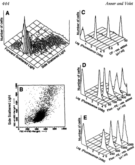
FACS is a convenient technique to assess the uptake of fluores- cent particles by cells (15). By appropriate window setting of the flow cytometer, the lymphocytes can be seen separately in three-dimen- sional (Fig. 1A) or two-dimensional (Fig. 1B,C) representation. When lymphocytes had been incubated with increasing concentrations of CF-vesicles (Fig. 1D) or Fluo-PE-vesicles (Fig. 1E,F ) the lymphocyte peak shifts progressively to the right on the Log scale as the fluores- cence associated with the cells increased by I to 3 orders of magni- tude. No cells remained at the initial fluorescence level indicating that all the vesicles enter into all lymphocytes. Also, there is no appearance of cell debris or of additional peaks when cells have taken up vesicles, reflecting that their morphology and viability are pre- served. In agreement with this observation, the Trypan blue analysis of the cells after incubation with fluorescent vesicles was 98.3% for the control preparation incubated without liposomes, 93.6, 85.4, 93.8, and 82.7% for cells incubated with increasing concentrations of Fluo- vesicles (Fig. 1E) and 86.4% for the control preparations and 83.8, 83.6, and 71.6% for the cells incubated with increasing concentra- tions of Fluo-vesicles shown in Fig. 1F. The data document that all isolated human lymphocytes take up fluorescent vesicles in a dose- dependent fashion without apparent toxicity.
Temperature Dependence of Vesicle Entry
Recently, we discovered that the 200 mM CF used to label the vesicles in their water phase, in conjunction with the high Na con- centration used to neutralize the acid CF, provoked multilayering of the vesicles and augmented their diameter from the 60 nm of Na,K-
ATPase-free vesicles to 330 nm (10). We exploited the fact to have
small single-walled and large, multilayered vesicles to see whether there was a difference in the temperature dependence of their uptake
444 Anner and Volet
!
,
=
No
" %W.o,._ 2"C-<".~.
# ~
% . 4 0 . . . . . ....~... ; ; : ~ . , . ~ : . . ./ t~
9~7;:~~
.z
9 " . " " . ' , . ~ , ~ , ~ . x ' : " " 9 - " . ~::::.!.X~.'.r .- % ~ , . ~oo#,
.. ".~.;:~,;~:r~:: " . . "-'*o,~o: y , r - x , ~ 00 ~." 9 " . . : " ' , " ' , " - . " - e e l 4 0 o , ? . a ' e~, .-"-.,~:~-...',- .'....-."" , ~ j ,~,,~ ~"
.~ : ~
""':"~:;'"
:'-;'"; .... ~ ,'" " ~," " ;~/'" ~k/"" ~ " i ~ o 0 0 0Fig. 1. Increase of cellular fluorescence with increasing vesicle/cell ratio. Freshly isolated human lymphocytes were incubated with increasing amounts of fluorescent vesicle for 30 rain at 37~ The vesicle quantity added is indicated in the figure in ~tL of suspension that had been pelleted and added to 1 mL cell suspension in the conditions described in Methods; 106 cells in 1 mL of PBS were added to the flow cytometer. The lympho- cytes were sorted by specific window settings. Three-dimensional (A) or two-dimensional (B,C) representations of the 104 events (cells) analyzed by scattered light shows a relatively homogenous lymphocytes popula- tion. (D) FACS analysis of lymphocytes incubated with increasing con- Cell Biochemistry and Biophysics Volume 30, 1999
because this physical parameter is informative as to the possible mechanisms involved. The uptake of the two types of vesicles by cells incubated at 4, 20, or 37~ was very close (Fig. 2A) as seen clearly by the similar shifts of the fluorescence of CF-vesicles (Fig. 2B) and Fluo-vesicles (Fig. 2C) by a temperature increase form 4 to 37~ The Q10 of the uptake was 1.46 + 0.7 (S.E.M., n = 4). The tem- perature coefficient (Q10) of a reaction is the ratio of the velocity measured at a given temperature (~ to the velocity measured at 10
degrees below (24). The Q10 of enzymatic reactions is generally
around 2, i.e., the velocity doubles with a 10~ increase of the tem- perature.
Such energy-requiring processes are described by an equation
derived from the Van't Hoff equation (25) stating that the reaction
rate k is proportional to a transmission coefficient (probability that a molecule in a transition state will give the product of the reaction),
and proportional to kT/h, which indicates the rate at which a mol-
ecule in the transition state is transformed into product (= Boltzmann constant x Absolute temperature/Planck constant). By contrast, if a purely physical process were implicated such as liposome adsorp- tion to the cell surface, the cellular fluorescence would be inversely related to temperature. According to the Gibb's law, the difference of the adsorbed amount (AA) of a compound is a function of its con- centration (AC). The concentration gradient within the adsorbed layer (a) = -(1/RT) x (AA/AC) where R is the gas constant and T is the absolute temperature. Thus, the high temperature dependence speaks against vesicle adsorption on the cell surface as a cause of the cell-associated fluorescence in line with the other analyses of data obtained by FACS and confocal microscopy presented herein. Na, K-A TPase in the Vesicles Does Not Interfere
with Their Cellular Uptake
Considering the rapid vesicle uptake by the isolated lympho- cytes, we wished to see whether the presence of Na,K-ATPase in the vesicle membrane altered their uptake. No statistically significant differences in the cellular fluorescence measured by FACS was observed when the cells were incubated with CF-vesicles or Fluo-
centrations of CF-vesicles added to 3.7 x 10 6 cells in 200 ~tL RPMI-1640.
(E,F) Lymphocytes incubated with increasing concentrations of Fluo-PE- vesicles in the experimental conditions described in Methods.
120 1 0 0 80 60 40 2 0 8 r G) o o _= | ._> (D n" 4 2 0 3 7 Temperature (~
B
oC
to o 7 '~e-oo, 3Fig. 2. Temperature-dependence of vesicle entry; 1.5 x 10⁶ cells were incubated in 100 ~tL RPMI-1640 with 50 ~tL/mL CF-vesicles (A,B) or with 50 ~tL/mL Fluo-vesicles (A,C) in 80 ~tL PBS for 30 min at 37~ Results are expressed in percentage of the maximum fluorescence obtained in the experiment. After the incubation, cells were washed once in PBS and ana- lyzed by FACS. Results are expressed in percentage of the maximum fluo- rescence obtained in the experiment. Data represent mean of two or three measurements (+ S.E.M.).
v e s i c l e s w i t h or w i t h o u t r e c o n s t i t u t e d N a , K - A T P a s e (data n o w shown).
Confocal microscopy was used to visualize the fluorescent vesicles taken up with or without reconstituted Na,K-ATPase. This n e w technique analyzes the fluorescence by a laser b e a m combined with powerful software for virtual sections across the cells yielding finally a three-dimensional m a p of the intracellular fluorescence. As the same cell is seen in fluorescent or transmission m o d e the intrac- ellular fluorescence can be attributed to cellular structures in the lim- its of the microscopic resolution.
W h e n cells were incubated with Fluo-vesicles without Na,K- ATPase for 5 (Fig. 3A) or 30 (Fig. 3B) min, they became strongly fluo- rescent (left panels). By comparison with the transmission images (right panels), it is evident that the vesicles have entered all cells. The same result is seen with cells incubated for 5 (Fig. 3C) or 30 (Fig. 3D) m i n with vesicles reconstituted with Na,K-ATPase. Obviously, the presence of renal Na,K-ATPase in the vesicles does not prevent their uptake by all lymphocytes, in agreement with the data obtained b y FACS.
That the fluorescence is not absorbed on the cell surface and is contained within the cells is shown in a typical scanned cell that has been incubated with Na,K-ATPase-vesicles for 20 m i n (Fig. 4) and was then analyzed in three dimensions by nine virtual I ~tm cross-sections. The first scan, on top of the cell, reveals no fluorescence; two small fluorescent spots became apparent on the next scanned plane; the next section, deeper within the cell, uncovers more fluorescence. The maxi- mal fluorescence was seen in the four sections going through the middle of the cell, sparing apparently the nuclear region.
Quantification of the
UptakeTo evaluate the importance of the Na,K-ATPase-vesicle entry into lymphocytes, it was important to quantify the process: how m a n y vesicles enter and how m u c h foreign material is imported with them expressed as a fraction of intrinsic? To answer this question, the size and composition of the vesicles as well as the size of the cell must be k n o w n and a method to quantify the internalized mem- branes is required. The ultrastructure of our vesicles with and with-
out Na,K-ATPase is k n o w n (8,9) and from there their content in
phospholipids and Na,K-ATPase molecules was calculated as shown in Table 1. W h e n the n u m b e r of phospholipids per vesicle is known, the internal fluorescent phospholipid can be extracted from the cell, its quantity determined by spectrofluorometry, and the vesicle n u m -
,11 0 9 ~ r
:~ ~
~ .~
~ ~ ~
~ o
-~~
~ ~ " ~
,.~ •: i ~ I
- o
.~ ~ 5 89 ~ ...
|
o ~
Fig. 4. Three-dimensional demonstration of intracellular fluorescence resulting from Na,K-ATPase-vesicle uptake. Virtual I B m z-sectioning of a typical cell that has been incubated with fluorescent vesicles for 20 min in the conditions described for Fig. 3.
ber taken up per cell calculated. However, the lymphocytes are con- taminated by monocytes, which are phagocytic cells and take up more vesicles than lymphocytes. The average percentage of mono- cytes present in the preparation was determined by FACS in a series of three different preparations (+ S.E.M.) to which 5 ktL CF-liposomes
h a d been added (to 2 x 10 6 cells in 100 BL RPMI-1640 at 37~ 8.3 +
0.1%, 8.7 + 0.6%, 9.1 + 0.1%, 9.4 + 0.3%, and 10.3 + 0.3% after 5, 10, 30, 60, and 120 rain of vesicle addition, respectively. An average value of 9% was chosen for the calculations.
With regard to the vesicle uptake, the following m o n o c y t e / l y m - phocyte fluorescence ratios were obtained in triplicates (+ S.E.M.) in six different preparations that incorporated, respectively, 36 + 1.5, 28 + 1, 27 + 3, 17 + 0.3, 23 + 0.2, and 10 + 0.3 times more fluorescence
within 60 m i n at 37~ the average of the six separate determina-
tions was 23. On the basis of the 9% monocyte fraction and the 23- fold difference of u p t a k e b y l y m p h o c y t e s a n d monocytes, the distribution of Fluo-PE-liposomes between monocytes and lympho-
450 Anner and Volet
Table 1
Parameters Used for Quantifying Vesicle Uptake Average diameter Phospholipid No. Surface Volume Lymphocyte Na,K-ATPase-vesicle total
Inner compartment (-4 nm membrane) per 100 nm vesicle outer leaflet
inner leaflet total Lymphocyte Na,K-ATPase-vesicle PC headgroup Lymphocyte Na,K-ATPase-vesicles 10 ~ t m a 100 n m b 92 nm 44,882 37,988 82,870 3.14 x 1 0 - 1 ~ 2 3.14 x 10⁻¹⁴m2 0.7 x 10⁻¹⁸m 2c 5.24 x 1 0 - 1 6 m 3 4.08 x 10 -22 m 3
aThe average cell size was estimated by optical and electronic microscopy and found com- patible with published data (28).
bThe vesicle size was determined by freeze fracture (8,9) and transmission electronic microscopy (10).
CThe diameter of a phosphatidylcholine headgroup was found in the work of Mimms et al. (29).
cytes was calculated considering that 9% of the cells (monocytes) take up 23 parts of the fluorescence and 91% of the cells (lympho- cytes) take up one part, which is equivalent to 72% of vesicles taken up by monocytes and 28% by lymphocytes. W h e n this correction ismade, the n u m b e r of vesicles incorporated per lymphocytes at increasing vesicle/cell ratios amounts to the values listed in Table 2. By combining these data with the data of Table 1, the a m o u n t of internalized m e m b r a n e and volume was also calculated, as well as the n u m b e r of Na,K-ATPase molecules (Table 2).
In previous work, the vesicular Na,K-ATPase was radiolabeled
by 11~ The cells were then incubated with the 11~
Na,K-ATPase-vesicles, washed in presence of external dimercapto- propanesulfonic acid (DMPS) to remove all external silver a n d the
11~ taken up by the cells was counted. According to previous
determinations, there were four molecules Na,K-ATPase per lipo- some at the p r o t e i n / l i p i d ratio used and, in conjunction with the value of 2 silver ions b o u n d per Na,K-ATPase molecule, the n u m b e r of liposomes incorporated was found to be 3114 + 215 (S.E.M., n = 21) for an incubation period of 120 rain at 35~ at a liposome/cell
ratio of 3.1. x 105 (26,27). Thus, two entirely different methods yield
Table 2
Foreign Material Brought to a Lymphocyte by Vesicle Uptake for 30 Min Vesicles
entered Foreign membrane Foreign volume Foreign Na,K-ATPase
(% of (% of % of
Number ( m 2) intrinsic) ( m 3) intrinsic) No. intrinsic
1910 a 2.158 x 10 -11 7 1.406 x 10 -19 0.03 11,460 38 4122 a 4.658 x 10 -11 15 3.035 • 10 -19 0.06 24,732 82 8748 a 9.885 • 10 -11 32 6.440 • 10 -19 0.12 52,488 175 16,087 ~ 1.818 x 10 -1~ 60 1.184 x 10 -18 0.23 96,522 322 39,414 a 4.454 x 10 -1~ 142 2.902 x 10 -18 0.55 236,484 788 18,098 b 2.045 x 10 -1~ 65 1.332 x 10 -18 0.25 108,588 362 19,004 b 2.147 • 10 -10 68 1.399 x 10 -18 0.27 114,024 380 41,526 v 4.692 X 10 -l~ 149 3.057X 10 -18 0.58 249,156 830 106,982 b 1.208 x 10 .9 385 7.876 x 10 -18 1.50 641,892 2140
Two different Fluo-vesicle preparations (a) and (b) were pelleted and incubated with 1.4 x 10⁶ cells for 30 min at 37~ resulting in ratios of 0.27, 0.59, 1.58, 3.99, and 8.88 x 10⁵ (a) and 0.6, 1.1, 1.8, and 4.7 x 10⁶ vesicles (b) per cell. After the incubation, the cells were processed and the entrapped Fluo-PE measured by spectrofluorometry as described in Methods and calculated according to the data of Table 1. The number of 6 Na,K-ATPase molecules per vesicle was taken from previous deter- minations of freeze fractured preparation prepared in the same conditions. A number of 30,000 Na,K- ATPase molecules per lymphocyte had been determined by 3H-ouabain (30).
close v a l u e s for the n u m b e r of Na,K-ATPase-vesicles i n c o r p o r a t e d p e r cell.
It is i m p r e s s i v e that a l y m p h o c y t e i n c u b a t e d in the p r e s e n c e of 4.7 m i l l i o n e x t e r n a l l y a d d e d Na,K-ATPase vesicles h a s t a k e n u p 3.85 times its o w n m e m b r a n e surface, replaced 1.5% of its v o l u m e b y for- eign solution, a n d t a k e n u p m o r e t h a n 20 times m o r e foreign Na,K- ATPase as c o m p a r e d to the intrinsic n u m b e r (Table 2).
ACKNOWLEDGMENTS
The a u t h o r s t h a n k D o m i n i q u e W o h l w e n d of the Cytofluoro- g r a p h i c Center, G e n e v a U n i v e r s i t y Medical School, for assistance w i t h FACS analysis, Ch. Burrus a n d D. Lacotte for help w i t h confo- cal a n d electron microscopy, a n d M. M o o s m a y e r for assistance w i t h Na,K-ATPase purification a n d reconstitution. This research w a s sup- p o r t e d b y the Swiss N a t i o n a l Science F o u n d a t i o n , G r a n t s No. 31- 25666.88 a n d 31-37552.93 a n d b y the Swiss Public H e a l t h Office, G r a n t No. 90-7052.
4 5 2 Anner and Volet
REFERENCES
1. Hamm-Alvarez, A. F., Wei, X., Berndt, N., and Runnegar, M. (1996) Protein phosphatase independently regulate vesicle movement and microtubule subpopulations in hepatocytes. Am. J. Physiol. 271, C929-C942.
2. Sion, J.-P., Ivanov, I. E., Adesnik M., and Sabatini, D. D. (1996) The pro- duction of post-Golgi vesicles requires a protein kinase C-like molecule, but not its phosphorylating activity. J. Cell Biol. 135, 355-370.
3. Levenson, R. (1994) Isoforms of the Na,K-ATPase: family members in search of function. Rev. Physiol. Biochem. Pharmacol. 123, 1-45. 4. Blaustein, M. P. (1993) Physiological effects of endogenous ouabain:
control of intracellular Ca2+ stores and cell responsiveness. Am. J.
Physiol. 264, C1367-C138.
5. Lees, G. J. (1991) Inhibition of sodium-potassium-ATPase: a poten- tially ubiquitous mechanism contributing to central nervous system neuropathology. Brain Res. Rev. 16, 283-300.
6. Li, K. and Sperelakis, N. (1994) Electrogenic Na-K pump current in rat skeletal myoballs. J. Cell Physiol. 159, 181-186.
7. Goldin, S. M. (1974) Reconstitution of active transport catalyzed by the purified sodium and potassium ion-stimulated adenosine triphosphatase from canine renal medulla. J. Biol. Chem. 248, 5907-5915.
8. Anner, B. M., Ting-Beall, H. P., and Robertson, J. D. (1984) Charac- terization of (Na + K)-ATPase liposomes. I. Effect of enzyme con- centration and modification on liposome size, intramembrane particle formation and Na,K-transport. Biochim. Biophys. Acta 773, 253-261.
9. Anner, B. M., Robertson, J. D., and Ting-Beall, H. P (1984) Charac- terization of (Na + K)-ATPase liposomes. II. Effect of alpha-subunit digestion on intramembrane particle formation and Na,K-transport.
Biochim. Biophys. Acta 773, 262-270.
10. Volet, B., Lacotte, D., Moosmayer, M., and B. M. Anner. (1994) Na,K- ATPase or carboxyfluorescein alter vesicle formation in vitro.
Biochim. Biophys. Acta, 1191, 1-6.
11. Weinstein, J. N., Yoshikami, S., HeNa,K-ATPasert, P., Blumenthal, R. and W. A. Hagins. (1977) Liposome-cell interaction: transfer and intracellular release of a trapped fluorescent marker. Science 195, 489-492.
12. Blumenthal, R., Weinstein, J. N., Sharrow, S. O. and P. HeNa,K- ATPasert. (1977) Liposome-lymphocyte interaction: Saturable sites for transfer and intracellular release of liposome contents. Proc. Natl.
Acad. Sci. USA 74, 5603-5607.
13. Lee K.-D., Nir S., and D. Papahadjopoulos D. (1993) Quantitative analysis of liposome-cell interactions in vitro: rate constants of bind- ing and endocytosis with suspension and adherent J774 cells and human monocytes. Biochemistry 32, 889-899.
14. Lee, K-D., Hong, K., and Papahadjopoulos, D. (1992) Liposomes rec- ognition by cells: In vitro binding and endocytosis mediated by spe- cific headgroups and surface charge density. Biochim. Biophys. Acta 1103, 185-197.
15. Leroux, J.-C., Gravel, P., Balant, L., Volet, B., Anner, B. M., All6man, E., Doelker, E., and Gurny, R. (1994) Internalization of poly(D,L- lactic acid) nanoparticles by isolated human leukocytes and analy- sis of plasma proteins adsorbed onto the particles. J. Biomed. Mat.
Res. 28, 471-481.
16. Dzhandzhugazyan, K. N. and Jorgensen, P. L. (1985) Asymmetric orientation of amino groups in the ct-subunit and the ~-subunit of (Na § + K+) - ATPase in tight right-side-out vesicles of basolateral membranes from outer medulla. Biochim. Biophys. Acta 817,165-173. 17. Anner, B. M., Moosmayer, M., and Imesch, E. (1994) Na,K-ATPase
characterised in artifical membranes. 1. Predominant conformations and ion fluxes associated with active and inhibited states. Mol. Mem-
brane Biol. 11, 237-245.
18. Smith, P. K., Krohn, R. I., Hermanson, G. T., Mallia, A. K., Gartner, F. H., Provenzano, M. D., Fujimoto, E. K., Goeke, N. M., Olson, B. J., and D. C. Klenk. (1985) Measurement of protein using bicinchoninic acid. Anal. Biochem. 150, 76-85.
19. Szoka, F. C., Jacobson, K., and Papahadjopoulos, D. (1979) The use of aqueous space markers to determine the mechanism of interac- tion between phospholipid liposomes and cells. Biochim. Biophys.
Acta 551, 295-303.
20. Ralston, E., Blumenthal, R., Weinstein, J. N., Sharrow, S. O., and He, P. (1980) Na,K-ATPasert lysophosphatidylcholine in liposomal membranes. Enhanced permeability but little effect on transfer of a water-soluble fluorescent marker into human lymphocytes. Biochim.
Biophys. Acta 597, 543-551.
21. Collins, D., Litzinger, D. C., and Huang, L. (1990) Structural anf functional comparisons of pH-sensitive liposomes composed of phosphatidylethanolamine and three different diacylsuccinyl- glycerols. Biochim. Biophys. Acta 1025, 234-242.
22. Boyum A. (1968) Isolation of mononuclear cells and granulo- cytes from human blood. Scand. J. Clin. Lab. Invest. 21,Suppl. 97, 77-89.
23. Lenette, D. A. (1978) An improved mounting medium for immunof- luorescence microscopy. Am. J. Pathol. 69, 647-648.
454 24. 25. 26.
Anner and Volet Dawes, E. A. (1972) Quantitative Problems in Biochemistry. Williams and Wilkin, Baltimore, MD, pp. 165-183.
Gutfreund, H. (1975) Enzymes: Physical Principles. Wiley, London, New York. pp. 161-165.
Anner, B. M., Meneghini, E., Hussain, S., Lacotte, D. and Moosmayer, M. (1994). Interaction of radiolabeled Na,K-ATPase-liposomes with human peripheral blood mono-nuclear cells. Biochim. Biophys. Acta 1194, 345-348.
27. Hussain, S., Meneghini, E, Moosmayer,. M., Lacotte, D. and Anner, B. M. (1994) Potent and reversible interaction of silver with pure Na,K-ATPase and Na,K-ATPase-liposomes. Biochim. Biophys. Acta 1190, 402-408.
28. Falciola, J., Volet, B., Anner, R. M., Moosmayer, M., Lacotte, D., Anner, B. M. (1994) Role of cell membrane Na,K-ATPase for sur- vival of human lymphocytes in vitro. Biosci. Rep. 14, 189-204. 29. Mimms, L. T., Zampighi, G., Nozaki, Y., Tanford, Ch., and J. A.
Reynolds. (1981) Phospholipid vesicle formation and transmem- brane protein incorporation using octyl glucoside. Biochemistry 20, 833-840.
30. Pedersen, K. E., and N. A. Klitgaard. (1983) Influence of quinidine on the binding of [3H]-ouabain and [3H]-digoxin by human lym- phocytes. Eur. J. Clin. Pharmacol. 25, 263-270.