Cite this article as: Wiese MN, Kawel-Boehm N, Moreno de la Santa P, Al-Shahrabani F, Toffel M, Rosenthal Ret al. Functional results after chest wall stabilization with a new screwlessfixation device. Eur J Cardiothorac Surg 2015;47:868–75.
Functional results after chest wall stabilization with a new
screwless fixation device
Mark Nikolaj Wiese
a, Nadine Kawel-Boehm
b, Pablo Moreno de la Santa
c, Feras Al-Shahrabani
a,
Melanie Toffel
a, Rachel Rosenthal
d, Juliane Schäfer
d,e, Michael Tamm
f, Jens Bremerich
band Didier Lardinois
a,*
a Division of Thoracic Surgery, University Hospital Basel, Basel, Switzerland bDepartment of Radiology, University Hospital Basel, Basel, Switzerland c Division of Thoracic Surgery, POVISA Hospital, Vigo, Spain
dDivision of General Surgery, University Hospital Basel, Basel, Switzerland
e Basel Institute for Clinical Epidemiology and Biostatistics, University Hospital Basel, Basel, Switzerland f Division of Pneumology, University Hospital Basel, Basel, Switzerland
* Corresponding author. Division of Thoracic Surgery, University Hospital Basel, Spitalstrasse 21, 4031 Basel, Switzerland. Tel: +41-61-2657218; fax: +41-61-2657238; e-mail: [email protected] (D. Lardinois).
Received 22 January 2014; received in revised form 4 July 2014; accepted 13 July 2014
Abstract
OBJECTIVES: This is the experience with the Stratos system in two surgical centres for the management of two types of rib fractures:flail
chest and multiple dislocated rib fractures with significant chest wall deformity.
METHODS: From January 2009 to May 2012, 94 consecutive patients were included. Selected indications were extended anterolateralflail
chest (n = 68) and dislocated painful rib fractures (n = 26). The open reduction internal fixation (ORIF) system consists of flexible titanium
rib clamps and connecting plates. The postoperative course was assessed. Clinical and functional outcomes were evaluated at 6 months. Functional assessment consisted of measurement of the functional vital capacity (FVC) and magnetic resonance imaging (MRI)
examin-ation with determinexamin-ation of the radiological vital capacity (rVC) in patients with aflail chest.
RESULTS: The median operation time and length of hospital stay were 122 min and 19 days, respectively, in patients with aflail chest, and
67 min and 11 days, respectively, in patients with dislocated painful rib fractures. The morbidity rate was 6.4% and the overall 30-day mor-tality rate was 1.1%. Clinical evaluation and pulmonary function testing at 6 months revealed no deformity of the chest wall, symmetrical
shoulder girdle mobility in 88% and a feeling of stiffness on the operated side in 19% of the patients operated for aflail chest. Median ratio
of FVC was 88%, not suggesting any restriction after stabilization. MRI was performed in 53% (36 of 68) of the patients with aflail chest. The
analysis of the rVC showed, on average, no clinically relevant restriction related to the operation, with a mean rVC of the operated relative
to the non-operated side of 92% (95% confidence interval: 83, 100). Stabilization of more than four ribs was associated with a lower
median rVC than stabilization of four or less ribs.
CONCLUSIONS: Our results suggest that stabilization of the chest wall with this screwless ribfixation device can be performed with a low
morbidity and lead to early restoration of chest wall integrity and respiratory pump function, without clinically relevant functional
restric-tion. Owing to the simplicity of thefixation technique, indications for stabilization can be safely enlarged to selected patients with
dislo-cated and painful rib fractures.
Keywords:Chest wall• Trauma • Surgery techniques • Outcome • Imaging
INTRODUCTION
Blunt chest wall trauma is commonly associated with rib fractures
[1–3]. The number of consecutive fractured ribs has been found to
be an independent marker of injury severity, resulting in increased
morbidity and mortality, particularly in the elderly [1,4,5]. Flail
chest injuries are observed in about 10% of chest wall trauma and
are associated with a high mortality rate of 10–36%, partly due to
the high incidence of associated life-threatening extrathoracic
injuries, but also because of pneumonia and sepsis related to
pro-longed intubation [1–6]. Surgical management of aflail chest is still
controversial and there are no large adequate prospective
rando-mized trials comparing conservative and surgical management [1,
4,7]. However, several studies and reports have clearly indicated
that operative chest wall stabilization for a flail chest in highly
selected patients leads to a shorter duration of ventilation support and, consequently, to a reduced morbidity due to prolonged
mechanical ventilation, considerably less infections and
© The Author 2014. Published by Oxford University Press on behalf of the European Association for Cardio-Thoracic Surgery. All rights reserved.
ORIGINAL ARTICLE
septicaemia as well as to a shorter intensive care unit (ICU) stay [8–
10]. Moreover, stabilization does not seem to be associated with
re-striction of pulmonary function [4,11]. A review of nine published
studies, regarding a more recent indication for chest wall
stabiliza-tion in patients without aflail chest, but with consecutive dislocated
and painful rib fractures, suggested that open reduction internal
fix-ation (ORIF) of isolated multiple rib fractures improves outcome in terms of pain reduction, respiratory function, quality of life and
allows a shorter recovery period [12]. Patients with anterolateral
dis-locations are particularly prone to develop late restriction and
pseu-darthrosis, leading to partial or complete functional incapacity [4,6,
7]. The incidence of pseudarthrosis is certainly under-reported and
the consequences are underestimated [1,13,14]. To prevent
short-and long-term morbidity of multiple dislocated painful rib fractures, several centres have initiated stabilization procedures in selected
patients [12,15–17]. The present study was designed tofirst assess
the feasibility and the complication rate of chest wall stabilization
using a new screwless rib fixation device (Stratos™, MedXpert,
Heitersheim, Germany) for several indications in selected patients in two centres. The second intention of the study was to assess the functional lung capacity and the mobility of the chest wall after ORIF
in the subgroup of patients with aflail chest. The ethics committees
of both hospitals provided approval for this study. This was an investigator-initiated study with funding support from MedXpert, Germany.
PATIENTS AND METHODS
Patients
From January 2009 to May 2012, 94 selected patients underwent
chest wall stabilization using the new screwlessfixation device from
Stratos™ (Strasbourg Thoracic Osteosynthesis System, MedXpert) in
two thoracic centres, the POVISA Hospital of Vigo, Spain (50 patients) and the University Hospital of Basel, Switzerland (44 patients).
Patients with an indication for surgicalfixation could be divided into
those with extendedflail chest (68 of 94, 72%) and those without a
flail chest (26 of 94, 28%). In patients with a flail chest, indications were as follows: non-intubated patients with respiratory failure despite continuous epidural analgesia and aggressive clearance of
bronchial secretions (n = 16, 24%); patients with an extended
antero-lateral flail chest and progressive dislocation of the fractured ribs
(n = 27, 40%); intubated patients who did not require prolonged
in-tubation in the absence of severe pulmonary contusion or cerebral injuries, in order to reduce the use of mechanical ventilation when
the patient failed to wean (n = 19, 28%); and patients who required a
thoracotomy or thoracoscopy due to associated extended
haemothorax (n = 6, 9%). The group of patients without a flail chest
consisted of multiple severe dislocated and therapy-refractory painful rib fractures, particularly in young athletic patients or in old patients living alone and requiring a prompt recovery.
Twenty patients were women (21%) and 74 were men (79%), with a mean age at operation of 55 years, ranging from 22 to 88
years. Fifty-five patients (59%) had motor vehicle accidents, 20
patients (21%) were injured at work or during sports activities, and 19 patients (20%) fell at home. Associated injuries were observed in 72 patients (77%) and consisted of orthopaedic lesions, head
in-juries and abdominal lesions. Of the 68 patients with aflail chest,
20 (29%) were intubated before or upon admission, due to
re-spiratory insufficiency, associated cerebral injuries or hypovolemic
shock. Chest trauma was unilateral in 97% (91 of 94 patients) and bilateral in 3% of the patients (3 of 94 patients). In patients with a flail chest, the median number of fractured ribs was 8, ranging from 6 to 11.
Surgical technique and postoperative follow-up
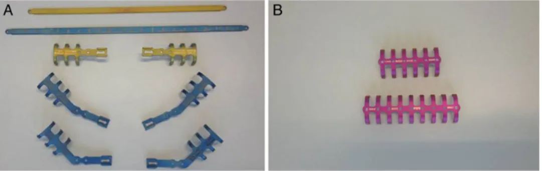
The new screwless Stratos device consists offlexible titanium rib
clips and connecting bars [16]. Because of the excellent pliability
of titanium, the rib clips and connecting bars can easily and pre-cisely be adapted to the shape of the ribs. The implants will rebound minimally after bending so that the material can be anchored tightly to the chest wall and resist against loosening. In
the case of aflail chest, one complete implant consists of two rib
clips that are either straight or angular (22.5° or 45° types) and a connecting bar to overbridge the unstable segment. The three dif-ferent angulations of the rib clips can be adapted to any anatomic-al situation by means of bending instruments and cover a
maximum range of−12° up to 57° (Fig.1A). Dislocated rib
frac-tures can also be stabilized by simple rib clips without connecting
bars (Fig.1B). Several straight or angled forceps can be used to
shape andfixate the titanium implants onto the ribs. The same
system can be used for reconstruction of the chest wall after partial resection and for correction of sternal deformities. In
patients with a flail chest, ORIF was performed through an
extended lateral approach. In patients without aflail chest, the
skin incision was shorter and made just over the fractures.
Through this single incision, it was possible to stabilize up tofive
ribs. Care was taken to avoid increasing injury of the contused soft tissue. The serratus anterior muscle was dissected and divided at
its insertion on the chest wall in patients with aflail chest or
divided within itsfibres in patients with dislocated rib fractures.
This technique provided a good exposure of the consecutive frac-tured ribs without causing extensive muscle damage. Care was
Figure 1:(A) The new screwless Stratos device consists offlexible titanium rib clips and connecting bars. This system is used for chest wall stabilization in patients with aflail chest. (B) Simple rib clips without connecting bars can be fixed to the ribs by grasping in patients with dislocated painful fractures.
THORA
C
IC
taken not to strip the periostium from the ribs and to prevent injury of the intercostal neurovascular bundle at the inferior
margin of the rib. In patients with aflail chest, rib clips were first
fixed medially and dorsally to the unstable segment. A connecting
bar was then shaped andfixed to the rib clips while the lung was
ventilated by the use of high tidal volumes in order to prevent
operation-related restriction. The unstable part of the rib wasfixed
to the connecting bar by the use of a resorbable thread of Vicryl®
(Ethicon) (Figs2and3). In patients without aflail chest, only
titan-ium rib clips were used without connecting bars. The fractures were reduced and stabilized with a minimum of three clips on each side
of the fracture line (Fig.4). Only the most dislocated ribs were
stabi-lized. Thefirst two ribs were not operated to avoid injury of the
sub-clavian vessels and the brachial plexus.
The postoperative course of all patients was prospectively assessed. For all patients, key data consisting of the number of sta-bilized ribs, operation time, duration of postoperative intubation,
length of hospital stay, morbidity and mortality were recorded. At 6 months following surgery, patients were clinically examined. The clinical evaluation included a subjective assessment of residual pain or discomfort of the chest wall and shoulder girdle, and was followed by an examination of the chest wall and shoulder girdle function. Functionality of the stabilized chest was assessed by lung function tests and by a dynamic magnetic resonance imaging (MRI) examination. MRI was only considered in the group of 68
patients withflail chest.
Six-month pulmonary function testing
We used functional vital capacity (FVC) in the evaluation of a pos-sible postoperative restriction. A restriction was noted if the
mea-sured FVC was <80% of the predicted value [18].
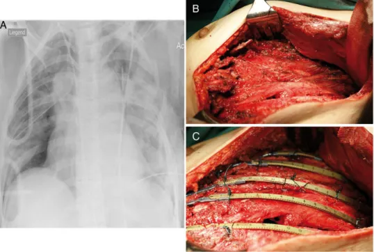
Figure 2:(A) Chest X-ray after a severe blunt chest trauma showing bilateral dislocated rib fractures and a left haemothorax. An extendedflail chest was diagnosed on the left side. (B) Intraoperative situs with severe dislocated rib fractures and impaction of the unstable segment. (C) Left chest wall after stabilization showing the rib clips and the connecting bars. The chest wall is well-shaped, avoiding postoperative restriction.
Figure 3:(A) Chest X-ray with dislocated rib fractures on the right side. The patient received analgesia with epidural catheter and was transferred to the normal ward. (B) Progressive shrinkage with reduction of the volume of the right hemithorax. The patient developed acute respiratory insufficiency. He was transferred to the inten-sive care unit and was intubated. (C) Chest X-ray after ORIF showing a well-shaped right hemithorax.
Six-month magnetic resonance imaging
examination
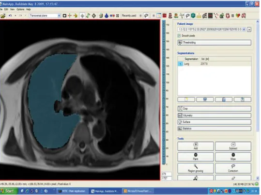
MRI exams were performed with a magneticflux density of 1.5 T. A
half-Fourier acquisition single-shot turbo spin echo sequence was acquired in the axial and coronal plane at inspiration and expiration with a slice thickness of 10 mm and no inter-slice gap covering the
entire thorax. All MRI findings were centrally interpreted at the
University Hospital of Basel by two experienced radiologists familiar with thoracic examinations. The endpoint of this analysis was the measured radiological vital capacity (rVC) on the operated side relative to the target rVC on the operated side. We derived the target rVC from the non-operated side by measuring the total volume of each side of the lung during maximal inspiration [radio-logical total lung capacity (rTLC)] and during maximal expiration [radiological residual volume (rRV)]. The difference between rTLC
and rRV was defined as the rVC (Fig.5). Measurements were
per-formed by manual tracing of the lung contour on each slice, for the right and left lung and by automatic calculation of the volume based on these measurements. The comparison of both sides was
performed under the assumption that, in a healthy adult, the
distri-bution of the total lung volume [18] and of the rVC is 55% on the
right side and 45% on the left side. In addition, we assumed that
the stabilization of one side of the chest wall does not influence
the mobility of the other side 6 months after surgery, when post-taumatic recovery is assumed to be established.
The following patients were excluded from the analysis: patients with no consent and lost to follow-up, claustrophobia, pregnancy, immobility and bilateral chest wall stabilization and contraindica-tions for MRI like metallic implants, pacemaker and old types of tattoo.
Statistical analyses
To assess the rVC of the operated compared with the non-operated
side, we calculated a two-sided 95% confidence interval (CI) for the
mean rVC of the operated side relative to its target value (derived from the non-operated side) (rVC %). We also report results of a sensitivity analysis with logarithm-transformed rVC % to achieve a better approximation to normality. The resulting CI on the logarith-mic scale was then transformed back to the original units, resulting in a CI for the geometric mean.
In an additional analysis, we describe functional results for patients with four or less stabilized ribs and for those with more than four stabilized ribs.
We used R version 3.1.0 (R Foundation for Statistical Computing,
Vienna, Austria) and the R add-on packagelattice version 0.20–29
for our analyses and for graphics.
RESULTS
Baseline and procedure characteristics of the 33 patients with a flail chest and unilateral chest wall stabilization, included in the
analysis of the rVC %, and of the 35 patients with aflail chest but
no MRI or bilateral chest wall stabilization, excluded from this
ana-lysis, are provided in Table1. In the 68 patients who underwent
sta-bilization for a flail chest, the median time from admission to
stabilization of the chest wall was 3.4 days (range, 0–17 days). The
median operation time in this group was 122 min, ranging from 70 to 205 min, and the median length of hospital stay was 19 days, with
a range of 4–84 days. In the 26 patients who underwent stabilization
for persistent dislocated and painful rib fractures, the median oper-ation time was 47 min, ranging from 28 to 95 min, and the median
length of hospital stay was 11 days, with a range of 3–74 days. The
overall 30-day mortality rate was 1.1%. The patient who died
under-went stabilization for aflail chest. The mortality rate in this group
was 1.5% (1 of 68 patients). The patient who died was a 76-year old
polytrauma patient who had undergone stabilization for aflail chest.
Several operations were required, which prolonged the intubation period and delayed operative stabilization. Postoperative
complica-tions were observed in 6 patients, all of whom were in theflail chest
group (6/68, 9%). These complications included pneumonia of the ipsilateral lung in 4 patients and wound infection in 2 patients. All 4 patients with postoperative pneumonia recovered after treatment combining bronchoscopic removal of secretion and antibiotics. In the 2 patients with infection, partial removal of the ORIF material was required 9 and 12 days after chest wall stabilization. These patients were diabetics and underwent a severe trauma with extended contusion of overlying soft tissue. Fortunately, the extent of the infection in the chest wall was well localized and only partial Figure 4:(A) Vehicle accident with dislocated rib fractures 4! 11 on the right
side. Despite analgesia with epidural catheter, the patient was unable to move and to cough. (B) Chest X-ray after stabilization using simple rib clips. The patient was discharged from the hospital 6 days after the operation.
THORA
C
IC
removal of the material was required, assuring further stability. Healing was achieved with drainage and antibiotics.
The six-month follow-up for clinical evaluation and pulmonary function testing was obtained in 75 of 93 surviving patients (81%),
including 56 patients with aflail chest and 19 patients with
dislo-cated and painful rib fractures. Nineteen patients were lost to follow-up or did not consent to further evaluation. In our clinical set-up, we did not observe any delayed fracture healing, including
the 2 patients who had wound infections. We did not notice any mechanical fatigue of the ORIF material and we only partially removed the ORIF material in 2 cases of local wound infection. Attenuated sensitivity of the anterior chest wall was noted in 9% of
the patients (8/93), all of whom initially suffered from aflail chest.
Thirteen patients reported feeling persistent pain and rigidness on the operated side following surgery. All these patients had been
operated for aflail chest (13 of 67, 19%). Shoulder girdle function
Figure 6:(A) Radiological vital capacity (rVC) of the operated side relative to its target value as derived from the non-operated side (rVC %) (n = 33). (B) Radiological vital capacity of the operated side relative to its target value as derived from the non-operated side (rVC %) against the ratio of the forced vital capacity (FVC %) for patients with aflail chest included in the main analysis and with an FVC % measurement available (n = 31). The smooth curve is a local average found using the default loess smoother in the R add-on packagelattice version 0.20–29.
Figure 5:MRI examination: primary endpoint consisting of determination of the radiological vital capacity on the operated right side (difference between radiological total lung capacity and radiological residual volume).
was symmetrical in 66 patients (88%), whereas in 9 patients ab-duction of the ipsilateral shoulder was limited to 90°. These patients also had associated injuries of the ipsilateral shoulder girdle. Chest wall deformity was observed in none of the patients. Radiological evaluation at follow-up showed no dislocation of the material in any of the patients. Pulmonary function testing 6 months following surgery revealed a median ratio of the FVC of 88% [interquartile range (IQR): 79, 95; range, 61, 124], suggesting that ORIF was not associated with postoperative restriction in the majority of patients.
Six-month magnetic resonance imaging results
Of the 68 patients who underwent chest wall stabilization for aflail
chest, 36 patients (53%) underwent MRI. Thirty-two patients were initially excluded due to the criteria mentioned above and did not undergo MRI. Three additional patients who underwent MRI were excluded from the main analysis due to bilateral chest wall stabil-ization. Overall, there were no apparent differences between
patients with aflail chest included in the main analysis and those
excluded from this analysis (Table1).
The MRI showed no paradoxical chest wall movement. The analysis of the rVC of the operated side relative to its target value as derived from the non-operated side (measurement and comparison of the lung volume on both sides) yielded a median
of 87% (IQR: 73, 111) (Fig.6). The mean rVC % was 92% (95% CI:
83, 100).
The sensitivity analysis with logarithmic transformation of the rVC % yielded results comparable with those from the main
analysis, with a geometric mean rVC of the operated side relative to its target value as derived from the non-operated side of 89% (95% CI: 81, 97).
In an additional analysis, we assessed the rVC % separately for
patients with stabilization of up to four ribs (n = 22) versus those
with stabilization of more than four ribs (n = 11). We found a higher median rVC % in patients with stabilization of up to four ribs compared with those with stabilization of more than four ribs [88% (IQR: 79, 110) vs 80% (IQR: 71, 98)].
Correlation between magnetic resonance imaging
and pulmonary function
Finally, a scatter plot of the rVC of the operated side relative to its target value as derived from the non-operated side against the ratio of the FVC revealed a modest correlation, with a Spearman
rank correlation coefficient of 0.21 (Fig.6B).
DISCUSSION
The goals of surgical chest wall stabilization are a preservation of
chest wall mechanics and lung function [19]. ORIF might
theoretic-ally lead to a rigidity of the chest wall. However, it has been shown that chest wall stabilization with reconstruction plates does not lead
to impaired postoperative pulmonary function [4]. These results are
confirmed in our study with the Stratos™ material. Lung function
tests analysing the FVC at 6 months following surgery revealed no restriction, not only in the whole series of patients, but also in the
Table 1: Baseline and procedure characteristics of patients with a flail chest and those with painful and dislocated rib fractures
Flail chest Painful and dislocated rib fractures
MRI and unilateral chest
wall stabilization (n = 33) No MRI or bilateral chestwall stabilization (n = 35) No MRI foreseen (n = 26) Baseline characteristics
Study centre,n (%)
Basel, Switzerland 19 (58) 14 (40) 11 (42)
Vigo, Spain 14 (42) 21 (60) 15 (58)
Median age (IQR), years 57 (45, 68) 52 (44, 67) 52 (47, 68)
Female gender,n (%) 5 (15) 8 (23) 7 (27) Side,n (%) Left 11 (33) 16 (46) 11 (42) Right 22 (67) 16 (46) 15 (58) Both sides 0 (0) 3 (9) 0 (0) Trauma,n (%) Fall 14 (42) 6 (17) 10 (38) Traffic 18 (55) 26 (74) 11 (42) Other 1 (3) 3 (9) 5 (19)
Median number of fractured ribs (IQR) 8 (6, 10) 8 (7, 12) 6 (5, 10)
Preoperative mechanical ventilation,n (%) 13 (39) 20 (57) 5 (19)
Procedure characteristics
Median number of stabilized ribs (IQR) 4 (3, 5) 4 (4, 7) 4 (2, 5)
Median length of hospital staya(IQR), days 17 (10, 29) 22 (16, 33) 11 (7, 20)
30-day mortality,n (%) 0 (0) 1 (3) 0 (0)
Patients with a flail chest were further subdivided into patients with MRI and unilateral chest wall stabilization and those with no MRI or bilateral chest wall stabilization.
MRI: magnetic resonance imaging; IQR: interquartile range. a
Available in 32 (97%), 33 (94%) and 26 (100%) patients with MRI and unilateral chest wall stabilization, with no MRI or bilateral chest wall stabilization and painful dislocated rib fractures, respectively.
THORA
C
IC
subgroup of 56 patients who suffered from more severe chest wall
injuries and extendedflail chest [median FVC: 86% (IQR: 79, 93;
range, 61, 124)]. Studies indicate that both FVC and total lung cap-acity (TLC) measurements are equally suitable for evaluation of
pul-monary restriction [18]. In this study, we have chosen the FVC since
spirometry is easier to perform during follow-up.
Our aim was to investigate the functional results following sta-bilization in greater detail than with simple lung function testing. The reason for this additional analysis is that patients who undergo rib stabilization often describe a feeling of heaviness, comparable with wearing a corset, on the operated side. MRI examination was performed in a subgroup of 33 patients with
extended injury of the thorax and a severeflail chest. The analysis
of the rVC of the operated side in comparison with the non-operated side seems to exclude a clinically relevant restriction due to stabilization.
A further analysis was then performed to evaluate if the number of stabilized ribs is associated with the mobility of the chest wall. In our series, the median number of stabilized ribs was 4. This number was arbitrarily chosen as a threshold for this evaluation. The measurement of rVC % showed a potential tendency to re-striction in the subgroup of 11 patients with more than four stabi-lized ribs with a median rVC % of 80 compared with 88% in the subgroup of patients with four or less stabilized ribs. In the 13 patients presenting with a feeling of rigidity on the operated side, there was no difference in the number of stabilized ribs. Although this analysis was purely descriptive and the subgroups were small, these results are an important suggestion that stabilization of all fractured ribs is not necessary to obtain a good lung capacity and that ORIF of a large number of ribs might lead to a relative
func-tional stiffness of the chest wall [1,20,21]. However, patient
dis-comfort seems to be rather caused by the severity of the trauma than by the operation itself. This observation was already men-tioned in previous studies, revealing that the feeling of stiffness was frequently encountered in patients with severe thoracic
injur-ies who did not undergo stabilization [4]. In this respect, we do not
advise the removal of thefixation device when patients complain
of residual chest wall discomfort.
This study shows that the use of the Stratos™ device in two
centres with extensive experience is practicable and associated with a very low complication and morbidity rate. In our study, we had no dislocation of the material. An important point is the possi-bility to obtain a stable ORIF without devascularizing the pericos-tal structures. In 2 patients, partial removal of the ORIF material was required 9 and 12 days after chest wall stabilization due to infection. These patients were diabetics and underwent a severe trauma with extensive soft-tissue contusion. Fortunately, the extent of the infection to the chest wall was well localized and only partial removal of the material was required, assuring further stability. The in-hospital mortality rate of the entire series of patients was 1.1%. The patient who died suffered from an
extendedflail chest. The mortality in this group was 1 of 68 (1.5%)
patients, which is relatively low compared with other reports (up
to 12% after stabilization) [1,4,8,21]. We are aware that the
popu-lation of patients included in this study is relatively heterogeneous
and consists not only of patients with aflail chest. We agree that
therapy for rib fractures should be conservative in most cases; of
note, out of about 300–400 patients annually treated in our
hos-pital for serial rib fractures, there were approximately 80 patients
with aflail chest and only a small percentage of patients were
sta-bilized by ORIF, but we think that indications for stabilization should not be restricted to selected patients with an unstable
chest wall. Severe dislocated and painful rib fractures are
frequent-ly accompanied by a significant limitation of functional capacity
and are often associated with an incapacity to work and social
impairments [12, 15]. In addition, young athletes could benefit
from early chest wall stabilization, since this form of operative therapy could allow them to continue training as soon as possible following surgery. Furthermore, older patients with a good quality
of life, who are living alone could also benefit from early
stabiliza-tion, since they often need to recover as rapidly as possible, so that they can continue to be active and independent. The low
morbidity observed with the Stratos™ material in our study might
justify the use of this system for such‘lesser-known’ indications for
chest wall stabilization.
We are planning a randomized controlled study to evaluate if
rib stabilization for dislocated painful fractures with the Stratos™
system is associated with less analgesia, a faster recovery and an earlier return to normal activities than conservative therapy.
In conclusion, our study shows that chest wall stabilization with
the screwless fixation device Stratos™ can be performed easily,
safely and in a manner that conserves tissue. The same device not
only allows stabilization of an extended flail chest, but also of
severe dislocated or painful rib fractures. In patients with aflail
chest, our results suggest that chest wall stabilization with this device does, on average, not lead to impaired postoperative pul-monary function or to a functional restriction as measured by MRI. ORIF of all fractured ribs is most likely not necessary to obtain good cosmetic and functional results. Mobility of the chest wall might be reduced after stabilization of a high (>4) number of ribs. These results reinforce our opinion that surgical chest wall
stabilization has a place in the multimodal therapy for aflail chest.
If we consider that operative stabilization may provide better cos-metic results and considerable savings due to a reduction in the use of ICU and general hospital resources when compared with conservative therapy in selected cases, while preserving pulmon-ary function and quality of life, chest wall stabilization might be
underutilized [22].
Conflict of interest: none declared.
REFERENCES
[1] Lafferty PM, Anavian J, Will RE, Cole PA. Operative treatment of chest wall injuries: indications, technique, and outcomes. J Bone and Joint Surg 2011;93:97–110.
[2] Liman ST, Kuzucu A, Tastepe Al. Chest injury due to blunt trauma. Eur J Cardiothorac Surg 2003;23:374–8.
[3] Ziegler DW, Agarwal NN. The morbidity and mortality of rib fractures. J Trauma 1994;37:975–9.
[4] Lardinois D, Krueger T, Dusmet M, Ghisletta N, Gugger M, Ris HB. Pulmonary function testing after operative stabilisation of the chest wall forflail chest. Eur J Cardiothorac Surg 2001;20:496–501.
[5] Athanassiadi K, Gerazounis M, Theakos N. Management of 150flail chest injuries: analysis of risk factors affecting outcome. Eur J Cardiothorac Surg 2004;26:373–6.
[6] Velmahos GC, Vassiliu P, Chan LS, Murray JA, Bren TV, Demetriades D. Influence of fail chest on outcome among patients with severe thoracic cage trauma. Int Surg 2002;87:240–4.
[7] Nirula R, Diaz J, Trunkey D, Mayberry J. Rib fracture repair: indications, technical issues, and future directions. World J Surg 2009;33:14–22. [8] Granetzny A, Abd El-Aal M, Emam E, Shalaby A, Boseila A. Surgical versus
conservative treatment offlail chest. Evaluation of the pulmonary status. Interact CardioVasc Thorac Surg 2005;4:583–7.
[9] Tanaka H, Yukioka T, Yamaguti Y. Surgical stabilization of internal pneu-matic stabilization? A prospective randomized study of management of severeflail chest patients. J Trauma 2002;52:727–32.
[10] Bhatnagar A, Mayberry J, Nirula R. Rib fracturefixation for flail chest: what is the benefit? J Am Coll Surg 2012;215:201–5.
[11] Mouton W, Lardinois D, Furrer M, Regli B, Ris HB. Long-term follow-up of patients with operative stabilisation of aflail chest. Thorac Cardiovasc Surg 1997;45:242–4.
[12] Girsowicz E, Falcoz PE, Santelmo N, Massard G. Does surgical stabilization improve outcomes in patients with isolated multiple distracted and painful non-flail rib fractures? Interact CardioVasc Thorac Surg 2012;14: 312–5.
[13] Cacchione RN, Richardson JD, Seligson D. Painful non-union of multiple rib fractures managed by operative stabilization. J Trauma 2000;48: 319–21.
[14] Ng AB, Giannoudis PV, Bismil Q, Hinsche AF, Smith RM. Operative stabilisation of painful non-united multiple rib fractures. Injury 2001;32: 637–9.
[15] de Moya M, Bramos T, Agarwal S, Fikry K, Janjua S, King DRet al. Pain as an indicator for ribfixation: a bi-institutional pilot study. J Trauma 2011;71: 1750–4.
[16] Moreno de la Santa P, Dolores Polo Otero M, Delgado Sanchez-Gracian C, Lozano Gomez M, Toscano Novella A, Calatayud Moscoso del Prado J et al. Surgical fixation of rib fractures with clips and titanium bars (STRATOS™ System). Preliminary Experience. CIR ESP 2010;88:180–6. [17] Khandelwal G, Mathur RK, Shukla S, Mahrshwari A. Prospective single
center study to assess the impact of surgical stabilization in patients with rib fracture. Int J Surg 2011;9:478–81.
[18] Aaron SD, Dales RE, Cardinal P. How accurate is spirometry at predicting restrictive pulmonary impairment? Chest 1999;115:869–73.
[19] Engel C, Krieg JC, Madey SM. Operative chest wallfixation with osteo-synthesis plates. J Trauma 2005;58:181–6.
[20] Fitzpatrick DC, Denard PJ, Phelan D, Long WB, Madey SM, Bottlang M. Operative stabilization offlail chest injuries: review of literature and fix-ation options. Eur J Trauma Emerg Surg 2010;36:427–33.
[21] Mayberry J, Kroeker AD, Ham B, Mullins RJ, Trunkey DD. Long term mor-bidity, pain and disability following repair of severe chest wall injury. Am Surg 2009;75:389–94.
[22] Richardson JD, Franklin GA, Heffley S, Seligson D. Operative fixation of chest wall fractures: an underused procedure? Am Surg 2007;73:591–6.
THORA
C
IC