Research Paper
The Tim-3-galectin-9 Secretory Pathway is Involved in the Immune
Escape of Human Acute Myeloid Leukemia Cells
Isabel Gonçalves Silva
a,1, Inna M. Yasinska
a,1, Svetlana S. Sakhnevych
a, Walter Fiedler
b, Jasmin Wellbrock
b,
Marco Bardelli
c, Luca Varani
c, Rohanah Hussain
d, Giuliano Siligardi
d, Giacomo Ceccone
e, Steffen M. Berger
f,
Yuri A. Ushkaryov
a,⁎
, Bernhard F. Gibbs
a,g,⁎
, Elizaveta Fasler-Kan
f,h,⁎
, Vadim V. Sumbayev
a,⁎
aSchool of Pharmacy, University of Kent, Chatham Maritime, UK
bDepartment of Oncology, Hematology and Bone Marrow Transplantation with Section Pneumology, Hubertus Wald University Cancer Center, University Medical Center Hamburg-Eppendorf,
Germany
c
Institute for Research in Biomedicine, Universita' della Svizzera italiana (USI), Bellinzona, Switzerland
d
Beamline 23, Diamond Light Source, Didcot, UK
e
European Commission Joint Research Centre, Ispra, Italy
f
Department of Pediatric Surgery and Department of Clinical Research, Children's Hospital, Inselspital, University of Bern, Switzerland
gDepartment of Dermatology, University of Oldenburg, Germany h
Department of Biomedicine, University of Basel, Switzerland
a b s t r a c t
a r t i c l e i n f o
Article history: Received 16 June 2017
Received in revised form 12 July 2017 Accepted 17 July 2017
Available online 19 July 2017
Acute myeloid leukemia (AML) is a severe and often fatal systemic malignancy. Malignant cells are capable of es-caping host immune surveillance by inactivating cytotoxic lymphoid cells. In this work we discovered a funda-mental molecular pathway, which includes ligand-dependent activation of ectopically expressed latrophilin 1 and possibly other G-protein coupled receptors leading to increased translation and exocytosis of the immune receptor Tim-3 and its ligand galectin-9. This occurs in a protein kinase C and mTOR (mammalian target of rapamycin)-dependent manner. Tim-3 participates in galectin-9 secretion and is also released in a free soluble form. Galectin-9 impairs the anti-cancer activity of cytotoxic lymphoid cells including natural killer (NK) cells. Soluble Tim-3 prevents secretion of interleukin-2 (IL-2) required for the activation of cytotoxic lymphoid cells. These results were validated in ex vivo experiments using primary samples from AML patients. This pathway provides reliable targets for both highly specific diagnosis and immune therapy of AML.
© 2017 Published by Elsevier B.V. This is an open access article under the CC BY-NC-ND license (http://creativecommons.org/licenses/by-nc-nd/4.0/). Keywords:
Acute myeloid leukemia Tim-3
Galectin-9 NK cells
Anti-leukemia immunity
1. Introduction
Acute myeloid leukemia (AML) is a blood/bone marrow cancer orig-inating from self-renewing malignant immature myeloid precursors, which rapidly becomes a systemic malignancy. It is often a fatal disease because malignant cells are capable of suppressing anti-cancer immuni-ty by impairing the functional activiimmuni-ty of natural killer (NK) cells and cy-totoxic T cells (Golden-Mason et al., 2013; Wang et al., 2007; Khaznadar et al., 2014). Recent evidence clearly demonstrated an involvement of the T cell immunoglobulin and mucin domain 3 (Tim-3) - galectin-9 pathway in this immune escape mechanism (Golden-Mason et al., 2013; Kikushige et al., 2015; Gonçalves Silva et al., 2016). Galectin-9 is aβ-galactoside-binding lectin, which has a tandem structure and
contains two carbohydrate recognition domains (CRDs) fused together by a peptide (Delacour et al., 2009). Galectin-9 has a specific receptor on AML cells known as Tim-3 which also could act as its possible traf-ficker (galectin-9 as all other galectins lacks a signal sequence required for transport into the endoplasmic reticulum (ER) and thus requires a trafficking protein for its secretion (Hughes, 1999; Delacour et al., 2009)). However, the mechanisms underlying the activation of biosyn-thesis of the components of the Tim-3-galectin-9 autocrine loop, galectin-9 secretion and its effects on cytotoxic lymphocytes (NK cells and T cells) remain poorly understood.
Recently, we discovered that human AML cells– but not healthy leu-kocytes – express physiologically active latrophilin 1 (LPHN1;
Sumbayev et al., 2016). LPHN1, an adhesion G-protein-coupled recep-tor, is highly expressed in neuronal axon terminals and in many secre-tory cells (Davletov et al., 1998; Silva and Ushkaryov, 2010). In all cells expressing this receptor, LPHN1 activation by its most potent agonist, α-latrotoxin (LTX) from black widow spider venom (Ushkaryov, 2002), triggers intracellular Ca2 +signaling and exocytosis of neuro-transmitters and hormones (Volynski et al., 2003). Similarly,
ligand-⁎ Corresponding authors.
E-mail addresses:[email protected](Y.A. Ushkaryov),
[email protected](B.F. Gibbs),[email protected](E. Fasler-Kan),
[email protected](V.V. Sumbayev).
1
IGS and IMY contributed equally to this work.
http://dx.doi.org/10.1016/j.ebiom.2017.07.018
2352-3964/© 2017 Published by Elsevier B.V. This is an open access article under the CC BY-NC-ND license (http://creativecommons.org/licenses/by-nc-nd/4.0/).
Contents lists available atScienceDirect
EBioMedicine
induced activation of LPHN1 in AML cells facilitates exocytosis of cyto-kines and growth factors (Sumbayev et al., 2016). Production of LPHN1 in AML cells is controlled by the mammalian target of rapamycin (mTOR) (Sumbayev et al., 2016), a highly conserved serine/threonine kinase that acts as a central regulator of growth and metabolism in healthy and malignant human myeloid cells (Yasinska et al., 2014). To function in cell-cell interactions and cell signaling, LPHN1 can interact with at least two endogenous ligands, Lasso/teneurin-2 (Silva et al., 2011) andfibronectin leucine rich transmembrane protein 3 (FLRT3) (Boucard et al., 2014), although only FLRT3 seems to be expressed in pe-ripheral tissues. In addition to triggering exocytosis by increasing cyto-solic Ca2+, LPHN1 can enhance the sensitivity of the release machinery by activating protein kinase C (Liu et al., 2005), which is also thought to be involved in galectin-9 secretion (Chabot et al., 2002). Based on these observations, we hypothesized that activation of LPHN1 by its ligands can induce secretion of galectin-9, thus protecting AML cells against NK and cytotoxic T cells. This hypothesis has been studied experimen-tally in the present study.
Here we report that the Tim-3-galectin-9 autocrine loop is activated in AML cells through protein kinase C (PKC)/mTOR pathways. These pathways trigger translation of both Tim-3 and galectin-9 and induce high levels of galectin-9 secretion as well as the release of soluble Tim-3. Importantly, this effect was also verified in the AML patients studied. Galectin-9 was found to impair AML cell killing by primary human NK cells. Soluble Tim-3 reduced the ability of T cells to secrete IL-2, a cytokine, which is required for the activation of both NK cells and cytotoxic T cells (Dhupkar and Gordon, 2017). Blood plasmas of AML patients contained significantly lower amounts of IL-2 compared to those of healthy donors. We confirmed that PKC activation occurred in AML cells in a LPHN1-dependent manner. The LPHN1 agonist LTX and natural ligand FLRT3 upregulated the Tim-3-galectin-9 autocrine loop in a PKC-dependent manner. Based on ourfindings, we conclude that LPHN1/PKC/mTOR/Tim-3-galectin-9 is a biosynthetic and secretory pathway which is operated by human AML cells resulting in a decrease of immune surveillance and promotion of disease progression.
2. Materials and Methods 2.1. Materials
RPMI-1640 medium, fetal bovine serum and supplements and basic laboratory chemicals were purchased from Sigma (Suffolk, UK). Maxisorp™ microtitre plates were provided either by Nunc (Roskilde, Denmark) and Oxley Hughes Ltd. (London, UK). Mouse monoclonal an-tibodies directed against mTOR andβ-actin, as well as rabbit polyclonal antibodies against phospho-S2448 mTOR, galectin-9, HRP-labelled rab-bit anti-mouse secondary antibody were purchased from Abcam (Cam-bridge, UK). Mouse monoclonal antibody against FLRT3 was obtained from Santa Cruz Biotechnology (Heidelberg, Germany). The polyclonal rabbit anti-peptide antibody (PAL1) against LPHN1 was described pre-viously (Davydov et al., 2009). LTX was purified as previously described (Ashton et al., 2000). Goat anti-mouse and goat anti-rabbitfluorescence dye-labelled antibodies were obtained from LI-COR (Lincoln, Nebraska USA). ELISA-based assay kits for the detection of galectin-9, Tim-3 and IL-2 were purchased from Bio-Techne (R&D Systems, Abingdon, UK). Anti-Tim-3 mouse monoclonal antibody, its single chain variant as well as human Ig-like V-type domain of Tim-3 (amino acid residues 22–124), expressed and purified from E. coli (Prokhorov et al., 2015) were used in our work. Secondary antibodies for confocal laser microscopy and imagingflow cytometry (goat anti-mouse and goat anti-rabbit Alexa 488, Alexa 555 and Alexa 647) were from Invitrogen (Carlsbad, USA). All other chemicals purchased were of the highest grade of purity.
2.2. Cell Lines and Primary Human Cells
THP-1 human myeloid leukemia monocytes, K562 chronic myeloge-nous leukemia cells and Jurkat T cells were obtained from the European Collection of Cell Cultures (Salisbury, UK). Renal clear cell carcinoma RCC-FG1 cells were obtained from CLS Cell Lines Service (Eppelheim, Germany). Cells were cultured in RPMI 1640 media supplemented with 10% fetal bovine serum, penicillin (50 IU/ml) and streptomycin sulfate (50 μg/ml). LAD2 mast cells were kindly provided by A. Kirshenbaum and D. Metcalfe (NIH, USA). Cells were cultured in Stem-Pro-34 serum-free media in the presence of 100 ng/ml SCF (Kirshenbaum et al., 2003).
Primary human AML mononuclear blasts (AML-PB001F, newly diag-nosed/untreated) were purchased from AllCells (Alameda, CA, USA) and handled in accordance with the manufacturer's instructions. Primary human NK cells were purified from buffy coat blood (prepared from healthy donors) obtained from the National Health Blood and Transfu-sion Service (NHSBT, UK) following ethical approval (REC reference: 16-SS-033). Primary CD34-positive HSCs were obtained from Lonza (Basel, Switzerland).
Femur bones of six-week-old C57 BL16 mice (25 ± 2.5 g, kindly pro-vided by Dr. Gurprit Lall, School of Pharmacy, University of Kent) were used for the experiments following approval by the Institutional Animal Welfare and Ethics Review Body. Animals were handled by authorized personnel in accordance with the Declaration of Helsinki protocols. Bone marrow was isolated from femur bone heads as described before (Swamydas and Lionakis, 2013) and whole extracts (1 mg protein/ml) were then obtained.
2.3. Primary Human Plasma Samples
Blood plasma of healthy donors was obtained from buffy coat blood (originated from healthy donors undergoing routine blood donation) which was purchased from the National Health Blood and Transfusion Service (NHSBT, UK) following ethical approval (REC reference: 16-SS-033). Primary human AML plasma samples were obtained from the sample bank of University Medical Centre
Hamburg-Eppendorf (Ethik-Kommission der Ärztekammer
Hamburg, reference: PV3469).
2.4. Western Blot Analysis
Tim-3, galectin-9, FLRT3, LPHN1 and Gαq were analyzed by Western blot and compared toβ-actin in order to verify equal pro-tein loading, as previously described (Yasinska et al., 2014). Briefly, cells were lysed using lysis buffer (50 mM Tris–HCl, 5 mM EDTA, 150 mM NaCl, 0.5% Nonidet-40, 1 mM PMSF, pH 8.0). After centrifu-gation, the protein content in the supernatants was analyzed. Finally, samples were added to the same volume of 2 × sample buffer (125 mM Tris–HCl, 2% sodium dodecyl sulfate (SDS), 10% glycerine, 1 mM dithiothreitol (DTT), 0.002% bromophenol blue, pH 6.9) and boiled for 5 min. Proteins were resolved using SDS–polyacrylamide gels followed by blotting onto nitrocellulose membranes. Molecular weights were calibrated in proportion to the running distance of rainbow markers. For all primary antibodies (seeMaterialssection) a 1:1000 dilution was used, except those against LPHN1 and FLRT3 (where a 1:500 dilution was used).β-actin staining was used to confirm equal protein loading as described previously (Yasinska et al., 2014). LI-COR goat secondary antibodies (dilution 1:2000), conjugated withfluorescent dyes, were used in accordance with manufacturer's protocol to visualize target proteins (using a LI-COR Odyssey imaging system). Western blot data were quantitatively analyzed using Odyssey software and values were subsequently normalized against those ofβ-actin.
2.5. Characterization of Tim-3 and Galectin-9 in Tissue Culture Medium Secreted Tim-3 and galectin-9 were characterized in the RPMI-1640 medium used to culture THP-1 cells. The proteins werefirst precipitated on Maxisorp ELISA plates (seeMaterialssection). For this purpose ELISA plates were coated overnight using single-chain antibody against Tim-3. Plates were then blocked with 2% BSA. Tissue culture medium was then applied and incubated for 4 h at room temperature, followed by exten-sive washing with TBST buffer. Proteins were then extracted using 0.2 M glycine-HCl buffer (pH 2.0). Extracts were neutralized using lysis buffer and subjected to Western blot analysis using mouse anti-Tim-3 and rab-bit anti-galectin-9 antibodies as described before (Gonçalves Silva et al., 2016) and above.
2.6. Enzyme-linked Immunosorbent Assays (ELISAs)
Galectin-9, sTim-3 and IL-2 were measured by ELISA using R&D Sys-tems kits according to manufacturer's protocols. In all cases the proce-dure involves specific detection of captured target proteins using biotinylated detection antibody. The interaction was then analyzed using streptavidin conjugated with horseradish peroxidase (HRP) ac-cording to the manufacturer's protocol. Tim-3-galectin-9 complex was also analyzed by ELISA. Single-chain antibody (described above, dilution 1:100) was used to capture the complex and biotinylated goat R&D Sys-tems antibody against galectin-9 (detection antibody) was used to de-tect galectin-9 bound to Tim-3. HRP-labelled streptavidin was then used to perform quantitative analysis according to the R&D Systems protocol for the galectin-9 assay kit. Phosphorylation of mTOR was ana-lyzed by ELISA as previously described (Yasinska et al., 2014). 2.7. In Cell Assays and in Cell Westerns
We employed a standard LI-COR in-cell Western (ICW) assay (meth-anol was used as permiabilization agent) to analyze total Tim-3 and galectin-9 expressions in the studied cells. The in-cell (ICA, also called on-cell) assay was employed to characterize Tim-3 and galectin-9 sur-face presence in the studied cells. We also used this assay to visualize binding of LAD2 cells to NK cells. IgE-sensitized LAD2 cells were exposed for 5 min to 1μg/ml, carefully washed with sterile PBS and exposed to LI-COR goat anti-mouse labelled secondary antibody. Following wash-ing with PBS, cells were scanned uswash-ing a LI-COR Odyssey imagwash-ing sys-tem (Gonçalves Silva et al., 2016).
2.8. Confocal Microscopy and Imaging Flow Cytometry
THP-1 cells were grown on 12 mm cover glasses in 24-well plates. Cells were treated (o/n) with PMA and thenfixed/permeabilized for 20 min with ice-cold MeOH or MeOH/acetone. Alternatively cells were fixed in a freshly prepared 2% paraformaldehyde, washed 3 times with PBS and then permeabilized with 0.1%TX-100. Cover glasses were blocked for 1 h at RT with 10% goat serum in PBS. 1μg/ml anti-Tim-3 an-tibody and anti-galectin-9 anan-tibody were used as primary antibodies and incubated o/n at 4 °C. Goat-anti-mouse Alexa Fluor 488 and goat-anti-rabbit Alexa Fluor 555 were used as secondary antibodies. Cells were incubated with secondary antibodies for 45 min at RT. The prepa-rations were examined on Olympus laser scanning confocal microscope as described (Prokhorov et al., 2015; Fasler-Kan et al., 2010). Images were collected and analyzed using proprietary image acquisition soft-ware. Imagingflow cytometry was performed in accordance with a pre-viously described protocol (Fasler-Kan et al., 2016). Briefly, permiabilized cells were stained with mouse anti-Tim-3 and rabbit galectin-9 antibodies for 1 h at room temperature. Goat anti-mouse Alexa Fluor 647 and goat-anti-rabbit Alexa Fluor 488 were used as secondary antibodies. Images were collected and analyzed using IDEAS analytical software on ImageStream X mark II (Amnis-EMD-Millipore, USA).
2.9. Synchrotron Radiation Circular Dichroism Spectroscopy
Human recombinant Tim-3, human recombinant galectin-9 and Tim-3-galectin-9 complex were analyzed using SRCD spectroscopy at beam line 23, Diamond Light Source (Didcot, UK). SRCD measurements were performed using 0.2μg/ml of samples in 10 cm path length cell, 3 mm aperture diameter and 800μl capacity using Module B with 1 nm increment, 1 s integration time, 1.2 nm bandwidth at 23 °C (Hussain et al., 2012a, 2012b; Siligardi and Hussain, 2015). The results obtained were processed using CDApps (Hussain et al., 2015) and OriginLab™.
2.10. PKCα Activity Assay
The catalytic activity of PKCα was measured as described before based on its ability to phosphorylate specific substrate in a reaction buff-er containing 20 mM Tris-HCl (pH 7.5), 20μM ATP, 5 mM MgCl2and 200 μM CaCl2(Micol et al., 1999). Phosphate groups attached to the sub-strate were detected using colorimetric assay (Abooali et al., 2014). 2.11. Cell Viability Assay
Cell viability was analyzed using the Promega UK Ltd. (Southamp-ton, UK) assay kit. We used an MTS colorimetric assay for assessing cell metabolic activity. NAD(P)H-dependent cellular oxidoreductase en-zymes playing crucial role in human myeloid cell survival (Sumbayev and Nicholas, 2010), reflect the number of viable cells present. Cells
were incubated with
3-(4,5-dimethylthiazol-2-yl)-5-(3-carboxymethoxyphenyl)-2-(4-sulfophenyl)-2H-tetrazolium (MTS) and then absorbance was measured at 490 nm in accordance with the manufacturer's protocol.
2.12. Leukemia Cell Protection Assay
K562 and NK cells were cultured separately or as a 1:2 co-culture (K562:NK) for 16 h, at 37 °C, in the absence or presence of 0.5– 5 ng/ml of galectin-9. The unfixed cell cultures were then imaged under an inverted microscope (TE200, Nikon), using phase-contrast lighting, a digital camera and the WinFluor image acquisition software (J. Dempster, University of Strathclyde). Raw images were analyzed using the ImageJ software (Schindelin et al., 2015), including illumina-tion correcillumina-tion, background subtracillumina-tion, overlapping cells separaillumina-tion, edge artefacts elimination, and particle size optimization (based on the size difference between K562 and NK cells). The selected areas were then applied to the raw images for automatic cell counting. 2.13. Statistical Analysis
Each experiment was performed at least three times and statistical analysis when comparing two events at a time was conducted using a two-tailed Student's t-test. Multiple comparisons were performed using ANOVA test. Post-hoc Bonferroni correction was applied. Statisti-cal probabilities (p) were expressed as * where pb 0.05; **, p b 0.01 and *** when pb 0.001.
3. Results
3.1. Differential Proteolytic Enzymes are Involved in the Secretion of the Tim-3 and Galectin-9 Complex in Human AML Cells
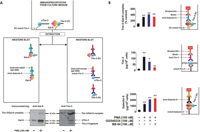
We investigated differential proteolytic shedding of free and galectin-9-bound Tim-3 from the surface of human AML cells as a pos-sible mechanism for the secretion of these proteins. Firstly, we exam-ined the medium used to culture THP-1 human AML cells with or without 16 h exposure to 100 nM phorbol 12-myristate 13-acetate (PMA) known to activate proteolytic shedding of Tim-3 (
Moller-Hackbarth et al., 2013). We then immunoprecipitated Tim-3 from the medium and extracted the precipitate as outlined in the Materials and Methods. Extracts were subjected to Western blot analysis followed by specific detection of galectin-9 and Tim-3. Specific galectin-9 bands appeared at around 32 kDa (molecular weight of galectin-9) as well as 52 kDa (Fig. 1A). Interestingly, the 52 kDa band was also detectable by anti-Tim-3 antibody (Fig. 1A), suggesting that this band corresponds to the unbroken Tim-3-galectin-9 complex. Furthermore, specific 3 bands appeared at around 33 kDa (molecular weight of soluble Tim-3– sTim-3) and around 20 kDa. This 20 kDa band is likely to be a fragment of Tim-3 shed together with galectin-9 being released from the complex during the Western blot procedure (Fig. 1A). This suggests that the Tim-3 protein fragment complexed with galectin-9 might be shed at different cleavage site(s). Interestingly, the amount of all the proteins detected was clearly higher in PMA-treated samples.
It has recently been found that Tim-3 can be shed from the cell sur-face by a disintegrin and metalloproteinase domain-containing proteins (ADAM) 10/17 (Moller-Hackbarth et al., 2013). We therefore investi-gated whether these proteases are associated with release of free Tim-3 and/or of the galectin-9-Tim-Tim-3 complex. We exposed THP-1 cells for 16 h to 100 nM PMA, after which the PMA-containing medium was re-moved and replaced with the same medium containing 100 μM GI254023X (ADAM 10 and 17 inhibitor) or 100μM BB-94, a matrix me-talloproteinase inhibitor. The cells were incubated for 4 h and levels of Tim-3 and galectin-9 were then measured in the culture medium by ELISA. We also measured soluble Tim-3-galectin-9 complex by captur-ing Tim-3 uscaptur-ing a scaptur-ingle-chain antibody and then detectcaptur-ing galectin-9 using a biotinylated anti-galectin-9 antibody. We found that PMA
treatment significantly upregulated sTim-3 release as well as the release of galectin-9 (a similar increase was observed in the Tim-3-galectin-9 complex,Fig. 1B). GI254023X and BB-94 decreased PMA-induced sTim-3 release but did not affect the release of either galectin-9 or the Tim-s Tim-3-galectin-9 complex (Fig. 1B), suggesting that this complex is differentially shed from the cell surface.
3.2. Protein Kinase C is Involved in the Activation of Tim-3 and Galectin-9 co-secretion by AML Cells
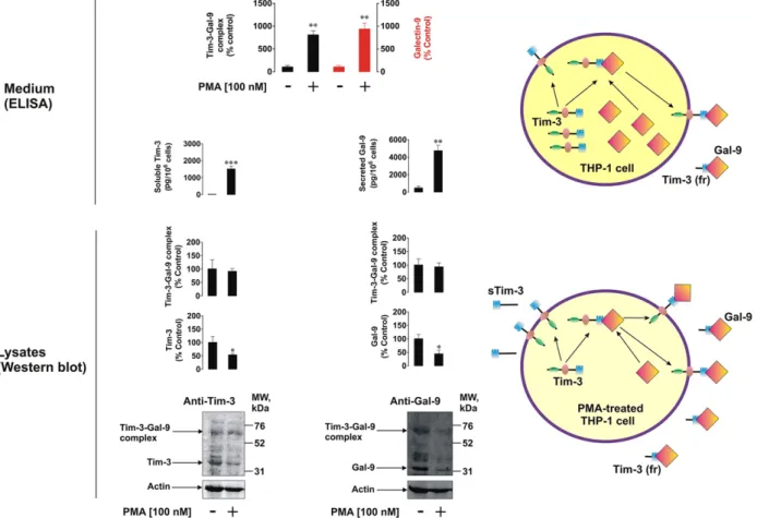
We considered the levels of Tim-3 and galectin-9 remaining in THP-1 cells following 16 h of exposure to specific PKC activator PMA. It was found that, despite the levels of released sTim-3, galectin-9 and Tim-3-galectin-9 complex were increased in PMA-treated cells, the levels of respective cell-associated proteins decreased (Fig. 2). Interestingly, a specific band in the range of 70 kDa detectable by both anti-Tim-3 and anti-galectin-9 antibodies was present in all the assays (Fig. 2). This molecular weight corresponds to a sum of those of uncleaved Tim-3 and galectin-9. This indicates that a complex between full Tim-3 and galectin-9 is first formed before undergoing shedding, which results in a release of its soluble form corresponding to the 52 kDa species, as described above. Our observations were confirmed by co-localization assays using confocal microscopy (Fig. 3). Following 24 h exposure to 100 nM PMA, paraformaldehyde-fixed non-permeabilized and methanol-permeabilized THP-1 human AML cells were investigated. We found that both galectin-9 and Tim-3 were present on the cell surface. In permeabilized cells there was clear evidence of co-localization
Fig. 1. Free and galectin-9-bound Tim-3 is shed differentially from the cell surface. THP-1 cells were exposed for 16 h to 100 nM PMA; medium was then exchanged for fresh PMA-free medium and cells exposed to the indicated concentrations of GI254023X (ADAM10/17 inhibitor) and BB-94 (matrix metalloproteinase inhibitor). Non treated THP-1 cells were incubated for 16 h after which medium was changed and cells incubated for further 4 h and used as a control. Western blot characterization of galectin-9 and Tim-3 variants (20 kDa fragment (Tim-3 (fr)) and 33 kDa (sTim-3)) was performed in medium collected afterfinal 4 h of incubation of resting and PMA-pre-treated THP-1 cells as outlined in theMaterials and Methods(A). All the samples were subjected to ELISA-based detection of galectin-9, soluble Tim-3 and Tim-3-galectin-9 complex (B). Images are from one experiment representative of six which gave similar results. Quantitative data represent mean values ± SEM of six independent experiments; *pb 0.05; **p b 0.01 vs. control.
of both proteins. Thesefindings were confirmed using imaging flow cytometry (Supplementary Fig. 1). In non-permeabilized cells we saw sectors full of either Tim-3 or galectin-9, without substantial co-localiza-tion. Given that galectin-9 is soluble, it can remain on the cell surface only if it is bound to its receptor, Tim-3 (Fig. 3). Taken together our findings suggest that Tim-3 is either externalized on its own or acts as a trafficker for galectin-9 (which lacks the signal domain required for secretion and thus requires a trafficker). Given that PMA, a specific PKC activator, significantly increases Tim-3 and galectin-9 secretion, it is likely that PKC is involved in the Tim-3 and galectin-9 co-secretion process.
3.3. Levels of Soluble Tim-3 and Galectin-9 are Highly Increased in the Blood Plasma of AML Patients: Characterization of the Tim-3-galectin-9 Complex in Human Blood Plasma
We then sought to confirm our findings in primary samples collected from AML patients. We analyzed plasma samples from 98 AML patients versus healthy donors and found that galectin-9 and Tim-3 levels were strikingly increased in blood plasma of AML patients (Fig. 4A, B, E and F). Five randomly selected plasma samples from the group of studied AML patients andfive from healthy donors were then subjected to detection
Fig. 2. PMA activates Tim-3 and galectin-9 production and release as well as generation of Tim-3-galectin-9 complex. 1 cells were treated with 100 nM PMA for 16 h. Non-treated THP-1 cells were used as a control. Cells were then harvested and galectin-9 as well as Tim-3 were analyzed in whole cell extracts by Western blot. Both proteins and Tim-3-galectin-9 complexes were analyzed by ELISA in the medium used to treat the cells. The bar diagram on the top shows the comparative analysis (expressed in % control) of galectin-9 and Tim-3-galectin-9 complex levels released by non-treated and PMA-treated THP-1 cells. Images are from one experiment representative of three which gave similar results. Quantitative data are the mean values ± SEM of three independent experiments; *pb 0.05; **p b 0.01; ***p b 0.001 vs. control.
Fig. 3. Co-localization of Tim-3 and galectin-9 in PMA-activated THP-1 cells. Co-localization of Tim-3 and galectin-9 was analyzed in non-permeabilized and permeabilized THP-1 cells following 24 h of exposure to 100 nM PMA using confocal microscopy (seeMaterials and Methodsfor details). Images are from one experiment representative of six which gave similar results.
of Tim-3-galectin-9 complex by ELISA as described above. The level of increase in Tim-3-galectin-9 complex in AML samples was similar to that of galectin-9 (Fig. 4C and G). We then randomly chosefive plasma samples from the group of studied AML patients and analyzed Tim-3 and galectin-9 levels by Western blot (Fig. 4D). Prior to loading onto the SDS-PAGE, samples were sonicated and boiled for 5 min at 95 °C. We found that sTim-3 and galectin-9 were clearly detectable. We could also see a clear band (probably representing the soluble form of the Tim-3-galectin-9 complex) at around 52 kDa (detectable by both anti-Tim-3 and anti-galectin-9 antibodies). This suggests that the com-plex released by THP-1 cells (Fig. 1) and the one found in blood plasma is unlikely to be formed after secretion. If this had been the case, its mo-lecular weight would have been around 65 kDa (33 kDa for sTim-3 and 32 kDa for galectin-9) rather than 52 kDa. A specific band was also de-tectable at around 20 kDa (Fig. 4D). These results are in line with those obtained for soluble forms of Tim-3, galectin-9 and Tim-3-galectin-9 complex released by THP-1 cells confirming that sTim-3 and Tim-3 complexed with galectin-9 are likely to be differentially shed from plasma membranes of AML cells. Interestingly, there is a clear evidence of a correlation between Tim-3 and galectin-9 levels in the plasma of both healthy donors and AML patients and these correla-tion levels were very similar to each other (Fig. 4H and I) suggesting a co-release of both proteins in both cohorts.
3.4. Tim-3 Binding Alters the Conformation of Galectin-9
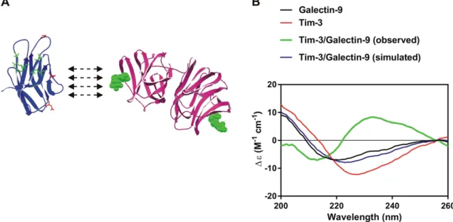
In order to assess the biophysical properties of Tim-3, galectin-9 and the Tim-3-galectin-9 complex we investigated them using synchrotron radiation circular dichroism (SRCD) spectroscopy at Diamond Light Source (Beam Line 23, Supplementary Fig. 2). Structural organization of Tim-3 and galectin-9 as well as their interaction are schematically presented in theFig. 5A. Galectin-9 interacts with non-glycosylated Tim-3 with nanomolar affinity (Kd = 2.8 × 10⁻⁸M); the binding can be further strengthened by interaction of galectin-9 with glycosylated Tim-3 (Prokhorov et al., 2015). Indeed, the complex is detectable by Western blot, which means that interaction between a lectin and
sugar is taking place. SRCD spectroscopy was also performed on galectin-9 and Tim-3 mixed to a stoichiometry of 1:1 molar ratio (Fig. 5B). Galectin-9 when mixed with Tim-3 showed a CD spectrum signi fi-cantly different from the simulated spectrum indicating that the inter-action of galectin-9 with Tim-3 causes significant conformational change of the proteins with a clear increase inβ-strand component. Based on the above, one might speculate that Tim-3 binding could alter the conformation of galect9, resulting in increased ability to in-teract with receptors in target cells. Since galectin-9 is a tandem protein with two sugar binding domains, one domain could bind Tim-3 (or other proteins) and leave the other domain open for interaction with a receptor molecule associated with the plasma membrane of a target cell (for example membrane associated Tim-3).
3.5. Latrophilin 1, Protein Kinase C and mTOR-Dependent Translation Play a Crucial Role in Tim-3 and Galectin-9 Production and Secretion
LPHN1 mRNA was found in primary human CD34-positive stem cells (Maiga et al., 2016). We were able to detect LPHN1 protein in them (at a slightly higher molecular weight than in THP-1 cells (around 140 kDa)), while in THP-1 it is detectable at 130 kDa (Supplementary Fig. 3) as well as in primary AML cells (Sumbayev et al., 2016). No Tim-3 or galectin-9 protein expression was detectable in primary human CD34-positive stem cells (Supplementary Fig. 3).
For this experimental set-up we used THP-1 cells and exposed them to 100 nM PMA or 250 pMα-latrotoxin (LTX, a highly specific and potent ligand of LPHN1 (Sumbayev et al., 2016)). We found that both PMA and LTX downregulated intracellular Tim-3 and galectin-9 levels (though not significantly) and significantly increased activat-ing phosphorylation of the mammalian target of rapamycin (mTOR) at S2448 (Fig. 6A and B). One hour pre-treatment of THP-1 cells with 70 nM Gö6983 (PKCα inhibitor) before exposure to PMA or LTX led to attenuation of stimulus-induced mTOR activation and downregulation of intracellular Tim-3 and galectin-9 levels. Interestingly, in the cells exposed just to Gö6983, phospho-S2448 mTOR and intracellular Tim-3/galectin-9 levels were not different
Fig. 4. Levels of galectin-9 and soluble Tim-3 are highly increased in blood plasma of AML patients. Galectin-9 and Tim-3 were measured by ELISA in blood plasma obtained from healthy donors and AML patients (A, B, E and F). The levels of Tim-3-galectin-9 complex were measured by ELISA in blood plasma offive randomly picked healthy donors and AML patients (C and G). Tim-3 and galectin-9 were characterized by Western blot in blood plasma fromfive randomly chosen AML patients (D). Correlations between Tim-3 and galectin-9 as well as between galectin-9 and Tim-3-galectin-9 complex was then determined (H, I, J and K). Images are from one experiment representative offive which gave similar results. Quantitative data represent mean values ± SEM of three independent experiments; *pb 0.05; **p b 0.01; ***p b 0.001 vs. control.
from the control. Both PMA and LTX highly upregulated release of both sTim-3 and galectin-9 from THP-1 cells. Gö6983 completely attenuated this increase in both cases, but did not change basic levels of Tim-3 and galectin-9 secretion, which suggests that basic (background) release of galectin-9 and Tim-3 does not depend on PKCα (Fig. 6D).
In summary, both PMA and LTX induce production of both Tim-3 and galectin-9 in THP-1 cells. We confirmed that THP-1 cells express Gαq (Supplementary Fig. 4A) and PMA as well as LTX induce highly sig-nificant upregulation of PKCα kinase activity (Supplementary Fig. 4B). Pre-treatment of THP-1 cells with 10μM AZD2014 (a highly specific mTOR inhibitor) before exposure to PMA or LTX reduced intracellular Tim-3 and galectin-9 levels as well as release of both proteins (Fig. 6C and D). This indicates that PMA or LTX-induced translation of both pro-teins depends on the mTOR pathway. Importantly, the solvents used to
dissolve pharmacological inhibitors had no effect on any of the studied protein levels or their secretion (data not shown).
These results were validated using primary human AML cells. For this purpose we exposed primary human AML mononuclear blasts AML-PB001F for 24 h to LTX followed by detection of secreted galectin-9 and Tim-3. We found that AML-PB0011F expressed LPHN1 and the secreted levels of both proteins were significantly increased in LTX-treated AML cells (Supplementary Fig. 5) confirming the findings obtained in THP-1 cells.
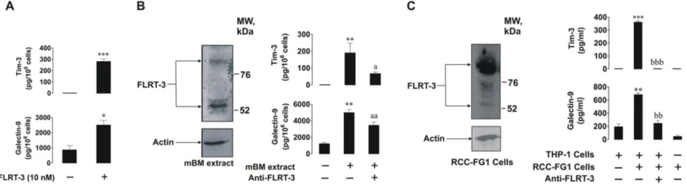
To confirm the physiological role of LPHN1 in galectin-9 release we exposed THP-1 cells to FLRT3, which is one of physiological ligands of LPHN1 (Boucard et al., 2014). We found that 10 nM FLRT3 induced sig-nificant upregulation of galectin-9 and sTim-3 release (Fig. 7A, a scheme of the experiment is presented in Supplementary Fig. 6A); it also upreg-ulated PKCα activity in THP-1 cells (Supplementary Fig. 4B). To confirm
Fig. 5. Interaction of Tim-3 with galectin-9 leads to major conformational changes increasing solubility of the protein complex. (A) The schematic structural models of Tim-3 extracellular domain (left) and galectin-9 (right). In the Tim-3 structure, amino acid residues involved in galectin-9-independent binding are highlighted in green. Residues, which are potential targets for glycosylation, are highlighted in red. In galectin-9, sugar molecules, which could potentially bind the protein, located close to the carbohydrate binding sites are shown in green. (B) The SRCD spectroscopy of Tim-3, galectin-9 and Tim-3-galectin-9 interaction (both simulated and real curves are presented).
Fig. 6. LPHN1, PKCα and mTOR pathways are involved in Tim-3 and galectin-9 production and secretion in AML cells. THP-1 cells were exposed to the indicated concentrations of PMA or LTX for 16 h with or without 1 h pre-treatment with the PKCα inhibitor Gö6983 (A, B, D) or the mTOR inhibitor AZD2014 (C, D). Cellular levels of Tim-3 and galectin-9 were analyzed by Western blot. Released Tim-3 and galectin-9 were detected by ELISA. Images are from one experiment representative of three which gave similar results. Quantitative data are the mean values ± SEM of three independent experiments; *pb 0.05; **p b 0.01; ***p b 0.001 vs. control. Symbols “a” or “b” are used instead of “*” to indicate differences vs. PMA and LTX-treated cells, respectively.
that this effect was physiologically relevant, we exposed THP-1 cells for 16 h to mouse bone marrow (mBM) extracts (10μg protein/ml, which contain FLRT3,Fig. 7B, a scheme of the experiment is presented in Sup-plementary Fig. 6B) obtained as outlined in theMaterials and Methods. Treatments were conducted with or without 1 h pre-treatment with 5 μg/ml FLRT3 neutralizing mouse antibody. We found that mBM extracts significantly upregulated galectin-9 and sTim-3 secretion in THP-1 cells. FLRT3 neutralizing mouse antibody reduced the effects of mBM extracts but did not block them (Fig. 7B). This means that BM contains several activators of galectin-9 secretion in AML cells. Finally, we co-cultured THP-1 cells with RCC-FG1 renal carcinoma cells (which are highly ad-herent) in the ratio 1 THP-1:2 RCC-FG1. RCC-FG1 cells express high levels of FLRT3 and release almost undetectable amounts of galectin-9 (Fig. 7C, a scheme of the experiment is presented in Supplementary
Fig. 6C). Cells were kept together for 16 h in the absence or presence of 5μg/ml FLRT3 neutralizing antibody and then galectin-9 and sTim-3 secretion levels were analyzed. We found that the presence of RCC-FG1 cells significantly increased galectin-9 and sTim-3 release and FLRT3 neutralization attenuated these effects. The presence of RCC-FG1 cells significantly upregulated PKCα activity, an effect that was also attenuated by neutralization of the FLRT3 (Supplementary Fig. 4C). These results suggest that FLRT3 stimulates the release of galectin-9 from AML cells.
3.6. Galectin-9 and sTim-3 Attenuate AML Cell Killing Activity of NK Cells Recent evidence suggested that galectin-9 (either soluble or cell sur-face associated) can interact with Tim-3 or possibly other receptors on
Fig. 7. FLRT3, a physiological ligand of LPHN1, induces galectin-9 and Tim-3 secretion. (A) THP-1 cells were exposed for 16 h to 10 nM extracellular domain of human recombinant FLRT3 followed by measurement of released Tim-3 and galectin-9 by ELISA. (B) THP-1 cells were exposed to mouse bone marrow (mBM) extracts for 16 h with or without 1 h pre-treatment with 5μg/ml anti-FLRT3 antibody. The presence of FLRT3 in mBM extracts was confirmed by Western blot analysis. Secreted Tim-3 and galectin-9 were measured by ELISA. (C, left) RCC-FG1 cells express FLRT3 as confirmed by Western blotting. (C, right) RCC-FG1 cells were co-cultured with THP-1 cells at a ratio of 1 THP-1:2 RCC-FG1 with or without 1 h pre-treatment with 5 μg/ml FLRT3 neutralizing antibody. Secreted galectin-9 and Tim-3 were measured by ELISA. Images are from one experiment representative of three which gave similar results. Quantitative data depict mean values ± SEM of three independent experiments; *pb 0.05; **p b 0.01; ***p b 0.001 vs. control. Symbols “a” or “b” are used instead of “*” to indicate differences vs. cells treated with mBM extracts or co-cultured with RCC-FG1 cells, respectively.
Fig. 8. LAD2 cells express and externalize Tim-3 and galectin-9. Left panel: surface-based and total Tim-3 and galectin-9 were measured in LAD2 human mast cell sarcoma cells using LI-COR in cell assay (ICA, non-permeabilized cells) and in cell Western (ICW, permeabilized cells). Right panel: protein levels of Tim-3 and galectin-9 were measured in resting and IgE-sensitized LAD2 cells by Western blot. Galectin-9 release was characterized using ELISA. Images are from one experiment representative of three which gave similar results. Quantitative data show mean values ± SEM of three independent experiments; ***pb 0.001 vs. control.
cytotoxic lymphoid cells including NK cells and cytotoxic T cells (Gleason et al., 2012). It may be proposed that Tim-3-galectin-9 interac-tion is involved in the creainterac-tion of immunological synapses between tar-get cells and cytotoxic lymphoid cells. To investigate this we used LAD2 human mast cell sarcoma cells kindly provided by Prof. Metcalfe and Dr. Kirshenbaum (NAID, NIH, USA;Kirshenbaum et al., 2003). These cells express both Tim-3 and galectin-9 with both proteins located mostly on the cell surface (Fig. 8) and not rapidly shed. This can thus be used to visualize the formation of immunological synapses between the two cell types. They also express high affinity IgE receptors (FcɛRI) which are not expressed by NK cells and thus can be used to distinguish between the two cell types.
Resting LAD2 cells do not release detectable amounts of galectin-9 and sensitization with IgE (which was used in order to label the cells for visualization) does not augment galectin-9 secretion considerably (Fig. 8). We therefore immobilized primary human NK cells isolated from buffy coats of human blood on ELISA plates as outlined in Materials and Methods. NK cells express Tim-3 (several glycosylation variants, Supplementary Fig. 7) but do not produce detectable amounts of galectin-9 protein. We applied IgE-sensitized LAD2 cells (Sumbayev et al., 2012) to the NK cells at a ratio of 1:1 with or without 15 min pre-in-cubation with galectin-9 neutralizing antibody. Isotype control antibody was also used instead of galectin-9 antibody to rule out the IgG effect. LAD2 cells were thenflagged using mouse IgM anti-IgE followed by vi-sualization using anti-mouse LI-COR secondary antibody (which recog-nizes IgM, see Materials and Methods for further details). We found that LAD2 cells were binding to NK cells and the presence of galectin-9 neu-tralizing antibody (but not isotype control antibody) abrogated this ef-fect (Fig. 9, a scheme of the experiment is presented in Supplementary Fig. 8). These results confirm that galectin-9 produced by LAD2 cells participates in their interactions with NK cells. Furthermore, abrogation of the effect by anti-galectin-9 antibodies may indicate that the Tim-3-galectin-9 interaction is the only pathway through which these cells could interact. This is most likely a result of IgE sensitization of LAD2 cells which highly increases the presence of galectin-9 on their surface. We then used K562 chronic myeloid leukemia cells which do not re-lease detectable amounts of galectin-9 (as confirmed by ELISA). K562 cells were exposed to PMA for 24 h in 96 well Maxisorp plates. Medium was replaced with PMA-free RPMI-1640 medium containing isolated primary human NK cells at a ratio of 1 K562:2 NK in the absence or pres-ence of 5 ng/ml human recombinant galectin-9. Cells were co-incubated for 16 h and their viability was then assessed using an MTS test. We
found that the presence of NK cells significantly reduced the viability of K562 cells however, the presence of galectin-9 attenuated K562 kill-ing effect (Fig. 10A). Viability of NK cells was not affected in any of the cases (Fig. 10A). Interestingly, the cytotoxic attack by NK cells also led to a dramatic change in the behavior of K562 cells, causing their massive aggregation. Using phase contrast microscopy, we determined the effect of galectin-9 on cell aggregation in individual or combined K562 and NK cell cultures. In the absence of galectin-9, there was clear evidence of K562 cells aggregating in the presence of NK cells (Fig. 10B). Galectin-9, in a dose-dependent manner, decreased the aggregation of K562 cells by NK cells, such that no K562 cell aggregation was detectable in the presence of 5 ng/ml galectin-9 (Fig. 10C). Galectin-9 itself had no visible effect on either of the two cell types alone. Thus, galectin-9 clear-ly protects myeloid leukemia cells from being killed by NK cells.
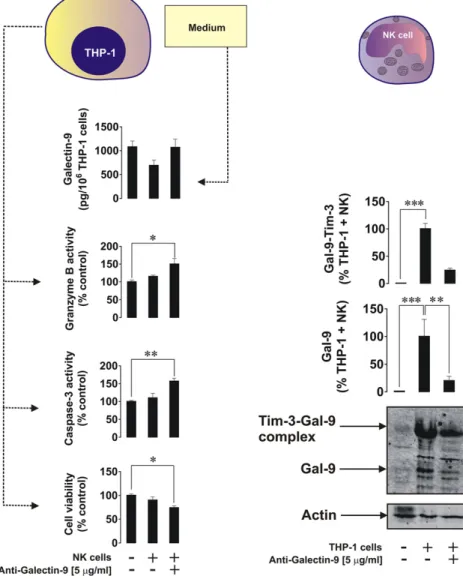
We then investigated the interactions between AML THP-1 cells and primary human NK cells. THP-1 cells were exposed to 100 nM PMA for 16 h. The medium was then replaced with PMA-free medium contain-ing NK cells at a ratio of 2 NK cells:1 THP-1 cells and left for 6 h in the absence or presence of 5μg/ml galectin-9-neutralizing antibody. Tim-3 and galectin-9 were then measured in the NK cells by Western blot analysis, and viability of THP-1 cells, activities of granzyme B, caspase-3 and galectin-9 release were monitored. We found that THP-1 cell via-bility was reduced when galectin-9 was neutralized (Fig. 11). This was in line with increased caspase-3 activity and granzyme B activities. Galectin-9 release was not affected (Fig. 11, galectin-9 bound to neutral-izing antibody is detectable in our system). We confirmed that resting NK cells did not produce detectable amounts of galectin-9. However, this protein on its own, and in the form of unbroken Tim-3-galectin-9 complex, was detectable in NK cells co-cultured with THP-1 cells and was reduced in the presence of galectin-9 neutralizing antibody. This suggests that THP-1 cells were the source of galectin-9, which was most likely bound to Tim-3 on the surface of NK cells, preventing the de-livery of NK cell-derived granzyme B into THP-1 cells and inhibiting the caspase-3-dependent apoptotic pathway.
Recently, a possible reciprocal link between levels of sTim-3 and IL-2, a cytokine, which activates cytotoxic activity of NK cells and T cells, was reported (Geng et al., 2006). We also found that in the plasma of healthy donors the levels of sTim-3 were significantly lower compared to AML patients (Fig. 4) whereas the levels of IL-2 were significantly higher (Fig. 12A and B). To investigate a possible direct influence of sTim-3, we exposed Jurkat T cells (resting Jurkat T cells produce detect-able amounts of IL-2) to increasing concentrations of Tim-3 for 24 h. We found a striking sTim-3 concentration-dependent and significant reduc-tion of IL-2 release from Jurkat T cells. This indicates that sTim-3 is capa-ble of binding a target protein (or a group of target proteins) and reducing IL-2 production thus preventing induction of NK cell and T lymphocyte anti-cancer activities.
Taken together, our results demonstrate a pathobiochemical path-way in AML cells. It is associated with activation of PKCα by LPHN1 (or any other receptors with similar activity) leading to the expression and exocytosis of sTim-3 and galectin-9, which prevent the activation of cytotoxic lymphocytes and impair their malignant cell killing activity. 4. Discussion
AML is a malignancy affecting bone marrow and blood and is a se-vere, and often fatal, systemic disease. AML cells escape host immune attack involving NK and cytotoxic T cells by impairing their activity (Golden-Mason et al., 2013; Kikushige et al., 2015; Gonçalves Silva et al., 2016). However, the biochemical mechanisms underlying the im-mune escape of malignant white blood cells remain unclear. Recently, it was shown that AML cells express high levels of the immune receptor Tim-3 and release galectin-9 which impairs the activity of NK cells and cytotoxic T cells (Gonçalves Silva et al., 2016). We have also suggested that Tim-3, as a membrane associated glycoprotein, might act as a traf-ficker for galectin-9 (Gonçalves Silva et al., 2016). As for all galectins,
Fig. 9. Galectin-9 participates in the formation of an“immunological synapse” between NK cells and LAD2 cells. Primary human NK cells were immobilized on the surface of Maxisorp plates. Cells were then co-incubated for 30 min with LAD2 cells with or without 30 min pre-treatment of LAD2 cells with 5μg/ml galectin-9 neutralizing antibody (or the same amount of isotype control antibody). LAD2 cells were then visualized using LI-COR assay as outlined inMaterials and Methods. Images are from one experiment representative of five which gave similar results. Quantitative data represent mean values ± SEM of five independent experiments; *pb 0.05; **p b 0.01.
galectin-9 is synthesized on free ribosomes and since it lacks the signal domain required for secretion it thus needs a trafficker in order to be re-leased (Delacour et al., 2009). When on the cell surface, Tim-3 is known to be shed by ADAM 10/17 proteolytic enzymes thus producing sTim-3, the function of which remains unknown (Moller-Hackbarth et al., 2013). We found, that Tim-3 could be shed in its free form as well as in complex with galectin-9; however, differential shedding is taking place. The Tim-3 fragment in the complex is about 20 kDa molecular weight, while sTim-3 is around 33 kDa. SRCD analysis of the complex suggests that the interaction between Tim-3 and galectin-9 proteins leads to major conformational change, possibly increasing the ability
of galectin-9 to interact with the target proteins. Since galectin-9 is a tandem protein containing two domains (Delacour et al., 2009), one of them might be interacting with Tim-3, while the other one could bind to a target receptor molecule, for example another molecule of Tim-3 associated with the plasma membrane of the target cell (Nagae et al., 2006). This may explain the high efficiency of galectin-9 in triggering Tim-3 on NK cells, which do not express galectin-9 and thus contain un-occupied Tim-3 on their surface.
Since we can observe the Tim-3-galectin-9 complex on Western blots following denaturing SDS-gel electrophoresis (Figs. 1, 2 and 4; ~ 52 kDa soluble form and ~ 70 kDa cell-derived form), it is likely that
Fig. 10. Galectin-9 protects myeloid leukemia K562 cells from being killed by primary human NK cells. (A) K562 cells were co-cultured for 16 h with primary human NK cells (at a ratio of 1 K562:2 NK) in the absence or presence of 5 ng/ml galectin-9. Viability of K562 and NK cells was then measured using an MTS test. Images are from one experiment representative of three which gave similar results. Quantitative data represent mean values ± SEM of three independent experiments; ***pb 0.001 vs. control. (B) K562 cells were co-cultured for 16 h with primary human NK cells (at a K562:NK ratio of 1:2) in the presence of different concentrations of galectin-9 (0–5 ng/ml). Cells were imaged using phase-contrast microscopy. The images are from one representative experiment of six (n = 6), which gave similar results. Scale bar (the same for all images), 50μm. (C) The NK cell-induced aggregation of K562 cells was quantified as a function of galectin-9 concentration. Left panel: percent of cells found in aggregates in individual cultures and in co-culture. Right panel: the size of cell aggregates in individual cultures and in co-culture. The data represent the mean values ± SD of six independent experiments; *, pb 0.05; **, p b 0.01; ****, p b 0.0001.
the binding between proteins is further strengthened by the interaction of galectin-9 with Tim-3-associated glycosides. Interestingly, the com-plex is detectable by Western blot with both Tim-3 and anti-galectin-9 antibodies. However, when these antibodies are sequentially applied to the same blot, the second antibody fails to detect the respec-tive protein in the same band (unless thefirst antibody is stripped off), due to steric hindrance. This effect explains why Tim-3 located on the
cell surface and covered by galectin-9 cannot be co-stained by the anti-body in confocal microscopy co-localization analysis (Fig. 3). Another point supporting this conclusion is that there was also clear evidence of co-localization of Tim-3 and galectin-9 in permiabilized THP-1 cells upon exposure to PMA (Fig. 3, Supplementary Fig. 1).
Previously it was reported that the release of both Tim-3 and galectin-9 depends on PKCα and proteolysis (Chabot et al., 2002). Our
Fig. 11. Cell-derived galectin-9 attenuates AML cell killing activity of primary human NK cells. THP-1 cells were co-incubated with primary human NK cells (ratio– 1 THP-1:2 NK) for 6 h followed by detection of THP-1 cell viability by the MTS test, measurement of activities of granzyme B and caspase 3 in THP-1 cell lysates and released galectin-9 (left panel). Galectin-9 levels from NK cells were determined by Western blot (right panel). Images are from one experiment representative of three which gave similar results. Quantitative data show mean values ± SEM of three independent experiments; *pb 0.05; **p b 0.01; ***p b 0.001 vs control.
Fig. 12. Soluble Tim-3 attenuates IL-2 release. (A and B) IL-2 levels were measured by ELISA in blood serum of healthy donors and AML patients. (C) Jurkat T cells were exposed to the increasing concentrations of Tim-3 for 24 h followed by detection of secreted IL-2 by ELISA. Data show mean values ± SEM of three independent experiments; *pb 0.05; **p b 0.01.
results confirmed these findings. PMA treatments induced PKCα activa-tion, which activated exocytosis of Tim-3 and galectin-9 as well as their mTOR-dependent production in THP-1 AML cells (Fig. 6).
Interestingly, natural and exogenous ligands of LPHN1, a G-protein coupled neuronal receptor expressed also in CD34-positive human stem cells (Supplementary Fig. 3) and AML cells but not healthy white blood cells, activated the PKCα pathway. They also induced both mTOR-dependent translation of Tim-3 and galectin-9 as well as their exocytosis. The effect was observed in THP-1 and primary human AML blasts. PKCα is known to provoke agglomeration of SNARE complex re-sponsible for exocytosis (Stockli et al., 2011; Morgan et al., 2005). Since FLRT3, one of natural ligands of LPHN1, is present in bone marrow (Fig. 7) it might explain how LPHN1 causes PKCα activation. Interestingly, constitutively active PKCα in malignant primary AML cells correlates with a very poor prognosis and high mortality rate of patients (Kurinna et al., 2006). This suggests that AML cells constantly release high levels of Tim-3 and galectin-9. Bone marrow also expresses other PKCα-activating proteins. When we exposed THP-1 cells to mouse bone marrow extracts, galectin-9 release was significantly higher
compared to resting THP-1 cells. FLRT3 neutralizing antibody signi fi-cantly reduced but did not abolish FLRT3 induced PKCα-dependent galectin-9 release. This suggests that galectin-9 and Tim-3 are synthe-sized and exocytozed by AML cells in a PKCα and mTOR-dependent manner, using available plasma membrane-associated PKCα activating receptors (for example LPHN1) to induce the whole pathway.
Galectin-9 prevents the delivery of granzyme B into AML cells (this is a perforin and mannose-6-phosphate receptor-dependent process (Supplementary Fig. 9)). Inside AML cells granzyme B performs cleav-age of the protein Bid into tBid, thus inducing mitochondrial dysfunc-tion and cytochrome C release followed by caspase 3 activadysfunc-tion. Proteolytic activation of caspase 3, in addition to the classic pathway, might also be directly catalyzed by granzyme B (Lee et al., 2014). Our re-sults with galectin-9 confirmed this concept (Fig. 11). Recently it was reported that galectin-9 induces interferon-gamma (IFN-γ) release from NK cells (Gleason et al., 2012). IFN-γ interacts with AML cells in-ducing the activity of indoleamine 2,3-dioxygenase (IDO1), an enzyme which converts L-tryptophan into formyl-L-kynurenine, which is then converted into L-kynurenine and released (Corm et al., 2009; Folgiero
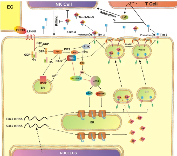
Fig. 13. AML cell-based pathobiochemical pathway showing LPHN1-induced classic activation of PKCα, which triggers translation of Tim-3 and galectin-9 as well as their secretion which is required for immune escape. The interaction of FLRT3 located on the surface of endothelial cells (EC) with LPHN1 leads to the activation of PKCα through the classic Gq/PLC/Ca2+
pathway. Ligand-bound LPHN1 activates Gq, which in then stimulates PLC. This leads to phosphatidyl-inositol-bisphosphate (PIP2) degradation and production of inositol-trisphospate (IP3) and
diacylglycerol (DAG). IP3interacts with ER-associated IP3receptor (IP3R) leading to Ca2+mobilization. PKCα is activated by DAG and Ca2+activates mTOR translational pathway
through downregulation of TSC1/TSC2. mTOR controls translation of Tim-3 and galectin-9. PKCα also phosphorylates Munc18 exocytosis regulator protein which provokes formation of SNARE complexes that tether vesicles to the plasma membrane. This pre-activates the release machinery, and elevated cytosolic Ca2+
lead to exocytosis of free and galectin-9-complexed Tim-3. Both types of Tim-3 are differentially shed from the cell surface by proteolytic enzymes. Soluble Tim-3 prevents IL-2 secretion required for activation of NK cells and cytotoxic T cells. Galectin-9 impairs AML cell killing activity of NK cells (and other cytotoxic lymphocytes).
et al., 2015; Mabuchi et al., 2016). L-kynurenine affects the ability of NK cells to kill AML cells, an effect which was seen in our experiments and presented in Supplementary Fig. 9. Soluble Tim-3 was shown to significantly downregulate production of IL-2, a cytokine required for activation of NK cells and cytotoxic T lymphocytes.
Taken together, our results show that human AML cells possess a se-cretory pathway which leads to the production and release of sTim-3 and galectin-9. Both proteins prevent the activation of NK cells and im-pair their AML cell-killing activity. This pathway, which involves the LPHN1-dependent activation of Tim-3 and galectin-9 production is summarized inFig. 13. The described pathway presents both bio-markers for AML diagnostics and potential targets (both sTim-3 and galectin-9) for AML immune therapy and thus can be considered as a fundamental discovery.
Grant Support
This work was supported by a Daphne Jackson Trust postdoctoral fellowship (to IMY), University of Kent Faculty of Sciences Research Fund (to VVS and YAU), Batzebär grant (to EFK and SB) and Oncosuisse grant KFS-3728-08-2015 (to LV and MB). Funders had no role in study design, data collection, data analysis, interpretation or writing of the report.
Acknowledgements
We thank Prof. Michelle D. Garrett (School of Biosciences, University of Kent, UK) for generously providing us with AZD2014. We are grateful to Dr. Gurprit S. Lall (School of Pharmacy, University of Kent, UK) for kindly providing us with biological materials for bone marrow extrac-tion. Antibody against Gαq was generously provided by Dr. Emma Veale, School of Pharmacy, University of Kent, UK. We are most grateful to Dr. Natasha S. Barteneva, School of Science and Technology, Nazarbayev University, Astana, Kazakhstan for her generous help with imagingflow cytometry. We thank Diamond Light Source for access to B23 beamline (SM12578).
Conflict of Interest
The authors declare no potential conflicts of interest. Author Contributions
IGS and IMY conducted most of the experiments, analyzed the data and contributed to manuscript writing, data interpretation andfigure assembly. SSS conducted the experiments reported inFigs. 2 and 7as well as Supplementary Figs. 4 and 6, analyzed the data, contributed to manuscript writing andfigure assembly. WF and JW were collecting plasma/providing blood plasma samples obtained from AML patients. MB and LV generated antibodies against Tim-3 and human recombinant Tim-3 protein fragment used in the study. RH, GS and GC conducted the experiments associated with SRCD and analyzed the data. SB contribut-ed to the study design and concept development. YAU and BFG designcontribut-ed the experiments associated with cell-cell interactions, analyzed and interpreted the data, contributed to the concept development and man-uscript writing. EFK designed and interpreted co-localization experi-ments, strongly contributed to design of experiments associated with Tim-3 and galectin-9 secretion, concept development and manuscript writing. VVS– designed the whole study, strongly participated in all the data collection, data analysis and interpretation, developed the con-cept and wrote the manuscript.
Appendix A. Supplementary data
Supplementary data to this article can be found online athttp://dx. doi.org/10.1016/j.ebiom.2017.07.018.
References
Abooali, M., Lall, G.S., Coughlan, K., Lall, H.S., Gibbs, B.F., Sumbayev, V.V., 2014.Crucial in-volvement of xanthine oxidase in the intracellular signalling networks associated with human myeloid cell function. Sci. Rep. 4, 6307.
Ashton, A.C., Rahman, M.A., Volynski, K.E., Manser, C., Orlova, E.V., Matsushita, H., Davletov, B.A., van Heel, M., Grishin, E.V., Ushkaryov, Y.A., 2000.Tetramerisation of α-latrotoxin by divalent cations is responsible for toxin-induced non-vesicular re-lease and contributes to the Ca2+
-dependent vesicular exocytosis from synapto-somes. Biochimie 82, 453–468.
Boucard, A.A., Maxeiner, S., Sudhof, T.C., 2014.Latrophilins function as heterophilic cell-adhesion molecules by binding to teneurins: regulation by alternative splicing. J. Biol. Chem. 289, 387–402.
Chabot, S., Kashio, Y., Seki, M., Shirato, Y., Nakamura, K., Nishi, N., Nakamura, T., Matsumoto, R., Hirashima, M., 2002.Regulation of galectin-9 expression and release in Jurkat T cell line cells. Glycobiology 12, 111–118.
Corm, S., Berthon, C., Imbenotte, M., Biggio, V., Lhermitte, M., Dupont, C., Briche, I., Quesnel, B., 2009.Indoleamine 2,3-dioxygenase activity of acute myeloid leukemia cells can be measured from patients' sera by HPLC and is inducible by IFN-gamma. Leuk. Res. 33, 490–494.
Davletov, B.A., Meunier, F.A., Ashton, A.C., Matsushita, H., Hirst, W.D., Lelianova, V.G., Wilkin, G.P., Dolly, J.O., Ushkaryov, Y.A., 1998.Vesicle exocytosis stimulated by α-latrotoxin is mediated by latrophilin and requires both external and stored Ca2+
. EMBO J. 17, 3909–3920.
Davydov, I.I., Fidalgo, S., Khaustova, S.A., Lelyanova, V.G., Grebenyuk, E.S., Ushkaryov, Y.A., Tonevitsky, A.G., 2009.Prediction of epitopes in closely related proteins using a new algorithm. Bull. Exp. Biol. Med. 148, 869–873.
Delacour, D., Koch, A., Jacob, R., 2009.The role of galectins in protein trafficking. Traffic 10, 1405–1413.
Dhupkar, P., Gordon, N., 2017.Interleukin-2: old and new approaches to enhance im-mune-therapeutic efficacy. Adv. Exp. Med. Biol. 995, 33–51.
Fasler-Kan, E., Barteneva, N., Ketterer, S., Wunderlich, K., Huwyler, J., Gygax, D., Flammer, J., Meyer, P., 2010.Activation of the JAK-STAT intracellular pathway in human retinal pigment epithelial cell line ARPE-19. Int. J. Interf. Cytokine Mediat. Res. 2, 127–136.
Fasler-Kan, E., Baiken, Y., Vorobjev, I.A., Barteneva, N.S., 2016.Analysis of nucleocytoplasmic protein shuttling by imagingflow cytometry. Methods Mol. Biol. 1389, 127–137.
Folgiero, V., Cifaldi, L., Li Pira, G., Goffredo, B.M., Vinti, L., Locatelli, F., 2015.TIM-3/Gal-9 interaction induces IFNgamma-dependent IDO1 expression in acute myeloid leuke-mia blast cells. J. Hematol. Oncol. 8, 36.
Geng, H., Zhang, G.M., Li, D., Zhang, H., Yuan, Y., Zhu, H.G., Xiao, H., Han, L.F., Feng, Z.H., 2006.Soluble form of T cell Ig mucin 3 is an inhibitory molecule in T cell-mediated immune response. J. Immunol. 176, 1411–1420.
Gleason, M.K., Lenvik, T.R., McCullar, V., Felices, M., O'Brien, M.S., Cooley, S.A., Verneris, M.R., Cichocki, F., Holman, C.J., Panoskaltsis-Mortari, A., et al., 2012.Tim-3 is an induc-ible human natural killer cell receptor that enhances interferon gamma production in response to galectin-9. Blood 119, 3064–3072.
Golden-Mason, L., McMahan, R.H., Strong, M., Reisdorph, R., Mahaffey, S., Palmer, B.E., Cheng, L., Kulesza, C., Hirashima, M., Niki, T., et al., 2013.Galectin-9 functionally im-pairs natural killer cells in humans and mice. J. Virol. 87, 4835–4845.
Gonçalves Silva, I., Ruegg, L., Gibbs, B.F., Bardelli, M., Fruehwirth, A., Varani, L., Berger, S.M., Fasler-Kan, E., Sumbayev, V.V., 2016.The immune receptor Tim-3 acts as a trafficker in a Tim-3/galectin-9 autocrine loop in human myeloid leukemia cells. Oncoimmunology 5, e1195535.
Hughes, R.C., 1999.Secretion of the galectin family of mammalian carbohydrate-binding proteins. Biochim. Biophys. Acta 1473, 172–185.
Hussain, R., Javorfi, T., Siligardi, G., 2012a.Circular dichroism beamline B23 at the Dia-mond Light Source. J. Synchrotron Radiat. 19, 132–135.
Hussain, R., Jávorfi, T., Siligardi, G., 2012b.Spectroscopic analysis: synchrotron radiation circular dichroism. Compr. Chiral. 8, 438–448.
Hussain, R., Benning, K., Myatt, D., Javorfi, T., Longo, E., Rudd, T.R., Pulford, B., Siligardi, G., 2015.CDApps: integrated software for experimental planning and data processing at beamline B23, Diamond Light Source. J. Synchrotron Radiat. 22, 862.
Khaznadar, Z., Henry, G., Setterblad, N., Agaugue, S., Raffoux, E., Boissel, N., Dombret, H., Toubert, A., Dulphy, N., 2014.Acute myeloid leukemia impairs natural killer cells through the formation of a deficient cytotoxic immunological synapse. Eur. J. Immunol. 44, 3068–3080.
Kikushige, Y., Miyamoto, T., Yuda, J., Jabbarzadeh-Tabrizi, S., Shima, T., Takayanagi, S., Niiro, H., Yurino, A., Miyawaki, K., Takenaka, K., et al., 2015.A TIM-3/Gal-9 autocrine stimulatory loop drives self-renewal of human myeloid leukemia stem cells and leu-kemic progression. Cell Stem Cell 17, 341–352.
Kirshenbaum, A.S., Akin, C., Wu, Y., Rottem, M., Goff, J.P., Beaven, M.A., Rao, V.K., Metcalfe, D.D., 2003.Characterization of novel stem cell factor responsive human mast cell lines LAD 1 and 2 established from a patient with mast cell sarcoma/leukemia; acti-vation following aggregation of FcepsilonRI or FcgammaRI. Leuk. Res. 27, 677–682.
Kurinna, S., Konopleva, M., Palla, S.L., Chen, W., Kornblau, S., Contractor, R., Deng, X., May, W.S., Andreeff, M., Ruvolo, P.P., 2006.Bcl2 phosphorylation and active PKC alpha are associated with poor survival in AML. Leukemia 20, 1316–1319.
Lee, J., Lee, S.J., Lim, K.T., 2014.ZPDC glycoprotein (24 kDa) induces apoptosis and enhances activity of NK cells in N-nitrosodiethylamine-injected Balb/c. Cell. Immunol. 289, 1–6.
Liu, J., Wan, Q., Lin, X., Zhu, H., Volynski, K., Ushkaryov, Y., Xu, T., 2005.α-Latrotoxin mod-ulates the secretory machinery via receptor-mediated activation of protein kinase C. Traffic 6, 756–765.
Mabuchi, R., Hara, T., Matsumoto, T., Shibata, Y., Nakamura, N., Nakamura, H., Kitagawa, J., Kanemura, N., Goto, N., Shimizu, M., et al., 2016.High serum concentration of L-kynurenine predicts unfavorable outcomes in patients with acute myeloid leukemia. Leuk. Lymphoma 57, 92–98.
Maiga, A., Lemieux, S., Pabst, C., Lavallee, V.P., Bouvier, M., Sauvageau, G., Hebert, J., 2016.
Transcriptome analysis of G protein-coupled receptors in distinct genetic subgroups of acute myeloid leukemia: identification of potential disease-specific targets. Blood Cancer J. 6, e431.
Micol, V., Sanchez-Pinera, P., Villalain, J., de Godos, A., Gomez-Fernandez, J.C., 1999. Corre-lation between protein kinase C alpha activity and membrane phase behavior. Biophys. J. 76, 916–927.
Moller-Hackbarth, K., Dewitz, C., Schweigert, O., Trad, A., Garbers, C., Rose-John, S., Scheller, J., 2013.A disintegrin and metalloprotease (ADAM) 10 and ADAM17 are major sheddases of T cell immunoglobulin and mucin domain 3 (Tim-3). J. Biol. Chem. 288, 34529–34544.
Morgan, A., Burgoyne, R.D., Barclay, J.W., Craig, T.J., Prescott, G.R., Ciufo, L.F., Evans, G.J., Graham, M.E., 2005.Regulation of exocytosis by protein kinase C. Biochem. Soc. Trans. 33, 1341–1344.
Nagae, M., Nishi, N., Murata, T., Usui, T., Nakamura, T., Wakarsuki, S., Kato, R., 2006.Crystal structure of the galectin-9 N-terminal carbohydrate recognition domain from Mus musculus reveals the basic mechanism of carbohydrate recognition. J. Biol. Chem. 281, 35884–35893.
Prokhorov, A., Gibbs, B.F., Bardelli, M., Ruegg, L., Fasler-Kan, E., Varani, L., Sumbayev, V.V., 2015.The immune receptor Tim-3 mediates activation of PI3 kinase/mTOR and HIF-1 pathways in human myeloid leukemia cells. Int. J. Biochem. Cell Biol. 59, 11–20.
Schindelin, J., Rueden, C.T., Hiner, M.C., et al., 2015.The ImageJ ecosystem: an open plat-form for biomedical image analysis. Mol. Reprod. Dev. 82, 518–529.
Siligardi, G., Hussain, R., 2015.CD Spectroscopy: An Essential Tool for Quality Control of Protein Folding. RJ Owen, ed. Methods Mol. Biol 1261. Springer, NY, pp. 255–276.
Silva, J.P., Ushkaryov, Y.A., 2010.The latrophilins,“split-personality” receptors. Adv. Exp. Med. Biol. 706, 59–75.
Silva, J.P., Lelianova, V.G., Ermolyuk, Y.S., Vysokov, N., Hitchen, P.G., Berninghausen, O., Rahman, M.A., Zangrandi, A., Fidalgo, S., Tonevitsky, A.G., Dell, A., Volynski, K.E.,
Ushkaryov, Y.A., 2011.Latrophilin 1 and its endogenous ligand Lasso/teneurin-2 form a high-affinity transsynaptic receptor pair with signaling capabilities. Proc. Natl. Acad. Sci. U. S. A. 108, 12113–12118.
Stockli, J., Fazakerley, D.J., James, D.E., 2011.GLUT4 exocytosis. J. Cell Sci. 124, 4147–4159.
Sumbayev, V.V., Nicholas, S.A., 2010.Hypoxia-inducible factor 1 as one of the“signaling drivers” of toll-like receptor-dependent and allergic inflammation. Arch. Immunol. Ther. Exp. 58, 287–294.
Sumbayev, V.V., Yasinska, I., Oniku, A.E., Streatfield, C.L., Gibbs, B.F., 2012.Involvement of hypoxia-inducible factor-1 in the inflammatory responses of human LAD2 mast cells and basophils. PLoS One 7, e34259.
Sumbayev, V.V., Gonçalves Silva, I., Blackburn, J., Gibbs, B.F., Yasinska, I.M., Garrett, M.D., Tonevitsky, A.G., Ushkaryov, Y.A., 2016.Expression of functional neuronal receptor latrophilin 1 in human acute myeloid leukemia cells. Oncotarget 7, 45575–45583.
Swamydas, M., Lionakis, M.S., 2013.Isolation, purification and labeling of mouse bone marrow neutrophils for functional studies and adoptive transfer experiments. JoVE e50586.
Ushkaryov, Y., 2002.α-Latrotoxin: from structure to some functions. Toxicon 40, 1–5.
Volynski, K.E., Capogna, M., Ashton, A.C., Thomson, D., Orlova, E.V., Manser, C.F., Ribchester, R.R., Ushkaryov, Y.A., 2003.Mutantα-latrotoxin (LTXN4C) does not form
pores and causes secretion by receptor stimulation: this action does not require neurexins. J. Biol. Chem. 278, 31058–31066.
Wang, F., He, W., Zhou, H., Yuan, J., Wu, K., Xu, L., Chen, Z.K., 2007.The Tim-3 ligand galectin-9 negatively regulates CD8+ alloreactive T cell and prolongs survival of skin graft. Cell. Immunol. 250, 68–74.
Yasinska, I.M., Gibbs, B.F., Lall, G.S., Sumbayev, V.V., 2014.The HIF-1 transcription complex is essential for translational control of myeloid hematopoietic cell function by maintaining mTOR phosphorylation. Cell. Mol. Life Sci. 71, 699–710.