Epidemiol. Infect. (1992). 108. 315 322 3 1 5 Printed in (heat Britain
Reactivity of human serum antibody with lipopolysaccharide O 78
antigen from enterotoxigenic Escherichia coli
H. BRUSSOW AND J. SIDOTI
Nestle Research Centre. Xestec Ltd., Vers-chez-les-Blanc, CH-1000 Lausanne 26, Switzerland
(Accepted 1 October 1991) SUMMARY
Fifteen and five of 20 volunteers challenged with the enterotoxigenic Escherichia coli strain 0 78 • H11 showed a fourfold titre increase of serum ELISA antibody to the homologous O 78 and the heterologous 0 8 lipopolysaccharide antigen, respectively. Sixty-three of 191 sera from 1- to 48-month-old German children showed serum antibody reactive with O 78 antigen, all but two of these O 78-positive sera also showed reactivity with at least one further 0 antigen. Only 14 of the 0 78 reactive sera also showed antibody to heat-labile enterotoxin. In addition, soluble 0 8 antigen could inhibit the binding of serum antibody to absorbed 0 78 in 68% of the German children. Antibody reactive with 0 78 antigen is thus not a reliable serological marker for enterotoxigenic E. coli infection in German children.
INTRODUCTION
Prospective community-based investigations showed that enterotoxigenic Escherichia coli (ETEC) are common pathogens associated with childhood diarrhoea in developing countries, but are rare in industrialized parts of the world [1]. In accordance with these epidemiological data we observed a high and a low prevalence of IgG antibody to the heat-labile enterotoxin (LT) in Ecuadorian and German children, respectively [2], Antibody to pooled lipopolysaccharides (LPS) from the most common O serogroups identified in ETEC diarrhoea were however as frequently observed in Ecuadorian as in German children [2]. This observation raised doubts about the specificity of these antibodies. In the present report we investigated the possibility whether antibody to an individual 0 antigen (0 78) could be used as a serological marker for ETEC strain infection. O 78 antigen was chosen because sera from volunteers orally challenged with ETEC strain O 78 • HI 1 were available to us [3] and because serogroup O 78 was the most frequently isolated ETEC strain in the area where the seroprevalence study was conducted 14].
MATERIAL AND METHODS
LPS antigens from E. coli serogroups O 8, 0 9. O 55, O 78 and O 128 (obtained from B. Rowe. Central Public Health Laboratory. Colindale. LTK) were prepared
O O O • ' o O O 3 0 2 0 1 4 -(«) (b)
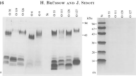
Fig. 1. Analysis of lipopolysaccharides in 1 3 % polyacrylamide gels by silver staining (a) and Coomsassie staining (b). The indicated purified 0 antigens and a protein molecular-weight standard (St: Pharmacia) were analysed by SDS-gel electiophoresis. Molecular weights in kilodaltons (kDa) are given for the standard.
by heating in saline a suspension of washed E. coli grown on veal-infusion agar (Difco) for 1 h at 100 °C [5]. After centrifugation. the supernatant was filtered on a 0-45 fim membrane. To remove contaminating RXA and protein, the heat-stable extract underwent ribonuclease A (Sigma) treatment (10/yg/ml for 1 h at 37 °C). followed by protease K (Merck) treatment (500 fig/ml. 2 h at 37 °C in the presence of 0"8% sodium dodecyl sulphate), three phenol:water extractions and finally dialysis against water. The lipopolysaccharides of the individual 0 antigen preparations were then analysed by gel electrophoresis [6]. Coomassie staining confirmed the absence of contaminating protein. Silver staining confirmed the presence of LPS molecules with the orderly spaced bands characteristic of the repeating units in their O side chain (Fig. 1). The individual LPS preparations were diluted to 5 fig LPS/ml in carbonate-bicarbonate coating buffer (pH 9-6) and added to flat-bottom 96-well polyvinyl microtiter plates (Dynatech. Chantilly, VA). Core glycolipid of E. coli strain Jo (Sigma Chemical. St Louis) was added to control plates. Sera of the American volunteers were diluted in a twofold dilution series, starting with a 1:10 dilution. Bound antibody was revealed with affinity-purified antibody to human IgG coupled to alkaline phosphatase (Sigma). Sigma 104 substrate was used and absorbance was read in a MR 5000 Dynatech ELISA plate reader. Cut-off level was an absorbance of 0-1 o.d. over the buffer blank, which gave only very low absorbance values.
Hundred microlitre samples of appropriately diluted serum predetermined to give an o.d. of O4-1-0 against the absorbed 0 78 antigen was incubated in the presence of 100 fil of soluble competitor LPS at a concentration of 5 //g/ml. Inhibition was defined if the competitor reduced the o.d. reading in ELISA by 50% or more in comparison with control reading of the serum in the absence of competitor. Samples were measured in duplicate.
Enterotoxigenic Escherichia coli
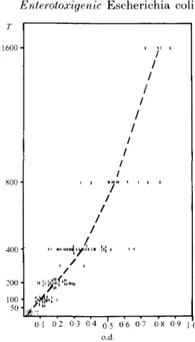
317 1600 -200 100 50 01 0 2 0-3 0 4 0*5 0-6 0-7 0-8 0-9 J O o.d.Fig. 2. LPS-speeifie IgM E L I S A a n t i b o d y t i t r e of 117 serum samples from B o c h u m G e r m a n y d e t e r m i n e d by end-point t i t r a t i o n T (twofold dilution series s t a r t i n g with 1 :50 dilution) were plotted against t h e o.d. value of t h e s a m e serum s a m p l e o b t a i n e d at a fixed 1 :200 serum dilution in E L I S A .
Table 1. Geometric mean titre and seroconversion of serum IgG ELISA antibody to homologous and heterologous 0 antigens in 20 North American volunteers challenged with 0 78-HU ETEC strain
O antigen 0 78 54 422 75 0 8 33 78 25 0 9 32 36 5 0 55 170 219 5 0 128 130 151 0 Jo' 30 35 0 Preimmune sera. GMT Immune serat. GMT SeroeonversionJ (in %)
* Core glycolipid of the rough E. coli strain Jo. t Sera were obtained 28 days after the challenge.
% Fourfold or greater titre increase.
University Children's Hospital of Bochum. Germany [7]. These sera were diluted 1:200 in PBS with 0-05% Tween 20. At that dilution the o.d. readings were linearly proportional to the relative antibody concentration measured by end-point titration (Fig. 2). Bound antibody was revealed with affinity purified
IgM antibody was measured in the children's sera to avoid confusion with passively acquired maternal IgG antibody. For the prevalence analysis, a serum sample was considered positive if the absorbance on the test plate was twice that on the control plate coated with core glycolipid of E. coli strain J5 (Sigma) and at least 0-1 o.d. greater than the o.d. of the control plate. The choice of a cut-off value of 0-1 o.d. is arbitrary. It is recognised that children negative by this criterion may have serum IgM antibody to LPS at a lower serum dilution: however their antibody titre must be low as 15 out 17 randomly selected sera with < (M o.d. had titres of < 50 by endpoint titration (Fig. 2).
RESULTS
Fifteen of the 20 North American volunteers (75%) showed a fourfold titre increase to the 0 antigen of the challenge strain O 78-H11 (Table 1, Fig. 3). Five (25%) showed also a fourfold titre increase to 0 8 antigen, whereas only one. one and none showed significant titre increases to 0 9. 0 55 and 0 128 antigen, respectively (Table 1). Soluble 0 78, but not 0 8. 0 9. 0 55 and 0 128 antigen inhibited binding of serum antibody from the American volunteers to the absorbed 0 78 antigen in inhibition ELISA.
IgM antibody against O 78 antigen was not detected in 45 cord sera and 19 sera from newborns from Bochum, Germany. The prevalence was 12 % in the first year of life and increased to 20, 49 and 6 3 % in the second, third and fourth year of life (Fig. 4a). Overall 63 out of 191 (33 %) 1- to 48-month-old German children showed serum antibody reactive with O 78 antigen. Fourteen of these 63 0 78-reactive sera also showed IgG antibody against the heat-labile enterotoxin (LT) at a cut-off level of 0'3 o.d. in the LT-specific ELISA described by Levine and colleagues (8) (Fig. 4^4). Sixty-one out of 63 sera reactive with O 78 antigen showed reactivity with at least one more of four 0 antigens tested (O 8, O 9, 0 55 and 0 128, Fig. 4B). Fig. 5 shows the age-related seroprevalence of IgM antibody to these four 0 antigens in comparison with antibody to O 78 antigen. Table 2 shows the reactivity pattern of LPS-specific IgM antibody in the different age groups of German children. Forty-five per cent of seropositive children younger than 1 year showed serum IgM antibody reactive with only one of the five 0 antigens tested and none reacted with all five 0 antigens tested. The percentage of sera reactive with only one of the five 0 antigens tested decreased gradually with increasing age (P = 0-0007, test for linear trend with increasing age). Conversely the percentage of sera reactive with all five O antigens tested increased gradually with increasing age (Table 2, P — 0-0001, test for linear trend with increasing age). In the oldest age group analysed (4-year-old children) only 10% reacted with only one, whereas 40% reacted with all of the five O antigens included into the analysis.
In inhibition ELISA soluble 0 8, 0 9 and O 128 antigens inhibited the binding of 0 78 reactive antibody to the absorbed 0 78 antigen in 68. 46 and 18% of the children's sera, respectively. This cross-inhibition was reciprocal: in 24. 27 and 32% of the children's sera the binding of O 8-, O 9- and 0 128-reactive antibody to the homologous antigen absorbed to the ELISA plate could be inhibited by
Enterotoxigenic Escherichia coli
319
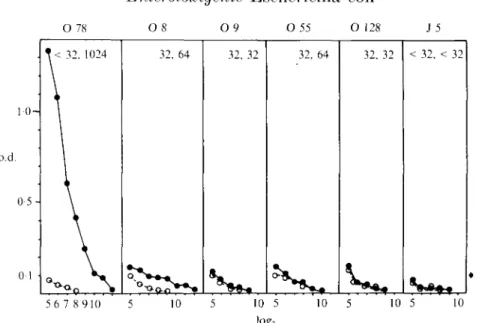
O 78 O 8 O9 O 55 Oi; J 5 1 0 -o.d. 0 5 -< 32. 1024 32. 64 32, 32 32, 64 32, 32 -< 32. -< 32 56 7 8 910 10 10 5 log; 10 5 10 5 10 Fig. 3. Serum TgG ELISA antibody response to homologous and heterologous O antigens of an adult volunteer challenged with ETEC strain O 78. o.d. reading in ELISA using the indicated O antigen or core glvcolipid of E. coli strain J 5 for coating the KLISA plates were plotted against the log2 dilution of the preimmune (O) or immune serum ( # ) . Titres for preimmune/immune sera are indicated in the upper right cornei'.45
5 0
-c 1 4 8 121624303642 48 Age (months)
Fig. 4. (a) Prevalence of KLISA IgJI antibody to lipopolysaecharide antigen 0 78 ( —) and () 8 {—) in different age groups of German children. Age groups are indicated by the upper age limit in months (C. cord sera), n. number of children in each age interval. The hatched area represents sera showing both IgM antibody to 0 78 antigen and IgG antibody to heat-labile enterotoxin. (b) Prevalence of ELISA IgM antibody to 0 78 antigen classified according to the reactivity of the sera with the indicated number of four further () antigens (O 8. O 9. O 55. 0 128). • , None: 0 . one: H. two: 0 . three: EL four.
50 50 -50" 50" O 55 O 78 0 1 4 812162430364248 Months
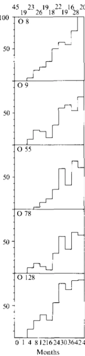
Fig. 5. Prevalence of IgM antibody to lipopolysaecharide of indicated Esche.richia coll 0 serogroup in German children of different age groups. 0. Cord sera: 1. infants > 1 : 4. infants > 1 and < 4 months of age and so forth, n. Number of children in each age group.
T a b l e 2. Reactivity of serum IgM antibody of German children against five 0 antigens of E . coli (0 8,0 9,0 55, 0 78, 0 128) in ELISA
No. of sera reactive with the indicated no. of five different LPS antigens
(% of LPS positive sera) Age-group (years) 0-1 1-2 2-3 3-4 No. of sera tested 87 40 35 48 No. of sera reactive with at least
one O antigen (% of total sera) 22 (25) 23 (58) 32 (91) 48(100) 1 3 10 (45) 2 (9) 5 (23) 7 (30) 5 (22) 2 (9) 6(19) 3(9) 6(19) 5(10) 6(13) 5(10) 5 (23) 0 6 (26) 3 (13) 9 (28) 8 (25) 13(27) 19(40)
Enterotoxigenic Escherichia coli 321 soluble O 78 antigen. In contrast soluble O 55 and O 78 antigen did not inhibit the binding of sera to absorbed 0 78 and O 55 antigen, respectively.
DISCUSSION
Thirty-three per cent of the sera from German children showed IgM antibody reactive with 0 78 antigen. Does this prevalence actually reflect exposure of these children to O 78 serogroup of E. coli ? Enterotoxigenic E. coli strains are only an infrequent cause of gastroenteritis in the geographical area where the sera were collected. Only 9 out of 60 identified E. coli isolates from gastroenteritis patients hospitalized in Bochum/Germany belonged to enterotoxigenic E. coli serogroups (eight isolates were in fact of O 78 serogroup [4], whereas 51 isolates belonged to enteropathogenic E. coli serogroups [4.9]. In addition only 22% of the sera reactive with 0 78 antigen showed IgG antibody to the heat-labile enterotoxin according to the criteria of Levineand colleagues.[8] Finally only 2 out of the 63 sera reacted only with O 78 antigen, but not with four other 0 antigens. The latter observation does not necessarily argue against the specificity of these antibodies. An age-related increase in sera with multiple 0 antigen reactivities would be expected if children undergo infections with different E. coli serogroups to which they mount a serogroup-specific serum immune response. However inhibition EL1SA showed for the sera of these German children a marked cross-reaction between 0 8 and 0 78 antigen. Interestingly, challenging adult volunteers with enterotoxigenic E. coli strain 0 7 8 H 1 1 induced a cross-reacting serum antibody response to O 8 antigen in 25% of the volunteers. Cross-reactions between 0 8 and 0 78 antigens have not been observed with animal hyperimmune sera [10, 11].
Our results contrast with those of two previous seroprevalence studies which failed to detect cross-reacting serum antibody to 0 antigen in children [12, 13]. It thus seems premature to use the reactivity of human sera with an individual 0 antigen as a serological marker for an infection with the specified E. coli serogroup. More must be learned about the cross-reactions demonstrated by human serum antibodies against 0 antigen to allow a meaningful serological analysis. Such studies might also be relevant for the development of an E. coli vaccine strain inducing cross-reacting antibodies after human vaccination.
A( KXOWLEDGEMENT
We thank B. Rowe for providing us E. coli serogroups, C. Tacket for sharing sera from US volunteers. H. Werchau and the late C. Mietens for sera from Bochum. FRG; V. K. Saudan for technical help, H. Link for technical advice, H. Rahim for statistical analysis and D. Massacand for typing the manuscript.
REFERENCES
1. l)u Pont H. Escherichia coli diarrhoea. In: Evans AS. Feldman HA. eds. Bacterial infections of humans. Epidemiology and control. Xew York: Plenum. 1984: 219-34.
2. Briissow H. Sidoti .1. Link H. Hoang Y. Barclay D. Dirren H, Freire WB. Age-specific prevalence of antibody to enterotoxigenic Escherichia coli in Ecuadorian and German children. .1. Tnfect Di.s 1990: 162: 974-7.
by milk immunoglobulin concentrate against oral challenge with enterotoxigenic Esch-erichia coli. New Engl J Med 1988; 318: 1240-3.
4. Mietens C. Kleinhorst C. Hilpert H, Gerber H. Amster H. Pahud J.J. Treatment of infantile E. coli gastroenteritis with specific bovine anti-.fi1. coli milk immunoglobulins. Eur J Pediatr
1979; 132: 239-52.
5. Roberts RS. Preparation of endotoxin. Nature 1966; 209: 80.
6. Tsai CM. Frasch CE. A sensitive silver stain for detecting lipopolysaccharides in polyacrylamide gels. Anal Biochem 1982; 119: 115-9.
7. Brussow H, Werchau H. Liedtke W. Lerner L. Mietens C. Sidoti .J. Sotek J. Prevalence of antibodies to rotavirus in different age-groups of infants in Boehum. West Germany. J Infect Dis. 1988; 157: 1014-22.
8. Levine MM, Young CR, Black RE. Takeda Y. Kingelstein RA. Enzyme-linked immunosorbent assay to measure antibodies to purified heat-labile enterotoxins from human and porcine strains of Escherichia coli and to cholera toxin: application in serodiagnosis and seroepidemiology. J Clin Microbiol 1985: 21: 174-9.
9. Robins-Brown R. Traditional enteropathogenic Escherichia coli of infantile diarrhoea. Rev Infect Dis 1987; 9: 28-53.
10. Edwards PR, Ewing WH. Identification of Enterobacteriaceae. Burgess Publishing ('ompany 1972 Minneapolis, chapter 5. pp 76—9.
11. 0rskov I. 0rskov F. Jann B. Jann K. Serology. chemistry and genetics of () and K antigens of Escherichia coli. Bacteriol Rev 1977; 4 1 : 667-710.
12. Kunin CM. Antibody distribution against nonenteropathogenic E. coli. Arch Intern Med 1962: 110: 676-86.
13. Xeter E, Westphal 0 , Luderitz 0 , Gino RM. Gorzvuski EA. Demonstration of antibodies against enteropathogenic Escherichia coli in sera of children of various ages. Pediatrics