© The Author 2013. Published by Oxford University Press on behalf of the Society of Toxicology.
This is an Open Access article distributed under the terms of the Creative Commons Attribution Non-Commercial License (http://creativecommons.org/licenses/by-nc/3.0/), which permits non-commercial re-use, distribution, and reproduction in any medium, provided the original work is properly cited. For commercial re-use,
please contact journals.permissions@oup.com.
Review
Mitochondria as a Target of environmental Toxicants
Joel N. Meyer,
*
,1Maxwell C. K. Leung,
*
John P. Rooney,
*
Ataman Sendoel,
†
Michael O. Hengartner,
†
Glen E. Kisby,
‡
and Amanda S. Bess
*
*Nicholas School of the Environment, Duke University, Durham, North Carolina; †Institute of Molecular Life Sciences, University of Zurich, Zurich, Switzerland; and ‡Department of Basic Medical Sciences, Western University of Health Sciences, Lebanon, Oregon
1To whom correspondence should be addressed at Nicholas School of the Environment, Duke University, Box 90328, A354 LSRC, Research Dr, Durham,
NC 27708-0328. Fax: (919) 668-1799. E-mail: joel.meyer@duke.edu. Received January 23, 2013; accepted April 23, 2013
Enormous strides have recently been made in our understand-ing of the biology and pathobiology of mitochondria. Many dis-eases have been identified as caused by mitochondrial dysfunction, and many pharmaceuticals have been identified as previously unrecognized mitochondrial toxicants. A much smaller but grow-ing literature indicates that mitochondria are also targeted by environmental pollutants. We briefly review the importance of mitochondrial function and maintenance for health based on the genetics of mitochondrial diseases and the toxicities resulting from pharmaceutical exposure. We then discuss how the principles of mitochondrial vulnerability illustrated by those fields might apply to environmental contaminants, with particular attention to fac-tors that may modulate vulnerability including genetic differences, epigenetic interactions, tissue characteristics, and developmental stage. Finally, we review the literature related to environmental mitochondrial toxicants, with a particular focus on those toxicants that target mitochondrial DNA. We conclude that the fields of environmental toxicology and environmental health should focus more strongly on mitochondria.
Key Words: contaminants; mitochondria; mitochondrial DNA;
mitochondrial toxicity; mitochondrial disease.
MitochoNDriA AND MitochoNDriAl DNA
Mitochondria are essential organelles best known for ATP
generation and their involvement in apoptosis. They also play
critical roles in other key processes including calcium, copper,
and iron homeostasis; heme and iron-sulfur cluster assembly;
synthesis of pyrimidines and steroids; thermogenesis and fever
response; and calcium signaling. The great majority of the
~1000–1500 (
Calvo and Mootha, 2010
) proteins that carry out
these functions are imported from the cytoplasm via
mitochon-dria targeting sequences or other mechanisms (
Bolender et al.,
2008
), and the proteins present vary significantly with tissue
(
Johnson et al., 2007
). This variability probably reflects
exten-sive variability in function, ranging from energy production in
muscle mitochondria to steroid synthesis in adrenal
mitochon-dria (
Vafai and Mootha, 2012
). A small number (13 in humans)
is encoded in the mitochondrial genome, along with the tRNA
and rRNAs required for their synthesis. These proteins are
components of complexes I, III, IV, and V of the electron
trans-port chain (ETC). Despite their small number, they are essential
and are transcribed at high rates: mtRNA represents ~5% of
total cellular RNA in many tissues but as high as 30% in heart
cells (
Mercer et al., 2011
).
Mitochondrial morphology also varies with cell type,
developmental stage, and environment (
Jansen and de Boer,
1998
;
Rube and van der Bliek, 2004
;
Vafai and Mootha, 2012
)
(
Fig. 1
). Morphology ranges from highly fragmented to highly
networked, is regulated via the processes of mitochondrial fusion
and fission, and is responsive to a variety of stressors (
Jendrach
et al
., 2008
;
Twig and Shirihai, 2011
). Morphological dynamics
are critical for maintenance of mitochondrial function (
Green and
Van Houten, 2011
;
Youle and van der Bliek, 2012
). The amount
of cellular mitochondria and their content are regulated via
mitochondrial biogenesis, nuclear signaling–mediated nuclear
and mitochondrial transcription, autophagy, and intraorganellar
degradation processes (
Diaz and Moraes, 2008
;
Mijaljica
et al
., 2007
;
Piantadosi and Suliman, 2012
;
Scarpulla, 2008
).
“Mitophagy” can selectively remove damaged mitochondria
(
Kim and Lemasters, 2011
;
Kim et al., 2007
), and damaged
cellular components can be degraded inside the mitochondria
or exported to lysosomes and peroxisomes via
mitochondria-derived vesicles (
Fischer et al., 2012
;
Nunnari and Suomalainen,
2012
). Mitochondria do not merely receive instructions from the
nucleus, however, but rather can initiate signaling in a variety
of ways. These include “retrograde signaling” (i.e., from the
mitochondria to the nucleus;
Chae et al., 2013
;
Liu and Butow,
2006
) to coordinate nuclear transcription with mitochondrial
needs via pathways including AMP-activated protein kinase,
sirtuins, calcium signaling, peroxisome proliferator–activated
receptor g coactivator 1a protein, and others (
Piantadosi and
Suliman, 2012
); mitochondrial-nuclear reactive oxygen species
(ROS)–mediated signaling (
Storz, 2006
); cell cycle arrest
signaling that may involve proteins with similar functions in the
nucleus (
Green et al., 2011
;
Koczor et al., 2009
;
Kulawiec et al.,
2009
); and initiation of apoptosis (
Tann et al., 2011
;
Raimundo
et al
., 2012
), as opposed to the better-known amplification of
an apoptotic process initiated elsewhere. There is evidence for
a “mitocheckpoint” that senses mitochondrial dysfunction and
triggers cell cycle arrest (
Singh, 2006
) although the mechanism
for this response is not fully understood.
Mitochondrial DNA (mtDNA) in many animal species is a
cir-cular intron-free genome consisting of 14,000–17,000 base pairs
(16,569 in humans) although it is much larger and noncircular and
contains introns in some species (
Anderson et al., 1981
;
Burger
et al
., 2003
). It is typically maternally inherited and present in
multiple copies within each cell; the mtDNAs are grouped in
DNA-protein complexes referred to as nucleoids (
Bogenhagen,
2012
;
Kukat et al., 2011
) that are anchored to the inner
mito-chondrial membrane. Most somatic cells in mammals have 10
3–
10
4mtDNAs (
Shoubridge and Wai, 2007
), but this number varies
significantly by cell type and developmental stage, as discussed
at more length below. mtDNA replication occurs independently
of the cell cycle. mtDNA half-life has been reported as 2–4 days
(
Kai et al., 2006
;
Lipsky and Pedersen, 1981
) but has not been
characterized in a wide range of cell types. mtDNA replication
and transcription are regulated in part by Tfam (Mitochondrial
transcription factor A), a major protein component of the
nucle-oid (
Campbell et al., 2012
). From an evolutionary perspective,
mtDNA has a high mutation rate compared with nuclear DNA
(nDNA). Normally, all of the mtDNAs in all of the cells of an
organism are of the same sequence; the presence of more than
one sequence in a cell is referred to as “heteroplasmy.”
Recognition of the key role of mtDNA integrity and
mitochon-drial function in health has grown greatly in recent years (
Chan,
2007
;
Chan et al., 2006
;
Christodoulou, 2000
;
Cohen, 2010
;
de
Moura et al., 2010
;
DiMauro and Davidzon, 2005
;
Hirano et al.,
2001
;
Jansen and Burton, 2004
;
Kwong et al., 2006
;
Manfredi
and Beal, 2000
;
Nunnari and Suomalainen, 2012
;
Schapira, 2012
;
Schmidt, 2010
;
Shaughnessy et al., 2010
;
Taylor and Turnbull,
2005
;
Wallace, 2005
), partly due to our increased understanding
and awareness of mitochondrial diseases.
MitochoNDriAl DisEAsEs
mtDNA and nDNA Mutations Can Cause Mitochondrial
Disease
Leber’s Hereditary Optical Neuropathy and mitochondrial
myopathies were in 1988 the first diseases demonstrated to
be caused by mtDNA mutations (
Holt et al., 1988
;
Wallace
et al
., 1988
). There are now more than 200, including
MELAS (myopathy, encephalopathy, lactic acidosis,
stroke-like episodes), MERRF (myoclonus epilepsy with ragged red
fibers), and many others (
Holt, 2010
). Importantly, cellular
dysfunction and clinical disease do not occur until a threshold
proportion of mtDNAs carrying mutations (heteroplasmy) is
reached (
DiMauro and Schon, 2003
;
Rossignol et al., 2003
).
Together, these diseases affect at least 1/10,000 people clinically
(
Chinnery et al., 2012
), establishing mtDNA mutations as a
major cause of disease.
Because most mitochondrial proteins are nuclear encoded,
it is not surprising that mutations in many nuclear genes also
cause mitochondrial diseases. For example, mutations in
DNA polymerase γ, the only mtDNA polymerase and thus
Fig. 1. Mitochondrial morphology. Panel (A) shows the mitochondrial network (MitoTracker Red-stained) containing many mtDNA nucleoids (PicoGreen-stained) surrounding the nucleus (Hoescht-(PicoGreen-stained) of a human primary fibroblast (photo: Amanda Bess). Panels (B) and (C) show a transgenic strain of C. elegans that expresses a mitochondrial matrix-targeted green fluorescent protein in body wall muscle cells. The image in panel (B) is from a control nematode, and the image in panel (C) is from a nematode exposed to 1µM carbonyl cyanide m-chlorophenyl hydrazone, a potent ionophore and inhibitor of oxidative phosphoryla-tion that leads to mitochondrial fragmentaphosphoryla-tion; the mitochondrial matrix was visualized via expression of green fluorescent protein (photo: John Rooney).
responsible for all mtDNA replication and repair, cause a wide
range of mitochondrial diseases including progressive external
ophthalmoplegia, Alper’s syndrome, and Leigh’s syndrome
(
Copeland, 2012
). The disease Friedreich’s ataxia is caused by
mutations in the gene encoding frataxin, a protein involved in
iron-sulfur cluster assembly (
Marmolino, 2011
).
Although individually rare, diseases caused by mtDNA and
nDNA mutations are estimated to collectively have an
inci-dence of ~1/4000 individuals (
Chinnery et al., 2004
;
DiMauro
and Davidzon, 2005
;
Howell et al., 2005
;
Wallace, 2005
).
There are currently no pharmaceutical cures for any
mitochon-drial disease (
Nunnari and Suomalainen, 2012
).
Mitochondrial Involvement in Common Diseases:
Causation Versus Correlation
The diseases described above are unambiguously caused by
mitochondrial dysfunction. There are also many very common
diseases for which there is strong correlative evidence,
suggest-ing that mitochondrial dysfunction is at least partially
causa-tive (
Wallace, 2005
). These are frequently diseases of old age
or energy homeostasis and include neurodegenerative diseases
such as Parkinson’s Disease and Alzheimer’s Disease (
Coskun
et al
., 2012
), many cancers (
Brandon et al., 2006
;
Kulawiec
et al
., 2010
;
Penta et al., 2001
), diabetes (
Rolo and Palmeira,
2006
), metabolic syndrome (
Mercer et al., 2010
),
cardiovascu-lar disease (
Ballinger, 2005
), and others (
Schon et al., 2012
).
Considerable effort is currently devoted to investigate whether
these relationships are causal. There is experimental evidence
demonstrating that defective mtDNA replication and repair
can accelerate organismal aging (
Bratic and Larsson, 2013
;
Trifunovic et al., 2004
).
Gene-Environment Interactions
In addition to the genetic diseases described so far, several
gene-environment interactions have been identified (
Cohen,
2010
;
Finsterer and Segall, 2010
). For example, valproic acid is
fatal in the context of some mitochondrial diseases (
Krähenbühl
et al
., 2000
;
Silva et al., 2008
), aminoglycosides (e.g.,
genta-mycin) can cause deafness in the context of specific mtDNA
mutations (
Guan, 2011
), and cigarette smoking and alcohol
intake are major risk factors for vision loss in people who are
homoplasmic for a mutation causing Leber’s hereditary optic
neuropathy (
Kirkman et al., 2009
).
Lessons from Mitochondrial Diseases
The existence of so many mitochondrial diseases illustrates
the critical importance of maintenance of mitochondrial and
mtDNA integrity for health. It also raises an important
ques-tion with implicaques-tions for environment-mediated mitochondrial
toxicity: Why did it take us so long to realize that many
dis-eases are in fact mitochondrial disdis-eases? Part of the answer is
surely that these diseases are very complicated. Although they
often target tissues that have high energy use or contain
non-replicating (and therefore irreplaceable) cells, this is somewhat
oversimplified (
Vafai and Mootha, 2012
), and there is
signifi-cant variability in their presentation. Some mtDNA mutations
result in single-tissue diseases, others affect a wide spectrum of
tissues, and others affect different tissues in different patients,
with varying age of onset (
Vafai and Mootha, 2012
;
Nunnari
and Suomalainen, 2012
). Furthermore, as illustrated by the
valproic acid and aminoglycoside examples, the same
environ-mental exposure may be innocuous in some people but deadly
in others, depending on genetic background. These
complica-tions suggest that identifying the etiology of an environmentally
induced mitochondrial dysfunction is likely to be challenging
with both laboratory and epidemiological approaches, as borne
out by the observation that, currently, the causes of only ~50%
of severe cases of mitochondrial dysfunction are identified
(
Vafai and Mootha, 2012
).
Drug-iNDucED MitochoNDriAl AND MtDNA toxicity
Drugs Can Cause Many Off-Target Mitochondrial Effects
Recognition that mitochondrial toxicity is a key “off-target”
effect of many drugs is also a relatively recent development.
Drugs have now been identified that inhibit and uncouple the
ETC; inhibit mitochondrial transport pathways, fatty acid
oxi-dation or uptake, the citric acid cycle, mtDNA replication, and
mitochondrial protein synthesis (a common mode of toxicity of
antibiotics that target bacterial ribosomes, to which
mitochon-drial ribosomes are similar due to mitochondria’s
evolution-ary origins); generate (or exacerbate generation of) ROS; and
more (
Begriche et al., 2011
;
Cohen, 2010
;
Dykens and Will,
2007
;
Kovacic et al., 2005
;
Mehta et al., 2008
;
Neustadt and
Pieczenik, 2008
;
Pacheu-Grau et al., 2010
;
Rowe et al., 2001
).
One well-studied example is adriamycin (doxorubicin), a
chemotherapeutic whose clinical use is limited by the fact that it
also causes irreversible and cumulative cardiomyopathy (
Wallace,
2007
). It appears to act largely by poisoning mitochondria, both via
redox cycling to generate ROS and by inhibiting ATP production
via uncoupling of oxidative phosphorylation. Although its
antineoplastic function is largely a result of DNA intercalation
and topoisomerase II inhibition in the nucleus, a substantial
amount reaches mitochondria, where it has a high binding affinity
for cardiolipin, a lipid found only in the mitochondrial inner
membrane (
Mustonen and Kinnunen, 1993
).
Another example is the nucleoside reverse transcriptase
inhibitors (NRTIs) that are used to combat human
immuno-deficiency virus (HIV) infection. NRTIs act by inhibiting the
reverse transcriptase activity required for viral replication. They
have been highly successful in treating adults and in preventing
transmission of HIV from pregnant mothers to their children, but
unfortunately many NRTIs also inhibit the mtDNA polymerase
γ. This has resulted mtDNA depletion- and mutation-mediated
mitochondrial toxicity, and even death, in patients and in animal
models (
Benhammou et al., 2007
;
Blanche et al., 1999
;
Chan,
2007
;
Claessens et al., 2003
;
Divi et al., 2010
;
Kohler and
Lewis, 2007
). Similar effects have been observed with
nucle-oside analogs intended for other viruses as well (
McKenzie
et al
., 1995
). Thus, chemicals that damage mtDNA or alter its
copy number can have very serious health consequences.
Such toxicities have led to failed clinical trials, “Black Box”
warnings and withdrawal of drugs from the market, and have
driven the pharmaceutical industry to develop better
meth-ods for detection of drug-induced mitochondrial dysfunction
(
Begriche et al., 2011
;
Dykens et al., 2007
;
Marroquin et al.,
2007
;
Nadanaciva et al., 2009
;
Pereira et al., 2009
).
Lessons from Drug-Induced Mitochondrial Toxicity
Mechanisms of drug-induced toxicity offer lessons in terms
of how other chemicals might damage mitochondria; these are
discussed at more length below. As in the case of mitochondrial
diseases, it is instructive to consider why it took us so long
to realize that many drugs target mitochondria. One reason is
that frequently only a small percentage of people have adverse
reactions (
Dykens and Will, 2007
). For example, only ~1% of
children of HIV-treated mothers showed mitochondrial
toxic-ity (
Barret et al., 2003
). The reason for this variability is not
entirely clear. In some cases, gene-environment interactions
are likely the explanation. In others, environment-environment
interactions, epigenetic factors (see below), or the
stochastic-ity associated with mtDNA replication and allocation may be
involved. In any case, it is sobering from an environmental
toxi-cology perspective that it took years to recognize the
mitochon-drial toxicity of many drugs because drugs are in general much
better studied for toxicity than most environmental
contami-nants. This suggests that many environmental pollutants may
be undiscovered mitochondrial toxicants.
rEAsoNs For MitochoNDriAl VulNErAbility, AND FActors thAt MAy MoDulAtE
VulNErAbility
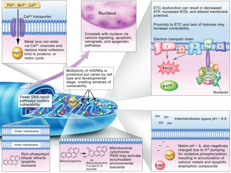
Reasons for mitochondrial vulnerability are presented
graph-ically in
Figure 2
and discussed in more detail below.
Accumulation of Pollutants
The high lipid content of mitochondrial membranes facilitates
accumulation of lipophilic compounds such as polycyclic
aro-matic hydrocarbons (PAHs) (
Backer and Weinstein, 1982
) and
some alkylating agents (
Wunderlich et al., 1972
). Cationic metals,
such as lead, cadmium, mercury, and manganese, have also been
shown to accumulate in mitochondria preferentially (
Atchison and
Hare, 1994
;
Bucio et al., 1999
;
Castellino and Aloj, 1969
;
Gavin
et al
., 1992
;
Sokolova et al., 2005a
). These metals may
accumu-late in mitochondria due to both entry via calcium transporters
(i.e., molecular mimicry) and chemical behavior resulting from
their interactions with mitochondrial pH and charge. The matrix
has a negative charge and slightly alkaline pH (~8 vs. ~6.9 for
the intermembrane space) due to the proton pumping associated
with oxidative phosphorylation. This also results in accumulation
of certain organic chemicals, particularly amphiphilic xenobiotics
including well-known mitochondrial toxicants such as ethidium
bromide, paraquat, 1-methyl-4-phenylpyridinium (MPP
+), and
others (
Cohen, 2010
;
Mehta et al., 2008
). It is also a property
that is exploited to target toxic (
Modica-Napolitano and Aprille,
2001
), diagnostic (
Ross et al., 2005
), or therapeutic (
Murphy,
2008
;
Murphy and Smith, 2007
) chemicals to mitochondria.
Ability to Metabolically Activate Pollutants
Another factor contributing to the mitochondrial
susceptibil-ity is the presence of cytochrome P450s in mitochondria, which
can activate chemicals that are relatively nonreactive prior to
metabolism, such as PAHs and mycotoxins (
Dong et al., 2009
;
Genter et al., 2006
;
Omura, 2006
). In addition, the existence
of close contacts between mitochondria and the endoplasmic
reticulum (
de Brito and Scorrano, 2010
) raises the possibility
that metabolites generated in the endoplasmic reticulum could
reach the mitochondria.
mtDNA Exhibits Unique Vulnerabilities But May Be
Buffered by High Copy Number
mtDNA is more vulnerable than nDNA to some
environmental genotoxins although the opposite is also true
in some cases (see below). A number of explanations for this
observation have been presented, including mtDNA’s physical
location, its reduced protein packaging compared with nDNA,
and its reduced repair capacity. These, along with the potential
for protection due to high copy number, are described in more
detail below.
Physically, mtDNA nucleoids are anchored to the matrix side
of the inner membrane, in close proximity to the ETC and many
proteins that contain transition metal ions. The ETC normally
generates a significant amount of ROS by electron loss; this has
been estimated at ~0.1% of O
2converted to superoxide anion
(
Beckman and Ames, 1998
;
Fridovich, 2004
) although the
amount may decrease with mild uncoupling (
Miwa and Brand,
2003
). Much of this appears to be produced by Complex I, with
release to the matrix, and Complex III, with release to the matrix
and intermembrane space (
Brand et al., 2004
). Manganese
superoxide dismutase (SOD) in the matrix, and copper-zinc
SOD in the intermembrane space, convert superoxide anion to
the longer lived and uncharged hydrogen peroxide; additional
antioxidant enzymes are present and regulated as well (
Valle
et al
., 2005
). Mitochondrial H
2O
2may interact with nearby
metals to produce hydroxyl radicals (
Giulivi et al., 1995
) in
close proximity to mtDNA. The many chemicals, disease states,
etc., that cause an increase in ROS generation may increase the
amount of superoxide produced: This includes redox cycling
agents that can intercept electrons such as adriamycin;
inhibi-tors of the ETC such as antimycin A and sodium azide that
lead to a super-reduced state of upstream complexes, increasing
the tendency to release electrons; dietary and exercise-related
ETC dysfunction; hyperoxia and hypoxia-reoxygenation; and
others (
Bailey et al., 2006
;
Barrett et al., 2004
;
Begriche et al.,
2006
;
Davies and Doroshow, 1986
;
Hasegawa et al., 1990
;
Indo
et al
., 2007
;
Ishiguro et al., 2001
;
Li et al., 2003a
;
Powell and
Jackson, 2003
;
Senft et al., 2002a, b
;
Turrens et al., 1982a, b
).
Furthermore, the Fenton reaction may be facilitated by redox
cycling of transition metal ions that are either chelated (
Dean
and Nicholson, 1994
) or released via ROS-induced degradation
of metal-containing proteins (
Mladenka et al., 2006
).
mtDNA lacks histones, which has frequently been cited as
a potential reason for increased vulnerability. However, recent
work (
Bogenhagen, 2012
) has shown that in fact a large number
of proteins associate with mtDNA, some of which are explicitly
protective (
Valle et al., 2005
), so mtDNA vulnerability to
reactive chemicals due to “nakedness” may be less than
previously speculated. Recent work has shown that nucleoid
structure is not static but can also be remodeled in response
to exposure to DNA intercalators including ethidium bromide
and doxorubicin (
Ashley and Poulton, 2009a, b
) and oxidative
stress (
Rebelo et al., 2009
).
Rebelo et al. (2009)
presented
evidence suggesting that different areas of the mitochondrial
genome were differentially packaged, so different regions may
be more or less “naked.” The nucleoid remodeling observed
by
Ashley and Poulton (2009a)
was protective against mtDNA
depletion that resulted from intercalator exposure. Furthermore,
the remodeling was p53 mediated, increasing the evidence for
a role of p53 in mitochondrial homeostasis (
Szczepanek et al.,
2012
). mtDNA occurs in a surprising array of structures in
cells from a wide range of tissues and species (
Kolesar et al.,
2012
). This may reflect to some extent significant variation in
the packaging function of proteins such as Tfam (
Campbell
et al
., 2012
) and the transcriptional status of the individual
genomes (
Kolesar et al., 2012
). Finally,
Chen et al. (2005)
showed that aconitase is a nucleoid component that assists in
mtDNA maintenance and protection against ethidium bromide
exposure, raising the possibility that nucleoid repackaging
may respond to bioenergetics and redox perturbations via
aconitase mobilization (
Shadel, 2005
). Thus, mtDNAs likely
have differential susceptibility to damage, both in a cell type–
specific fashion and even within single cells.
A very large difference between nDNA and mtDNA is the
relative lack of repair pathways present in mitochondria, at
least in humans (there is species variability). Although our
understanding of mtDNA repair pathways is evolving and
includes more proteins and pathways than previously
recog-nized (
Croteau et al., 2012
;
Kamenisch et al., 2010
;
Liu and
Demple, 2010
;
Scheibye-Knudsen et al., 2012
;
Sykora et al.,
2011
;
Valentin-Vega et al., 2012
), many types of damage are
either irreparable or only very slowly repaired. Base excision
repair is present in mitochondria, but nucleotide excision repair
(NER) is not, and recombinational and mismatch repairs are
either absent or quite limited (
Druzhyna et al., 2008
;
Gredilla
et al
., 2010
;
Kazak et al., 2012
;
Kraytsberg et al., 2004
).
Furthermore, mtDNA is a major target for oxidative stress
(
Van Houten et al., 2006
), despite the relatively robust
capac-ity for repair of oxidative damage. The sole replicative mtDNA
polymerase, DNA polymerase γ, appears to have very little
translesion synthesis capacity (
Graziewicz et al., 2004, 2007
;
Kasiviswanathan et al., 2012
), so that this damage tolerance
mechanism is also lacking or very limited. The absence of NER
is of particular relevance from an environmental toxicology
perspective because it means that many important
environmen-tal genotoxicants including some PAHs, mycotoxins, and
ultra-violet light will cause damage that is irreparable in mtDNA.
Furthermore, because a subset of DNA damage caused by ROS
is repaired by NER (
Brooks, 2008
;
Cline et al., 2010
), any of
the many factors that increase ROS in mitochondria (see above)
could also create some highly persistent damage.
What might the effects of persistent mtDNA damage be? In
addition to inhibiting replication, there is strong evidence that
such damage inhibits RNA transcription (
Cline, 2012
) and
mito-chondrial function (
Bess et al., 2012
;
Brar et al., 2012
;
Furda
et al
., 2012b
;
Leung et al., 2013
;
Rachek et al., 2009
). Although
most of the measures of mitochondrial function so far have
focused on energetics (ATP levels, oxygen consumption, etc.),
it may be useful to consider other endpoints. For example, might
mitochondrial dysfunction in the context of steroid-producing
cells result in endocrine disruption? mtDNA damage likely
con-tributes to generation of mtDNA mutations although this
pro-cess is currently poorly understood (
Copeland, 2012
;
Krishnan
et al
., 2008
;
Kulawiec et al., 2010
;
Loeb et al., 2005
). Improved
sequencing methods support the relatively high (compared with
the rest of the mtDNA) mutability of the D loop region (
Schmitt
et al
., 2012
) and will improve our understanding of both
mutagenesis and normal occurrence of heteroplasmy, which is
now detectable at much lower levels than previous methods
per-mitted. The “vicious cycle” hypothesis suggests that
mitochon-drial damage can lead to greater mitochonmitochon-drial dysfunction, in
turn leading to greater release of ROS and damage, etc. (
Yakes
and Van Houten, 1997
).
On the other hand, the mitochondrial genome is present in
multiple copies, potentially conferring buffering via redundancy.
As described earlier, mitochondrial dysfunction resulting from
mtDNA mutations is not observed until a certain threshold of
heteroplasmy is reached. This likely results from the combination
of multiple copies and the fact that mitochondrial fusion permits
swapping of contents, sometimes referred to as complementation
or functional buffering. Presumably, such buffering would also
occur for mtDNA damage. We recently found that similar levels
of mtDNA damage led to much greater inhibition of
mitochon-drial function in live Caenorhabditis elegans than cells in culture
(
Bess et al., 2012, 2013b
;
Leung et al., 2013
). Although there are
multiple possible explanations for this observation, it could result
from the much higher mtDNA copy number in human cells in
culture compared with C. elegans. The high copy number also
means that irreparable—or even repairable—DNA damage can
be handled by degrading damaged mtDNAs by one of a number
of possible mechanisms including degradation of the genomes
themselves or of entire mitochondria along with mtDNA (i.e.,
autophagy or mitophagy)(
Bess et al., 2012, 2013a, b
;
Rouzier
et al
., 2012
;
Shokolenko et al., 2009
). mtDNAs that have been
removed can then be replaced by mtDNA replication. The high
copy number and potential functional buffering also suggests that
mtDNA damage may be an excellent early biomarker of
mito-chondrial damage: High levels of mtDNA damage will occur and
persist and therefore be detectable, yet will precede late-stage
col-lapse of mitochondrial function associated with clinical detection.
Further complicating the picture is evidence that inhibition
of mitochondrial function may, at intermediate levels and under
some circumstances, have at least some beneficial effects. For
example, mild inhibition of mitochondrial function extends
lifespan and increases resistance to a variety of stressors in
C. elegans
(
Dillin et al., 2002
;
Rea et al., 2007
) and mice (
Liu
et al
., 2005
). In many cases, this effect is only observed if the
inhibition occurs during development and not if it occurs later in
life. However, it is important to note that greater inhibition leads
to pathological effects, and in general, developmental exposures
that alter programming of mitochondrial function later in life
are not always be beneficial (
Knudsen and Green, 2004
).
Cell Type Modulates Sensitivity
Mitochondria vary dramatically from tissue to tissue (
Calvo
and Mootha, 2010
;
Vafai and Mootha, 2012
), so it is not
surpris-ing that evidences from mitochondrial disease and mitochondrial
drug toxicity indicate that different cell types are differentially
susceptible to mitochondrial toxicants. There are many factors
that may sensitize specific cell types to mitochondrial
toxic-ity. For example, high reliance on mitochondrial function may
mean that dysfunction more easily causes cell death (e.g., as
often seen in mitochondrial diseases that target high energy-use
cells) because the increased function may result in higher ROS
production in some circumstances. An interesting example here
is the aforementioned case of adriamycin, which causes
princi-pally mitochondrial toxicity in heart, where much electron flow
occurs in mitochondria, and much more endoplasmic reticulum
damage in liver, where electron flow is much higher in the ER
(
Berthiaume and Wallace, 2007
;
Brown and Borutaite, 2012
).
Certain cell types (e.g., some neurons) also have low antioxidant
defenses and/or high inherent oxidative stress (
Crouch et al.,
2007
). Proliferating cells, both cancerous and noncancerous,
generate a great deal of ATP via aerobic glycolysis rather than
oxidative phosphorylation, apparently in order to
preferen-tially utilize carbon for biosynthesis required for cell division
(
Vander Heiden et al., 2009
). Any functional deficits
associ-ated with mitochondrial dysfunction in such cells may result
more from loss of biosynthetic than energy-generating
capac-ity. Finally, there is evidence that mtDNA repair capacity varies
with cell type, with brain tissue in particular showing low repair
(
Karahalil et al., 2002
;
Szczesny et al., 2010
). Nonetheless, it
is clear that the full spectrum of factors that result in cell-type
sensitivity is not well understood, and short-term effects may
differ from long-term effects, in part due to the occurrence of
secondary responses (
Vafai and Mootha, 2012
).
Developmental Stage Modulates Sensitivity
Some developmental stages are likely more sensitive to
mitochondrial toxicants due to age-dependent differences in
mitochondrial content, mitochondrial metabolism, and
mito-chondrial defense/repair mechanisms.
Mitochondrial count/volume and mtDNA copy number vary
dramatically with age in humans and other species that have
been examined. Although most cells thus far examined have
10
3–10
4copies of mtDNA, somatic and primordial germ cells
in the early embryo may contain as few as 10s or 100s of
cop-ies, constituting a “bottleneck” in mtDNA copy number and/
or transferral (
Carling et al., 2011
;
Jansen, 2000
;
Jansen and de
Boer, 1998
;
Pikó and Taylor, 1987
;
Shoubridge and Wai, 2007
).
Lower copy number is likely to reduce the protective effect of
redundant copies described above. However, the bottleneck
may also offer an opportunity for removal of severely damaged
mitochondria or aberrant mtDNAs (
Fan et al., 2008
;
Stewart
et al
., 2008
), although it is not entirely clear when this process
of purifying selection acts or how it may vary by mutation type
(
Freyer et al., 2012
), in part because the mitochondrial genome
is not believed to be very active early in development when the
bottleneck occurs. Furthermore, although newly fertilized eggs
have a very high mtDNA copy number (~10
5(
Shoubridge and
Wai, 2007
); but highly variable (
May-Panloup et al., 2007)
),
the evidence suggests that in many species those mtDNAs are
simply allocated into daughter cells during early development,
without additional replication, resulting in much lower copy
number after initial rounds of cell division (
Artuso et al., 2012
;
Bratic et al., 2009
;
Chase and Dawid, 1972
;
Leung et al., 2013
;
Rubenstein et al., 1977
;
Tsang and Lemire, 2002
;
Wai et al.,
2008
). Because mtDNA allocation into daughter cells at mitosis
(“mitotic segregation”) is apparently random, and the bottleneck
phenomenon results in low copy number at least in some cell
types (most have not been examined), it is possible for some
cells to end up with a high percentage of damaged or mutant
mtDNAs after multiple cycles of cell division. This high level of
heteroplasmy or damaged mtDNAs would then be inherited by
all of the cells derived from the progenitor cell. As previously
pointed out in other contexts (
Krishnan et al., 2008
),
mitochon-drial alterations such as mutations in cell types that do undergo
a bottleneck would not manifest until the second (in the case of
somatic cells) or even third (in the case of primordial germ cells)
generation, relative to an exposed mother. The effects might be
somatic alterations or fertility difficulties in the children of the
exposed mother or mitochondrial defects in the grandchildren.
There is also a lower copy number in old age (
Hebert et al.,
2010
;
Meyer et al., 2007
), suggesting vulnerability in aged
individuals.
Age-related changes in mitochondrial metabolism are also
well documented. Early life stages often display more anaerobic
metabolism and spherical morphologies, and older individuals
typically show signs of mitochondrial dysfunction although
these effects are frequently tissue specific (
Braeckman et al.,
2002
;
Brand et al., 2004
;
Brys et al., 2007, 2010
;
Dillin et al.,
2002
;
Hebert et al., 2010
;
Jansen and de Boer, 1998
;
Knudsen
and Green, 2004
;
Meyer et al., 2007
;
Tsang and Lemire, 2003
;
Tsang et al., 2001
;
Yasuda et al., 2006
). How reduced aerobic
mitochondrial function will influence the effect of mitochondrial
toxicants is not certain. On the one hand, reduced dependence
on oxidative phosphorylation can result in less sensitivity to
mitochondrial toxicity, as observed in at least some cells in
culture (
Marroquin et al., 2007
). On the other hand, reduced
capacity to generate energy might inhibit repair processes and
stress responses. Furthermore, the age dependence of
nonenergy-related mitochondrial functions (calcium homeostasis, heme
and iron-sulfur cluster assembly, synthesis of pyrimidines and
steroids, etc.) is less well understood. Finally, there is evidence
for an age-related decrease in base excision repair capacity in
mtDNA (
Anson et al., 2006
;
Druzhyna et al., 2008
;
Gredilla
et al
., 2010
;
Maynard et al., 2009
;
Szczesny and Mitra, 2005
;
Szczesny et al., 2003
).
We speculate that these early-life vulnerabilities make
mito-chondria important candidates for playing a role in diseases and
pathologies resulting from early environmental exposures, an
important area of current environmental health research (
Kai
et al
., 2006
;
Knudsen and Green, 2004
;
Landrigan et al., 2005
).
Similarly, late-life vulnerabilities suggest that environmental
toxicants may play an important role in the mitochondrial
dys-function commonly observed in aging and diseases of aging
(
Ames et al., 1995
;
Chomyn and Attardi, 2003
;
Fridovich,
2004
;
Melov et al., 1995
;
Nicholls, 2002
).
Conclusion: Vulnerability and Robustness
Many of the physical, chemical, and biological
characteris-tics of mitochondria make them vulnerable to toxicant
expo-sure (
Fig. 2
). At the same time, mitochondria are generally
robust (
Vafai and Mootha, 2012
) and are protected in a
num-ber of ways, including great redundancy of content, ability to
replace defective components, and complementation. However,
genetic deficiencies in the mitochondrial processes
responsi-ble for these defenses may sensitize some people to exposures
(
Meyer and Bess, 2012
).
MitochoNDriAl suscEptibility to ENViroNMENtAl toxicANts
The pathologies associated with mitochondrial disease and
drug-induced toxicity highlight the importance of mitochondrial
function for health. The demonstrated mitochondrial effects of
many drugs along with the theoretical considerations described
above lead to the concern that mitochondria may be an important
target for many environmental toxicants, presumably via many
or all of the same mechanisms described above for drugs. The
possibility that environmental contaminants may target
mito-chondria has been noted previously (
Shaughnessy et al., 2010
).
Nonetheless, which contaminants are important is far from clear
(
Schmidt, 2010
). We review some of the literature on pollutants
that target mitochondria, focusing in particular on mtDNA as a
target. We also discuss promising research approaches and
pro-pose important directions for future research.
Environmental Mitochondrial Toxicants
Although environmental contaminants have in general been
less well studied than drugs from the mitochondrial toxicity
perspective, there are nonetheless some well-understood
exam-ples. Paraquat, for example, appears to act largely by redox
cycling in a fashion analogous to adriamycin (
Martinez and
Greenamyre, 2012
). Rotenone is a Complex I inhibitor that
results in increased ROS production, mtDNA damage, and
apoptosis (
Ishiguro et al., 2001
;
Li et al., 2003b
;
Shokolenko
et al
., 2009
). Carbon monoxide and cyanide act as Complex
IV inhibitors (
Ninomiya-Tsuji, 2008
). An example of an
envi-ronment-environment interaction is provided by the
unfortu-nate co-occurrence of folate deficiency and exposure to either
cyanide or methanol, which appears to have resulted in
mito-chondrial toxicity–mediated optic neuropathy in ~50,000
peo-ple (
Sadun, 1998
). There are also many other contaminants that
are less well characterized as mitochondrial toxicants but are
reported to cause at least some of their effects via mitochondrial
toxicity. These include particulate matter (
Janssen et al., 2012
;
Hou et al., 2010
;
Xia et al., 2004
), lipopolysaccharide (
Suliman
et al
., 2003
), cadmium and copper (
Garceau et al., 2010
;
Sokolova et al., 2005a, b
), PAH quinones (
Babu et al., 2005
),
dioxin (
Biswas et al., 2008
;
Shertzer et al., 2006
), acrolein
(
Jia et al., 2007
), acrylamide (
Lee et al., 2012
), perfluorinated
compounds (
Starkov and Wallace, 2002
;
Walters et al., 2009
),
usnic acid (
Guo et al., 2008
;
Joseph et al., 2009
), methoxychlor
(
Gupta et al., 2006
), 3-nitropropionate (
Sabri, 1998
), cigarette
smoke (
Ballinger et al., 1996
;
Cakir et al., 2007
;
Westbrook
et al
., 2010
), manganese and lead (
Bowman et al., 2011
;
Sabri,
1998
;
Zheng et al., 1998
), arsenic (
Dopp et al., 2008
;
Echaniz-Laguna et al., 2012
;
Naranmandura et al., 2011
), mercury
(
Belyaeva et al., 2012
;
Farina et al., 2012
;
O’Hara et al., 2002
),
and pentachlorophenol (
Valmas et al., 2008
). It is important to
note that many if not all of the exposures are likely to affect
tar-gets other than mitochondria, and in some cases, mitochondrial
effects may be secondary to effects elsewhere.
However, although many of these chemicals cause effects
other than mitochondrial toxicity, the importance of the
mito-chondrial effects is in some cases supported by the fact that
there is overlap in the pathologies observed after exposures
and mitochondrial diseases. For example, the optic
neuropa-thy described by
Sadun (1998)
was very similar to Leber’s
Hereditary Optical Neuropathy, and rotenone, paraquat, and
manganese are all associated with Parkinson’s Disease or
related symptoms (
Cannon and Greenamyre, 2011
;
Tanner
et al
., 2011
).
Environmental Mitochondrial Genotoxicants
Supplementary table 1
lists studies that have directly
compared nDNA with mtDNA damage after chemical exposure.
A few studies are included that examined only mtDNA damage
or deletions. Studies that have examined chemical effects
on mtDNA copy number only, while important, were too
numerous to tabulate. Although a full review of the observed
effects is beyond the scope of this review, it is important to note
that chemical exposures have resulted in both decreased and
increased mtDNA copy number, and this may depend not just
on the chemical but also on the dose and time point assessed.
For example, oxidative stress appears to be able to stimulate
mitochondrial biogenesis (including mtDNA replication) at
low doses but depletion at higher doses. This may be a function
of competing effects of ROS, resulting in both removal of
damaged genomes (
Shokolenko et al., 2009
) and induction
of biogenesis (
Suliman et al., 2005
). In addition, short-term
depletion can result in compensatory responses (
Suliman et al.,
2003
;
Leung et al., 2013
) over the longer term.
Some genotoxicants cause more damage in mtDNA (e.g.,
PAHs and oxidative stress), whereas others (e.g.,
methylmeth-anesulfonate) cause more damage in nDNA (
Supplementary
table 1
). In some cases (e.g., cisplatin, aflatoxin B
1), both have
been reported (
Supplementary table 1
). For some toxicants, the
degree of differential vulnerability has been quite variable in
different reports. In a dramatic example, PAHs and aflatoxin
B
1have been reported to cause more mtDNA than nDNA
dam-age, but the ratio has been as low as less than 2:1 and as high
as 500:1 (
Supplementary table 1
). Similarly, mtDNA:nDNA
damage ratios after cisplatin exposure have varied from much
less than 1 to several hundred. The reasons for these large
dif-ferences are unclear but may reflect different experimental
designs, methods for measuring DNA damage, or cell type
dif-ferences such as nucleoid packaging (
Campbell et al., 2012
).
Of note, the number of genotoxicants that have been tested for
relative mitochondrial versus nuclear genotoxicity is relatively
limited.
The differential mtDNA versus nDNA susceptibilities likely
reflect a combination of the different factors reviewed earlier:
toxicokinetics (i.e., where the contaminant and/or its
metabo-lites compartmentalize within the cell), differential packaging
of the two genomes, lack of repair of some types of damage in
mtDNA, etc.
Approaches for Investigating mtDNA Damage
As can be seen from the examples above, it can be
chal-lenging to determine that a specific chemical causes toxicity
only via mitochondrial effects. Most chemicals, even those that
clearly directly cause mitochondrial toxicity, will also cause
some other effects. In some cases, mitochondrial toxicity may
be a secondary effect. How then can we isolate the effects of
mitochondrial or mtDNA toxicity?
Several approaches have been used. One is to remove mtDNA
from the picture altogether via the use of rho zero
(mtDNA-depleted) cells (
Brar et al., 2012
;
Davermann et al., 2002
;
Huang
et al
., 2012
). This approach is clean but has the disadvantage
of creating cells that are highly abnormal. A second approach is
to create mtDNA damage by targeting a restriction
endonucle-ase to the mitochondria, so that it will only cut mtDNA. This is
also a clean approach, with the disadvantage that it only permits
the study of doublestrand breaks, rather than any of the types of
DNA damage that are more commonly caused by toxicant
expo-sure. A third approach is genetic; for example,
Tann et al. (2011)
recently used siRNA to specifically deplete the mtDNA repair
enzyme ExoG,
Green et al. (2011)
studied the effects of p53
defi-ciency on cellular response to rotenone, and
Simsek et al. (2011)
compared the rescue capacity of nuclear- versus
mitochondrial-targeted DNA Ligase III in DNA Ligase III–deficient mice.
Several groups have demonstrated mtDNA-mediated effects by
targeting DNA repair enzymes to mitochondria, improving DNA
repair, and reducing or rescuing the toxic effects observed (
Cai
et al
., 2005
;
Koczor et al., 2009
;
Rachek et al., 2009
;
Shokolenko
et al
., 2010
). The use of a suite of toxicants that all impact a
similar mitochondrial target (e.g., ETC complex I) has also been
employed (
Kulawiec et al., 2009
). Finally, it is possible to use
a genotoxicant that causes much more damage to mtDNA than
nDNA (
Anson et al., 1998, 2006
;
Herraiz et al., 2003
;
Rachek
et al
., 2009
;
van der Eb et al., 2003
) or that is repaired in nDNA
but not in mtDNA (
Bess et al., 2012
;
Leung et al., 2013
). These
approaches are more environmentally realistic but less clean
because it is never possible to entirely avoid some damage to
macromolecules other than mtDNA.
A methodological prerequisite for such studies is an unbiased
method for comparing DNA damage in the two genomes. One
approach is to measure specific adducts after extracting and
iso-lating two genomes. However, this may lead to artifacts (
Anson
et al
., 2000
). An alternative approach is to use a sequence-based
approach that permits analysis of damage in the two genomes
without differential extraction. The two best described methods
are the Southern blot (
Anson et al., 2006
) and QPCR (
Furda
et al
., 2012a
;
Hunter et al., 2010
;
Meyer, 2010
) assays.
Another critical methodological consideration, especially for
in vitro
(cell culture) work, is ensuring that the cells are actually
using their mitochondria.
Marroquin et al. (2007)
showed that
cells grown on typical high glucose media, which derive nearly
all of their ATP from anaerobic metabolism, show much less
susceptibility to a variety of mitochondrial toxicants than do
the same cells grown on galactose, which forces them to use
oxidative phosphorylation. This also means that the typical
concerns about using cancer cells are especially pertinent
because many cancer cells are not only grown on glucose but
exhibit very abnormal mitochondrial metabolism in any case
(
Nakajima and Van Houten, 2012
).
Future Directions
There are important areas of basic mitochondrial biology
that are growing rapidly and represent important
opportuni-ties for interdisciplinary research for environmental health
scientists. For example, the growing understanding of
nuclear-mitochondrial signaling and coordination described earlier has
important implications for mitochondrial responses to
stress-ors. Three additional emerging research areas are addressed
briefly below.
Our understanding of repair and removal of mtDNA
dam-age has increased greatly. Nonetheless, much remains to be
learned about repair pathways per se (
Gredilla et al., 2010
;
Liu and Demple, 2010
), how mtDNA damage leads to mtDNA
mutations (
Copeland, 2012
;
Krishnan et al., 2008
;
Loeb et al.,
2005
), and the role of mitochondrial dynamics in mtDNA
maintenance. Recent results demonstrate that in addition to
functioning in removing mtDNA damage (
Bess et al., 2012
,
2013a, b
;
Rouzier et al., 2012
) and mtDNA mutations (
Fan
et al
., 2008
;
Stewart et al., 2008
), mitochondrial dynamics
pro-cesses may be critical even for normal function of DNA repair
pathways that are present in the mitochondria (
Rouzier et al.,
2012
) and for maintenance of nDNA integrity (
Qian et al.,
2012
). As discussed above, a key future area of research will
be to investigate how these functions vary by cell type and
developmental stage.
Second, there is growing understanding of the importance
of epigenetic-mitochondrial interactions (
Chinnery et al.,
2012
;
Wallace and Fan, 2010
). Most directly, there is now
evi-dence for methylation and hydroxymethylation at some CpGs
in mtDNA, probably carried out via a mitochondrial-targeted
form of DNA methyltransferase 1 (DNMT1), where it may
serve as both a de novo and maintenance methyltransferase
(
Shock et al., 2011
). Furthermore, the levels of mitochondrial
DNMT1 were regulated by stress-responsive transcription
fac-tors involved in mitochondrial biogenesis (
Shock et al., 2011
),
and one study reports that exposure to metal-rich particulate
matter can alter mtDNA methylation (
Byun et al., in press
). It
will be important to clarify both how common
pollutant-medi-ated alterations in mtDNA methylation are and what the
func-tional consequences of mtDNA methylation are. More indirect
evidence for a mitochondria-epigenetics relationship was
pro-vided by
Carugno et al. (2012)
, who found in an
epidemiologi-cal study that benzene exposure resulted in increased mtDNA
copy number, and that this correlated with altered methylation
patterns in four nuclear loci (LINE-1, Alu, p15, and MAGEA1).
There are also examples of nuclear-encoded mitochondrial
pro-teins whose epigenetic regulation affects mitochondrial
homeo-stasis and mitochondrial changes affecting nDNA methylation.
For example, in vitro methylation of the Tfam promoter site led
to reduced transcription of mtDNA (
Choi et al., 2004
); Tfam
function is absolutely required for mtDNA maintenance and
mitochondrial function, as shown using various mouse Tfam
knockout strains (
Dogan and Trifunovic, 2011
). Methylation
of the mtDNA polymerase γ regulates mtDNA copy number
(
Kelly et al., 2012
). Loss of mtDNA altered nDNA methylation
at multiple specific loci in cancer cell lines (
Smiraglia et al.,
2008
;
Xie et al., 2007
). The mechanisms for
mitochondrial-nuclear influences are unclear but may include retrograde
signaling (
Bungard et al., 2010
), oxidative stress–induced
alter-ations in DNA methylation (
Chia et al., 2011
), and alterations
in mitochondrial function that reduce availability of
mitochon-dria-produced molecules required and potentially rate
limit-ing for epigenetic modification (e.g., methyl groups for DNA
methylation or cofactors such as flavin adenine dinucleotide,
NAD
+, acetyl-CoA, and α-ketoglutarate required for histone
modification;
Cyr and Domann, 2011
;
Donohue and Bultman,
2012
;
Minocherhomji et al., 2012
).
Finally, in addition to laboratory work, we hope that
epide-miologists will consider mitochondrial endpoints. Although
there are diseases where we know mitochondrial dysfunction
is causal, more research is needed for many more where there
is only correlative evidence or where such association may be
theoretical only. Epidemiological work may be particularly
dif-ficult in the context of mitochondrial dysfunction for a variety
of reasons, including (1) the unusual patterns of heritability, (2)
heteroplasmy and the requirement that a threshold be reached
prior to clinical detection of effects, (3) the potential for latency
of effects, especially when a damaging event occurs early in
development, and (4) potential epigenetic influences.
In conclusion, we argue that mitochondria are likely an
under-appreciated target of environmental contaminants and hope that
future work from the environmental toxicology and
environ-mental health communities will focus more on mitochondria.
supplEMENtAry DAtA
Supplementary data are available online at
http://toxsci.
oxfordjournals.org/
.
FuNDiNg
National Institute of Environmental Health Sciences
(1R01-ES017540-01A2, P42 ES010356-10A2).
rEFErENcEs
Ames, B. N., Shigenaga, M. K., and Hagen, T. M. (1995). Mitochondrial decay in aging. Biochim. Biophys. Acta 1271, 165–170.
Anderson, S., Bankier, A. T., Barrell, B. G., de Bruijn, M. H., Coulson, A. R., Drouin, J., Eperon, I. C., Nierlich, D. P., Roe, B. A., Sanger, F., et al. (1981).
Sequence and organization of the human mitochondrial genome. Nature 290, 457–465.
Anson, R. M., Croteau, D. L., Stierum, R. H., Filburn, C., Parsell, R., and Bohr, V. A. (1998). Homogenous repair of singlet oxygen-induced DNA damage in differentially transcribed regions and strands of human mitochondrial DNA. Nucleic Acids Res. 26, 662–668.
Anson, R. M., Hudson, E., and Bohr, V. A. (2000). Mitochondrial endogenous oxidative damage has been overestimated. FASEB J. 14, 355–360.
Anson, R. M., Mason, P. A., and Bohr, V. A. (2006). Gene-specific and mitochondrial repair of oxidative DNA damage. Methods Mol. Biol. 314, 155–181.
Artuso, L., Romano, A., Verri, T., Domenichini, A., Argenton, F., Santorelli, F. M., and Petruzzella, V. (2012). Mitochondrial DNA metabolism in early development of zebrafish (Danio rerio). Biochim. Biophys. Acta 1817, 1002–1011.
Ashley, N., and Poulton, J. (2009a). Anticancer DNA intercalators cause p53-dependent mitochondrial DNA nucleoid re-modelling. Oncogene 28, 3880–3891.
Ashley, N., and Poulton, J. (2009b). Mitochondrial DNA is a direct target of anti-cancer anthracycline drugs. Biochem. Biophys. Res. Commun. 378, 450–455.
Atchison, W. D., and Hare, M. F. (1994). Mechanisms of methylmercury-induced neurotoxicity. FASEB J. 8, 622–629.
Babu, T. S., Tripuranthakam, S., and Greenberg, B. M. (2005). Biochemical responses of the aquatic higher plant Lemna gibba to a mixture of copper and 1,2-dihydroxyanthraquinone: Synergistic toxicity via reactive oxygen species. Environ. Toxicol. Chem. 24, 3030–3036.
Backer, J. M., and Weinstein, I. B. (1982). Interaction of benzo(a)pyrene and its dihydrodiol-epoxide derivative with nuclear and mitochondrial DNA in C3H10T ½ cell cultures. Cancer Res. 42, 2764–2769.
Bailey, S. M., Robinson, G., Pinner, A., Chamlee, L., Ulasova, E., Pompilius, M., Page, G. P., Chhieng, D., Jhala, N., Landar, A., et al. (2006). S-adenosylmethionine prevents chronic alcohol-induced mitochondrial dysfunction in the rat liver. Am. J. Physiol. Gastrointest. Liver Physiol. 291, G857–G867.
Ballinger, S. W. (2005). Mitochondrial dysfunction in cardiovascular disease. Free Radic. Biol. Med. 38, 1278–1295.
Ballinger, S. W., Bouder, T. G., Davis, G. S., Judice, S. A., Nicklas, J. A., and Albertini, R. J. (1996). Mitochondrial genome damage associated with ciga-rette smoking. Cancer Res. 56, 5692–5697.
Barret, B., Tardieu, M., Rustin, P., Lacroix, C., Chabrol, B., Desguerre, I., Dollfus, C., Mayaux, M. J., and Blanche, S. (2003). Persistent mitochondrial dysfunction in HIV-1-exposed but uninfected infants: Clinical screening in a large prospective cohort. AIDS 17, 1769–1785.
Barrett, M. J., Alones, V., Wang, K. X., Phan, L., and Swerdlow, R. H. (2004). Mitochondria-derived oxidative stress induces a heat shock protein response. J. Neurosci. Res. 78, 420–429.
Beckman, K. B., and Ames, B. N. (1998). Mitochondrial aging: Open ques-tions. Ann. N. Y. Acad. Sci. 854, 118–127.
Begriche, K., Igoudjil, A., Pessayre, D., and Fromenty, B. (2006). Mitochondrial dysfunction in NASH: Causes, consequences and possible means to prevent it. Mitochondrion 6, 1–28.
Begriche, K., Massart, J., Robin, M. A., Borgne-Sanchez, A., and Fromenty, B. (2011). Drug-induced toxicity on mitochondria and lipid metabolism: Mechanistic diversity and deleterious consequences for the liver. J. Hepatol. 54, 773–794.
Belyaeva, E. A., Sokolova, T. V., Emelyanova, L. V., and Zakharova, I. O. (2012). Mitochondrial electron transport chain in heavy metal-induced neu-rotoxicity: Effects of cadmium, mercury, and copper. ScientificWorldJournal. 2012, 136063.
Benhammou, V., Tardieu, M., Warszawski, J., Rustin, P., and Blanche, S. (2007). Clinical mitochondrial dysfunction in uninfected children born to