TNFR1 signalling is critical for the development of
demyelination and the limitation of T-cell
responses during immune-mediated CNS disease
Lesley Probert,
1Hans-Pietro Eugster,
2Katerina Akassoglou,
1* Jan Bauer,
4Karl Frei,
3Hans Lassmann
4and Adriano Fontana
21Department of Molecular Genetics and Immunology, Correspondence to: Adriano Fontana, Section of Clinical Hellenic Pasteur Institute, Athens, Greece,2Section of Immunology, Department of Internal Medicine, University Clinical Immunology, Department of Internal Medicine and Hospital Zurich, Ha¨ldeliweg 4, CH-8044 Zurich,
3Department of Neurosurgery, University Hospital Zurich, Switzerland
Switzerland and4Division of Neuroimmunology, Brain E-mail: [email protected] Research Institute, University of Vienna, Austria
*Present address: Department of Pharmacology, State University of New York, Stony Brook, NY 11794-8651, USA
Summary
In this review we summarize the essential findings about the function of tumour necrosis factor (TNF) and its cognate receptors TNFR1 and TNFR2, and lymphotoxinα (LT-α) ligands in immune-mediated CNS inflammation and demyelination. The advent of homologous recombination technology in rodents provides a new method which has been used during the last 5 years and has led to insights into the pathophysiology of experimental autoimmune encephalomyelitis (EAE) in an unprecedented way. Studies with knockout mice in which
Keywords: tumour necrosis factorα; tumour necrosis factor α receptor type 1; demyelination; inflammation; experimental autoimmune encephalomyelitis
Abbreviations: EAE⫽ experimental autoimmune encephalomyelitis; IL ⫽ interleukin; IFN ⫽ interferon; LT ⫽ lymphotoxin; MBP ⫽ myelin basic protein; MOG ⫽ myelin oligodendrocyte glycoprotein; TNF ⫽ tumour necrosis factor; TNFR ⫽ TNF receptor
Introduction
The myelin sheath that enfolds many of the larger axons in the CNS is formed by oligodendrocytes. Selective myelin loss occurs in apparently very different diseases of the CNS and the mechanisms of demyelination are various. Loss of myelin may be due to damage of myelin, for example by toxins, or result from impairment of oligodendrocyte function and survival. Demyelination is one of the hallmarks of experimental autoimmune encephalomyelitis (EAE), an inflammatory disease of the CNS that serves as an animal model for multiple sclerosis. EAE can be induced in a number of genetically susceptible animal species by immunization with myelin or the myelin components, including myelin basic protein (MBP), proteolipid protein and myelin oligodendrocyte glycoprotein (MOG) (for review, see © Oxford University Press 2000
genes of the TNF ligand/receptor superfamily are not expressed and studies with transgenic mice overexpressing TNF and TNFR reveal the critical role of the TNFR1 signalling pathway in the control of CNS demyelination and inflammation. These studies provide novel findings and at the same time shed light on the complex pathophysiology of EAE. Together, these findings may contribute to better understanding of EAE and open new avenues in experimental therapies for multiple sclerosis.
Wekerle et al., 1994). Several essential features of human multiple sclerosis are reflected in MOG-induced EAE: the chronic, relapsing clinical disease course, and the pathohistological triad of inflammation, reactive gliosis and the formation of large, confluent demyelinated plaques with a topographical distribution similar to that in multiple sclerosis. EAE can be transferred by MBP-, proteolipid protein- or MOG-specific T cells that express CD4 (a marker of T-helper cells) and produce interferon γ (IFN-γ) but little interleukin (IL) 4, IL-10 or TGF-β (transforming growth factor β) (for review, see Olsson, 1995). As a consequence of the invasion of the CNS by such autoreactive T-helper 1 cells, the perivascular macrophages and glial cells, including intraparenchymal microglia cells and astrocytes, become
activated. The induction of demyelination, however, is more complicated. Depending upon the genetic background of the animals and the properties of the autoantigen, demyelination is induced by different immunological effector mechanisms. In rats and primates little demyelination is observed when the disease is induced by adoptive transfer of myelin-specific CD4⫹ T cells. Massive demyelination ensues when, in addition to the encephalitogenic T-cell response, demyelinating anti-MOG antibodies are present in the lesions (Linington et al., 1988; Storch et al., 1998; Genain et al., 1999). In contrast, demyelinated plaques can be induced in mice in the absence of a demyelinating antibody response: B-cell-deficient mice develop a chronic form of EAE with early onset of disease and pronounced demyelination (Eugster et al., 1998; Hjelmstrom et al., 1998). Furthermore, nitric oxide can be excluded as a crucial effector molecule in demyelination because inactivation of the inducible nitric oxide synthase gene in mice immunized with MOG does not prevent T-cell-mediated demyelination (Sahrbacher et al., 1998).
First data supporting a role for TNF-
α and
LT-
α in the aetiopathogenesis of CNS
inflammation and demyelination
Tumour necrosis factor (TNF)-α and lymphotoxin (LT)-α are both considered to be proinflammatory, cytotoxic cytokines (Appendix I) and critical mediators of inflammatory, autoimmune pathologies such as multiple sclerosis and EAE. Evidence comes from four types of experiments. First, these cytokines are detectable in lesions of multiple sclerosis and EAE. In EAE they are identified, together with IFN-γ and IL-2, at the onset of CNS lesions, but not during repair, which is paralleled by the predominance of so-called T-helper 2 (Th-2) cells producing IL-10, IL-4 and TGF-β (for review, see Olsson, 1995). The encephalitogenicity of T cells is associated with their ability to synthesize TNF-α and LT-α (Powell et al., 1990). Apart from being produced by infiltrating lymphocytes and macrophages, TNF-α and LT-α are also expressed by activated CNS parenchymal cells, microglial cells (Frei et al., 1987; Renno et al., 1995) and astrocytes (Liebermann et al., 1989; Chung and Benenveniste, 1990). The capacity for TNF-α expression is much higher in astrocytes isolated from T-cell-mediated EAE-susceptible Lewis rats than from resistant Brown Norway rats (Chung et al., 1991). In multiple sclerosis patients, TNF-α is overproduced in the serum and cerebrospinal fluid (Sharief and Hentges, 1991). In lesions, members of the TNF-α ligand and receptor families are expressed extensively. LT-α is expressed by lymphocytes and microglia, TNF-α by microglia/macrophages, occasional astrocytes and endothelial cells, TNFR1 by oligodendrocytes around lesions, and LTβR by astrocytes (Hofman et al., 1989; Selmaj et al., 1991; Canella et al., 1997; Raine et al., 1998). Furthermore, T cells isolated from multiple sclerosis patients positive for
HLA-DR2 produce significantly more TNF-α and LT-α (but not IFN-γ) than T cells from DR2– donors (Zipp et al., 1995). However, none of four polymorphic loci situated at the TNF-α locus was found to be associated with the incidence of the relapsing–remitting form of multiple sclerosis (Roth et al., 1994).
Secondly, further to the well-documented pro-inflammatory effects of TNF-α and LT-α, including the induction of adhesion molecules (Bevilacqua, 1993), chemokines (Butcher et al., 1996) and macrophage activation (Philip and Epstein, 1986), both cytokines can induce selective cytotoxicity of rodent, bovine and human primary oligodendrocytes and oligodendrocyte cell lines (Selmaj and Raine, 1988; D’Souza et al., 1995) and induce myelin damage in CNS slices (Selmaj and Raine, 1988). They have therefore been directly implicated in the demyelinating process. This has been a somewhat controversial issue in culture. When the transfected oligodendrocyte cell line Oli-neu was used, TNF-α-mediated cell lysis was observed in immature but not mature cells (Malipiero et al., 1997). However, in vivo, TNF-α/TNFR1 signalling in the CNS of transgenic mice was shown to selectively induce the apoptosis of mature myelinating oligodendrocytes, myelin vacuolation and the development of primary demyelinating lesions (Akassoglou et al., 1998a). In addition to affecting oligodendrocyte survival, TNF-α may also hinder remyelination by augmenting the effect of IFN-γ in hindering oligodendrocyte development (Agresti et al., 1996).
Thirdly, direct evidence for the involvement of TNF-α in chronic EAE comes from in vivo studies. On the one hand, administration of TNF-α prolongs EAE and induces relapses (Kuroda and Shimamoto, 1991; Crisi et al., 1995); on the other hand, blockade of TNF-α and LT-α by antibodies or TNFR1 preparations prevents or ameliorates clinical disease (Ruddle et al., 1990; Baker et al., 1994; Martin et al., 1995; Selmaj et al., 1995; Willenborg et al., 1995; Klinkert et al., 1997; Korner et al., 1997a). Amelioration was correlated with substantially reduced CNS inflammation in some studies (Ruddle et al., 1990; Martin et al., 1995; Selmaj et al., 1995). Interestingly, in two other studies a soluble TNFR1–IgG fusion protein was shown to inhibit clinical symptoms without hindering cellular infiltration into the CNS (Klinkert et al., 1997; Korner et al., 1997a), but the activation state of inflammatory leucocytes and microglia isolated from the treated animals was found to be reduced (Korner et al., 1997a). The reason certain anti-TNF-α antibody preparations result in disease exacerbation remains unclear (Willenborg et al., 1995), but additional emerging roles for TNF-α in the suppression of the effector phase of the autoimmune response may underlie such effects (Appendix I) (for review, see Cope, 1998).
Are TNF-
α, LT-α or both cytokines required
for EAE?
In C57BL/6 mice, sensitization with MOG peptide 35–55 leads to a chronic relapsing inflammatory demyelinating
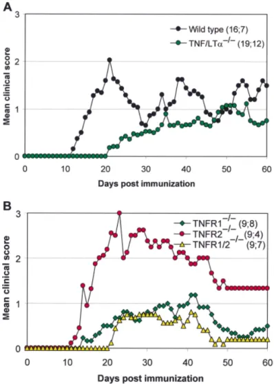
Fig. 1 Clinical course of MOG-induced EAE from wild-type, TNF/LT-α–/–, TNFR1–/–, TNFR2–/–and
TNFR1/TNFR2–/–mice. The acute phase of clinical symptoms is clearly controlled either by the
absence of both ligands (TNF-α and LT-α) or by the absence of TNFR1. On the other hand, EAE in TNFR2–/–mice results in the exacerbation of clinical symptoms during the acute phase of EAE.
Double TNFR1/TNFR2–/–mice had a clinical course similar to that of TNFR1–/–mice. The ascending
paralysis was scored from 0 to 5 (0⫽ no clinical signs; 1 ⫽ limp tail; 2 ⫽ partial hind limb paralysis; 3⫽ complete hind limb paralysis; 4 ⫽ tetraplegia; 5 ⫽ death). Numbers in parentheses indicate the total number of animals in study; number remaining at end of study.
disease. Demyelination in this model is independent of demyelinating antibodies, at least in the initial stages, since the humoral response to MOG peptide 35–55 does not recognize intact MOG in vivo. This model is therefore a good tool to study antibody-independent demyelination in EAE. Study of the function of TNF-α and LT-α in EAE became possible with the generation of gene knockout mice. When immunized with MOG peptide 35–55, mice produced by backcrossing TNF/LT-α–/– mice onto the C57BL/6 background do not develop an acute disease but show a slowly progressive form of EAE (Eugster et al., 1999) (Fig. 1). Clinical symptoms of EAE in rodents (ascending paralysis) are the consequences of mainly two pathological features affecting the spinal cord: inflammation (involving macrophages and T lymphocytes) and demyelination. Demyelination, however, is not necessarily the predominant
factor in the induction of clinical signs. In our studies with MOG-induced EAE in C57BL/6 mice, the loss of myelin follows the initial invasion of lymphocytes and macrophages and the onset of clinical signs with a delay of more than 1 week. The recruitment of T cells into the CNS of TNF/ LT-α–/– mice was found to be unimpaired, and at later time points of the disease (day 60 after immunization) was even increased compared with controls. However, despite the intense inflammation, only minimal demyelination develops (Fig. 2A–C) (Eugster et al., 1999). This indicates that the clinical symptoms of EAE in TNF/LT-α–/– mice are mainly the result of inflammation. Indeed, the clinical features of EAE are well known to develop in experimental animals in the absence of demyelination. This is seen, for example, in rats with EAE after immunization with myelin autoantigens or the transfer of myelin-specific T cells (for review, see
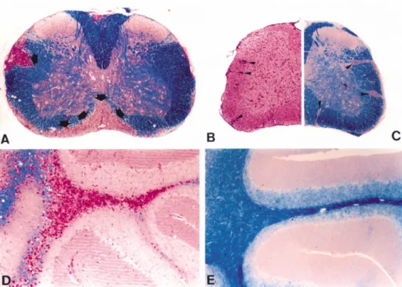
Fig. 2 Demyelination in MOG-induced EAE and TNF transgenic mice depends on the expression of
TNFR1. (A) Large demyelinated areas (arrows) are found in the spinal cord of wild-type animals with MOG-induced EAE (day 60, Klu¨ver–Barrera luxol fast blue stain, magnification⫻48). (B and C) In TNF/LT-α–/–mice with MOG-EAE (day 60), inflammation is prominent (haematoxylin–eosin,
arrowheads), whereas demyelination (C) is minimal (magnification⫻48). (D) In Tg6074 TNF transgenic mice (8 weeks old), large demyelinated plaques with periodic acid-Schiff-positive macrophages are formed in the cerebellum (magnification⫻99). (E) Tg6074 transgenics on a TNFR1–/–background, however, do not show pathology (magnification⫻99).
Wekerle et al., 1994). Numerous in vitro and in vivo studies have delineated the complicated role of cytokines, oxygen radicals, nitric oxide and proteases in affecting the integrity of the blood–brain barrier and in impairing neuronal functions. The latter role may be due to direct effects on neurones or may result from effects of the aforementioned molecules on astrocytes and oligodendrocytes, cells that are critically involved in the survival and function of neurones. Controversial data have been published on the effect of cytokines on neurones. For example, IL-6, which is detected at the onset of EAE, enhances the response of neurones to excitatory amino acids, induces the persistence of calcium-induced calcium release and, upon overexpression in transgenic animals, leads to neurodegeneration (Holliday et al., 1995; Qiu et al., 1995; Heyser et al., 1997). IL-1 suppresses the calcium channel current in hippocampal neurones, and thereby may lead to progression of neurodegenerative processes (Plata-Salaman and ffrench-Mullen, 1994). On the other hand, TNF-α, IL-6 and IL-1 have been reported to increase neuronal survival after a toxic influx of calcium mediated by excitotoxins. The effects of IL-6 and IL-1 may be due, at least in part, to the induction of release of neurotrophogenic factors such as nerve growth factor (Frei et al., 1989; Carlson et al., 1999). The data available indicate that the clinical symptoms induced by a
low level of demyelination, as seen in TNF/LT-α–/– mice, may be aggravated by oedema formation due to alterations of the blood–brain barrier and by abnormal functions of neurones in inflammatory lesions.
Our recent data have demonstrated that the increase in T-cell infiltration of the CNS seen in these mice is a result of impaired elimination of the T cells present in the CNS tissue. It is known that (i) T-cell apoptosis in the CNS precedes clinical remission in EAE, and (ii) depending upon the disease model analysed, Fas ligand (FasL) and/or TNF-α can mediate the apoptosis of activated T cells. In studying the different possibilities, it was found that the extent of T-cell apoptosis is diminished by 50% in EAE lesions of TNF/LT-α–/– mice (Bachmann et al., 1999). This contrasts with lpr and gld mice, which have a genetic defect of Fas-mediated apoptosis and which show no decrease in T-cell apoptosis in EAE lesions compared with controls (Bachmann et al., 1999).
The individual roles of TNF-α and LT-α in the effects seen in TNF/LT-α–/–mice, namely the delay of MOG-induced EAE, the striking inflammation associated with reduction of T-cell apoptosis and the limited extent of demyelination, are now beginning to emerge after intensive research efforts. A critical requirement for TNF-α in the initiation of the neurological deficit of EAE has been demonstrated elegantly
by Sedgwick and co-workers, using MOG peptide 35–55 as an antigen in TNF-α–/–mice that had been generated directly on a C57BL/6 genetic background (Korner et al., 1997a). These mice showed a significant delay (6 days) in the onset of clinical disease relative to wild-type mice. This delay correlated with impaired migration of leucocytes within the CNS and the formation of cuffs. At later time points, disease was severe and associated with widespread perivascular inflammation in the CNS, but primary demyelination appeared to be reduced overall (Korner et al., 1997a). These results demonstrate that TNF-α plays a unique role in promoting disease initiation by controlling initial leucocyte movement within the CNS, possibly through its known effect on the induction of adhesion molecules and chemokines.
In wild-type mice, TNF-α was already present in the CNS by day 7 after immunization with MOG 35–55, peaked at day 20, and then waned. At day 7, TNF-α-producing cells were predominantly microglia (Juedes et al., 2000). TNF-α induces expression of T-cell chemokines, including IP-10 (interferon-inducible protein 10) and MCP-1 (monocyte chemotactic protein 1) in astrocytes. Indeed IP-10, MCP-1 and the chemokine RANTES (regulated upon activation, normal T-cell expressed and secreted chemokine) were detected in the CNS by day 8 after immunization with MOG 35–55 (Juedes et al., 2000). These data make it very likely that TNF-α triggers the expression of chemokines in the CNS, which leads to invasion by T cells and monocytes. In the absence of TNF-α in TNF-α/LT-α or in TNF-α gene knockout mice, invasion of the CNS by immune cells is delayed, but later develops extensively. This indicates that other cytokines, such as IFN-γ, may at some time points compensate for the absence of TNF-α. Indeed, astrocytes treated with IFN-γ release IP-10, a potent chemokine for activated but not resting T cells (Oh et al., 1999). In BALB/c mice with a knockout of the IFN-γ gene, an increase in the development of EAE was noted, the effect being associated with induction of the chemokines MCP-1 and GRO-α (Glabinski et al., 1999). The redundancy of both the signals inducing chemokine production and the chemokines activating T cells and monocytes, together with the expression of chemokine genes in different types of cells, allows compensation for a single defect in the whole cascade, leading to invasion of the CNS by inflammatory cells. Furthermore, genetic influences mediated by both MHC (major histocompatibility complex) and non-MHC genes play a significant role.
In a recent study of mice produced by backcrossing TNF-α–/–animals onto a C57BL/6 background, no delay in the onset of MOG-induced EAE was evident, but the duration of disease was prolonged and was paralleled by higher levels of mononuclear cell infiltrates and extensive demyelination (Liu et al., 1998). Furthermore, inactivation of the TNF-α gene converted MOG-resistant mice of the 129/Sv strain to a state of high susceptibility. This study confirmed that TNF-α is not an obligatory mediator of inflammation and demyelination in EAE, but in addition provided the first
evidence for a potent anti-inflammatory role of TNF-α in limiting the extent and duration of clinical disease. Our own studies in TNF-α–/– mice (backcrossed onto C57BL/6) immunized with MOG confirm the data of Sedgwick and co-workers (Korner et al., 1997a), the difference from wild-type controls being mainly a delay in disease onset (H. P. Eugster, K. Frei and H. Fontana, unpublished observation). Interestingly, the finding that TNF-α is required for the normal initiation of clinical EAE has been confirmed in a different EAE model, in which MBP is used as an antigen to induce CNS inflammation and demyelination in susceptible SJL mice. Immunization of TNF-α–/– mice on the SJL/J genetic background, and appropriate SJL (H-2b) congenic
controls with MBP showed a significant delay (5 days) in the onset of neurological deficits, but the eventual development of severe EAE with perivascular inflammation and demyelination was equal to that in controls (Kassiotis et al., 1999a).
An independent role for LT-α in EAE has been more difficult to evaluate. An apparently clear picture was painted in a comparative study in which MOG was used as an antigen in LT-α–/–and LT-β–/– mice generated initially on a 129 ⫻ C57BL/6 background and backcrossed at least four times onto C57BL/6. Whereas LT-β–/–mice developed EAE, LT-α–/– mice exhibited an EAE-resistant phenotype, and a major role for homotrimeric LT-α in EAE pathogenesis was deduced (Suen et al., 1997). A major criticism of autoimmunity studies using LT-α–/–(LT-α–/–and TNF/LT-α–/–) and LT-β–/– mice has been that these animals show severe structural and functional immune deficiencies, including the absence of lymph nodes and Peyer’s patches, as well as a disorganized splenic microarchitecture that is associated with decreased numbers of splenic dendritic cells (De Togni et al., 1994; Banks et al., 1995; Eugster et al., 1996; Koni et al., 1997) (Appendix I). Nevertheless, evidence was obtained for successful MOG-induced T-cell priming in LT-α–/– and LT-β–/–mice (Suen et al., 1997). TNF/LT-α–/–mice are susceptible to MOG-induced EAE, demonstrating that, despite severe immune system abnormalities, primary autoimmunity to myelin can be induced in these mice (Eugster et al., 1999). As stated by Steinman and ourselves, however, compensatory mechanisms may overcome structural and functional deficiencies in these gene knockout mice (Frei et al., 1997; Steinman, 1997). Furthermore, gene inactivation-dependent changes may occur, such as significantly reduced TNF-α levels in LT-α–/– mice (Alexopoulou et al., 1998). It is interesting to note here that these animals were found to be significantly more susceptible than wild-type controls to the transfer of EAE by T-cell lines or spleen cells from MOG-immunized mice (Suen et al., 1997). This finding further supports an anti-inflammatory effect of TNF-α and LT-α during CNS autoimmunity. The role of LT-α derived from haemopoiesis (the major source of this cytokine) was addressed elegantly by the use of bone marrow chimaeras. Bone marrow from LT-α–/– mice was transferred into immunodeficient mice in which the recombinase activation
Table 1 TNFR1-dependent demyelination: evidence from different animal models of disease
Animal model Mouse strain Disease index* Inflammation† Demyelination† EAE: immunization with MOG Wild-type 347 ⫹⫹ ⫹⫹⫹ peptide 35–55 TNFR1–/– 114 ⫹⫹⫹ ⫹ TNFR2–/– 565 ⫹⫹ ⫹⫹⫹ TNFR1/2–/– 71 ⫹⫹ ⫹ TNF/LT-α–/– 81 ⫹⫹⫹ ⫹ TNF-α–/– 153 ⫹⫹(⫹) ⫹⫹(⫹) LT-α–/–‡ 61 (560)‡ (⫹) –
Tg6074 TNF transgenic mice TNFR1/2⫹/⫹ Ataxia/paralysis ⫹⫹⫹ ⫹⫹⫹
TNFR1–/– Normal – –
TNFR2–/– Ataxia/paralysis ⫹⫹⫹ ⫹⫹⫹ *Animals were monitored and weighed daily, and disease severity was scored on a scale from 0 to 5, with 0.5 points for intermediate clinical findings. The scores were as follows: 0⫽ no detectable signs of EAE; 1⫽ loss of tail tone; 2 ⫽ definitive tail and partial hind limb paralysis; 3 ⫽ hind limb paralysis; 4⫽ hind and fore limb paralysis; 5 ⫽ death. The disease index was calculated by adding all the daily average disease scores (of all animals in a group), dividing by the average day of disease onset (of all animals in a group) and multiplying by 100.†Histopathology was assessed at day 60 after immunization with MOG and at 8 weeks in Tg6074 TNF-α transgenic mice.‡Data from Suen and
colleagues (Suen et al., 1997); disease index calculated at 30 days; control disease index in brackets.
gene Rag-1 had been inactivated. EAE in these mice showed disease onset, rate of progression and peak severity similar to those in RAG-1–/–mice that had been reconstituted with bone marrow from wild-type mice (Rimington et al., 1998). As a positive control, the transfer of TNF-α–/–bone marrow into RAG-1–/–recipients delayed the onset of actively induced EAE and altered the movement of inflammatory leucocytes within the CNS. These results show that bone marrow-derived LT-α does not play a unique role in EAE. However, it remains to be determined whether CNS-specific LT-α, demonstrated in total RNA from whole CNS by RT–PCR (reverse transcriptase polymerase chain reaction) (Rimington et al., 1998), represents a disease-relevant level of LT-α by parenchymal cells such as astrocytes or resident microglia. While a specific role for LT-α in EAE remains to be defined, one possibility is that it can upregulate EAE susceptibility genes. It may be that EAE in TNF/LT-α–/– mice, which develop the disease in spite of the absence of LT-α, is due to loss of the function of TNF-α in suppressing these undefined, LT-α-controlled EAE susceptibility genes.
Absence of TNFR1 mimics the effect of TNF-
α
and LT-
α deficiency: inflammation but no
demyelination
TNF-α and LT-α bind as homotrimers to both the TNFR1 (p55 TNF-α receptor) and the TNFR2 (p75 TNF-α receptor) (Appendix I). On the basis of the observation of a profound effect of the absence of TNF/LT-α on MOG-induced EAE, we questioned which of the two TNF-α receptors is involved in the disease. TNFR2–/– mice develop a chronic form of EAE with a very severe onset (Fig. 1). It remains to be determined whether this effect is a direct result of the elevated TNF-α levels that have been measured in these mice (Peschon
et al., 1998), or whether the TNFR2 can mediate a disease-protective effect in the presence of a functional TNFR1. The absence of TNFR2 also results in the absence of soluble TNFR2, the concentration of which has been shown to dramatically exceed that of soluble TNFR1 under inflammatory conditions (Carpenter et al., 1995). Thus, the lack of soluble TNFR2, which can scavenge TNF (Aderka et al., 1992) and block its action, may result in stronger TNFR1 signalling. In contrast, TNFR1–/– and TNFR1/ TNFR2–/– mice resemble the phenotype of TNF/LT-α mice (Table 1). Both TNFRs are upregulated in the high endothelial venules of the lymph nodes and the CNS during the preclinical phase of EAE (Kahn et al., 1999). However, it is not known whether the absence of one of the two TNFRs leads to altered expression of the remaining receptor. Nevertheless, it is clear that TNFR1 signalling is intact in TNFR2–/– mice (Peschon et al., 1998). The chronic progressive disease, which developed without being preceded by an acute phase, was not associated with extensive demyelination but rather with profound inflammation in the CNS at late (day 60 after immunization) stages of disease (Eugster et al., 1999). This inflammation was paralleled by a reduction in T-cell apoptosis in cellular infiltrates (Bachmann et al., 1999). In TNFR1/ TNFR2–/– mice, inflammation was less striking than in TNFR1–/–mice. Thus, demyelination in MOG-induced EAE depends largely on TNFR1-mediated signalling. It remains to be determined whether TNF-α, LT-α or a still unidentified ligand of the TNFR1 is responsible for inducing this effect. In contrast, the TNFR1 is not essential for the development of CNS inflammation but plays a major role in the removal of T cells from lesions, and it may therefore be involved in disease resolution. The TNFR1 has been shown to play an important role not only in EAE but also in other experimental autoimmune diseases, such as collagen-induced arthritis
(Mori et al., 1996), myosin-induced myocarditis (Bachmaier et al., 1997) and autoimmune diabetes in NOD (nonobese diabetic) mice (Pakala et al., 1999). These findings allocate an important mediator function to TNFR1 in experimental autoimmune disease.
TNFR1-dependent demyelination in mice
overexpressing TNF-
α in the CNS
Potent roles for TNF-α/TNFR1 signalling in the pathogenesis of CNS inflammation and primary demyelination have been demonstrated further in another, non-autoimmune-mediated model of multiple sclerosis in which disease is triggered spontaneously by the local expression of murine or human TNF-α transgenes by CNS glia. Two independent lines of transgenic mice have been described. The Tg6074 line has a high level of expression of a murine TNF-α transgene, under the control of its own promoter, specifically in the CNS, probably by virtue of the chance integration site of the transgene into the murine genome. Expression of this transgene was detectable from the first postnatal week of age in immature glial precursor cells (Probert et al., 1995; Akassoglou et al., 1998a). The TgK21 line shows astrocyte-specific expression of a mutant human TNF-α transgene, which leads to the production of an uncleavable, transmembrane TNF protein under the control of the promoter of the glial fibrillary acid protein gene (Akassoglou et al., 1997). Both transgenic lines spontaneously develop oligodendrocyte apoptosis, myelin vacuolation and primary demyelination, which proceeds in the presence of large numbers of T cells and recruited phagocytic macrophages (Akassoglou et al., 1998a) (Fig. 2D). Lesions progress to chronic (Tg6074) or acute (TgK21) multiple sclerosis-like plaques, with secondary axonal damage. Clinical symptoms differ in the two transgenic lines, and this is likely to reflect differences in the site of transgene expression and therefore the primary location of the lesions. Tg6074 mice, in which the lesions affect mainly the white matter of the cerebellum, develop ataxia between 3 and 4 weeks of age, followed by kyphosis, limb paresis and paralysis (Probert et al., 1995). TgK21 mice, which develop severe inflammatory lesions in the spinal cord, show ascending paralysis similar to that seen in EAE (Akassoglou et al., 1997). A dominant role for the TNFR1 in signalling both inflammation and demyelination in these models was demonstrated by the finding that disease was completely abrogated when transgenic mice were backcrossed onto a TNFR1-deficient background (Akassoglou et al., 1998a) (Fig. 2E). A clue to the mechanism by which TNF-α induces oligodendrocyte death has also come from transgenic studies. Astrocyte-specific expression of a biologically active, uncleavable membrane TNF-α protein in transgenic mice selectively triggers oligo-dendrocyte apoptosis and demyelination via TNFR1-mediated signalling (Akassoglou et al., 1998a), even in the absence of endogenous TNF-α (K. Akassoglou and
L. Probent, unpublished observations). In contrast, oligo-dendrocytes remain completely unaffected by the neurone-specific expression of membrane or wild-type (membrane and soluble) TNF-α molecules in young animals, even though CNS inflammation is severe in the latter case, particularly in the meninges and perivascular space (K. Akassoglou and L. Probent, unpublished observations). Oligodendrocyte and myelin damage is observed only in old animals. These results indicate that TNF- α/TNFR1-mediated demyelination is mechanistically independent of TNF-α/TNFR1-mediated CNS inflammation in vivo in transgenic mice. Furthermore, they show that TNF-α can mediate oligodendrocyte and myelin damage through cell contact-dependent mechanisms involving membrane TNF-α, when expressed as a transgene by appropriate cells such as astrocytes. It remains to be determined, however, whether this action of TNF-α is due to a direct effect on oligodendrocytes or whether it is mediated by an indirect mechanism mediated by TNFR1 on microglia/macrophages, for example.
Therefore, in both TNF-α transgenic mice and MOG-induced EAE, the TNFR1 has been found to be important for the development of demyelination and the initiation of the inflammatory process. In contrast to MOG-induced EAE, CNS inflammation has also been shown to require TNFR1 for its development and progression in TNF-α transgenic mice. It is important to note here that the contribution of inflammation to disease appears to be completely different in these two models (Fig. 3). In EAE, activated T cells drive disease and the TNFR1 is important in mediating T-cell apoptosis within the CNS and clinical remission once disease has been initiated (Bachmann et al., 1999). In TNF-α transgenic mice, clinical symptoms and histopathological changes develop independently of adaptive immunity (Kassiotis et al., 1999b). Histopathological lesions begin with TNFR1-dependent oligodendrocyte apoptosis. Lesions progress to fulminant demyelinating plaques, and in sharp contrast to EAE, remyelination does not occur. In this latter model the multiple pro-inflammatory effects of continuous TNF-α/TNFR1 signalling within the CNS, for example in the activation of endothelial cells, astrocytes and microglia/macrophages, appear to dominate a chronic– progressive disease and vanish in the absence of TNFR1. Disease induced by the murine TNF-α transgene in Tg6074 mice developed fully in the absence of the TNFR2 (Akassoglou et al., 1998a). While the TNFR2 does not appear to contribute significantly to pathology in the Tg6074 system, we have recently obtained evidence that transmembrane TNF-α expressed on astrocytes can specifically signal pro-inflammatory effects through the TNFR2 in the CNS of transgenic mice when this receptor is overexpressed on a TNFR1–/– background. Specifically, transmembrane TNF-α/TNFR2 interactions lead to endo-thelial cell activation and the upregulation of adhesion molecules, with the development of chronic vascular inflammation and perivascular deposition of connective
Fig. 3 Critical points in TNF/TNFR functioning in the pathogenesis of CNS inflammation and
demyelination, as understood from the study of animal models. Disease in EAE (right) is initiated and driven by autoreactive T lymphocytes (T). Invasion of the CNS by activated T cells results in breakdown of the blood–brain barrier (BBB), glial cell activation and local release of cytokines within the CNS microenvironment. Primary demyelination is induced by different immune effector
mechanisms depending upon the genetic background of the animal and the antigen used, and involves the phagocytosis of myelin by activated microglia and recruited blood monocytes (Mg/M). Primary demyelination proceeds with little damage to the myelin-forming oligodendrocyte (OL) and with maintenance of the potential for remyelination. Aberrant local TNF expression on the other hand, as it occurs in TNF-transgenic mice (left), can induce oligodendrocyte apoptosis and myelin vacuolation, as well as primary inflammatory demyelination, which may be initiated by the pro-inflammatory effects of TNF on local CNS glia and endothelial cells. In this model, demyelination proceeds in the presence of activated phagocytic Mg/Mφ, T and B cells, considerable oligodendrocyte depletion, and the absence of remyelinating events. The pattern of demyelination observed in TNF transgenics closely resembles that observed in a subgroup of multiple sclerosis patients whose lesions are characterized by primary oligodendropathy (Lucchinetti et al., 1996).
tissue fibres (fibrosis), the latter representing a footprint of the extensive and chronic inflammatory process (Akassoglou et al., 1998b). This adds to previous evidence that the TNFR2 is an important mediator of vascular inflammation, as seen in the murine model of cerebral malaria (Lucas et al., 1997), and indicates that under certain circumstances TNF-α/TNFR2 interactions may contribute to CNS disease.
TNFR1, the ideal target for treatment?
TNF-α and LT-α have been implicated as key mediators in chronic inflammatory and autoimmune diseases such as EAE and multiple sclerosis, and have been identified as important therapeutic targets for more than a decade. It is now clear, through the study of this ligand/receptor family in vivo in transgenic and knockout animals, that their diverse and potent
effects in the pathogenesis of disease extend well beyond their long-established pro-inflammatory effects. This newly emerging evidence is expected to have a significant impact upon the design of any therapeutic protocols that aim to block TNF/LT-α signalling pathways or downstream mediators for the treatment of human autoimmune diseases such as multiple sclerosis.
A critical role for TNF-α in the initiation of the neurological deficits of EAE and in leucocyte movement within the CNS has been defined, specifically in the formation of perivascular cuffs (Korner et al., 1997a). An important role for TNF-α in orchestrating cell movement is also manifest in the altered splenic architecture that characterizes TNF-α–/–mice (Appendix I). TNF-α is known to induce the expression of adhesion molecules (Bevilacqua, 1993) and chemokines (Butcher and Picker, 1996), and both types of molecule are potentially important secondary mediators of TNF-α-dependent effects on cell movement. Consistent with this concept, recent evidence has shown that the production of T- and B-cell chemoattractants is reduced in the spleen and lymph nodes of TNF-α–/– and LT-α/β–/– mice (Appendix I) (Ngo et al., 1999). It will be interesting to identify such TNF-α-inducible molecules from the CNS during EAE.
Furthermore, a potent anti-inflammatory role during EAE has been ascribed to TNF-α, following the observation that neurological signs of disease are significantly increased in its absence (Liu et al., 1998). A variety of TNF-α-dependent processes may underlie this effect. For example, there is good evidence to show that TNF-α can downregulate systemic and organ-specific autoimmunity and that T cells are important targets (Appendix I). Although the mechanisms behind this effect are not yet known, there is recent evidence that TNF-α can attenuate T-cell receptor signalling. Alternatively, TNF-α may signal apoptosis directly in activated CD4⫹and CD8⫹cells in vivo. Indeed, a major role for the TNFR1 in inducing T-cell apoptosis and lymphocyte clearance from the CNS during EAE, and therefore in the limitation of disease, has been demonstrated recently (Bachmann et al., 1999).
Finally, studies in TNF-α transgenic mice clearly show that TNF-α/TNFR1 signalling can induce oligodendrocyte apoptosis, myelin vacuolation and primary, macrophage-mediated demyelination (Akassoglou et al., 1998a). Unfortunately, quantitative data concerning experimentally induced demyelination in TNF-α–/–mice are not yet available, but MOG-induced demyelination in TNFR1–/– mice is significantly reduced (Eugster et al., 1999), indicating that this signalling pathway is also critically involved in demyelination in EAE. It remains to be determined whether TNF-α, LT-α or an as yet unidentified ligand of the TNFR1 signals this effect. Nevertheless, the finding that demyelination can proceed even in the absence TNF-α/ TNFR1 signalling in the EAE model shows that alternative pathways exist to mediate this process. In MOG-immunized TNF/LT-α–/– mice, initially generated on the 129/Sv ⫻ C57BL/6 (H-2b) genetic background, backcrossed to SJL/J
(H-2s) mice, the extent of demyelination and inflammation
was greater, equivalent to that seen in wild-type mice of the C57BL/6 background (Frei et al., 1997). Since TNF/LT-α–/– SJL mice carried the MHC H-2bhaplotype from the parental
C57BL/6 mice, these results indicate that non-MHC genes in SJL mice compensate for the absence of TNF-α and LT-α in mediating demyelination. The importance of non-MHC genes in mediating EAE in SJL mice has also been demonstrated in MBP-induced EAE, in which a comparison between SJL/J inbred mice and SJL H-2bmice [generated as
congenic controls for TNF-α–/– mice (H-2b) that had been
backcrossed for five generations onto SJL], showed no difference in the development of neurological deficits, inflammation or demyelination despite the presence of a non-susceptible haplotype (H-2b) in the latter mice (Kassiotis et al.,
1999b). Identification of the genes that link inflammation with demyelination in MOG-induced EAE in TNF/LT-α–/– SJL mice might open new avenues of experimental therapy in EAE. Linkage analysis of crosses between EAE susceptible SJL/J (H-2s) and resistant B10s (H-2s) mice revealed disease
susceptibility loci at the telomeric end of chromosome 2 and near the centre of chromosome 3 (Encinas et al., 1996). However, these loci were found to map with inflammatory loci and clinical disease. It will be interesting to take demyelination as a readout in genetic studies in the future.
With the increased understanding of the roles of TNFR1-mediated signalling in leucocyte recruitment into the CNS, demyelination and T-cell apoptosis and clearance, potential selective applications of soluble TNFR1 in multiple sclerosis can be envisaged. Indeed, in EAE, the parenteral administration of TNFR1 inhibits the development of neurological deficits in Lewis rats and SJL/J mice immunized with MBP (Martin et al., 1995; Selmaj et al., 1995; Klinkert et al., 1997; Korner et al., 1997b) if treatment is before the onset of clinical disease. This is consistent with the demonstrated role of TNF-α in the initiation of CNS inflammation. However, established disease is relatively refractory to this treatment. A recent multicentre study on multiple sclerosis patients with relapsing–remitting disease treated with TNFR1 had to be stopped because of a significant increase in the frequency of relapse (Van Osten et al., 1996). In the light of recent finding that, in multiple sclerosis patients, various subgroups can be found with apparently different mechanisms of demyelination (Lucchinetti et al., 1996), it is perhaps not surprising that TNF blockade did not give the expected results. Moreover, with our present knowledge of the role of TNFR1 in the clearance of T cells from the CNS and in disease limitation, it is clear that any treatment aimed at blocking the action of TNF would need to be selective. A combination of soluble TNFR treatment with antagonists that block the adherence of T cells or their trafficking through the blood–brain barrier, for example inhibitors of matrix metalloproteases, is more likely to influence disease activity. Nonetheless, the identification of the non-MHC genes that influence TNFR1-independent
demyelination, as seen in SJL mice, is expected to open exciting new opportunities for therapy in multiple sclerosis.
Acknowledgements
We thank Horst Bluethmann (F. Hoffmann-La Roche Ltd, Basel, Switzerland) and Mark Moore (Genentech Inc., South San Francisco, Calif., USA) for providing the TNF receptor knockout mice and Ju¨rg Tschopp and Michael Hahne (Institute of Biochemistry, University of Lausanne, Epalinges, Switzerland) and George Kollias (Laboratory of Molecular Genetics, Hellenic Pasteur Institute, Athens, Greece) for TNF knockout mice. L.P. also thanks George Kollias for providing Tg6074 and TgK21 transgenic mice and for his participation in their characterization, and Roly Foulkes (Celltech, Slough, Berks, UK) for the TN3 monoclonal anti-TNF antibodies necessary for the maintenance of Tg6074 mice. This work was supported by the Swiss National Science Foundation (grant 31-42900.95), the Swiss National Multiple Sclerosis Society and EU Biotechnology Programme BIO4-CT96-0174.
References
Aderka D, Engelmann H, Maor Y, Brakebusch C, Wallach D. Stabilization of the bioactivity of tumor necrosis factor by its soluble receptors. J Exp Med 1992; 175: 323–9.
Agresti C, D’Urso D, Levi, G. Reversible inhibitory effects of interferon-gamma and tumour necrosis factor-alpha on oligo-dendroglial lineage cell proliferation and differentiation in vitro. Eur J Neurosci 1996; 8: 1106–16.
Akassoglou K, Probert L, Kontogeorgos G, Kollias G. Astrocyte-specific but not neuron-Astrocyte-specific transmembrane TNF triggers inflammation and degeneration in the central nervous system of transgenic mice. J Immunol 1997; 158: 438–45.
Akassoglou K, Bauer J, Kassiotis G, Pasparakis M, Lassmann H, Kollias G, et al. Oligodendrocyte apoptosis and primary demyelination induced by local TNF/p55TNF receptor signaling in the central nervous system of transgenic mice: models for multiple sclerosis with primary oligodendrogliopathy. Am J Pathol 1998a; 153: 801–13.
Akassoglou K, Pasparakis M, Bauer J, Lassmann H, Probert L, Kollias G. Probing for the cellular specificity of the p55 TNFR in TNF-α-induced inflammatory demyelination: conditional gene targeting of the p55 TNFR in astrocytes. J Interferon Cytokine Res 1998b; 18: A-86.
Alexopoulou L, Pasparakis M, Kollias G. Complementation of lymphotoxin alpha knockout mice with tumor necrosis factor-expressing transgenes rectifies defective splenic structure and function. J Exp Med 1998; 188: 745–54.
Alimzhanov MB, Kuprash DV, Kosco-Vilbois MH, Luz A, Turetskaya RL, Tarakhovsky A, et al. Abnormal development of secondary lymphoid tissues in lymphotoxin beta-deficient mice. Proc Natl Acad Sci USA 1997; 94: 9302–7.
Bachmaier K, Pummerer C, Kozieradzki I, Pfeffer K, Mak TW, Neu N, et al. Low-molecular-weight tumor necrosis factor receptor
p55 controls induction of autoimmune heart disease. Circulation 1997; 95: 655–61.
Bachmann R, Eugster HP, Frei K, Fontana A, Lassmann H. Impairment of TNF-receptor-1 signaling but not fas signaling diminishes T-cell apoptosis in myelin oligodendrocyte glycoprotein peptide-induced chronic demyelinating autoimmune encephalo-myelitis in mice. Am J Pathol 1999; 154: 1417–22.
Baker D, Butler D, Scallon BJ, O’Neill JK, Turk JL, Feldmann M. Control of established experimental allergic encephalomyelitis by inhibition of tumor necrosis factor (TNF) activity within the central nervous system using monoclonal antibodies and TNF receptor– immunoglobulin fusion proteins. Eur J Immunol 1994; 24: 2040–8. Banks TA, Rouse BT, Kerley MK, Blair PJ, Godfrey VL, Kuklin NA, et al. Lymphotoxin-alpha-deficient mice. Effects on secondary lymphoid organ development and humoral immune responsiveness. J Immunol 1995; 155: 1685–93.
Bevilacqua MP. Endothelial–leukocyte adhesion molecules. [Review]. Annu Rev Immunol 1993; 11: 767–804.
Black RA, Rauch CT, Kozlosky CJ, Peschon JJ, Slack JL, Wolfson MF, et al. A metalloproteinase disintegrin that releases tumour-necrosis factor-alpha from cells. Nature 1997; 385: 729–33. Butcher EC, Picker LJ. Lymphocyte homing and homeostasis. [Review]. Science 1996; 272: 60–6.
Cannella B, Sizing ID, Benjamin CD, Browning JL, Raine CS. Antibodies to lymphotoxin alpha (LT alpha) and LT beta recognize different glial cell types in the central nervous system. J Neuroimmunol 1997; 78: 172–9.
Carlson NG, Wieggel WA, Chen J, Bacchi A, Rogers SW, Gahring LC. Inflammatory cytokines IL-1 alpha, IL-1 beta, IL-6, and TNF-alpha impart neuroprotection to an excitotoxin through distinct pathways. J Immunol 1999; 163: 3963–8.
Carpenter A, Evans TJ, Buurman WA, Bemelmans MH, Moyes D, Cohen J. Differences in the shedding of soluble TNF receptors between endotoxin-sensitive and endotoxin-resistant mice in response to lipopolysaccharide or live bacterial challenge. J Immunol 1995; 155: 2005–12.
Chung IY, Benveniste EN. Tumor necrosis factor-alpha production by astrocytes. Induction by lipopolysaccharide, IFN-gamma, and IL-1 beta. J Immunol 1990; 144: 2999–3007.
Chung IY, Norris JG, Benveniste EN. Differential tumor necrosis factor alpha expression by astrocytes from experimental allergic encephalomyelitis-susceptible and -resistant rat strains. J Exp Med 1991; 173: 801–11.
Cope AP. Regulation of autoimmunity by proinflammatory cytokines. [Review]. Curr Opin Immunol 1998; 10: 669–76. Cope AP, Liblau RS, Yang XD, Congia M, Laudanna C, Schreiber RD, et al. Chronic tumor necrosis factor alters T cell responses by attenuating T cell receptor signaling. J Exp Med 1997; 185: 1573–84. Crisi GM, Santambrogio L, Hochwald GM, Smith SR, Carlino JA, Thorbecke GJ. Staphylococcal enterotoxin B and tumor-necrosis factor-alpha-induced relapses of experimental allergic encephalo-myelitis: protection by transforming growth factor-beta and interleukin-10. Eur J Immunol 1995; 25: 3035–40.
Crowe PD, VanArsdale TL, Walter BN, Ware CF, Hession C, Ehrenfels B, et al. A lymphotoxin-beta-specific receptor. Science 1994; 264: 707–10.
D’Souza S, Alinauskas K, McCrea E, Goodyer C, Antel JP. Differential susceptibility of human CNS-derived cell populations to TNF-α-dependent and independent immune-mediated injury. J Neurosci 1995; 15: 7293–300.
De Togni P, Goellner J, Ruddle NH, Streeter PR, Fick A, Mariathasan S, et al. Abnormal development of peripheral lymphoid organs in mice deficient in lymphotoxin. Science 1994; 264: 703–7. Douni E, Kollias G. A critical role of the p75 tumor necrosis factor receptor (p75TNF-α) in organ inflammation independent of TNF, lymphotoxin alpha, or the p55TNF-R. J Exp Med 1998; 188: 1343–52.
Elliott MJ, Maini RN, Feldmann M, Long-Fox A, Charles P, Bijl H, et al. Repeated therapy with monoclonal antibody to tumour necrosis factor alpha (cA2) in patients with rheumatoid arthritis. Lancet 1994; 344: 1125–7.
Encinas JA, Lees MB, Sobel RA, Symonowicz C, Greer JM, Shovlin CL, et al. Genetic analysis of susceptibility to experimental autoimmune encephalomyelitis in a cross between SJL/J and B10.S mice. J Immunol 1996; 157: 2186–92.
Eugster HP, Muller M, Karrer U, Car BD, Schnyder B, Eng VM, et al. Multiple immune abnormalities in tumor necrosis factor and lymphotoxin-alpha double-deficient mice. Int Immunol 1996; 8: 23–36.
Eugster HP, Frei K, Kopf M, Lassmann H, Fontana A. IL-6-deficient mice resist myelin oligodendrocyte glycoprotein-induced autoimmune encephalomyelitis. Eur J Immunol 1998; 28: 2178–87. Eugster HP, Frei K, Bachmann R, Bluethmann H, Lassmann H, Fontana A. Severity of symptoms and demyelination in MOG-induced EAE depends on TNFR1. Eur J Immunol 1999; 29: 626–32. Flynn JL, Goldstein MM, Chan J, Triebold KJ, Pfeffer K, Lowenstein CJ, et al. Tumor necrosis factor-alpha is required in the protective immune response against Mycobacterium tuberculosis in mice. Immunity 1995; 2: 561–72.
Frei K, Siepl C, Groscurth P, Bodmer S, Schwerdel C, Fontana A. Antigen presentation and tumor cytotoxicity by interferon-gamma-treated microglial cells. Eur J Immunol 1987; 17: 1271–8. Frei K, Malipiero UV, Leist TP, Zinkernagel RM, Schwab ME, Fontana A. On the cellular source and function of interleukin 6 produced in the central nervous system in viral diseases. Eur J Immunol 1989; 19: 689–94.
Frei K, Eugster HP, Bopst M, Constantinescu CS, Lavi E, Fontana A. Tumor necrosis factor alpha and lymphotoxin alpha are not required for induction of acute experimental autoimmune encephalomyelitis. J Exp Med 1997; 185: 2177–82.
Futterer A, Mink K, Luz A, Kosco-Vilbois MH, Pfeffer K. The lymphotoxin beta receptor controls organogenesis and affinity maturation in peripheral lymphoid tissues. Immunity 1998; 9: 59–70. Genain CP, Cannella B, Hauser SL, Raine CS. Identification of autoantibodies associated with myelin damage in multiple sclerosis. Nat Med 1999; 5: 170–5.
Glabinski AR, Krakowski M, Han Y, Owens T, Ransohoff RM.
Chemokine expression in GKO mice (lacking interferon-gamma) with experimental autoimmune encephalomyelitis. J Neurovirol 1999; 5: 95–101.
Gray PW, Aggarwal BB, Benton CV, Bringman TS, Henzel WJ, Jarrett JA, et al. Cloning and expression of cDNA for human lymphotoxin, a lymphokine with tumor necrosis activity. Nature 1984; 312: 721–24.
Grewal IS, Grewal KD, Wong FS, Picarella DE, Janeway CA Jr, Flavell RA. Local expression of transgene encoded TNF alpha in islets prevents autoimmune diabetes in nonobese diabetic (NOD) mice by preventing the development of auto-reactive islet-specific T cells. J Exp Med 1996; 184: 1963–74.
Herbein G, Montaner LJ, Gordon S. Tumor necrosis factor alpha inhibits entry of human immunodeficiency virus type 1 into primary human macrophages: a selective role for the 75-kilodalton receptor. J Virol 1996; 70: 7388–97.
Heyser CJ, Masliah E, Samimi A, Campbell IL, Gold LH. Progressive decline in avoidance learning paralleled by inflammatory neurodegeneration in transgenic mice expressing interleukin 6 in the brain. Proc Natl Acad Sci USA 1997; 94: 1500–5.
Hjelmstrom P, Juedes AE, Fjell J, Ruddle NH. B-cell-deficient mice develop experimental allergic encephalomyelitis with demyelination after myelin oligodendrocyte glycoprotein sensitization. J Immunol 1998; 161: 4480–3.
Hofman FM, Hinton DR, Johnson K, Merrill JE. Tumor necrosis factor identified in multiple sclerosis brain. J Exp Med 1989; 170: 607–12.
Holliday J, Parsons K, Curry J, Lee SY, Gruol DL. Cerebellar granule neurons develop elevated calcium responses when treated with interleukin-6 in culture. Brain Res 1995; 673: 141–8. Jacob CO, Aiso S, Michie SA, McDevitt HO, Acha-Orbea H. Prevention of diabetes in nonobese diabetic mice by tumor necrosis factor (TNF): similarities between TNF-α-alpha and interleukin 1. Proc Natl Acad Sci USA 1990; 87: 968–72.
Juedes AE, Hjelmstrom P, Bergman CM, Neild AL, Ruddle NH. Kinetics and cellular origin of cytokines in the central nervous system: insight into mechanisms of myelin oligodendrocyte glycoprotein-induced experimental autoimmune encephalomyelitis. J Immunol 2000; 164: 419–26.
Kahn MA, Dopp JM, Liva S, MacKenzie-Graham AJ, Chang R, Huang A, et al. Temporal kinetics and cellular phenotype of TNF p55/p75 receptors in experimental allergic encephalomyelitis. J Neuroimmunol 1999; 95: 19–34.
Kassiotis G, Bauer J, Akassoglou K, Lassmann H, Kollias G, Probert L. A tumor necrosis factor-induced model of human primary demyelinating diseases develops in immunodeficient mice. Eur J Immunol 1999a; 29: 912–7.
Kassiotis G, Pasparakis M, Kollias G, Probert L. TNF accelerates the onset but does not alter the incidence and severity of myelin basic protein-induced experimental autoimmune encephalomyelitis. Eur J Immunol 1999b; 29: 774–80.
Klinkert WE, Kojima K, Lesslauer W, Rinner W, Lassmann H, Wekerle H. TNF-α-alpha receptor fusion protein prevents experimental auto-immune encephalomyelitis and demyelination in
Lewis rats: an overview. [Review]. J Neuroimmunol 1997; 72: 163–8.
Koni PA, Sacca R, Lawton P, Browning JL, Ruddle NH, Flavell RA. Distinct roles in lymphoid organogenesis for lymphotoxins alpha and beta revealed in lymphotoxin beta-deficient mice. Immunity 1997; 6: 491–500.
Kontoyiannis D, Pasparakis M, Pizarro TT, Cominelli F, Kollias G. Impaired on/off regulation of TNF biosynthesis in mice lacking TNF AU-rich elements: implications for joint and gut-associated immunopathologies. Immunity 1999; 10: 387–98.
Korner H, Sedgwick JD. Tumour necrosis factor and lymphotoxin: molecular aspects and role in tissue-specific autoimmunity. [Review]. Immunol Cell Biol 1996; 74: 465–72.
Korner H, Riminton DS, Strickland DH, Lemckert FA, Pollard JD, Sedgwick JD. Critical points of tumor necrosis factor action in central nervous system autoimmune inflammation defined by gene targeting. J Exp Med 1997a; 186: 1585–90.
Korner H, Lemckert FA, Chaudhri G, Etteldorf S, Sedgwick JD. Tumor necrosis factor blockade in actively induced experimental autoimmune encephalomyelitis prevents clinical disease despite activated T cell infiltration to the central nervous system. Eur J Immunol 1997b; 27: 1973–81.
Kratz A, Campos-Neto A, Hanson MS, Ruddle NH. Chronic inflammation caused by lymphotoxin is lymphoid neogenesis. J Exp Med 1996; 183: 1461–72.
Kuroda Y, Shimamoto Y. Human tumor necrosis factor-alpha augments experimental allergic encephalomyelitis in rats. J Neuroimmunol 1991; 34: 159–64.
Le Hir M, Bluethmann H, Kosco-Vilbois MH, Muller M, di Padova F, et al. Differentiation of follicular dendritic cells and full antibody responses require tumor necrosis factor receptor-1 signaling. J Exp Med 1996; 183: 2367–72.
Lieberman AP, Pitha PM, Shin HS, Shin ML. Production of tumor necrosis factor and other cytokines by astrocytes stimulated with lipopolysaccharide or a neurotropic virus. Proc Natl Acad Sci USA 1989; 86: 6348–52.
Linington C, Bradl M, Lassmann H, Brunner C, Vass K. Augmentation of demyelination in rat acute allergic encephalo-myelitis by circulating mouse monoclonal antibodies directed against a myelin/oligodendrocyte glycoprotein. Am J Pathol 1988; 130: 443–54.
Liu J, Marino MW, Wong G, Grail D, Dunn A, Bettadapura J, et al. TNF is a potent anti-inflammatory cytokine in autoimmune-mediated demyelination. Nat Med 1998; 4: 78–83.
Lucas R, Juillard P, Decoster E, Redard M, Burger D, Donati Y, et al. Crucial role of tumor necrosis factor (TNF) receptor 2 and membrane-bound TNF in experimental cerebral malaria. Eur J Immunol 1997; 27: 1719–25.
Lucchinetti CF, Bruck W, Rodriguez M, Lassmann H. Distinct patterns of multiple sclerosis pathology indicate heterogeneity on pathogenesis. [Review]. Brain Pathol 1996; 6: 259–74.
Malipiero U, Frei K, Spanaus KS, Agresti C, Lassmann H, Hahne M, et al. Myelin oligodendrocyte glycoprotein-induced autoimmune encephalomyelitis is chronic/relapsing in perforin knockout mice,
but monophasic in Fas- and Fas ligand-deficient lpr and gld mice. Eur J Immunol 1997; 27: 3151–60.
Marino MW, Dunn A, Grail D, Inglese M, Noguchi Y, Richards E, et al. Characterization of tumor necrosis factor-deficient mice. Proc Natl Acad Sci USA 1997; 94: 8093–8.
Martin D, Near SL, Bendele A, Russell DA. Inhibition of tumor necrosis factor is protective against neurologic dysfunction after active immunization of Lewis rats with myelin basic protein. Exp Neurol 1995; 131: 221–8.
Matsumoto M, Mariathasan S, Nahm MH, Baranyay F, Peschon JJ, Chaplin DD. Role of lymphotoxin and the type I TNF receptor in the formation of germinal centers. Science 1996; 271: 1289–91. McDermott MF, Aksentijevich I, Galon J, McDermott EM, Ogunkolade BW, Centola M, et al. Germline mutations in the extracellular domains of the 55 kDa TNF receptor, TNFR1, define a family of dominantly inherited autoinflammatory syndromes. Cell 1999; 97: 133–44.
McSorley SJ, Soldera S, Malherbe L, Carnaud C, Locksley RM, Flavell RA, et al. Immunological tolerance to a pancreatic antigen as a result of local expression of TNFalpha by islet beta cells. Immunity 1997; 7: 401–9.
Mori L, Iselin S, De Libero G, Lesslauer W. Attenuation of collagen-induced arthritis in 55-kDa TNF receptor type 1 (TNFR1)-IgG1-treated and TNFR1-deficient mice. J Immunol 1996; 157: 3178–82. Moss ML, Jin SL, Milla ME, Bickett DM, Burkhart W, Carter HL, et al. Cloning of a disintegrin metalloproteinase that processes precursor tumour-necrosis factor-alpha. Nature 1997; 385: 733–6. Nedospasov SA, Shakhov AN, Turetskaya RL, Mett VA, Azizov MM, Georgiev GP, et al. Tandem arrangement of genes coding for tumor necrosis factor (TNF-alpha) and lymphotoxin (TNF-beta) in the human genome. Cold Spring Harbor Symp Quant Biol 1986; 51: 611–24.
Nedwin GE, Naylor SL, Sakaguchi AY, Smith D, Jarrett-Nedwin J, Pennica D, et al. Human lymphotoxin and tumor necrosis factor genes: structure, homology and chromosomal localization. Nucleic Acids Res 1985; 13: 6361–73.
Ngo VN, Korner H, Gunn MD, Schmidt KN, Riminton DS, Cooper MD, et al. Lymphotoxin alpha/beta and tumor necrosis factor are required for stromal cell expression of homing chemokines in B and T cell areas of the spleen. J Exp Med 1999; 189: 403–12. Oh JW, Schwiebert LM, Benveniste EN. Cytokine regulation of CC and CXC chemokine expression by human astrocytes. J Neurovirol 1999; 5: 82–94.
Olsson T. Critical influences of the cytokine orchestration on the outcome of myelin antigen-specific T-cell autoimmunity in experimental autoimmune encephalomyelitis and multiple sclerosis. Immunol Rev 1995; 144: 245–68.
Pakala SV, Chivetta M, Kelly CB, Katz JD. In autoimmune diabetes the transition from benign to pernicious insulitis requires an islet cell response to tumor necrosis factor alpha. J Exp Med 1999; 189: 1053–62.
Pasparakis M, Alexopoulou L, Episkopou V, Kollias G. Immune and inflammatory responses in TNF alpha-deficient mice: a critical requirement for TNF alpha in the formation of primary B cell
follicles, follicular dendritic cell networks and germinal centers, and in the maturation of the humoral immune response. J Exp Med 1996; 184: 1397–411.
Pasparakis M, Alexopoulou L, Grell M, Pfizenmaier K, Bluethmann H, Kollias G. Peyer’s patch organogenesis is intact yet formation of B lymphocyte follicles is defective in peripheral lymphoid organs of mice deficient for tumor necrosis factor and its 55-kDa receptor. Proc Natl Acad Sci USA 1997; 94: 6319–23. Pennica D, Nedwin GE, Hayflick JS, Seeburg PH, Derynck R, Palladino MA, et al. Human tumour necrosis factor: precursor structure, expression and homology to lymphotoxin. Nature 1985; 312: 724–9.
Peschon JJ, Torrance DS, Stocking KL, Glaccum MB, Otten C, Willis CR, et al. TNF receptor-deficient mice reveal divergent roles for p55 and p75 in several models of inflammation. J Immunol 1998; 160: 943–52.
Philip R, Epstein LB. Tumour necrosis factor as immunomodulator and mediator of monocyte cytotoxicity induced by itself, gamma-interferon and interleukin-1. Nature 1986; 323: 86–9.
Plata-Salomon C, ffrench-Mullen J. Interleukin-1 beta inhibits Ca2⫹ channel currents in hippocampal neurons through protein kinase C. Eur J Pharmacol 1994; 266: 1–10.
Powell MB, Mitchell D, Lederman J, Buckmeier J, Zamvil SS, Graham M, et al. Lymphotoxin and tumor necrosis factor-alpha production by myelin basic protein-specific T cell clones correlates with encephalitogenicity. Int Immunol 1990; 2: 539–44.
Probert L, Akassoglou K, Pasparakis M, Kontogeorgos G, Kollias G. Spontaneous inflammatory demyelinating disease in transgenic mice showing central nervous system-specific expression of tumor necrosis factor alpha. Proc Natl Acad Sci USA 1995; 92: 11294–8. Qiu Z, Parsons KL, Gruol DL. Interleukin-6 selectively enhances the intracellular calcium response to NMDA in developing CNS neurons. J Neurosci 1995; 15: 6688–99.
Raine CS. Multiple sclerosis: TNF revisited, with promise. [Review]. Nat Med 1995; 1: 211–4.
Raine CS, Bonetti B, Cannella B. Multiple sclerosis: expression of molecules of the tumor necrosis factor ligand and receptor families in relationship to the demyelinated plaque. Rev Neurol (Paris) 1998; 154: 577–85.
Renno T, Krakowski M, Piccirillo C, Lin JY, Owens T. TNF-alpha expression by resident microglia and infiltrating leukocytes in the central nervous system of mice with experimental allergic encephalomyelitis. Regulation by Th1 cytokines. J Immunol 1995; 154: 944–53.
Riminton DS, Korner H, Strickland DH, Lemckert FA, Pollard JD, Sedgwick JD. Challenging cytokine redundancy: inflammatory cell movement and clinical course of experimental autoimmune encephalomyelitis are normal in lymphotoxin-deficient, but not tumor necrosis factor-deficient, mice. J Exp Med 1998; 187: 1517–28.
Roth MP, Nogueira L, Coppin H, Clanet M, Clayton J, Cambon-Thomas A. Tumor necrosis factor polymorphism in multiple sclerosis: no additional association independent of HLA. J Neuroimmunol 1994; 51: 93–9.
Rothe J, Lesslauer W, Lotscher H, Lang Y, Koebel P, Kontgen F, et al. Mice lacking the tumour necrosis factor receptor 1 are resistant to TNF-mediated toxicity but highly susceptible to infection by Listeria monocytogenes. Nature 1993; 364: 798–802.
Ruddle NH, Bergman CM, McGrath KM, Lingenheld EG, Grunnet ML, Padula SJ, et al. An antibody to lymphotoxin and tumor necrosis factor prevents transfer of experimental allergic encephalomyelitis. J Exp Med 1990; 172: 1193–200.
Sahrbacher UC, Lechner F, Eugster HP, Frei K, Lassmann H, Fontana A. Mice with an inactivation of the inducible nitric oxide synthase gene are susceptible to experimental autoimmune encephalomyelitis. Eur J Immunol 1998; 28: 1332–8.
Schuchmann M, Hess S, Bufler P, Brakebusch C, Wallach D, Porter A, et al. Functional discrepancies between tumor necrosis factor and lymphotoxin alpha explained by trimer stability and distinct receptor interactions. Eur J Immunol 1995; 25: 2183–9. Selmaj KW, Raine CS. Tumor necrosis factor mediates myelin and oligodendrocyte damage in vitro. Ann Neurol 1988; 23: 339–46. Selmaj K, Raine CS, Cannella B, Brosnan CF. Identification of lymphotoxin and tumor necrosis factor in multiple sclerosis lesions. J Clin Invest 1991; 87: 949–54.
Selmaj K, Papierz W, Glabinski A, Kohno T. Prevention of chronic relapsing experimental autoimmune encephalomyelitis by soluble tumor necrosis factor receptor I. J Neuroimmunol 1995; 56: 135–41. Sharief MK, Hentges R. Association between tumor necrosis factor-alpha and disease progression in patients with multiple sclerosis. N Engl J Med 1991; 325: 467–72.
Steinman L. Some misconceptions about understanding autoimmunity through experiments with knockouts. J Exp Med 1997; 185: 2039–41.
Storch MK, Stefferl A, Brehm U, Weissert R, Wallstrom E, Kerschensteiner M, et al. Autoimmunity to myelin oligodendrocyte glycoprotein in rats mimics the spectrum of multiple sclerosis pathology. Brain Pathol 1998; 8: 681–94.
Suen WE, Bergman CM, Hjelmstrom P, Ruddle NH. A critical role for lymphotoxin in experimental allergic encephalomyelitis. J Exp Med 1997; 186: 1233–40.
Sytwu HK, Liblau RS, McDevitt HO. The roles of Fas/APO-1 (CD95) and TNF in antigen-induced programmed cell death in T cell receptor transgenic mice. Immunity 1996; 5: 17–30. van Dullemen H, van Deventer S, Hommes DW, Bijl HA, Jansen J, Tytgat GN, et al. Treatment of Crohn’s disease with anti-tumor necrosis factor chimeric monoclonal antibody (cA2). Gastroenterology 1995; 109: 129–35.
Vandenabeele P, Declercq W, Vanhaesebroeck B, Grooten J, Fiers W. Both TNF receptors are required for TNF-mediated induction of apoptosis in PC60 cells. J Immunol 1995; 154: 2904–13.
van Oosten BW, Barkhof F, Truyen L, Boringa JB, Bertelsmann FW, von BB, et al. Increased MRI activity and immune activation in two multiple sclerosis patients treated with the monoclonal anti-tumor necrosis factor antibody cA2. Neurology 1996; 47: 1531–34. Vassalli P. The pathophysiology of tumor necrosis factors. [Review]. Annu Rev Immunol 1992; 10: 411–52.
Wallach D, Boldin M, Varfolomeev E, Beyaert R, Vandenabeele P, Fiers W. Cell death induction by receptors of the TNF family: towards a molecular understanding. [Review]. FEBS Lett 1997; 410: 96–106.
Ware CF, VanArsdale TL, Crowe PD, Browning JL. The ligands and receptors of the lymphotoxin system. [Review]. Curr Top Microbiol Immunol 1995; 198: 175–218.
Wekerle H, Kojima K, Lannes-Vierra J, Lassmann H, Linington C. Animal models. [Review]. Ann Neurol 1994; 36 Suppl: S47–53. Willenborg DO, Fordham SA, Cowden WB, Ramshaw IA. Cytokines and murine autoimmune encephalomyelitis: inhibition or enhance-ment of disease with antibodies to select cytokines, or by delivery
Appendix I
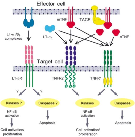
The MHC-encoded cytokines, tumour necrosis factor α (TNF-α) and lymphotoxinα (LT-α) (Gray et al., 1984; Nedwin et al., 1985; Pennica et al., 1985; Nedospasov et al., 1986), are proinflammatory, cytotoxic cytokines and critical mediators of host defence and immune regulation (Vassalli, 1992). TNF-α is expressed in response to injury or infection by a wide variety of haematopoietic and non-haematopoietic cells, mainly macrophages. It is produced as a 26 kDa membrane-associated protein which is cleaved and released as a soluble 17 kDa protein by specific metalloproteinases (Black
et al., 1997; Moss et al., 1997) (Fig. A1). Both the soluble 17 kDa
and the cell-associated 26 kDa molecular forms of TNF-α trimerize to form the bioactive ligand. They signal through two structurally related but functionally distinct receptors, the TNFR1 (p55 TNF-α receptor) and the TNFR2 (p75 TNF-α receptor), that are coexpressed on most cell types, to initiate and regulate cell death and survival pathways (Vandenabeele et al., 1995). TNF-α mediates functions that range from the recruitment of leucocytes at sites of inflammation to the regulation of cell death by proliferation, cytotoxicity or apoptosis (Wallach et al., 1997). The complexity of the action of TNF-α in vivo reflects factors such as the differential presence and activity of the soluble 17 and 26 kDa molecular forms, independent signalling by the TNFR1 and TNFR2, and the availability of shedded receptors which neutralize or augment TNF-α action. The majority of biological responses classically attributed to TNF-α are mediated by TNFR1. In contrast, TNFR2 has few specific functions; these include the induction of thymocyte proliferation in vitro and possibly a role in suppressing TNF-α-mediated inflammatory responses in vivo (Peschon et al., 1998). LT-α is expressed as a 25 kDa protein (Ware et al., 1995). It has no transmembrane sequence but is functional either as a soluble homotrimer (LT-α3) or in
association with LTβ (LT-α1β2) on the surface of activated T and
B cells and NK cells. Although TNF-α and the secreted form of LT-α (LT-α3) share receptors, they are produced by different types
of cells in response to different stimuli, and despite similarity in their binding affinity to TNF-α receptors, the cellular responses to binding appear to be different (Schuchmann et al., 1995). The two cytokines therefore function as distinct entities in vivo. LT-α1β2
heterotrimers have been shown recently to signal specifically through a distinct receptor, the LT-βR (Crowe et al., 1994), and this is now known to mediate distinct functional effects in vivo.
TNF-α and LT-α have been shown to be critical mediators of immune function and host defence (antiviral and antibacterial). Studies in gene-targeted mice deficient in the TNF-α/LT-α ligand and receptor system have revealed essential functions for these cytokines in the development and organization of lymphoid tissue and in immune function, and have shown that the effects of
of exogenous cytokines using a recombinant vaccinia virus system. Scand J Immunol 1995; 41: 31–41.
Zheng L, Fisher G, Miller RE, Peschon J, Lynch DH, Lenardo MJ. Induction of apoptosis in mature T cells by tumour necrosis factor. Nature 1995; 377: 348–51.
Zipp F, Weber F, Huber S, Sotgiu S, Czlonkowska A, Holler E, et al. Genetic control of multiple sclerosis: increased production of lymphotoxin and tumor necrosis factor-alpha by HLA-DR2⫹ T cells. Ann Neurol 1995; 38: 723–30.
Received February 3, 2000. Revised May 12, 2000. Accepted June 1, 2000
TNF-α and LT-α are largely non-overlapping. LT signalling through the LT-βR is crucial for the organogenesis of lymph nodes and Peyer’s patches, the development of normal spleen structure and the maturation of antibody affinity (De Togni et al., 1994; Matsumo
et al., 1996; Futterer et al., 1998). In contrast, lymphoid
organo-genesis in TNF-α and TNFR1 knockout mice remains intact, but these molecules are essential for the correct formation of splenic follicular structures and germinal centres, and B-cell follicles in all secondary lymphoid tissues (Le Hir et al., 1996; Pasparakis et al., 1996, 1997; Alimzhanov et al., 1997; Alexopoulou et al., 1998). Recent evidence indicates that these effects of TNF-α and of LT-α signalling through the LT-βR on B- and T-cell organization are mediated by the downstream induction of chemokines such as B-lymphocyte chemoattractant (BCL) and secondary lymphoid tissue chemokine (SLC) from stromal cells (Ngo et al., 1999). Comparative studies have indicated that LT-α3 may signal independently of LT-β in the development of mesenteric and cervical lymph nodes (Rothe et al., 1993; Ngo et al., 1999). Moreover, TNF-α and TNFR1 signalling is central to the successful host defence response to infection by various pathogens (Flynn et al., 1995; Marino et al., 1997), and a specific role for TNF-α/TNFR2 signalling in the inhibition of the entry of HIV-1 into human primary tissue-cultured macrophages has been shown (Herbein et al., 1996).
In contrast to their essential roles in lymphoid organ development and their beneficial effects during infection and tumorigenesis (Vassalli, 1992), TNF-α and LT-α have well-documented proinflammatory effects and have been implicated directly in the pathogenesis of human inflammatory and autoimmune diseases. Studies in knockout mice have shown that TNFR1 is a key mediator of endotoxic shock, and there is now considerable evidence to show that the overproduction or inappropriate production of TNF-α and LT-α, or the membrane accumulation of TNFR1, can be deleterious to the host and linked with the pathogenesis of distinct human diseases such as rheumatoid arthritis and multiple sclerosis (Raine, 1995; Korner and Sedgwick, 1996; McDermott et al., 1999). Interestingly, TNFR2 has been shown recently to signal ligand-independent pathology when overexpressed in transgenic mice (Douni and Killias, 1998). Both TNF-α and LT-α can influence disease at multiple levels. They are potently proinflammatory and their overexpression in transgenic or mutant mice triggers local inflammation and the development of complex disease phenotypes such as arthritis and Crohn’s-type disease (Kratz et al., 1996; Kontoyiannis et al., 1999). TNF-α and LT-α are also cytotoxic cytokines, and by triggering death in target cells such as oligodendrocytes (Selmaj and Raine, 1988) may initiate and exacerbate inflammation and autoimmunity. Finally, there is now compelling evidence to suggest that chronic TNF-α expression