HAL Id: tel-01378908
https://tel.archives-ouvertes.fr/tel-01378908
Submitted on 11 Oct 2016
HAL is a multi-disciplinary open access archive for the deposit and dissemination of sci-entific research documents, whether they are pub-lished or not. The documents may come from teaching and research institutions in France or abroad, or from public or private research centers.
L’archive ouverte pluridisciplinaire HAL, est destinée au dépôt et à la diffusion de documents scientifiques de niveau recherche, publiés ou non, émanant des établissements d’enseignement et de recherche français ou étrangers, des laboratoires publics ou privés.
Etude de la stabilité de la méthylation ADN chez
Arabidopsis thaliana et impact sur la transcription
Mélanie Rigal
To cite this version:
Mélanie Rigal. Etude de la stabilité de la méthylation ADN chez Arabidopsis thaliana et impact sur la transcription. Sciences agricoles. Université Blaise Pascal - Clermont-Ferrand II, 2014. Français. �NNT : 2014CLF22496�. �tel-01378908�
UNIVERSITE CLERMONT-FERRAND II – UNIVERSITE BLAISE PASCAL
N° D.U. 2496
ECOLE DOCTORALE DES SCIENCES DE LA VIE, SANTE, AGRONOMIE, ENVIRONNEMENT
N° d’Ordre 641
THESE
Présentée pour obtenir le grade deDOCTEUR D’UNIVERSITE
(spécialité Génétique et physiologie moléculaires)
Etude de la stabilité de la méthylation ADN
chez Arabidopsis thaliana et impact sur la
transcription
Présentée par
Mélanie RIGAL
Soutenue le 10 Octobre 2014
Directeur de thèse : Olivier MATHIEU
MEMBRES DU JURY :
Examinateur : Nicolas BOUCHE (Chargé de recherche, INRA, Versailles)
Rapporteur : Jean-Marc DERAGON (Professeur, Université de Perpignan Via Domitia, Perpignan)
Examinateur : Maria GALLEGO (Professeur, Université Blaise Pascal, Clermont-Ferrand) Rapporteur : Thierry LAGRANGE (Directeur de recherche CNRS, Perpignan)
Résumé
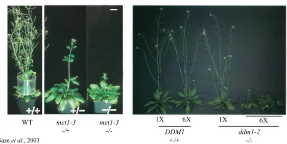
La maintenance de la méthylation ADN sur les sites CG joue un rôle crucial dans le silencing des éléments transposables (TE) et l’expression correcte des gènes. Chez Arabidopsis
thaliana, on observe, dans le mutant met1-3 déficient en méthylation CG, une apparition
ectopique de méthylation CHG sur de nombreux gènes, ainsi qu’une relocalisation de la diméthylation de la lysine 9 de l’histone H3 (H3K9me2) de l’hétérochromatine vers l’euchromatine. Nous avons démontré que ceci est lié à un défaut de transcription au niveau du grand intron du gène codant la déméthylase H3K9me2 IBM1. Nous avons également constaté que dans les épihybrides F1 issus du croisement de plantes met1-3 avec une plante sauvage la méthylation CHG présente dans l’intron de l’allèle IBM1 hérité du parent mutant était perdue. Afin de définir si la perte de méthylation affecte également d’autres loci génomiques, et plus globalement à l’échelle du génome entier l’impact de la rencontre de deux épigénomes différents, nous avons analysé les profils de méthylation, de siRNAs et de transcription des épihybrides F1. Nos données révèlent que l’union de deux méthylomes distincts au sein d’un même génome provoque une restructuration considérable des profils épigénétiques et transcriptionnels. La méthylation CHG apparaissant sur de nombreux gènes dans met1 tend à persister, créant ainsi de nouveaux épiallèles pouvant être hérités. Du côté des TE, nombre d’entre eux sont déméthylés et réactivés, tandis que d’autres sont immédiatement reméthylés et resilencés. Ainsi, ces résultats contribuent à la compréhension de la stabilité de la méthylation ADN et de son rôle dans le contrôle différentiel des gènes et des TE.
Abstract
Study of DNA methylation stability in Arabidopsis thaliana and impact on transcription Maintenance of DNA methylation at CG sites is crucial for silencing of transposable element (TE) and proper expression of genes. In Arabidopsis thaliana, met1-3 mutant, deficient in CG methylation, shows ectopic appearance of CHG methylation at numerous genes, as well as relocation of H3K9me2, from heterochromatin towards euchromatin. We have shown that this is due to a defect of the transcription of the large intron of the gene encoding the IBM1 H3K9me2 demethylase. We also found that, in the F1 epihybrids from the cross between
met1 and WT plants, CHG methylation at the intron of the met1-derived IBM1 allele is lost.
In order to define whether the loss of methylation at IBM1 also affects other genomic loci, and the impact of the union of two different epigenomes on methylation and transcription genome-wide, we analyzed the methylation, siRNA and transcription patterns of F1 epihybrids. Our data reveal that the union of two distinct methylomes within the same genome triggers considerable restructuring of epigenetic and transcriptional patterns. CHG methylation appearing in the mutant parent tends to persist in F1, creating new epialleles that can be inherited. On the TE side, lots of them are demethylated and reactivated while others are immediately remethylated and resilenced. Thus, our results provide new insights to the understanding of DNA methylation stability and its role in the differential control of genes and TEs.
Mots-clés : Arabidopsis, épigénétique, méthylation ADN, MET1, hybrides, IBM1, histone, H3K9me2, transcription, siRNA.
Keywords : Arabidopsis, epigenetic, DNA methylation, MET1, hybrids, IBM1, histone, H3K9me2, transcription, siRNA.
Remerciements :
Je tiens avant tout à remercier Olivier Mathieu de m’avoir permise de réaliser ces quatre années dans son laboratoire. Je n’aurai pu imaginer meilleur directeur de thèse, à la fois patient et exigent, obstiné et minutieux, et toujours accessible, formant le petit Padawan aux techniques, à l’interprétation des données obtenues, m’incitant à me remettre un peu en question, mais pas TOUT le temps.
Je remercierai aussi mes compagnes et compagnons de labeur,
Zoltan, auprès de qui j’ai commencé cette thèse, dont j’ai pu apprécié le tempérament hongrois, les paroles réconfortantes visant à calmer mes angoisses…
Yoko, my Japanese sister, with whom I had a lot of great time, in the lab and outside, keeping me company late in the evening and the week-ends…
Ma pretty woman, Leticia, plus angoissée encore que moi, mais toujours à l’écoute et avec un cœur immense dans une taille de guêpe.
Angeles, dont l’arrivée a valu une belle escapade en girl trio à Lyon et dont j’ai pu apprécié le tempérament zen et de feu en même temps, locura español…
Les garçons aussi, Steve, Anthony, en mode iPad à fond, l’arrivée des « Montpelliérains », Maxime puis Pierre, et Sébastien, qui ont chacun fait ou font leur petit bonhomme de chemin dans l’équipe…
Un énorme remerciement d’ailleurs à Pierre pour le choix d’un concert merveilleux, une affiche de thèse Indiana Jonesque, d’une gentillesse et intelligence scientifique rare…
Un Grand Merci tout particulier à Romain aussi, dont la venue a bien facilité nos analyses bioinformatiques, mais a bien égayé également le quotidien.
Merci aux 2 Pélissier, Mr Péloch, qui m’a donné moulte conseils techniques en tentant de garder patience quand j’ouvrais des yeux ronds et lui demandais de répéter pour la 4ème fois…
Un Immense merci aussi à Anne-Sophie, gestionnaire mais pas seulement, toujours à l’écoute, hyper efficace et adorable, à qui je ne peux que souhaiter d’atteindre une vie professionnelle stable et épanouissante.
Je remercie bien sûr aussi tous les autres membres du laboratoire que j’ai pu croiser durant ces quatre années de thèse, les étudiants comme les chercheurs des trois équipes du couloir.
Je remercierai aussi très sincèrement Detlef Weigel et Claude Becker pour notre collaboration, m’avoir accueilli dans le laboratoire, formé au séquençage ARN puis avoir contribué à l’analyse des données et à la rédaction du manuscrit.
Je dédierai ce manuscrit à Ma Famille, mes parents, mes frère et sœur et tous les autres, ayant subi mes absences chroniques, mon obsession pour le labo, mes angoisses et ma presque volatilisation durant ces quatre années… et Un Immense
Merci à mes petits choux, neveu et nièce, ayant décidé d’arriver pendant cette période et m’ayant ainsi offert des instants de bonheur, de rigolade et de maternage salvateurs.
Je remercierai aussi mes amies qui m’ont, je crois, pardonné mon absence et mon obsession, durant ces années, et qui m’ont suivi jusqu’à aujourd’hui…
En tout dernier point, je souhaite remercier les membres du jury d’avoir accepté notre invitation, lu le manuscrit dense et fastidieux durant les beaux jours d’été, et offert à ma soutenance des remarques qui nous ont particulièrement été utiles notamment pour la suite des analyses de séquençage et de la rédaction de l’article.
Merci à Vous …
TABLE DES MATIERES
I. INTRODUCTION ...2
I.A. Voies épigénétiques chez Arabidopsis thaliana ... 2
I.A.1. L’épigénétique... 2
I.A.2. Composition du génome d’Arabidopsis thaliana... 4
I.A.3. La transcription chez Arabidopsis thaliana... 6
I.A.4. Répression des éléments transposables... 8
I.A.5. Régulation épigénétique des gènes...24
I.A.4.a. La méthylation ADN...24
I.A.4.b. La déméthylation ADN...30
I.A.4.c. Les marques histones et variants d’histones...36
I.A.4.a.1. L’acétylation et la déacétylation des histones...36
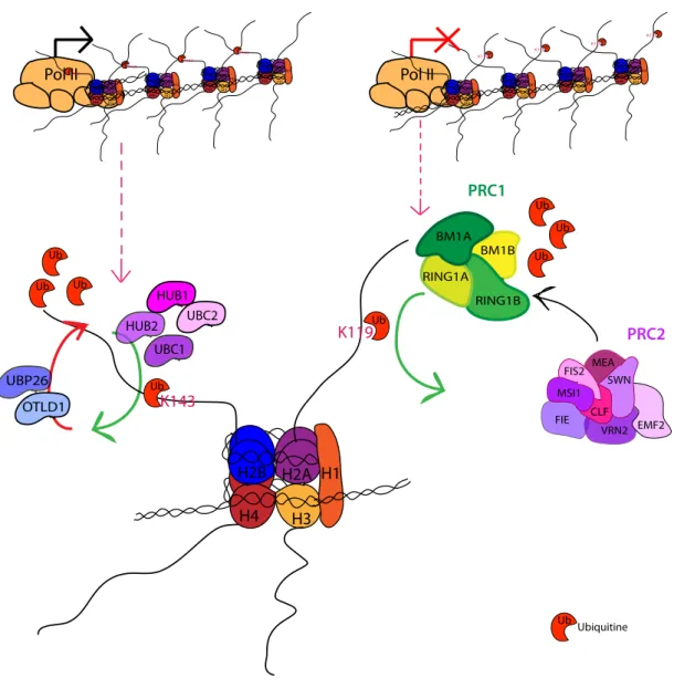
I.A.4.a.2. L’ubiquitination et déubiquitination des histones...38
I.A.4.a.3. La méthylation/déméthylation des histones...40
I.A.4.a.4. Les variants d’histones...48
I.A.4.b. La voie RNA-directed DNA methylation (RdDM)...50
I.A.4.c. Rôle de la méthylation CG sur les gènes...52
I.B. Stabilité de la méthylation ADN au cours des générations... 56
I.B.1. Les épiallèles naturels...56
I.B.2. Variabilité épigénétiques naturelle...60
I.B.2.a. Profils épigénétiques des populations naturelles...60
I.B.2.b Impact de la formation d’hybrides sur les profils épigénétiques...64
I.B.3. Epiallèles induits dans des mutants hypométhylés...68
I.B.4. Rétrocroisement de mutants hypométhylés...70
I.B.5. Analyse de lignées épigénétiques recombinantes...72
I.C. Objectifs de la thèse ... 76
II. CHAPITRE 1 : ETUDE DE LA REGULATION DU GENE IBM1
DANS LE MUTANT MET1...78
II.A. Introduction ... 80
II.B. Article « Rigal et al., 2012 » ... 82
III. CHAPITRE 2 : CONSEQUENCES IMMEDIATES DE LA
RENCONTRE DE PLANTES AUX GENOMES IDENTIQUES MAIS
AUX EPIGENOMES DISTINCTS – DYNAMIQUE DE LA
METHYLATION ADN ET DE LA TRANSCRIPTION DANS LES
EPIYBRIDES F1...132
III.A. Introduction... 134
III.B. Article « Rigal et al., 2015 »... 138
IV. DISCUSSION GENERALE ...234
INTRODUCTION
1
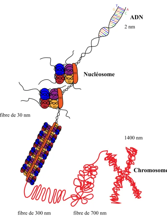
Figure I-1 : Représentation schématique de la structure de la chromatine.
La molécule d’ADN d’un diamètre de 2 nm est enroulée tout d’abord sur les octamères d’histones comportant deux dimères H2A-H2B et deux dimères H3-H4. Ceci forme le nucléosome. De plus, cette structure est stabilisée par la liaison de l’histone H1 à l’ADN internucléosomal, permettant le repliement du nucléofilament en fibre de 30 nm, elle-même compactée en boucles de 300 nm d’envergure. Ces boucles sont organisées en super-hélices formant ainsi la structure en chromosome de 1400 nm.
H1 H1 H2A H2B H4 H3 H2A H2B H4 H3 Nucléosome ADN fibre de 300 nm fibre de 700 nm 1400 nm fibre de 30 nm 2 nm A T G C Chromosome
I. INTRODUCTION
I.A. Voies épigénétiques chez Arabidopsis thaliana
I.A.1. L’épigénétique
La découverte de l’ADN comme support de l’information génétique par Watson et Crick il y a plus de 60 ans a représenté une avancée majeure dans la compréhension des mécanismes impliqués dans le développement et le fonctionnement des organismes vivants. Cependant, un mystère demeurait sous-jacent : comment toutes les cellules d’un individu peuvent-elles porter la même information génétique mais avoir des caractéristiques particulières, au cours du développement mais aussi selon les organes, les tissus ?
L’épigénétique a permis de lever le voile au moins partiellement sur ce paradoxe. La notion d’épigénétique est définie, dès les années 1950, comme l’étude des mécanismes moléculaires participant à la régulation de l’expression des gènes au cours du développement et de la différenciation cellulaire. Les modifications épigénétiques n’affectent pas la séquence d’ADN elle-même mais peuvent être transmises d’une génération à l’autre et être réversibles. De plus, elles peuvent être modulées par des facteurs environnementaux. Comme détaillé plus en avant dans les paragraphes suivants, elles jouent également un rôle crucial dans la régulation des éléments transposables, composants majeurs des génomes eucaryotes dont la mobilisation, pouvant engendrer des réarrangements chromosomiques et des mutations, menace l’intégrité du génome.
Que ciblent ces modifications épigénétiques ?
L’ADN est fortement compacté dans le noyau grâce à de nombreuses protéines, ce qui forme la chromatine, structure cible des modifications épigénétiques (Figure I-1).
Le premier niveau de compaction de l’ADN consiste en l’enroulement de la molécule autour d’octamères d’histones (deux dimères H3-H4 et deux dimères H2A-H2B), formant les nucléosomes (Luger et al. 1997). Chaque nucléosome contient environ 145 pb d’ADN et est séparé du nucléosome voisin par un fragment d’ADN de liaison de 10 à 80 bp. L’assemblage de ces histones dans la chromatine a lieu le plus souvent lors de la réplication, et implique des chaperones d’histones, telles que CAF-1.
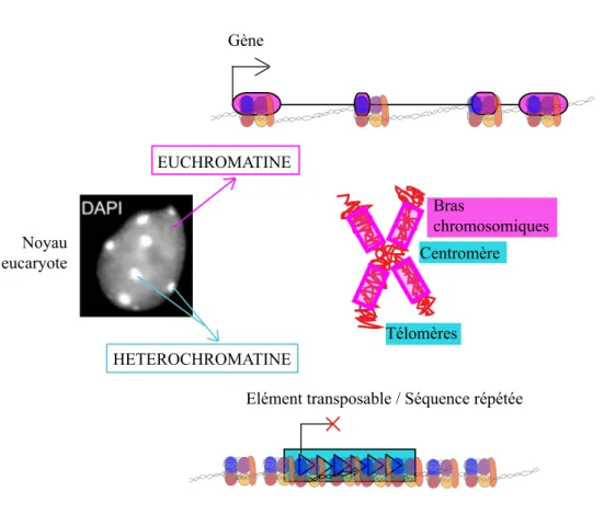
Figure I-2 : Deux types de chromatine sont retrouvés dans les noyaux eucaryotes.
L’euchromatine, peu condensée est riche en gènes et localisée principalement sur les bras chromosomiques chez A. thaliana. L’hétérochromatine, quant à elle, est très condensée, riche en histones H1 et enrichie en éléments transposables et séquences répétées, elle est retrouvée dans les zones péricentromériques et télomériques.
HETEROCHROMATINE EUCHROMATINE
Gène
Elément transposable / Séquence répétée Bras chromosomiques Noyau eucaryote Bras chromosomiques H1 H1 H2A H2B H4 H3 H2A H2B H4 H3 Nucléosome ADN fibre de 300 nm fibre de 700 nm 1400 nm fibre de 30 nm 2 nm A T G C Centromère Télomères
Secondairement, l’histone H1 s’associe à cet ADN de liaison et permet ainsi d’augmenter le degré de compaction et former une fibre de 30 nm. La fibre chromatinienne se condense à la mitose sous forme de chromosomes. Les analyses cytologiques mettent en évidence deux types de chromatine : l’euchromatine, peu condensée, riche en gènes, et l’hétérochromatine, très condensée grâce à la présence de l’histone H1, incluant chez la plante modèle
Arabidopsis thaliana les régions télomériques, et péricentromériques (Figure I-2).
I.A.2. Composition du génome d’Arabidopsis thaliana
La plante modèle Arabidopis thaliana possède un génome de 125 Mb, se répartissant sur cinq chromosomes ; il s’agit d’un des plus petits génomes du règne végétal d’après les données actuelles (2000). Les gènes codant des protéines occupent 50% du génome. Au nombre de 27 411, ils ont une taille moyenne de 2 kb, avec des exons de 296 pb et des introns de 165 pb en moyenne. Les éléments transposables représentent, eux, 24% du génome, une proportion faible en comparaison à d’autres plantes comme le Maïs par exemple (85% du génome) (Figure I-3A&B). Cette portion du génome inclut à 40% des éléments de classe I, qui transposent par un mécanisme de « copier/coller » via un intermédiaire ARN. Ces éléments sont majoritairement retrouvés au niveau des centromères.
Les 60% restants sont occupés en part égale par des transposons de classe II, se déplaçant, eux, par un mode de « couper/coller », et des Hélitrons, dont le mode de transposition par cercle roulant est encore mal connu. Ces transposons ont une distribution plus large et variable que les éléments de classe I, même si fortement regroupés dans les zones péricentromériques et les « knobs » hétérochromatiques. Les Miniature Inverted repeat Transposable Elements (MITEs), hobo, Activator, Tam3 (hAT) et Hélitrons sont aussi largement retrouvés sur les bras chromosomiques, riches en gènes (2000).
Figure I-3 : Distribution du génome d’Arabidopsis thaliana en gènes et éléments transposables.
A- Chez A. thaliana, sur les cinq chromosomes, les séquences répétées sont préférentiellement localisées dans les zones péricentromériques tandis que les gènes sont plus présents sur les bras chromosomiques.
B- Les gènes occupent la moitié du génome d’A. thaliana et les éléments transposables près d’un quart. Les éléments de classe I transposent par un mécanisme de « copier/coller » via un intermédiaire ARN, ainsi leur mobilisation provoque une augmentation de leur nombre de copies. Les éléments de classe II se déplacent par un mode de « copier/coller », changeant ainsi de localisation génomique. Les Hélitrons, eux, sont mobilisés via un mode de transposition peu connu à ce jour, qui mettrait en jeu une structure en cercle roulant.
Gènes codant des protéines Eléments transposables Classe I Classe II Hélitrons ARN TIR TIR 1 2345 1 2345 ADNc Rétro-transcription 1 2345 1 2345 LTR LTR TIR TIR LTR LTR LTR LTR Réplication Eléments de classe II Hélitrons Eléments de classe I GENOME 50% 24% 40% 30% 30% Zhang et al., 2006 A B
I.A.3. La transcription chez Arabidopsis thaliana
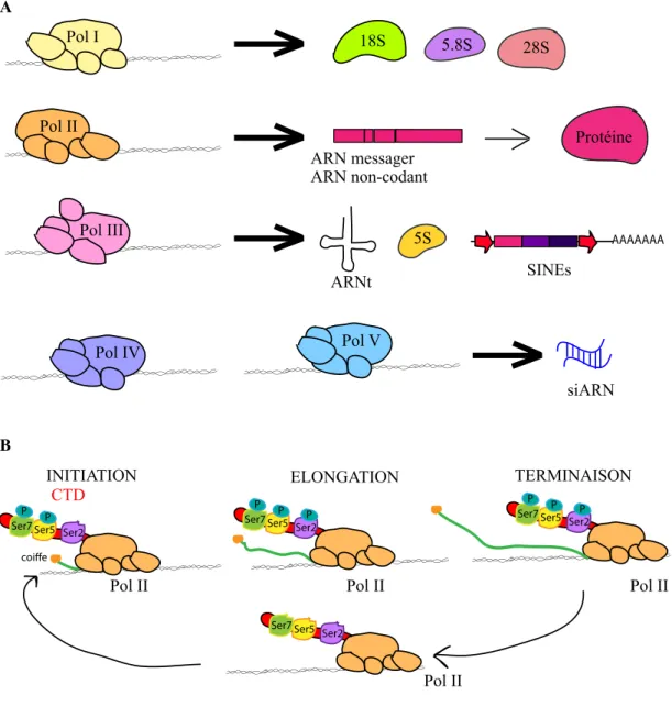
Chez Arabidopsis, cinq polymérases ARN ont été identifiées et caractérisées (Figure I-4A). La polymérase I (Pol I) est la plus spécialisée, elle transcrit les gènes codant les ARN ribosomaux (ARNr) 18S, 5.8S et 28S. La polymérase II (Pol II) transcrit un très grand nombre de gènes codant des protéines mais aussi des ARN non-codants (ARNnc) et des petits ARN modulant ou maturant les ARN messagers (ARNm) ou les ARNr. Enfin, la polymérase III (Pol III) transcrit diverses classes de petits ARN tels que les ARN de transfert (ARNt), l’ARNr 5S, de petits ARN régulateurs ainsi que les Short Interspersed Nuclear Elements (SINE).
Deux autres polymérases ARN spécifiques des Plantes, Pol IV et Pol V, jouent un rôle crucial dans la génération de petits ARN guidant la méthylation ADN de novo, dans un processus appelé voie RNA-directed DNA methylation (RdDM) (Haag et al. 2011).
La transcription par la polymérase II, nécessaire à l’expression des gènes et de la majorité des éléments transposables se déroule en trois phases, l’initiation, l’élongation et la terminaison. Ces étapes sont régulées grâce à des modifications post-traductionnelles du domaine Carboxy-Terminal (CTD) de la Pol II mais également grâce à l’intervention de divers acteurs épigénétiques, comme décrit dans les paragraphes suivants (Figure I-4B). Tout d’abord, le complexe de préinitiation de la transcription recrute la Pol II hypophosphorylée au niveau du promoteur. Le résidu Serine 5 du CTD est phosphorylé par le facteur de transcription TFIIH. Ceci facilite la coiffe en 5’ de l’ARNm nouvellement synthétisé. La Serine 7 est également phosphorylée par la cycline kinase CDKF;1 (Hajheidari et al. 2013).
Durant l’élongation, la phosphorylation du CTD sur la Serine 2 par les CDKC ;1 et CDKC ;2 permet aussi son interaction avec de nombreuses protéines impliquées dans l’épissage des transcrits primaires et ainsi le couplage de la transcription et l’épissage.
Chez Arabidopsis, les mécanismes de terminaison de la transcription sont encore peu clairs mais le CTD joue également un grand rôle dans le recrutement de la machinerie de maturation de l’extrémité 3’ des ARN. Après le clivage et la polyadénylation des ARN, la polymérase est rapidement déphosphorylée afin de pouvoir être recyclée pour un nouveau cycle de transcription.
Figure I-4 : La transcription chez Arabidopsis
A- Il existe quatre polymérases chez A. thaliana. Tandis que la polymérase II transcrit principalement les gènes codant des protéines ainsi que des ARN non-codants (ARNnc), la polymérase I transcrit les gènes codant les ARN ribosomiques (ARNr) 18S, 5.8 S et 28S. La polymérase III, quant à elle, transcrit les gènes codant les ARN de transfert (ARNt), l’ARNr 5S ainsi que les éléments transposables de classe I de type Short Interspersed Nuclear Elements (SINE). Les polymérases IV et V spécifiques des Plantes permettent la production de petits ARN guidant la méthylation ADN de novo via une voie nommée RNA-directed DNA Methylation (RdDM).
B- La transcription par la polymérase II se déroule en trois phases, l’initiation où le carboxy-terminal (CTD) est phosphoryle sur les Serines 5 et 7. La phosphorylation facilite le dépôt d’une coiffe sur le transcrit initié. Durant l’élongation, la Serine 2 est phosphorylée également, permettant le couplage de la transcription avec l’épissage. Lors de la terminaison, des protéines de maturation de l’extrémité 3’ sont recrutées. Après clivage et polyadénylation du transcrit généré, le CTD est déphosphorylé, la polymérase peut débuter un nouveau cycle de transcription. ARNt 5S AAAAAAA SINEs 5.8S 18S 28S ARN messager ARN non-codant Protéine siARN Pol II Pol III Pol V Pol IV Pol I Pol II CTD
INITIATION ELONGATION TERMINAISON
Pol II Pol II Ser5P Ser7 P Ser2P Pol II Ser5P Ser7 P Ser2P Ser5P Ser7 P Ser2 Ser5 Ser7 Ser2 coiffe A B
I.A.4. Répression des éléments transposables
Les mécanismes épigénétiques de répression des éléments transposables sont détaillés dans la revue suivante :
« A "mille-feuille" of silencing : epigenetic control of transposable elements. » Rigal M, Mathieu O. ; Biochimica et Biophysica Acta. 2011.
Pour cette revue, j’ai participé à l’étude bibliographique, à la réflexion autour de l’organisation et du contenu de la publication, ainsi qu’à la rédaction des différents paragraphes.
INTRODUCTION
Review
A “mille-feuille” of silencing: Epigenetic control of transposable elements☆
Mélanie Rigal, Olivier Mathieu⁎
Centre National de la Recherche Scientifique (CNRS), UMR 6247—GReD—INSERM U 931, Clermont Université, Aubière, France
a b s t r a c t a r t i c l e i n f o
Article history: Received 15 February 2011
Received in revised form 30 March 2011 Accepted 1 April 2011
Available online 12 April 2011 Keywords:
Transposable elements Silencing
Arabidopsis
Despite their abundance in the genome, transposable elements (TEs) and their derivatives are major targets of epigenetic silencing mechanisms, which restrain TE mobility at different stages of the life cycle. DNA methylation, post-translational modification of histone tails and small RNA-based pathways contribute to maintain TE silencing; however, some of these epigenetic marks are tightly interwoven and this complicates the delineation of the exact contribution of each in TE silencing. Recent studies have confirmed that host genomes have evolved versatility in the use of these mechanisms to individualize silencing of particular TEs. These studies also revealed that silencing of TEs is much more dynamic than had been previously thought and can be reversed on the genomic scale in particular cell types or under special environmental conditions. This article is part of a Special Issue entitled “Epigenetic control of cellular and developmental processes in plants”. © 2011 Elsevier B.V. All rights reserved.
1. Introduction
All genomes hold a genetic toolkit that is necessary to properly orchestrate the development of an organism. Genes can be crowded within genomes. Over the past 15 years, intensive sequencing efforts have revealed that most eukaryotic genomes do not simply contain genes but are filled with transposable elements (TEs). In fact, TEs can be the major constituents of a given genome, which leaves little space for genes; this finding is particularly striking in plants[1,2]. TEs are mobile pieces of DNA that have been viewed as genomic parasites that multiply and insert themselves at new genomic positions when activated. TEs have been classified into two classes based on whether their transposition strategy involves an RNA intermediate[3]. Class I TEs, known as retrotransposons, require reverse transcription of an RNA intermediate for duplication, while transposition of class II TEs, also called DNA transposons, does not rely on reverse-transcription of the TE transcripts. DNA transposons are further subdivided into two subclasses. Subclass I is composed of elements containing terminal inverted repeats and are transposed through a “classical” cut-and-paste mechanism. Subclass II contains the more recently identified helitrons, which do not contain terminal inverted repeats and appear to duplicate through a rolling-circle mechanism, which requires the TE-encoded helicase and replicase proteins. Class I elements generally represent the vast majority of the repetitive fraction of a given plant genome, likely due to their duplicative mode of transposition, which increases their copy number after each mobilization. Although most TEs are present in the form of truncated or mutated derivatives, genomes contain full-length TEs that retain an intact code
and the potential to transpose. Mobilization of these elements represents a threat to genome integrity, a fact that led to their discovery in maize by Barbara McClintock in the late 1940s[4]. Indeed, active TEs can be responsible for chromosome breakage and genomic rearrangements and can interrupt gene function through insertion. To restrain the harmful potential of TEs, host genomes have generated sophisticated mecha-nisms that counteract TE activation and maintain TEs in a silent state. Although they seem to be selfish pieces of DNA, TEs are thought to play a major role in genome evolution. TEs and the mechanisms that regulate their activity are involved in major cellular processes, such as gene regulation, centromere and telomere function, genomic imprinting and X chromosome inactivation[5,6]. These mechanisms are epigenetic in nature because they do not result from genetic mutations. Rather, they generate a repressive chromatin environment, which is faithfully inherited through mitosis and meiosis, but which can also be reversed in a developmentally regulated or environmentally induced manner. Repressive chromatin is commonly associated with specific small RNA signatures and with a range of epigenetic marks that affect DNA and histone proteins.
In this review, we discuss how these epigenetic processes contribute to the silencing of TEs and focus on recent studies that have expanded our understanding of these pathways.
2. Control of TE activity by DNA methylation
DNA methylation, which is the addition of a methyl group to DNA residues, occurs primarily at cytosines in eukaryotes. Apart from yeast (Schizosaccharomyces pombe and Saccharomyces cerevisiae) and some invertebrates (Caenorhabditis elegans, Drosophila melanogaster, Tribolium castaneum,and Oikopleura dioica), cytosine DNA methylation is present at detectable levels in all organisms that have been examined to date[7,8]. In mammals, the prevalent view is that cytosine DNA methylation occurs Biochimica et Biophysica Acta 1809 (2011) 452–458
☆ This article is part of a Special Issue entitled “Epigenetic control of cellular and developmental processes in plants”.
⁎Corresponding author.
E-mail address:olivier.mathieu@univ-bpclermont.fr(O. Mathieu). 1874-9399/$ – see front matter © 2011 Elsevier B.V. All rights reserved. doi:10.1016/j.bbagrm.2011.04.001
Contents lists available atScienceDirect
Biochimica et Biophysica Acta j o ur n a l h o m e p a ge : ww w. e l s ev i e r. c o m/ l o c a t e / b b a g r m
INTRODUCTION 11
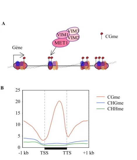
almost exclusively at CG dinucleotides, although a recent study has challenged this view by revealing that nearly one-quarter of methylation sites detected in undifferentiated human embryonic stem cells are in non-CG contexts[9]. In plants, non-CG methylation is common, and DNA methylation affects cytosines in all sequence contexts, including CG, CHG (H_A, T or C) and CHH.
Mammalian DNA methyltransferase 1 (DNMT1) and its ortholog in plants, methyltransferase1 (MET1), propagate CG methylation patterns following each round of DNA replication. These DNA methyltransferases function in combination with accessory proteins, UHRF1 in mammals and variant in methylation (VIM) in plants, which have strong affinities for hemimethylated CG sites and likely aid in the recruitment of DNMT1 and MET1 to replicating DNA [10–12]. The plant-specific protein chromomethylase3 (CMT3) primarily maintains CHG methylation, while domains rearranged methyltransferase2 (DRM2) ensures the persistence of CHH methylation. CMT3 is recruited to chromatin via interaction of its chromodomain with methylated histone H3 tails. Accordingly, the CMT3 chromodomain has been shown in vitro to bind directly to H3 tails trimethylated at lysine 9 and lysine 27[13]. DRM2, an ortholog of the mammalian DNMT3 methyltransferases, is guided to DNA by 24-nucleotide (nt) small interfering RNAs (siRNAs) in a pathway known as RNA-directed DNA methylation (RdDM)[14–16]. In addition, decrease in DNA methylation1 (DDM1), a SWI2/SNF2-related chromatin-remodeling factor, contributes to DNA methylation in all sequence contexts[17–19], although its precise molecular mode of action is still poorly understood.
The first evidence for a role of DNA methylation in controlling TE activity resulted from studies of class II TEs in maize, approximately 40 years after the discovery of TEs by B. McClintock. Molecular analyses of the Activator (Ac), Suppressor-mutator (Spm) and Mutator (Mu) elements[20–22]revealed that inactivation of these elements was correlated with the methylation of their DNA. In the active state, transposase promoter sequences from the Ac and Spm elements are hypomethylated, resulting in active transcription. Likewise, DNA methylation at the autonomous Mu-family elements MuDR is associated with transcriptional silencing [23,24]. Later studies in Arabidopsis showed that several classes of TEs become hypomethylated and transcriptionally reactivated in a ddm1-induced hypomethylation background[25–28]. These studies further reported transposition of two DNA transposons in ddm1: the MULE-like element AtMu1 and the CACTAelements[25,26]. More recently, the characterization of a ddm1-induced floral phenotype revealed that DNA transposons and ATGP3 retrotransposons of the gypsy class are mobilized in ddm1. Furthermore, hybridizing genomic DNA from both wild-type and ddm1 lines on high-density tilling array allowed the authors to detect increases in the copy number, and thus transposition, of several families of DNA transposons and retroelements in the mutant background[29]. Mutations in LSH1, the mouse ortholog of DDM1, also lead to hypomethylation and transcrip-tional reactivation of TEs[30,31]. These and other experimental data have led to the view that one important role of DNA methylation is to function as a genomic immune system that ensures the silencing of TEs. Recent high-throughput profiling studies of DNA methylation in Arabidopsishave confirmed that, similar to that observed in human and mouse, TEs and other types of repeated sequences represent the most highly methylated sequences in the genome[32–35]. In two large-scale studies that quantified DNA methylation in an additional set of 21 eukaryotic genomes in total, Zemach et al. and Feng et al. showed that this trend can be extended to all analyzed plant types
[7,8]. In addition to CG methylation, TEs are enriched in CHG and CHH methylation, which is consistent with their preferential association with H3K9me2 and their active targeting by the RdDM pathway
[32,33,36,37]. It may therefore be reasonable to expect that all three types of methylation cooperate or contribute to a certain degree in TE silencing. Accordingly, ATGP3 elements that are transcribed and mobilized in the CG and non-CG hypomethylated ddm1 mutant background remain transcriptionally silent in the met1 and cmt3
single mutants[29]. Transcription and transposition of ATGP3 was only detected in met1 cmt3 double mutants, which are deficient in both CG and CHG methylation. Although CACTA transcripts accumu-late to various degrees in the met1 and cmt3 single mutants, high-frequency mobility of this TE is achieved only in the met1 cmt3 double mutant[38]. Together, these observations illustrate that CG and CHG methylation are redundantly required to restrain TE activity at either the transcriptional (ATGP3) or transpositional levels (CACTA). These data also suggest that different types of TEs are controlled differen-tially by DNA methylation. Indeed, this effect was recently illustrated by the analysis of a copia-like retrotransposon named Evadé (EVD)
[39]. Transcription of EVD is reactivated upon loss of CG methylation in a met1 mutant, and EVD is progressively mobilized in successive generations of the mutant. In addition to CG methylation, low levels of CHH methylation are found on this TE. Mutations that affect CHH methylation do not release EVD silencing; however, in combination with met1, they lead to a synergistic increase in EVD transcription, indicating that in the absence of CG methylation, the RdDM pathway curbs the accumulation of EVD transcripts. Importantly, in one combination of mutations (met1 nrpe2, see below), EVD is the only TE mobilized, and other TEs, including CACTA and ATGP3, remain immobile. These findings underscore the idea that host genomes have evolved selectivity in the control of TE activity.
Unlike mammals, it has long been known that plant genomes likely do not globally reset the genomic DNA methylation patterns between sexual generations. Accordingly, loci that lose DNA methylation and the associated epigenetic silencing in met1 and ddm1 mutants, including TEs, retain activity and hypomethylation for several generations following removal of the mutations[17,27,40,41]. Epigenetic recombi-nant inbred lines (epiRILs) derived from met1 or ddm1 mutants show inheritance of DNA hypomethylation at some genomic regions. As expected, inheritance of hypomethylation at CACTA elements was found to result in transposition in ddm1-derived epiRILs that have inherited these hypomethylated CACTA epigenetic alleles (“epialleles”)[42]. In the case of met1-derived epiRILs, CACTA elements should remain immobile because they do not transpose in the parental met1 mutant background. Thus, it was unexpected when Reinders et al. reported CACTA mobilization in nearly 30% of the met1-derived epiRILs. CACTA transposition was triggered stochastically in some epiRIL individuals after several generations of propagation by self-pollination[42,43]. Although it was not directly tested, it can be inferred that CACTA mobilization in these lines results from the inheritance of met1-derived elements that are hypomethylated at CG sites and a concomitant loss of additional repressive epigenetic marks, such as non-CG methylation or H3K9me2. Indeed, it was previously shown that non-CG methylation patterns and H3K9me2 patterns are stochastically altered and unstably inherited upon met1-induced loss of CG methylation[44]. Similar to CACTA, the EVD retroelement was found to transpose after inbreeding of some met1-derived epiRILs, but the two types of elements were not necessarily simultaneously reactivated in the same lines, which once again underscores the apparent selective control of TEs by distinct epigenetic mechanisms. Investigating ddm1-derived epiRILs, Teixeira and colleagues also reported insights into this selectivity. They demonstrated that a subset of TEs, which is demethylated in ddm1, is progressively remethylated and resilenced over multiple generations after introduction into a wild-type background[45]. The propensity for remethylation correlates with the retention of CHH methylation, which appears to be independent of DDM1 at remethylatable TEs. In contrast, CHH methylation at non-remethylatable TEs displays a strong depen-dence on DDM1. Therefore, CHH methylation is clearly maintained by distinct mechanisms at various TEs.
3. Role of histone modifications
In addition to DNA methylation, TEs are associated with various post-translational histone modifications, which are characteristic of a
453 M. Rigal, O. Mathieu / Biochimica et Biophysica Acta 1809 (2011) 452–458
INTRODUCTION
repressive chromatin environment. In particular, TEs located in pericentric heterochromatin are associated with very high levels of H3K9me2 and H3K27me1 [13,36,46,47]. In Arabidopsis, several histone methyltrans-ferases are responsible for the propagation of H3K9me2, including kryptonite/SUVH4 (KYP), SUVH5 and SUVH6, which display functional redundancy at some genomic loci[48–52]. Alternatively, H3K27me1 is catalyzed by the histone methyltransferases Arabidopsis trithorax-related protein 5 (ATXR5) and ATXR6, which appear to be functionally redundant
[46].
H3K9me2 and H3K27me1 both appear to contribute to transcrip-tional silencing of some TEs. In Arabidopsis, TE reactivation was observed in kyp suvh5 suvh6 triple mutants[49]and atxr5 atxr6 double mutants
[46]. In rice, mutations in the histone H3K9 methyltransferases genes SGD714and SDG728 also lead to TE activation [53,54]. H3K27me1 functions in transcriptional silencing in a pathway that is clearly independent of DNA methylation and H3K9me2 [46], and TEs reactivated in atxr5 atxr6 mutants retain high levels of these two repressive marks. Moreover, H3K27me1 levels are not changed in met1 and ddm1 mutants, which show reduced DNA methylation, and the kyp mutant, which shows reduced H3K9me2 levels[13,47]. Recently, it was shown that the ATXR5/6 H3K27me1 pathway suppresses re-replication of TE-rich heterochromatic regions, which has led to the speculation that this pathway may have evolved to restrain excess DNA replication and reactivation of TEs[55].
The precise contribution of H3K9me2 to TE silencing is more difficult to dissect given that H3K9me2 is tightly interwoven with DNA methylation, in particular CHG methylation. At the genome level, approximately 90% of CHG methylation overlaps with H3K9me2-enriched regions[36], and TEs have high levels of CHG methylation
[28,33,35,56,57]. KYP and KYP SUVH5 SUVH6 mutants show reduced H3K9me2 levels at reactivated TEs and a significant reduction in CHG methylation at these sequences[48–52]. Moreover, some TEs show reduced H3K9me2 levels in mutants for CMT3, the maintenance DNA methyltransferase for CHG sites[58]. KYP, SUVH5 and SUVH6 contain SET and RING associated (SRA) domains, which have been shown to bind to DNA with methylated cytosines, and KYP and SUVH6 display preference for CHG methylation over CG methylation[51,59]. Alterna-tively, it has been proposed that CMT3 is recruited to chromatin via the interaction of its chromodomain with H3K9me2[60]. These findings suggest that H3K9me2 and CHG methylation are maintained through a self-reinforcing feedback loop, such that influencing one modification likely impacts the other. In addition, mutations in MET1 also reduce H3K9me2 in TE-rich regions[41,61], particularly for TEs with a high frequency of methylated CGs, where H3K9me2 is less dependent upon KYP and CMT3[62]. This result suggests that an additional pathway, which is dependent on CG methylation, maintains H3K9me2 at TEs. Remarkably, a null mutation in the increase in bonsai methylation 1 (IBM1) histone demethylase gene results in ectopic H3K9me2 and CHG DNA hypermethylation in a large number of genes, while TEs are completely unaffected[62–64]. Thus, IBM1 protects genes but not TEs from H3K9me2 and CHG methylation. Together with multiple pathways that maintain H3K9me2, this situation contributes to the perpetuation of a robust silenced state at TEs.
Interestingly, it has recently been demonstrated that KYP plays a role in restraining the mobilization of the EVD retroelement, specifically at the post-transcriptional level[39]. Indeed, while met1 kyp double mutants accumulate EVD transcripts at a level similar to met1 single mutants, extrachromosomal copies of this TE only accumulate in the double mutant. In an analogous manner to mammalian lysine methyltrans-ferases, which also methylate non-histone proteins[65–68], it has been proposed that KYP may use a substrate other than H3K9 to inactivate, through methylation, a TE-encoded protein required for the translation and/or reverse transcription of EVD transcripts[39].
Other histone modifications likely play a role in TE silencing; however, similarly to H3K9me2, the exact contributions of these modifications are difficult to delineate given the links between DNA
methylation and histone modifications. For instance, histone deacetyla-tion appears to be important for the maintenance of TE silencing. In support of this idea, an analysis of Arabidopsis histone deacetylase mutants, such as hda6, has revealed that several classes of TEs are transcriptionally activated[27]. Likewise, downregulation of the histone deacetylase gene OsSRT1 in rice results in upregulation of many genomic loci, primarily TEs[69]. Reactivation of these TEs is also correlated with a reduction in H3K9me2, the appearance of H3K4me2, and occasionally reduction in DNA methylation. Similar to histone acetylation, ubiquiti-nation of histone H2B is required to maintain chromatin in an open state
[70]. Histone H2B is monoubiquitinated in Arabidopsis (at lysine 143), animals and yeast[70–72]. Mutations in an Arabidopsis deubiquitination enzyme, UBP36, release transcriptional silencing of several different classes of TEs. Silent TEs are enriched for non-ubiquitinated H2B, and the deubiquitination of H2B seems to be required for H3K9me2 and subsequent non-CG DNA methylation[71]. Interestingly, this suggests that TEs are associated with active histone modification marks by default and that faithful maintenance of a repressive chromatin state depends on the continuous removal of these active marks.
4. Small RNAs and TE silencing
RNA silencing is widely used by eukaryotes, notably to control TE activity at the transcriptional and post-transcriptional levels. This mechanism involves small RNA molecules of 21- to 35-nt, which guide silencing by modulating chromatin states or targeting RNA degradation. Among the various classes of small RNAs, endogenous siRNAs (endo-siRNAs) and PIWI-associated RNAs (piRNAs) partici-pate in the silencing of TEs in both Drosophila and mice[73–75]. Endo-siRNAs originate from the cleavage of long double-stranded RNAs (dsRNAs) by a Dicer enzyme; whereas piRNAs are derived from single-stranded transcripts in a Dicer-independent pathway.
The piRNA pathway is mostly germline-specific while endo-siRNAs also repress TE activity in somatic tissues. The endo-siRNA and piRNA pathways have been shown to target heterochromatin formation and transcriptional silencing of TEs in Drosophila[76–78]. In mouse, there is also evidence that piRNAs are involved in transcriptional silencing of TEs through DNA methylation[79].
No piRNAs have been reported in plants, but there is abundant evidence that siRNAs are involved in RNA-directed DNA methylation and gene silencing[14]. Genome-wide studies in Arabidopsis have shown that siRNAs derived predominantly from TEs and other types of repeats are associated with dense DNA methylation and H3K9me2
[32,33,35,36,80].
The number of proteins identified from genetic screens as being involved in the RdDM pathway is continuously increasing. Proteins required for RdDM include RNA-dependent RNA polymerase2 (RDR2), which generates dsRNAs from single-stranded RNA matrix; the endonuclease DICER-LIKE3, which cleaves dsRNAs to produce 24-nt siRNAs; and Argonaute proteins, including AGO4 and AGO6, which bind siRNAs and play a role in recruiting DNA methyltransferase DRM2 to DNA. Paradoxically, transcription from silent genomic targets is necessary for RdDM. This transcription is achieved by two plant-specific RNA polymerases, Pol IV and Pol V. Pol IV is required for production of the vast majority of 24-nt siRNAs from methylated regions[81], and although Pol IV transcripts have not been identified, they are thought to be used as templates for generation of siRNAs through the successive actions of RDR2 and DCL3. Pol V transcribes intergenic regions, and Pol V transcripts interact with the AGO4/siRNA complexes to induce DNA methylation and H3K9me2[82,83].
Unlike the met1 or ddm1 mutants, which have drastic impacts on TE silencing, mutations in the RdDM pathway in Arabidopsis only result in selective reactivation of transcription at a subset of TEs [84–92]. Moreover, TEs are not heritably reactivated by mutations in the RdDM pathway, and the addition of the met1 mutation to RdDM mutants shows synergistic release of transcriptional silencing [39,44]. This
INTRODUCTION
finding underscores the idea that CG methylation is the primary epigenetic mark of heritable silencing, whereas RdDM-mediated non-CG methylation seems to be largely used to reinforce the pre-existing silencing at TEs. Although the molecular mechanism still needs to be elucidated, it has been recently shown that proteins involved in the RdDM pathway, such as NRPE2 (a common subunit of Pol IV and Pol V), also restrict the mobilization of the EVD retroelement at the post-transcriptional level after it has been post-transcriptionally activated[39].
Initiation of silencing at TEs is still poorly documented; however a role for siRNAs in the establishment of heritable silencing at a TE was clearly demonstrated through studies of the Mu killer (Muk) locus in maize. This locus naturally occurred as a result of the duplication and inversion of a portion of the autonomous Mu element MuDR[93]. Transcription of Muk produces long RNAs that fold into dsRNAs, thereby providing a template for Dicer-like activities to generate MuDR-matching siRNAs[93,94]. When a plant carrying muk is crossed with a plant carrying MuDR, Muk-derived ~25-nt siRNAs induce cytosine DNA methylation in all three sequence contexts at corre-sponding MuDR sequences, associated with an enrichment in H3K9me2 and transcriptional inactivation of MuDR[93–95].
Uniquely matching 24-nt siRNAs, which are complementary to a single location in the genome, are more consistently correlated with DNA methylation than siRNAs that match multiple genomic locations
[32]. Accordingly, transcription from TEs targeted by uniquely matching siRNAs is more efficiently silenced than TEs targeted by siRNAs that match multiple locations[96]. Hollister and colleagues have suggested that this difference in the efficacy of siRNA-mediated silencing may be explained by the dilution effect of siRNAs that match multiple locations across many targets relative to uniquely matching siRNAs. TEs in Arabidopsis lyrata, a close relative of Arabidopsis thaliana, are targeted by a lower fraction of uniquely matching siRNAs than in A. thaliana[96]. In addition, A. thaliana contains two- to threefold fewer TEs than A. lyrata, raising the interesting possibility that the variable strength of RdDM may contribute to differential TE proliferation among species[96].
Recently, it was shown that one of the ten Argonaute proteins encoded by the Arabidopsis genome, AGO9, controls female gamete formation by restricting the specification of a single megaspore mother cell, the female gametophyte precursor[97]. Wild-type plants develop a single megaspore mother cell that undergoes gametogen-esis and forms the female gametophyte, whereas AGO9 mutants have multiple differentiated gametic cells that are able to initiate gametogenesis. A similar phenotype was observed in several mutants of the RdDM pathway, including rdr2 and dcl3 and double mutants for Pol IV and Pol V. This phenotype, however, was also observed in mutants for RDR6 and SGS3, which are involved in another siRNA pathway (the trans-acting siRNA pathway) [97]. Interestingly, Olmedo-Monfil and colleagues showed that AGO9 preferentially interacts with 24-nt siRNAs derived from TEs and is necessary for the maintenance of TE silencing in female gametes. These results suggest that AGO9 is involved in an unorthodox, and potentially specific, siRNA-silencing pathway that is necessary for maintaining TE silencing in female gametes. AGO9 is also highly expressed in anthers
[97], where it may play a similar role. 5. Reversing TE silencing
Although TE silencing can be inherited over multiple generations, it is not static. Different stresses have been shown to counteract silencing, and recent reports have suggested that TE silencing is developmentally controlled and reversed in specific Arabidopsis cell types.
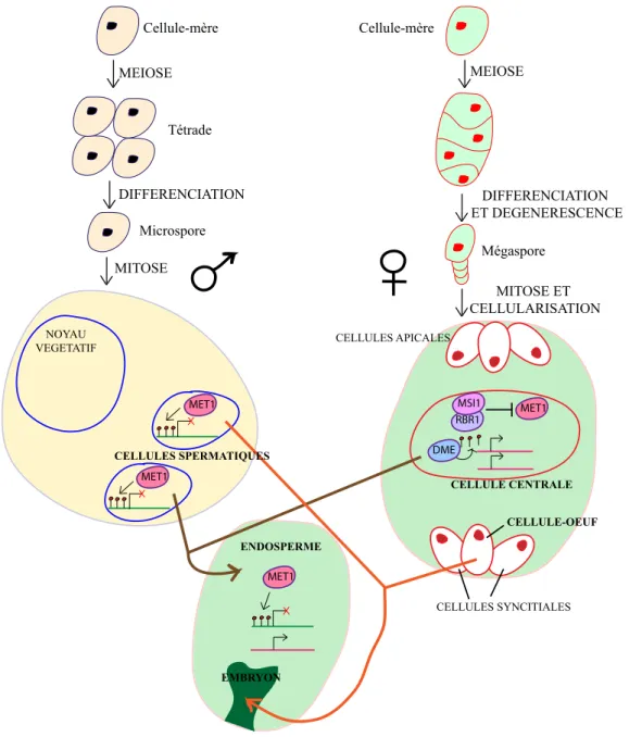
In angiosperms, fertilization involves several nuclei that are contained in both female and male gametophytes. On the female side of the reproductive system, gametogenesis leads to the formation of a haploid egg cell and a homodiploid central cell surrounded by synergid and antipodal haploid cells. The male gametophyte, or pollen grain,
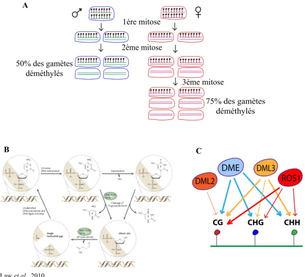
contains three haploid cells: two sperm cells embedded inside the cytoplasm of a larger vegetative cell. Once hydrated on the stigma, this large cell extends a tube that grows through the gynoecium due to attraction by the synergid cells and delivers the two sperm cells inside the female gametophyte. In the double fertilization process specific to angiosperms, one sperm cell fertilizes the haploid egg cell, giving rise to a diploid embryo, while the second sperm cell fertilizes the homodiploid central cell, producing a triploid endosperm that serves as a nurturing tissue for embryo development. Hence, the vegetative cell of the pollen grain and the central cell of the female gametophyte do not contribute to the next generation. Recent reports have shown that drastic changes in DNA methylation and siRNA accumulation take place in these “dead end” cells in Arabidopsis[98–101]. Two different technical approaches have revealed widespread reductions in DNA methylation at TEs and other repeats in the endosperm [99,100]. This decreased DNA methylation is in part due to the activity of DEMETER, a DNA glycosylase expressed in the central cell prior to fertilization that excises methylcytosines [99,100,102–104], and also possibly to reduced expression of MET1 during gametogenesis[105]. Hypomethylation occurs primarily at CG sites and is accompanied by increased methylation at CHH sites in repeated sequences, which are consistent with previous observations from met1 mutants that have lost CG methylation[44,99]. Methylation at CHH sites is a signature of RdDM, and consistent with this idea, Mosher and colleagues have reported a massive burst of 24-nt siRNAs from TEs and repeats. This burst begins in the central cell and continues in the endosperm during seed development[98]. Whether reduced DNA methylation results in the release of TE silencing in the endosperm has not been tested. Slotkin and colleagues, however, have shown transcriptional reactivation and mobilization of some TEs that are associated with reduced DNA methylation and a loss of 24-nt TE siRNAs in the vegetative cell of the pollen grain[101]. Hypomethylation in the vegetative nucleus is likely a consequence of both downregulation of genes required for the maintenance of DNA methylation, such as MET1 and DDM1, and of active demethylation by an unidentified DNA demethylase
[101,105,106]. Slotkin et al. also noticed an accumulation of 21-nt siRNAs from Athila retrotransposons, which may be produced in the vegetative nucleus and accumulate in sperm cells[101]. Similarly, because both 21- and 24-nt siRNAs are mobile in Arabidopsis[107,108], the burst of 24-nt siRNAs produced in the central cell/endosperm may migrate into and accumulate in the egg cell/embryo. This high level of 24-nt siRNAs may provide an additional layer of silencing at TEs in the egg cell/embryo by reinforcing the RdDM pathway. Consistent with this explanation, Hsieh and colleagues found that the levels of CHH methylation at TEs and repeats are elevated in the embryo relative to adult tissues[99]. On the male side, 21-nt siRNAs produced in the vegetative cell may also strengthen TE silencing in sperm cells at the post-transcriptional level to prevent TE mobilization. These 21- and 24-nt siRNAs may play similar roles in backup of TE silencing during fertilization[78].
Epigenetic reprogramming of TE silencing has also been docu-mented in maize, where MuDR silencing is reversed upon the change from juvenile to adult phase, in a tissue adjacent to the one that will produce the germ line[95].
Plants do not establish a germ cell line early during development. Instead, progenitor germ cells differentiate late during sporophyte development. The optimum window for a TE to mobilize and produce new insertions that will be subsequently inherited by the next generation is between the differentiation of gametophyte precursors and the formation of the early embryo. In this regard, accumulating extra layers of TE silencing in these cells would secure genome integrity. But, what could induce TE silencing relaxation? Obviously, as demonstrated experimentally, mutations in epigenetic regulators involved in the maintenance of CG methylation, such as MET1 or DDM1, may induce TE mobilization [25,26,29,39]. However, such mutations would be expected to occur relatively rarely. Perhaps more 455 M. Rigal, O. Mathieu / Biochimica et Biophysica Acta 1809 (2011) 452–458
INTRODUCTION
commonly, changes in environmental conditions or sudden environ-mental stresses could challenge TE silencing. Accordingly, two recent reports have demonstrated that heat stress can overcome TE silencing in Arabidopsis, at least at the transcriptional level [109,110]. Transcription of several classes of TEs and other heterochromatic targets, which are normally silent at ambient temperature, is significantly upregulated upon exposure to a heat stress at 37 °C. This release from silencing occurs across the genome[109]and is mainly transient. Interestingly, at the loci examined, stress-induced destabilization of silencing occurred without altering common repressive epigenetic marks, including DNA methylation and H3K9me2[109,110], indicating that these marks are not sufficient for efficient transcriptional silencing under these conditions. It is currently unknown whether heat stress produces an activating signal that overcomes the presence of these marks or interferes with their readout. Moreover, whether stresses have the same effect on the genomes of gametes, gamete precursors or the early embryo is not known. It would be of interest to determine if the “extra layer” of silencing present in these cell types/developmental stages, which are represented by high levels of specific siRNAs and the possible action of an unconventional siRNA pathway involving AGO9, provides these cells/developmental stages with relatively lower sensitivity to stress. In a recent study, Ito and colleagues showed that an LTR retro-element, named ONSEN, could be mobilized following heat stress-induced transcriptional stimulation[111]. Although stress was applied to young seedlings, transposition of ONSEN was found only in the progeny of stressed plants. Furthermore, new ONSEN insertions were detected only in the progeny of RdDM mutants subjected to stress but not in the progeny of stressed wild-type individuals. Using elegant genetic approaches, the authors demonstrated that in the absence of a functional RdDM pathway, “memory” of the heat-stress persists during plant development, resulting in ONSEN mobilization, which interest-ingly occurred prior to gametophyte development[111].
6. Conclusion
A combination of genomic and genetic studies in plants have revealed that TEs are the main targets of epigenetic silencing pathways and that TEs are controlled by several layers of silencing. Crosstalk among these layers has been observed, and as exemplified by specific bursts of 21- and 24-nt siRNAs and the role of AGO9, certain silencing layers seem to be devoted to specific cell types or particular developmental stages. In this “mille-feuille” of silencing, the precise contribution of each layer to the silencing efficiency for a given TE is highly variable (Fig. 1).
The purpose and evolutionary origin for this specificity remain elusive. Because TEs represent highly dynamic genomic components and because there are potentially deleterious consequences of TE mobilization, evolutionary forces have likely driven the production of
various epigenetic mechanisms to ensure TE silencing. Various factors may govern the specificity of TE silencing, including TE size, copy number, transposition competence, genomic location and local chromatin environment. Accumulating evidence has demonstrated that a variety of environmental stresses can challenge TE silencing and possibly lead to bursts of transposition. Interestingly, distinct stresses affect TEs differentially, suggesting that selectivity and complexity in TE silencing may have evolved as a consequence of stress exposure.
As we increase our knowledge of the molecular details of TE silencing, future studies will need to reveal how environmental factors interfere with these silencing mechanisms and thereby contribute to TE proliferation.
Acknowledgments
We thank Thierry Pélissier and Charles White for comments on the article and the anonymous reviewers for helpful suggestions. We apologize to colleagues whose research we did not have space to discuss. O.M. is supported by a Starting Independent Researcher grant from the European Research Council (260742–I2ST).
References
[1] M.I. Tenaillon, J.D. Hollister, B.S. Gaut, A triptych of the evolution of plant transposable elements, Trends Plant Sci. 15 (2010) 471–478.
[2] S.R. Wessler, Transposable elements and the evolution of eukaryotic genomes, Proc. Natl. Acad. Sci. USA 103 (2006) 17600–17601.
[3] T. Wicker, F. Sabot, A. Hua-Van, J.L. Bennetzen, P. Capy, B. Chalhoub, A. Flavell, P. Leroy, M. Morgante, O. Panaud, E. Paux, P. SanMiguel, A.H. Schulman, A unified classification system for eukaryotic transposable elements, Nat. Rev. Genet. 8 (2007) 973–982.
[4] B. McClintock, Mutable loci in maize, Carnegie Inst. Wash. Year b 47 (1948) 155–169.
[5] D. Lisch, Epigenetic regulation of transposable elements in plants, Annu. Rev. Plant Biol. 60 (2009) 43–66.
[6] R.K. Slotkin, R. Martienssen, Transposable elements and the epigenetic regulation of the genome, Nat. Rev. Genet. 8 (2007) 272–285.
[7] S. Feng, S.J. Cokus, X. Zhang, P.Y. Chen, M. Bostick, M.G. Goll, J. Hetzel, J. Jain, S.H. Strauss, M.E. Halpern, C. Ukomadu, K.C. Sadler, S. Pradhan, M. Pellegrini, S.E. Jacobsen, Conservation and divergence of methylation patterning in plants and animals, Proc. Natl. Acad. Sci. USA 107 (2010) 8689–8694.
[8] A. Zemach, I.E. McDaniel, P. Silva, D. Zilberman, Genome-wide evolutionary analysis of eukaryotic DNA methylation, Science 328 (2010) 916–919. [9] R. Lister, M. Pelizzola, R.H. Dowen, R.D. Hawkins, G. Hon, J. Tonti-Filippini, J.R. Nery,
L. Lee, Z. Ye, Q.M. Ngo, L. Edsall, J. Antosiewicz-Bourget, R. Stewart, V. Ruotti, A.H. Millar, J.A. Thomson, B. Ren, J.R. Ecker, Human DNA methylomes at base resolution show widespread epigenomic differences, Nature 462 (2009) 315–322. [10] M. Bostick, J.K. Kim, P.O. Esteve, A. Clark, S. Pradhan, S.E. Jacobsen, UHRF1 plays a
role in maintaining DNA methylation in mammalian cells, Science 317 (2007) 1760–1764.
[11] S. Liu, Y. Yu, Y. Ruan, D. Meyer, M. Wolff, L. Xu, N. Wang, A. Steinmetz, W.H. Shen, Plant SET- and RING-associated domain proteins in heterochromatinization, Plant J. 52 (2007) 914–926.
[12] H.R. Woo, T.A. Dittmer, E.J. Richards, Three SRA-domain methylcytosine-binding proteins cooperate to maintain global CpG methylation and epigenetic silencing in Arabidopsis, PLoS Genet. 4 (2008) e1000156.
STRESS cell-type specific AGO9 MET1 CMT3 mCG mCHG mCHH RdDM H3K9me2 H3K27me1 DRM2 (CMT3) KYP , SUVH5, SUVH6 ATXR5, ATXR6 Pol IV , Pol V , RDR2, DCL3, AGO4, AGO6 mCG mCHG mCHH H3K9me2 H3K27me1 siRNAs
Fig. 1. Representation of the different layers that constitute the “mille-feuille” of silencing (left), which ensures efficient silencing of TEs (right). Repression of TEs depends on the combined contribution of the different layers of silencing, and the importance of each layer is variable across TEs. In addition, crosstalk exists between some of the epigenetic marks that constitute these silencing layers (arrows on the left). Environmental stresses can overcome known repressive epigenetic marks and lead to reactivation of some TEs. 456 M. Rigal, O. Mathieu / Biochimica et Biophysica Acta 1809 (2011) 452–458
INTRODUCTION