HAL Id: hal-02993285
https://hal.inrae.fr/hal-02993285
Submitted on 6 Nov 2020
HAL is a multi-disciplinary open access archive for the deposit and dissemination of sci-entific research documents, whether they are pub-lished or not. The documents may come from teaching and research institutions in France or abroad, or from public or private research centers.
L’archive ouverte pluridisciplinaire HAL, est destinée au dépôt et à la diffusion de documents scientifiques de niveau recherche, publiés ou non, émanant des établissements d’enseignement et de recherche français ou étrangers, des laboratoires publics ou privés.
mycorrhizal fungus Rhizophagus irregularis
Simon Pons, Sylvie Fournier, Christian Chervin, Guillaume Bécard, Soizic
Rochange, Nicolas Frei Dit Frey, Virginie Puech Pagès
To cite this version:
Simon Pons, Sylvie Fournier, Christian Chervin, Guillaume Bécard, Soizic Rochange, et al.. Phy-tohormone production by the arbuscular mycorrhizal fungus Rhizophagus irregularis. PLoS ONE, Public Library of Science, 2020, 15 (10), pp.e0240886. �10.1371/journal.pone.0240886�. �hal-02993285�
RESEARCH ARTICLE
Phytohormone production by the arbuscular
mycorrhizal fungus Rhizophagus irregularis
Simon Pons1,2, Sylvie Fournier1,2, Christian Chervin3, Guillaume Be´card1,Soizic Rochange1, Nicolas Frei Dit Frey1*, Virginie Puech PagèsID1,2*
1 Laboratoire de Recherche en Sciences Ve´ge´tales, Universite´ de Toulouse, CNRS, UPS,
Castanet-Tolosan, France, 2 MetaboHub-Metatoul AgromiX, Laboratoire de Recherche en Sciences Ve´ge´tales, Universite´ de Toulouse, CNRS, UPS, Castanet-Tolosan, France, 3 Ge´nomique et Biotechnologie des Fruits, Universite´ de Toulouse, Toulouse INP, INRA, Castanet-Tolosan, France
*puech@lrsv.ups-tlse.fr(VPP);frei-dit-frey@lrsv.ups-tlse.fr(NFDF)
Abstract
Arbuscular mycorrhizal symbiosis is a mutualistic interaction between most land plants and fungi of the glomeromycotina subphylum. The initiation, development and regulation of this symbiosis involve numerous signalling events between and within the symbiotic partners. Among other signals, phytohormones are known to play important roles at various stages of the interaction. During presymbiotic steps, plant roots exude strigolactones which stimulate fungal spore germination and hyphal branching, and promote the initiation of symbiosis. At later stages, different plant hormone classes can act as positive or negative regulators of the interaction. Although the fungus is known to reciprocally emit regulatory signals, its potential contribution to the phytohormonal pool has received little attention, and has so far only been addressed by indirect assays. In this study, using mass spectrometry, we ana-lyzed phytohormones released into the medium by germinated spores of the arbuscular mycorrhizal fungus Rhizophagus irregularis. We detected the presence of a cytokinin (iso-pentenyl adenosine) and an auxin (indole-acetic acid). In addition, we identified a gibberellin (gibberellin A4) in spore extracts. We also used gas chromatography to show that R.
irregu-laris produces ethylene from methionine and theα-ketoγ-methylthio butyric acid pathway. These results highlight the possibility for AM fungi to use phytohormones to interact with their host plants, or to regulate their own development.
Introduction
Arbuscular mycorrhizal (AM) symbiosis is a 460 million-year-old interaction [1] between glo-meromycotina fungi and over 70% of land plants [2]. In angiosperms, AM fungi colonize the inner root cortex of their host to develop intracellular ramified structures called arbuscules. These arbuscules are the main site for nutrient exchange between the plant and the fungus. AM fungi provide their host plant with water and minerals, and in return receive carbon sources (mainly sugars and lipids) [3,4]. As AM fungi are obligate biotrophs, this interaction is essential for their growth, development and reproduction. On the plant side, this interaction is most often beneficial as it can improve nutrition and/or resistance to biotic and abiotic stresses [5–7]. a1111111111 a1111111111 a1111111111 a1111111111 a1111111111 OPEN ACCESS
Citation: Pons S, Fournier S, Chervin C, Be´card G,
Rochange S, Frei Dit Frey N, et al. (2020) Phytohormone production by the arbuscular mycorrhizal fungus Rhizophagus irregularis. PLoS ONE 15(10): e0240886.https://doi.org/10.1371/ journal.pone.0240886
Editor: Richard A. Wilson, University of
Nebraska-Lincoln, UNITED STATES
Received: July 29, 2020 Accepted: October 5, 2020 Published: October 16, 2020
Copyright:© 2020 Pons et al. This is an open access article distributed under the terms of the
Creative Commons Attribution License, which permits unrestricted use, distribution, and reproduction in any medium, provided the original author and source are credited.
Data Availability Statement: All relevant data are
within the paper and its Supporting Information files.
Funding: This work was partially financed by the
ANR Mycormones (ANR-18-CE20-0001), MetaboHUB network (ANR-11-INBS-0010), the Tulip LabEx (10-LABX-0041), and the French ministry of Higher Education, Research and Innovation.
Competing interests: The authors have declared
Prior to physical contact, the two partners of the AM symbiosis interact via signalling mole-cules [8,9]. Host roots release several types of compounds affecting the presymbiotic develop-ment of AM fungi, such as some flavonoids, phenolic compounds, hydroxy fatty acids [10,11]. Particular attention has been paid to the root-exuded strigolactone phytohormones: they stim-ulate the germination of AM fungal spores, the oxidative metabolism and branching of germi-nating hyphae, and finally root colonization [12–15]. In addition,N-acetylglucosamine-based
compounds could be exchanged in two directions. Lipochitooligosaccharides (LCOs) and chit-ooligosaccharides (COs) are released by germinating spores of AM fungi and stimulate the ini-tiation of the symbiosis via the activation of the common symbiosis signalling pathway and the activation of lateral root formation [16–18]; and a plant exporter of N-acetylglucosamine has been shown to be required for the first steps of the interaction [19]. Finally, additional, yet unidentified, signals of plant or fungal origin may act prior to root colonization [20–23].
Later stages of AM interactions are regulated by a number of factors, including nutrient exchange [23] and phytohormones [24,25]. Analysis of AM symbiosis regulation by phytohor-mones has revealed a complex pattern of modified hormonal contents or altered response to hormones in mycorrhizal plants. Reciprocal effects of exogenous hormone application on the symbiotic interaction have also been found. Although observations were made across a wide range of combinations of plant/fungal species and experimental conditions, it is possible to draw broad conclusions about the role of the different hormone families. Auxins (AUX), abscisic acid (ABA) and brassinosteroids (BRs) have been identified as positive regulators of the AM symbiosis [26–28]. On the contrary, gibberellins (GAs) and salicylic acid (SA) have been described as negative regulators of the interaction [29–31]. The effects of cytokinins have not yet been clearly established [32]. Finally, the role of ethylene (ET) and jasmonic acid (JA) seems to vary with their concentration [33–36].
Importantly, these studies have addressed the role of phytohormones in the AM symbiosis by two main approaches: the analysis of plant mutants affected in phytohormone synthesis or perception, and the treatment of mycorrhizal plants with exogenous hormones. The study of hormone perception mutants clearly addresses the effects of hormones on the plant. In con-trast, both exogenous treatments and hormone deficiency in the plant result in modified hor-monal contents in colonized roots, which could impact either or both symbionts. In spite of this, and because phytohormones are generally seen as plant signals, results of such studies are commonly interpreted exclusively in terms of impacts on the plant. Likewise, changes in monal contents measured in mycorrhizal plants are usually attributed to modifications of hor-monal metabolism in plant cells. This interpretation ignores a potential contribution of the fungal partner to the hormonal pool. Yet, many microorganisms can produce phytohormones and this could also be the case of AM fungi. Among soil microorganisms interacting with plants, plant growth-promoting rhizobacteria and fungi have been shown to produce auxin, cytokinins, ABA and gibberellins [37,38], and this can contribute to their growth-promoting effects [39]. In the fungal kingdom, phytohormone production has been documented in sym-bionts like ectomycorrhizal fungi [40,41], as well as in pathogens [37]. Ethylene is quite com-mon acom-mong phytohorcom-mones produced by fungi [42–45] and in some cases the biosynthetic pathways have been characterized. Theα-keto γ-methylthio butyric acid (KMBA) pathway, well described inBotrytis cinerea [44], requires the deamination of methionine into KMBA. Subsequently, KMBA can be oxidised into ethylene through different means. It can either be oxidized by hydroxyl radicals [46], by peroxidases [47], or by the photo-oxidation of flavins [48]. In contrast, the second ethylene-producing pathways in microorganisms involves a very specific enzyme. The Ethylene Forming Enzyme (EFE), described inPenicillium digitatum, or Fusarium oxysporum [42,49], produces ethylene through two simultaneous reactions using L-arginine and 2-oxoglutarate as co-substrates. Both pathways differ from the main pathway for
ethylene production in plants which involves a light-independent and methionine-dependent pathway requiring the aminocyclopropane-carboxylate (ACC) synthase (ACS) and ACC oxi-dase (ACO).
The fact that many plant-associated microorganisms produce phytohormones raises the possibility of a similar behaviour in AM fungi which have co-evolved with plants for over 400 million years. This question is challenging to address experimentally, essentially because of the obligate biotrophy of these fungi. This feature implies that isolated fungi can only be kept in culture for short periods of time, and limits the availability of biological material. Nevertheless, previous studies have provided indirect evidence for the presence of phytohormones in AM fungi. ELISA tests indicated that spores and hyphae ofRhizophagus (formerly Glomus) species
could contain aglycone and glycosylated ABA [50], while indirect bioassays suggested the pres-ence of gibberellin-like and cytokinin-like molecules [51]. A direct analysis by GC-MS of spore extracts revealed the presence of small amounts of IAA inGlomus intraradices, but in
this study spores were directly taken from maize pot cultures, hence not in axenic conditions, and may have been contaminated with root fragments or other microorganisms [52].
In this study, we analysed the presence of phytohormones in germinating spores, or in their exudates, of the model AM fungusRhizophagus irregularis grown axenically. We used a
combi-nation of gas/liquid chromatography and mass spectrometry to allow unambiguous com-pound identification. In the case of ethylene, we investigated the putative biosynthetic pathways through the use of metabolic precursors.
Materials & methods
Chemicals, reagents and standards
Phytohormone standards were purchased from Olchemim [isopentenyl adenine (iP), isopen-tenyl adenosine (iPR), isopenisopen-tenyl adenine-9-glucoside (iP9G), kinetine (Ki),meta-topoline
(mT), trans-zeatine (tZ), cis-zeatine (cZ), trans-zeatine riboside (tZR), cis-zeatine riboside
(cZR), dihydrozeatine (DHZ), benzyladenine (BAP), gibberellic acid 1 (GA1), indole-3-acetic
acid aspartate (IAA-Asp), abscisic acid glucose ester (ABA-GE), brassinolide (BL)], Fluka [indole-3-butyric acid (IBA), naphtalenic acetic acid (NAA)], Acros Organics [indole-3-acetic acid (IAA), indole-3-propionic acid (IPA), abscisic acid (ABA)], Sigma-Aldrich [indole-3-ace-tic acid alanine (IAA-Ala), jasmonic acid (JA), strigol], Fisher chemical [gibberellic acid 3 (GA3)] and Duchefa [gibberellic acid 4 (GA4)]. We prepared the standards following the
man-ufacturer’s recommendations, and stored solutions at -20˚C. L-Methionine and α-keto-γ-methylthio butyrate (KMBA) were purchased from Sigma-Aldrich. LC/MS-grade acetonitrile and HPLC-grade methanol were purchased from Fisher Chemical, formic acid from Acros Organics, and ammonium hydroxide from Sigma-Aldrich.
Fungal culture and exudate preparation
Rhizophagus irregularis DAOM 197198 sterile spores were purchased from Agronutrition
(Labège, France). The spores were produced in axenic conditions. Spore numbers were deter-mined by the supplier by counting spores in an aliquot of the sold suspension with a binocular microscope. Spores were rinsed from their storage buffer using a 40μm nylon cell strainer (VWR) by five washes with sterile UHQ water. Spores were resuspended in sterile UHQ water and stored at 4˚C before use.
For the production of germinated spore exudates (GSE), spores were germinated in sterile UHQ water in a CO2incubator (30˚C, 2% CO2) for 7 days with a concentration of 400 spores.
Macherey-Nagel, France), then frozen in liquid nitrogen and freeze-dried. Filtered spores were collected and stored at -80˚C.
Phytohormone and KMBA extraction
From germinated spore exudates. Freeze-dried GSE from 10,000 spores or 250,000
spores ofR. irregularis were reconstituted in 100 μL of 1 M formic acid and stored at -20˚C
before MS analysis.
From ground spores. The protocol of phytohormone extraction and separation by Solid
Phase Extraction (SPE) was adapted from Kojimaet al. [53] as follows. Two hundred and fifty thousand spores were hand-ground in liquid nitrogen with a mortar and pestle, resuspended in 1 mL of cold modified Bieleski’s solvent (methanol/water/formic acid 75:20:5, v:v:v; [54]) and left overnight at -20˚C to achieve complete extraction. The crude extracts were centrifuged for 15 min at 10,000 xg, at 4˚C. The pellet was reextracted in 200 μL modified Bieleski’s solvent
for 30 min at -20˚C, centrifuged 15 min at 10,000 xg, and the supernatant pooled with the first
one. Extracts were pre-purified on SPE Oasis HBL cartridges (1 mL per 30 mg, Waters). Car-tridges were conditioned in 1 mL methanol and equilibrated in 1 mL of 1 M formic acid. Two-mL samples were loaded and eluted with 1 Two-mL modified Bieleski’s solvent. Eluates containing phytohormones were dried under nitrogen stream and reconstituted in 1 mL of 1 M formic acid. The separation of phytohormones was achieved with SPE Oasis MCX cartridges (1 mL per 30 mg, Waters). Cartridges were conditioned in 1 mL methanol and equilibrated in 1 mL of 1 M formic acid. Samples were successively eluted by 1 mL methanol (fraction 1, containing neutral and acidic hormones), 1 mL of 0.35 M ammonium hydroxide (fraction 2) and 1 mL methanol/0.35 M ammonium hydroxide 60:40 (fraction 3, containing cytokinins) [53]. The three fractions were dried under nitrogen stream and kept at 4˚C before analysis. Fractions were reconstituted in 100μL 1 M formic acid and then analysed by LC-MS.
LC-MS analysis of phytohormones
Samples (GSEs or fractions obtained from ground spores as previously described) were ana-lysed by UHPLC-MS with two types of mass spectrometers, a Q-Trap 5500 (AB Sciex) in MRM mode for higher detection sensitivity, and a Q-Exactive Plus™ (Thermo Scientific) for higher mass accuracy by high resolution analysis, each of them having their own advantages. The Q-trap 5500 mass spectrometer allows to perform analysis in multiple reaction monitor-ing (MRM) mode, which improves selectivity and detection sensitivity for targeted metabolites [55], whereas the Q-Exactive plus is equipped with an orbitrap analyzer, allowing to measure them/z ratio of each ion with greater mass accuracy for structural confirmation [56].
Twenty-one biological replicates of GSE were used to perform phytohormone detection. IPR was consistently identified in GSE, but probably due to biological variability, IAA was detected in 76% of them. Thirteen samples were used to perform quantification. Twelve repli-cates of ground spores were used for detection of phytohormones. GA4was detected in 66% of
them.
Multiple reaction monitoring (MRM) analysis. A UHPLC system (Dionex Ultimate
3000, Thermo Scientific) was equipped with a Kinetex C18 column (100× 2.1 mm, 2.6 μm, 100Å, Phenomenex) heated at 45˚C. Five-μL samples were injected. Separation was performed at a constant flow rate of 0.3 mL.min-1, in a gradient of solvent A (water + 0.1% formic acid) and B (acetonitrile + 0.1% formic acid): 1 min 5% B; 11 min 5% to 100% B; 2 min 100% B, and re-equilibration to the initial conditions in 4 min. The Q-Trap 5500 mass spectrometer was equipped with an electrospray source. Curtain gas was set to 30 psi, nebulizer to 40 psi and turbo gas to 60 psi. Capillary voltage was set to 5.5 kV (positive mode) or -4.5 kV (negative
mode) on Electrospray Ionization (ESI) source (400˚C). Samples were monitored in positive and negative modes in scheduled Multiple Reaction Monitoring (MRM) mode (60s). Using 25 standards of free or conjugated hormones, in infusion mode (7μL.min-1), the best parameters for declustering potential, collision energy and collision cell exit potential were selected for precursor and product ions measurement. Ionization mode, selected MRM transitions, limit of detection (LOD) and retention time for each hormone are listed inS1 Table. Limits of detection (signal to noise ratio > 3) were determined using standards diluted from 0.1 mM to 1pM in methanol. By this approach, we could perform an approximate quantification of phy-tohormones in fungal samples. Data processing was performed using Analyst 1.6.2 software.
High resolution mass spectrometry (HRMS) analysis. A UHPLC system (Ultimate 3000
RSLC system, Thermo Scientific) was equipped with a Hypersil Gold aQ C18 column (100 mm x 2.1 mm, 1.9μm, 175 Å, Thermo Scientific #25302102130), heated at 35˚C. Five-μL sam-ples were injected. Separation was performed at a constant flow rate of 0.3 mL.min-1, in a gra-dient of solvent A (water + 0.05% formic acid) and B (acetonitrile + 0.05% formic acid): 1 min 5% B; 7 min 5% to 96% B; 1 min 96% B and re-equilibration to the initial conditions in 3 min. The Q-Exactive Plus™ mass spectrometer was equipped with a H-ESI II probe, heated at 256˚C. Sheath gas was set to 48, sweep gas to 2, auxiliary gas to 11, and heated at 413˚C. Capil-lary voltage was set to 3.5 kV in positive mode and -2.5 kV in negative mode. Ionization was performed in positive and negative modes, in full scan analysis (centroid), with a resolution of 35,000. Automatic Gain Control was set to 3.106, with a 50 to 600m/z scan range. A
Target-MS/MS scan of confirmation of the phytohormone, based on the specified inclusion list (5 ppm), was triggered when the mass spectrometer detected a known phytohormone in an MS spectrum. In this case, Automatic Gain Control was set to 2.105, and resolution to 17,500. Data processing was performed by Xcalibur 3.0 and Tracefinder 3.2 softwares.
Ethylene detection
20,000 spores in 2 mL of sterile UHQ water, either untreated, or supplemented with 10 mM of methionine or 1μM of KMBA, were incubated in a sterilized glass tube (Ø = 1.35 cm, H = 6.10 cm, V = 7.5 mL, dead volume = 5.5 mL) wrapped in tinfoil to avoid light exposure, and sealed with a porous silicone stopper (Hirschmann Instruments). They were germinated for three days in a CO2incubator (30˚C, 2% CO2). The stopper was then replaced by an air-tight rubber
stopper, and spores were confined for 1 day and exposed or not to light (100μmol photons m−2s−1, 21˚C).
The headspace ethylene content was assayed by gas chromatography as described previ-ously [57]. One mL of headspace gas was manually injected into a GC-FID (Agilent 7820a), equipped with a 80/100 alumina column (1/8” x 2 mm x 1.5 m, Agilent) and set with the fol-lowing parameters: oven temperature 70˚C, injector temperature 110˚C, N2vector gas flow
rate 28 mL.min-1, flame ionization detector temperature 250˚C. Ethylene peak area was mea-sured and normalized with the O2injection peak area. Its retention time and calibration were
determined with an external standard of 4 ppm of ethylene.
For ethylene production assays: darkness without spores n = 13, light without spores n = 16, darkness with spores n = 17, light with spores n = 18, light and methionine without spores n = 7, darkness and methionine with spores n = 9, light and methionine with spores n = 24, light and KMBA without spores n = 6, light and KMBA with spores n = 6.
KMBA detection by LC-MS
A UHPLC system (Dionex Ultimate 3000, Thermo Scientific) was equipped with a reverse-phase column Acquity UPLC BEH-C18 (2.1× 150 mm, 1.7 μm, Waters) heated at 45˚C.
Ten-μL samples were injected. Separation was performed at a constant flow rate of 0.3 mL.min-1, in a gradient of solvent A (water + 0.1% formic acid) and B (acetonitrile): 1 min 5% B; 8 min 5% to 100% B; 2 min 100% B, and re-equilibration to the initial conditions in 2 min.
A Q-Trap 4500 mass spectrometer (AB Sciex) was used with an electro-spray ionization source in the negative ion mode. Curtain gas was set to 30 psi, nebulizer to 40 psi and turbo gas to 60 psi. Capillary voltage was set to -3.5 kV (negative mode) on Electrospray Ionization (ESI) source (400˚C). Optimizations of the source parameters were done using the KMBA standard at 10−5M water by infusion at 7μL.min-1
, using a syringe pump. Three GSE sample were analysed for each condition. Data processing was performed using Analyst 1.6.2 software.
Sequence analysis
Glomeromycotina (taxid:214504) nucleotide and protein sequences were analysed using TBLASTN and BLASTP searches with default parameters on the NCBI website. The 2-oxoglu-tarate-dependent ethylene/succinate-forming enzyme fromPenicillium digitatum
XP_014538251.1 was chosen as query to identify putative EFEs inR. irregularis.
Statistical analysis
The version 4.0.0 of R [58], with the version 1.3–1 of package Agricolae [59] and the version 3.1.0 of package GGPlot2 [60] were used for statistical analysis. In order to compare ethylene production between all groups, a non-parametric analysis was carried out using Kruskal-Wal-lis test and pairwise comparisons were performed using FDR adjustment for multiple comparisons.
Results
Detection of phytohormones in germinated spore exudates and germinated
spore extracts
The aim of this study was to investigate the production of phytohormones by the model AM fungusR. irregularis. To avoid any contamination with plant-borne phytohormones, it was
crucial to start from pure fungal material. We used spores ofR. irregularis produced in axenic
conditions, and the spores, free of root debris, were carefully rinsed to eliminate the storage solution.
We started with an analysis of exudates produced byR. irregularis spores germinated in
water for seven days. At this stage, although not stimulated by root exudates, the germinating hyphae continue to grow a little and the spores resemble in all respects (shape, density, pres-ence of lipid droplets) to what they were on the first day. These germinated spore exudates (GSE) were concentrated and analysed by Liquid Chromatography (LC) coupled to Mass Spectrometry (MS). The detection of a total of 38 compounds, covering eight hormone fami-lies (S1 Table), was attempted. Synthetic standards were available for 25 of these compounds, allowing direct comparison of retention times and MS data. Two types of MS analyses were successively carried out. The highly sensitive Multiple Reaction Monitoring (MRM) mode was used to look for characteristic precursor-to-product-ionm/z transitions upon fragmentation.
The retention times associated with thesem/z transition signals were compared with the
reten-tion times of corresponding standards. To further ascertain hormone identificareten-tion, High-Res-olution Mass Spectrometry (HRMS) was then used to extract signals for precursor and product ions of the expected accuratem/z (+/- 5 ppm), and again was performed in
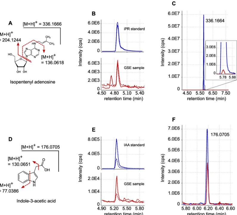
Using these two approaches, we identified two hormones in GSE samples produced by 10,000 spores: the cytokinin isopentenyl adenosine (iPR) and the auxin indole-acetic acid (IAA) (Fig 1A and 1D). In the MRM mode, iPR was detected with them/z transitions
Fig 1. Detection by LC-MS of iPR and IAA exuded byR. irregularis spores. (A) Structure and fragmentation pattern of iPR. (B) UPLC-MRM-MS chromatograms of iPR in positive mode. Blue lines represent the signals obtained for iPR external standard (100 nM). Red lines represent the signals obtained with GSE produced by 10,000R. irregularis spores. Plain lines are for m/z transition 336 > 204. Dashed lines are for m/z transition 336 > 136. (C) LC-HRMS extracted ion chromatogram
(XIC) form/z = 336.1666 (+/-5ppm). The blue line represents the signal obtained for iPR external standard (300 nM). The red line represents the signal obtained with
GSE produced by 250,000R. irregularis spores. (D) Structure and fragmentation pattern of IAA. (E) UPLC-MRM-MS chromatograms of IAA in positive mode. Blue
lines represent the signals obtained for IAA external standard (100 nM). Red lines represent the signals obtained with GSE produced by 10,000R. irregularis spores.
Plain lines are form/z transition 176 > 131. Dashed lines are for m/z transition 176 > 77. (F) LC-HRMS XIC for m/ z = 176.0705 (+/-5ppm). The blue line represents
the signal obtained for IAA external standard (300 nM). The red line represents the signal obtained with GSE produced by 250,000R. irregularis spores. Signal
intensities are displayed in counts per second (cps).
336 > 204 and 336 > 136; IAA was identified with them/z transitions 176 > 130 and 176 > 77
(Fig 1B and 1E). For both compounds, the observed retention times matched those of the cor-responding standards (Fig 1B and 1E). We were able to detect accurately iPR and IAA in 16 out of 21 biological replicates. The other phytohormones presented inS1 Tablewere not detected in the GSE samples. According to external standard curves, we estimated that one spore could on average exude 1.2 x 10⁻¹⁸mol of iPR (+/- 1.3 x 10⁻¹⁸mol) and 29 x 10−18mol of IAA (+/- 25 x 10−18mol), during seven days of germination. Compound identification was confirmed through HRMS using GSE from 250,000 spores. The precursor ion ofm/z 336.1662
for iPR, detected at a retention time of 5.78 min (Fig 1C), yielded after selection and fragmen-tation a product ion ofm/z 204.1246 (S1 Fig). The precursor ion ofm/z 176.0706 for IAA,
detected at a retention time of 6.18 min (Fig 1F), yielded after selection and fragmentation a product ion ofm/z 130.0650 (S2 Fig). The mass data, as well as retention times, matched those of the corresponding standards (Fig 1C and 1F).
To investigate whether additional hormones could be present inR. irregularis spores, but
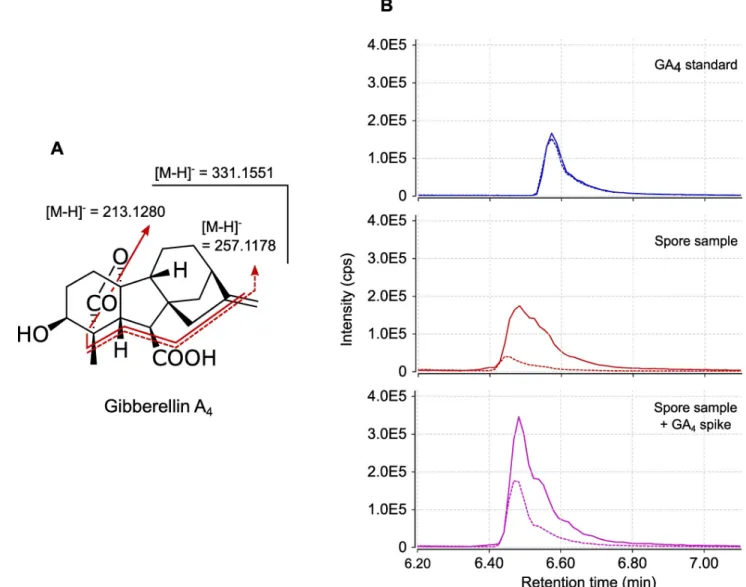
not released (or in very low amounts) into GSE, we next analysed extracts of 250,000 ground spores. Extracts were pre-purified through two solid-phase extraction (SPE) steps and the frac-tions where hormones were expected (in the first fraction for acidic and neutral hormones such as auxins and gibberellic acids, and in the third fraction, for basic hormones such as cyto-kinins) were analysed by LC-MS/MS. In these samples, IAA and iPR were detected, but as trace amounts, perhaps due to the complex nature of the matrix. However, this approach allowed the detection in MRM mode of MS-MS transition signals characteristic of a third phy-tohormone, gibberellic acid 4 (GA4). Transitionsm/z 331 > 257 and 331 > 213 (Fig 2A) were
detected in our samples, at almost the same retention time as the standard (retention time shift = -0.09 min,Fig 2B). To investigate whether this slight shift was due to matrix interactions [61], we spiked our sample with the GA4standard. This addition yielded a single
chro-matographic peak without any splitting, at the same retention time as the spore sample alone and with a doubled intensity. We can therefore attribute the slight retention time difference in
Fig 2Bto matrix interactions during chromatographic separation.
Production of ethylene by
R. irregularis germinated spores
The release of ethylene by germinating spores was analysed by gas chromatography. To this end, spores were first allowed to germinate for three days in water, in test tubes closed with a gas-permeable stopper. The stopper was then replaced by a gas-tight rubber stopper, and spores were incubated for an additional 24 hours. Gas in the headspace was then sampled for ethylene analysis. We used light dependency as a criterion to distinguish between ethylene bio-synthesis pathways. Indeed, light exposure allows to reveal KMBA-dependent ethylene pro-duction [44,45]. Therefore, ethylene production was assessed comparatively in spores protected or not from light.
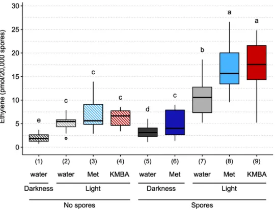
We noticed in control tubes containing no spores that a background quantity of ethylene was produced in the dark as well as in the light (Fig 3, conditions 1 and 2) [62]. The presence of spores slightly enhanced ethylene production in the dark (Fig 3, conditions 1 and 5). When exposed to light, germinating spores produced about 3 times more ethylene than in the dark, suggesting that this production was dependent on the KMBA pathway (Fig 3, conditions 5 and 7) [44,46,48].
To investigate whether the KMBA pathway for ethylene synthesis was used byR. irregularis,
we tested the effects of adding methionine into the incubation medium. In the dark, methio-nine addition increased ethylene production by 55% (Fig 3, conditions 5 and 6). While in the light, methionine addition did not increase ethylene production in the absence of spores (Fig
3, conditions 2 and 3), this addition in the presence of spores increased ethylene production by 56% (Fig 3, conditions 7 and 8). We then tested the effects of added KMBA. In the light and in the absence of spores, KMBA did not yield ethylene (Fig 3, conditions 2 and 4). In contrast, in the presence of spores, KMBA addition stimulated an ethylene production (Fig 3, conditions 7 and 9) similar to the methionine treatment.
Finally, we analysed by LC/MS the presence of KMBA in the exudates of spores germinated in the presence or not of methionine. This experiment was carried out in the dark to avoid light oxidation of KMBA. We could not detect the presence of KMBA in samples without methionine whereas we detected a strong KMBA signal in the methionine-treated samples, at a retention time of 3.7 min in MRM mode (m/z transitions 147 > 46 and 147 > 99) (S3 Fig).
Genomic sequences of Glomeromycotina were analysed for the presence of genes associ-ated with the different ethylene biosynthesis pathways. The KMBA pathway requires the
Fig 2. GA4detection in multiple reactions monitoring (MRM) mode. (A) Structure and fragmentation pattern of GA4. (B) UPLC-MRM-MS
chromatogram of GA4in negative mode. Top (blue lines): External standard (30 nM) of GA4. Middle (red lines): pre-purified SPE fraction from 250,000
ground spores ofR. irregularis. Bottom (purple lines): pre-purified SPE fraction from 250,000 ground spores of R. irregularis spiked with GA4standard
to a final concentration of 30 nM. Plain lines are form/z transition 331 > 213. Dashed lines are for m/z transition 331 > 257. Signal intensities are
displayed in counts per second (cps).
deamination of methionine into KMBA, which can be mediated by any transaminase, and the oxidation of KMBA into ethylene can be carried out non-specifically by peroxidases, or chemi-cally [46,63,64]. Therefore, sequence analyses are not suitable to investigate the existence of this pathway. To look for genes associated with the major ACC pathway used by plants, AtACS1, AtACS8 and AtACS7 were chosen as representative of the three ACS clades described in Arabidopsis [65]. In the three cases, a tyrosine aminotransferase (GLOIN_2v1675208) and a pyridoxal phosphate-dependent transferase (GLOIN_2v1486204) were found as the best hits inR. irregularis genome, with only a maximum of 24% identity at the amino acid level with the
query sequence. We also used AtACO2, AtACO1 and AtACO5 as representative of the three ACC oxidases (ACO) clades [66]. With these three queries, only hypothetical proteins with less than 26% of identity were found inR. irregularis datasets. To look for genes of the EFE
pathway, BLAST analysis using thePenicillium digitatum EFE protein as query allowed the
identification of two isoforms of a protein annotated as Fe (2+) and 2-oxoglutarate (2OG)-dependent dioxygenases inR. irregularis (GBC41587.1 and GBC41586.1). However, the
simi-larity with theP. digitatum protein was very low (23–24% of identity).
Discussion
Germinated spore exudates of AM fungi are known to trigger a number of symbiotically rele-vant responses in host root cells (e.g.: activation of nuclear calcium spiking in the root epider-mis, transcriptional reprogramming in root cells [67–69]). This indicates that these spore exudates contain molecular signals released by isolated AM fungal spores within a few days of
Fig 3. Ethylene production byR. irregularis in response to different treatments. 20,000 spores were germinated for three days in the dark, in the presence or absence of 10 mM methionine (Met) or 1μM α-keto γ-methylthio butyric acid (KMBA). Tubes were then sealed with a gas-tight stopper and exposed to light or darkness for 24h. One mL of the headspace gas was then analysed by gas chromatography. Different letters indicate different statistical groups (pairwise Kruskal-Wallis test with FDR correction,P < 0.05).
incubation. GSE form a matrix of relatively low complexity, which favours sensitive compound detection through mass spectrometry. We thus started our study by analysing GSE for the presence of a wide variety of phytohormones. The presence of iPR and IAA was unambigu-ously demonstrated by a combination of MRM and HRMS analyses, in GSE samples obtained from relatively small amounts of fungal material (10,000 spores). The estimated concentrations are in the same range as those already observed with ectomycorhizal fungal species or in plants [70,71]. Although the presence of cytokinin-like compounds and of IAA was already sus-pected or hypothesized in AM fungi [51,52,72], the present study is to our knowledge the first conclusive report for the release of these two phytohormones by an AM fungus. It is of course possible that other phytohormones are present in low amounts in GSE, and have escaped detection despite the high sensitivity of the MRM approach (seeS1 Tablefor detection limits).
Analysis of spore extracts was undertaken as a complementary approach to look for hor-mones that would not be released into GSE. This allowed the detection of MRM signals corre-sponding to GA4, at a retention time very close to that of the standard. Despite
pre-purification, the fractions of spore extracts remain a complex matrix, which can induce slight shifts in chromatographic retention times. Using a GA4standard to spike our sample allowed
us to observe that the retention times recorded in the spore sample for both MRM transitions were identical to those of the standard in this matrix (Fig 2). Unfortunately, complex matrices decrease the sensitivity of HRMS analysis. We were thus unable to confirm the identity of this compound through accuratem/z determination. Our preliminary observations are however
consistent with the previous bioassay-based detection of gibberellin-like compounds in AM fungi [51]. Given that gibberellins can be synthesised (and were actually discovered) in other fungi [73], it would make evolutionary sense to find them also in AM fungi.
In contrast with Eschet al. [50], we did not detect the presence of ABA or glycosylated ABA. This difference might be due to the use of different fungal materials. The study of Esch
et al. was carried out on an unspecified species of the genus Rhizophagus (formerly Glomus),
and perhaps more importantly, the analysed material consisted of extraradical mycelium and spores obtained non axenically after several weeks of culture in pots. Furthermore, in this study, ABA detection was based on indirect ELISA tests, which likely differ from mass spec-trometry in terms of sensitivity and specificity.
The detection of an auxin, a cytokinin and a gibberellin inR. irregularis does not mean that
this fungus is actually able to synthesize these molecules. They could have been produced by the host plant (here the hairy roots ofDaucus carota), imported by the fungus during the
sym-biotic exchanges between the two partners and stored in spores. Isotopic labelling experiments could be used to investigate the biosynthetic origin of these hormones, and these approaches could benefit from a very recent work revealing that myristate can be used as an exogenous carbon source and supportin vitro growth of R. irregularis [74].
Unlike the above hormones, there is no doubt about the fungal biosynthetic origin of ethyl-ene.De novo ethylene production was measured over a period of 24 h and could be stimulated
by the addition of methionine, a metabolic precursor. The addition of methionine strongly enhanced the synthesis of KMBA by the fungus (S3 Fig) and acted synergistically with expo-sure to light to promote ethylene production (Fig 2). Light- and methionine-dependency are characteristic features of the KMBA pathway described in other fungi [44]. Hence, although we cannot rule out the existence of additional ethylene biosynthetic pathways, our biochemical data support the KMBA pathway as being involved in the synthesis of ethylene inR. irregularis.
Interestingly, KMBA-derived ethylene was also demonstrated in the ectomycorrhizal fungi
Tuber brochii and T. melanosporum [45]. To further investigate the existence of alternative pathways for ethylene synthesis inR. irregularis, we analysed Glomeromycotina genomic
in the classical ACC pathway found in plants. Similarly, the best hits obtained when searching for a fungal EFE exhibited limited identity with the query sequence. The fact that enzymes in the EFE family can be involved in the biosynthesis of a multitude of products [75] sheds fur-ther doubt on the role of theirRhizophagus homologs in ethylene synthesis. Further studies
would be necessary to formally exclude the existence of these two biosynthetic pathways inR. irregularis, but in view of these initial investigations, their existence seems unlikely.
The observation that iPR, IAA and ethylene are released by germinating spores into their environment is consistent with a signalling role in the AM symbiosis. A similar hypothesis was proposed by Le Marqueret al. [76] with CLAVATA3/Embryo surrounding region-related (CLE) peptides, another type of plant hormone potentially produced and excreted by AM fungi. First, if these hormones are still released at late stages, they could contribute directly to changes in hormonal contents in mycorrhizal plants. For example, AM colonization has been shown to increase auxin concentration in roots ofM. truncatula, Zea mays and Glycine max
[52,77,78]. In tomato roots, the expression of the auxin-dependent reporter DR5-GUS was higher in arbuscule-containing cells than in the surrounding cells [26]. This higher auxin con-centration could be partly due to the AM fungus exudation and exportation to root tissues. Second, the release of these hormones by AM fungi could have profound effects on the symbi-osis itself, such as the positive effects observed upon auxin treatment, which improves mycor-rhization frequency and arbuscule abundance [26,79]. These hormones could also act through a modulation of plant development. For example, the simultaneous exudation of IAA and Myc-LCOs by the fungus could have synergistic effect on lateral root formation, as shown by Buendiaet al. [80] with exogenous treatments onBrachypodium distachyon. The possible
effects of ethylene released by the fungus are more difficult to predict. In arbuscular mycor-rhiza, ethylene is mostly described as a negative regulator [30,67,81,82]. This conclusion was mainly drawn from studies using plant mutants disturbed in the production or perception of ethylene. As already proposed, the negative downstream effect of these mutations on AM sym-biosis may also result from crosstalks with additional phytohormones and not directly from modifications in ethylene signallingper se [35]. Whether through direct or indirect mecha-nisms, the production of ethylene by AM fungi could serve to prevent excessive colonization of the root system. It is also important to note that ethylene inhibition of AM symbiosis was shown to be concentration-dependent [35] and that, in some cases, a low ethylene concentra-tion was able to stimulate root colonizaconcentra-tion [36].
In addition to hormonal signalling to the plant, it is also possible that AM fungi use phyto-hormones to regulate their own development. In support of this hypothesis, candidate genes putatively encoding ethylene and cytokinin receptors were recently identified in the genome ofR. irregularis [83] and await functional characterization. This study was focused on histidine kinases, and thus does not exclude the existence of other types of receptors for other phytohor-mones in AM fungi. Indeed, a variety of horphytohor-mones can affect AM fungal developmentin vitro
[12–14,36,84,85]. It can also be noted that phytohormone exudation by plant roots is not restricted to strigolactones, and has also been reported for auxin, abscisic acid, jasmonate, salicylate and a cytokinin [86–89]. Bringing together these observations, it is tempting to spec-ulate on the exchange of several hormonal signals in both directions during AM symbiosis, in addition to the well-known effects of phytohormones as internal regulators of plant physiol-ogy. The use of a common language has also been reported in other contexts of host-microbe interactions. For example, plant bacterial pathogens produce cytokinin and have evolved a cor-responding receptor [90], and gut bacteria produce and possess sensors for neuroendocrine hormones that were once thought to be specific of their host [91,92]. In this line, it would be tempting to reconsider the historical name “phytohormones” when these molecules trigger bidirectional cross-kingdom activities. Plants have lived with AM symbionts since they
colonized land, and the molecular language underlying this long-standing and intimate rela-tionship is only beginning to be unravelled. Deciphering the hormonal biosynthesis and per-ception pathways in AM fungi will certainly help to understand how this common language developed through evolution.
Supporting information
S1 Table. List of molecules analyzed by LC-MS in highly sensitive MRM mode. The
hor-mone family, molecule name, abbreviation, formula, retention time, preferential detection mode, precursor and product ions for MRM analysis and limit of detection (determined using direct infusion of standards diluted from 0.1 mM to 1 pM in methanol) are indicated. Grey lines correspond to theoretical fragmentation values when standards were not available [71,
93]. (XLSX)
S1 Fig. High resolution mass spectra (HRMS) of iPR in positive mode. (A) and (B) HRMS
spectra of iPR standard (300 nM), at 5.78 min. (C) and (D) HRMS spectra of 250,000R. irregu-laris GSE at 5.78 min. (A) and (C), MS spectra. (B) and (D) MS/MS (m/z 336.1666 +/- 0.5 Da)
spectra. (EPS)
S2 Fig. High resolution mass spectra (HRMS) of IAA in positive mode. (A) and (B) HRMS
spectra of IAA standard (300 nM) at 6.19 min. (C) and (D) HRMS spectra of 250,000R. irregu-laris GSE, at 6.19 min. (A) and (C) MS spectra. (B) and (D) MS/MS (m/z 176.0705 +/- 0.5Da)
spectra. (EPS)
S3 Fig. KMBA detection in multiple reaction monitoring (MRM) mode. (A) Structure and
fragmentation pattern of KBMA. (B) UPLC-MRM-MS chromatogram of KMBA in negative mode. Top (blue lines): External standard (500 nM) of KMBA. Middle (red lines): GSE pro-duced by 20,000 spores treated with 10 mM methionine. Bottom (green lines): GSE propro-duced by 20,000 untreated spores. Plain lines are form/z transition 147 > 47. Dashed lines are for m/ z transition 147 > 99. Signal intensity is displayed in counts per second (cps).
(EPS)
Acknowledgments
We thank Cyril Libourel, Marielle Aguilar and Helene San Clemente for their help with statis-tical analyses. We thank Maryne Laigle, Virginie Durand and Thibaut Perez for their help with mass spectrometry analysis.
Support for mass spectrometry analyses was provided by the ICT-Mass spectrometry and MetaboHUB-MetaToul- AgromiX Facilities.
Author Contributions
Conceptualization: Simon Pons, Sylvie Fournier, Christian Chervin, Guillaume Be´card, Soizic
Rochange, Nicolas Frei Dit Frey, Virginie Puech Pagès.
Data curation: Simon Pons. Formal analysis: Simon Pons.
Investigation: Simon Pons, Sylvie Fournier, Christian Chervin, Guillaume Be´card, Nicolas
Frei Dit Frey, Virginie Puech Pagès.
Methodology: Simon Pons.
Supervision: Christian Chervin, Guillaume Be´card, Nicolas Frei Dit Frey, Virginie Puech
Pagès.
Writing – original draft: Simon Pons, Christian Chervin, Guillaume Be´card, Nicolas Frei Dit
Frey, Virginie Puech Pagès.
Writing – review & editing: Simon Pons, Christian Chervin, Guillaume Be´card, Soizic
Rochange, Nicolas Frei Dit Frey, Virginie Puech Pagès.
References
1. Redecker D. Glomalean Fungi from the Ordovician. Science. 2000; 289: 1920–1921.https://doi.org/10. 1126/science.289.5486.1920PMID:10988069
2. Brundrett MC, Tedersoo L. Evolutionary history of mycorrhizal symbioses and global host plant diver-sity. New Phytol. 2018; 220: 1108–1115.https://doi.org/10.1111/nph.14976PMID:29355963
3. Smith S, Read D. Mycorrhizal Symbiosis. Mycorrhizal Symbiosis. Academic Press; 2008.https://doi. org/10.1016/B978-0-12-370526-6.X5001-6
4. Keymer A, Gutjahr C. Cross-kingdom lipid transfer in arbuscular mycorrhiza symbiosis and beyond. Curr Opin Plant Biol. 2018; 44: 137–144.https://doi.org/10.1016/j.pbi.2018.04.005PMID:29729528
5. Smith SE, Smith FA, Jakobsen I. Mycorrhizal fungi can dominate phosphate supply to plants irrespec-tive of growth responses. Plant Physiol. 2003; 133: 16–20.https://doi.org/10.1104/pp.103.024380
PMID:12970469
6. Pozo MJ, Azco´ n-Aguilar C. Unraveling mycorrhiza-induced resistance. Current Opinion in Plant Biol-ogy. 2007. pp. 393–398.https://doi.org/10.1016/j.pbi.2007.05.004PMID:17658291
7. Mohammadi K, Shiva Khalesro, Sohrabi Y, Heidari G. A Review: Beneficial Effects of the Mycorrhizal Fungi for Plant Growth. J Appl Environ Biol Sci. 2011; 1: 310–319.
8. Choi J, Summers W, Paszkowski U. Mechanisms Underlying Establishment of Arbuscular Mycorrhizal Symbioses. Annu Rev Phytopathol. 2018; 56: 135–160. https://doi.org/10.1146/annurev-phyto-080516-035521PMID:29856935
9. Maclean AM, Bravo A, Harrison MJ. Plant signaling and metabolic pathways enabling arbuscular mycorrhizal symbiosis. Plant Cell American Society of Plant Biologists; 2017 pp. 2319–2335.https:// doi.org/10.1105/tpc.17.00555PMID:28855333
10. Be´card G, Kosuta S, Tamasloukht M, Se´jalon-Delmas N, Roux C. Partner communication in the arbus-cular mycorrhizal interaction. Canadian Journal of Botany. 2004. pp. 1186–1197.https://doi.org/10. 1139/B04-087
11. Nagahashi G, Douds DD. The effects of hydroxy fatty acids on the hyphal branching of germinated spores of AM fungi. Fungal Biol. 2011; 115: 351–358.https://doi.org/10.1016/j.funbio.2011.01.006
PMID:21530917
12. Akiyama K, Matsuzaki K, Hayashi H. Plant sesquiterpenes induce hyphal branching in arbuscular mycorrhizal fungi. Nature. 2005; 435: 824–827.https://doi.org/10.1038/nature03608PMID:15944706
13. Besserer A, Be´card G, Jauneau A, Roux C, Se´jalon-Delmas N. GR24, a Synthetic Analog of Strigolac-tones, Stimulates the Mitosis and Growth of the Arbuscular Mycorrhizal Fungus Gigaspora rosea by Boosting Its Energy Metabolism. Plant Physiol. 2008; 148: 402–413.https://doi.org/10.1104/pp.108. 121400PMID:18614712
14. Besserer A, Puech-Pagès V, Kiefer P, Gomez-Roldan V, Jauneau A, Roy S, et al. Strigolactones Stimu-late Arbuscular Mycorrhizal Fungi by Activating Mitochondria. PLoS Biol. 2006; 4: e226.https://doi.org/ 10.1371/journal.pbio.0040226PMID:16787107
15. Gomez-Roldan V, Fermas S, Brewer PB, Puech-Pagès V, Dun EA, Pillot J-P, et al. Strigolactone inhibi-tion of shoot branching. Nature. 2008; 455: 189–194.https://doi.org/10.1038/nature07271PMID:
18690209
16. Maillet F, Poinsot V, Andre´ O, Puech-Page´s V, Haouy A, Gueunier M, et al. Fungal lipochitooligosac-charide symbiotic signals in arbuscular mycorrhiza. Nature. 2011; 469: 58–64.https://doi.org/10.1038/ nature09622PMID:21209659
17. Genre A, Chabaud M, Balzergue C, Puech-Pagès V, Novero M, Rey T, et al. Short-chain chitin oligo-mers from arbuscular mycorrhizal fungi trigger nuclear Ca2+ spiking in Medicago truncatula roots and their production is enhanced by strigolactone. New Phytol. 2013; 198: 190–202.https://doi.org/10.1111/ nph.12146PMID:23384011
18. Volpe V, Carotenuto G, Berzero C, Cagnina L, Puech-Pagès V, Genre A. Short chain chito-oligosac-charides promote arbuscular mycorrhizal colonization in Medicago truncatula. Carbohydr Polym. 2020; 229: 115505.https://doi.org/10.1016/j.carbpol.2019.115505PMID:31826410
19. Nadal M, Sawers R, Naseem S, Bassin B, Kulicke C, Sharman A, et al. An N-acetylglucosamine trans-porter required for arbuscular mycorrhizal symbioses in rice and maize. Nat Plants. 2017; 3.https://doi. org/10.1038/nplants.2017.73PMID:28548655
20. Gutjahr C, Gobbato E, Choi J, Riemann M, Johnston MG, Summers W, et al. Rice perception of symbi-otic arbuscular mycorrhizal fungi requires the karrikin receptor complex. Science. 2015; 350: 1521– 1524.https://doi.org/10.1126/science.aac9715PMID:26680197
21. Choi J, Lee T, Cho J, Servante EK, Pucker B, Summers W, et al. The negative regulator SMAX1 con-trols mycorrhizal symbiosis and strigolactone biosynthesis in rice. Nat Commun. 2020; 11: 2114.
https://doi.org/10.1038/s41467-020-16021-1PMID:32355217
22. Sun X guang, Bonfante P, Tang M. Effect of volatiles versus exudates released by germinating spores of Gigaspora margarita on lateral root formation. Plant Physiol Biochem. 2015; 97: 1–10.https://doi.org/ 10.1016/j.plaphy.2015.09.010PMID:26397199
23. Lanfranco L, Fiorilli V, Gutjahr C. Partner communication and role of nutrients in the arbuscular mycor-rhizal symbiosis. New Phytologist. 2018. pp. 1031–1046.https://doi.org/10.1111/nph.15230PMID:
29806959
24. Liao D, Wang S, Cui M, Liu J, Chen A, Xu G. Phytohormones regulate the development of arbuscular mycorrhizal symbiosis. International Journal of Molecular Sciences. 2018. p. 3146.https://doi.org/10. 3390/ijms19103146PMID:30322086
25. Gutjahr C. Phytohormone signaling in arbuscular mycorhiza development. Current Opinion in Plant Biology. 2014. pp. 26–34.https://doi.org/10.1016/j.pbi.2014.04.003PMID:24853646
26. Etemadi M, Gutjahr C, Couzigou JM, Zouine M, Lauressergues D, Timmers A, et al. Auxin perception is required for arbuscule development in arbuscular mycorrhizal symbiosis. Plant Physiol. 2014; 166: 281–292.https://doi.org/10.1104/pp.114.246595PMID:25096975
27. Martı´n-Rodrı´guez JA´ , Leo´n-Morcillo R, Vierheilig H, Ocampo JA, Ludwig-Mu¨ller J, Garcı´a-Garrido JM. Ethylene-dependent/ethylene-independent ABA regulation of tomato plants colonized by arbuscular mycorrhiza fungi. New Phytol. 2011; 190: 193–205.https://doi.org/10.1111/j.1469-8137.2010.03610.x
PMID:21232061
28. Bitterlich M, Kru¨gel U, Boldt-Burisch K, Franken P, Ku¨hn C. The sucrose transporter SlSUT2 from tomato interacts with brassinosteroid functioning and affects arbuscular mycorrhiza formation. Plant J. 2014; 78: 877–889.https://doi.org/10.1111/tpj.12515PMID:24654931
29. Foo E, Ross JJ, Jones WT, Reid JB. Plant hormones in arbuscular mycorrhizal symbioses: An emerg-ing role for gibberellins. Ann Bot. 2013; 111: 769–779.https://doi.org/10.1093/aob/mct041PMID:
23508650
30. Torres de Los Santos R, Vierheilig H, Ocampo JA, Garcı´a Garrido JM. Altered pattern of arbuscular mycorrhizal formation in tomato ethylene mutants. Plant Signal Behav. 2011; 6: 755–758.https://doi. org/10.4161/psb.6.5.15415PMID:21543888
31. Herrera Medina MJ, Gagnon H, Piche´ Y, Ocampo JA, Garcı´a Garrido JM, Vierheilig H. Root coloniza-tion by arbuscular mycorrhizal fungi is affected by the salicylic acid content of the plant. Plant Sci. 2003; 164: 993–998.https://doi.org/10.1016/S0168-9452(03)00083-9
32. Boivin S, Fonouni-Farde C, Frugier F. How auxin and cytokinin phytohormones modulate root microbe interactions. Frontiers in Plant Science. 2016.https://doi.org/10.3389/fpls.2016.01240PMID:
27588025
33. Landgraf R, Schaarschmidt S, Hause B. Repeated leaf wounding alters the colonization of Medicago truncatula roots by beneficial and pathogenic microorganisms. Plant Cell Environ. 2012; 35: 1344– 1357.https://doi.org/10.1111/j.1365-3040.2012.02495.xPMID:22329418
34. Ludwig-Mu¨ller J, Bennett RN, Garcı´a-Garrido JM, Piche´ Y, Vierheilig H. Reduced arbuscular mycor-rhizal root colonization in Tropaeolum majus and Carica papaya after jasmonic acid application can not be attributed to increased glucosinolate levels. J Plant Physiol. 2002; 159: 517–523.https://doi.org/10. 1078/0176-1617-00731
35. Khatabi B, Scha¨fer P. Ethylene in mutualistic symbioses. Plant Signal Behav. 2012; 7: 1634–1638.
36. Ishii T, Shrestha YH, Matsumoto I, Kadoya K. Effect of ethylene on the growth of vesicular-arbuscular mycorrhizal fungi and on the mycorrhizal formation of trifoliate orange roots. J Japanese Soc Hortic Sci. 1996; 65: 525–529.https://doi.org/10.2503/jjshs.65.525
37. Chanclud E, Morel J-B. Plant hormones: a fungal point of view. Mol Plant Pathol. 2016; 17: 1289–1297.
https://doi.org/10.1111/mpp.12393PMID:26950404
38. Ferguson BJ, Mathesius U. Phytohormone Regulation of Legume-Rhizobia Interactions. Journal of Chemical Ecology. 2014. pp. 770–790.https://doi.org/10.1007/s10886-014-0472-7PMID:25052910
39. Spaepen S, Bossuyt S, Engelen K, Marchal K, Vanderleyden J. Phenotypical and molecular responses of Arabidopsis thaliana roots as a result of inoculation with the auxin-producing bacterium Azospirillum brasilense. New Phytol. 2014; 201: 850–861.https://doi.org/10.1111/nph.12590PMID:24219779
40. Gay G, Normand L, Marmeisse R, Sotta B, Debaud JC. Auxin overproducer mutants of Hebeloma cylin-drosporum Romagnesi have increased mycorrhizal activity. New Phytol. 1994; 128: 645–657.https:// doi.org/10.1111/j.1469-8137.1994.tb04029.x
41. Raudaskoski M, Kothe E. Novel findings on the role of signal exchange in arbuscular and ectomycorrhi-zal symbioses. Mycorrhiza. 2015. pp. 243–252.https://doi.org/10.1007/s00572-014-0607-2PMID:
25260351
42. Chou TW, Yang SF. The biogenesis of ethylene in Penicillium digitatum. Arch Biochem Biophys. 1973; 157: 73–82.https://doi.org/10.1016/0003-9861(73)90391-3PMID:4716963
43. Graham JH, Linderman RG. Ethylene production by ectomycorrhizal fungi, Fusarium oxysporum f.sp. pini, and by aseptically synthesized ectomycorrhizae and Fusarium-infected Douglas-fir roots. Can J Microbiol. 1980; 26: 1340–1347.https://doi.org/10.1139/m80-222PMID:7214223
44. Chague´ V, Elad Y, Barakat R, Tudzynski P, Sharon A. Ethylene biosynthesis in Botrytis cinerea. FEMS Microbiol Ecol. 2002; 40: 143–149.https://doi.org/10.1111/j.1574-6941.2002.tb00946.xPMID:
19709221
45. Splivallo R, Fischer U, Go¨bel C, Feussner I, Karlovsky P. Truffles regulate plant root morphogenesis via the production of auxin and ethylene. Plant Physiol. 2009; 150: 2018–2029.https://doi.org/10.1104/pp. 109.141325PMID:19535471
46. Ogawa T, Takahashi M, Fujii T, Tazaki M, Fukuda H. The Role of NADH:Fe(III)EDTA Oxidoreductase in Ethylene Formation from 2-Keto-4-Methylthiobutyrate. J Ferment Bioeng. 1990; 69: 287–291.https:// doi.org/10.1016/0922-338X(90)90107-8
47. Yang SF, Ku HS, Pratt HK. Photochemical production of ethylene from methionine and its analogues in the presence of flavin mononucleotide. J Biol Chem. 1967; 242: 5274–5280. PMID:6065098
48. Billington DC, Golding BT, Pr1mroset SB, Primrose SB. Biosynthesis of ethylene from methionine. Iso-lation of the putative intermediate 4-methylthio-2-oxobutanoate from culture fluids of bacteria and fungi. Biochem J. 1979; 182: 827–836.https://doi.org/10.1042/bj1820827PMID:42392
49. Hottiger T, Boller T. Ethylene biosynthesis in Fusarium oxysporum f. sp. tulipae proceeds from gluta-mate/2-oxoglutarate and requires oxygen and ferrous ions in vivo. Arch Microbiol. 1991; 157: 18–22.
https://doi.org/10.1007/BF00245329
50. Esch H, Hundeshagen B, Schneider-Poetsch H, Bothe H. Demonstration of abscisic acid in spores and hyphae of the arbuscular-mycorrhizal fungus Glomus and in the N2-fixing cyanobacterium Anabaena variabilis. Plant Sci. 1994; 99: 9–16.https://doi.org/10.1016/0168-9452(94)90115-5
51. Barea JM, Azco´n-Aguilar C. Production of plant growth-regulating substances by the vesicular-arbuscu-lar mycorrhizal fungus Glomus mosseae. Appl Environ Microbiol. 1982; 43: 810–3.https://doi.org/10. 1128/AEM.43.4.810-813.1982PMID:16345991
52. Ludwig-Mu¨ller J, Kaldorf M, Sutter EG, Epstein E. Indole-3-butyric acid (IBA) is enhanced in young maize (Zea mays L.) roots colonized with the arbuscular mycorrhizal fungus Glomus intraradices. Plant Sci. 1997; 125: 153–162.https://doi.org/10.1016/S0168-9452(97)00064-2
53. Kojima M, Kamada-Nobusada T, Komatsu H, Takei K, Kuroha T, Mizutani M, et al. Highly sensitive and high-throughput analysis of plant hormones using ms-probe modification and liquid chromatographytan-dem mass spectrometry: An application for hormone profiling in oryza sativa. Plant Cell Physiol. 2009; 50: 1201–1214.https://doi.org/10.1093/pcp/pcp057PMID:19369275
54. Hoyerova K, Gaudinova A, Malbeck J, Dobrev PI, Kocabek T, Solcova B, et al. Efficiency of different methods of extraction and purification of cytokinins. Phytochemistry. 2006; 67: 1151–1159.https://doi. org/10.1016/j.phytochem.2006.03.010PMID:16678229
55. Vidova V, Spacil Z. A review on mass spectrometry-based quantitative proteomics: Targeted and data independent acquisition. 2017.https://doi.org/10.1016/j.aca.2017.01.059PMID:28351641
56. Makarov A, Scigelova M. Coupling liquid chromatography to Orbitrap mass spectrometry. J Chromatogr A. 2010; 1217: 3938–3945.https://doi.org/10.1016/j.chroma.2010.02.022PMID:20299023
57. Chen Y, Althiab Almasaud R, Carrie E, Desbrosses G, Binder BM, Chervin C. Ethanol, at physiological concentrations, affects ethylene sensing in tomato germinating seeds and seedlings. Plant Sci. 2020; 291: 110368.https://doi.org/10.1016/j.plantsci.2019.110368PMID:31928675
58. R core team. R: A language and environment for statistical computing. R Found Stat Comput Vienna, Austria. 2020. Available:http://www.r-project.org/
59. de Mendiburu F. agricolae: Statistical Procedures for Agricultural Research. 2020. Available:https:// myaseen208.github.io/agricolae/
60. Wickham H. ggplot2—Elegant Graphics for Data Analysis. Springer. Springer International Publishing; 2016.https://doi.org/10.1007/978-3-319-24277-4
61. Fang N, Yu S, Ronis MJJ, Badger TM. Matrix effects break the LC behavior rule for analytes in LC-MS/ MS analysis of biological samples. Exp Biol Med. 2015; 240: 488–497.https://doi.org/10.1177/ 1535370214554545PMID:25304311
62. Jacobsen J V, Mcglasson WB, Selby HB. Ethylene Production by Autoclaved Rubber Injection Caps Used in Biological Systems. Plant Physiol. 1970.
63. Primrose SB. Evaluation of the Role of Methional, 2-Keto-4-methylthiobutyric Acid and Peroxidase in Ethylene Formation by Escherichia coli. J Gen Microbiol. 1977; 98: 519–528.https://doi.org/10.1099/ 00221287-98-2-519
64. Yang SF. Biosynthesis of ethylene. Ethylene formation from methional by horseradish peroxidase. Arch Biochem Biophys. 1967; 122: 481–487.https://doi.org/10.1016/0003-9861(67)90222-6PMID:6066254
65. Booker MA, DeLong A. Producing the ethylene signal: Regulation and diversification of ethylene biosyn-thetic enzymes. Plant Physiol. 2015; 169: 42–50.https://doi.org/10.1104/pp.15.00672PMID:26134162
66. Houben M, Van de Poel B. 1-aminocyclopropane-1-carboxylic acid oxidase (ACO): The enzyme that makes the plant hormone ethylene. Frontiers in Plant Science. 2019. p. 695.https://doi.org/10.3389/ fpls.2019.00695PMID:31191592
67. Mukherjee A, Ane´ J-MM. Germinating Spore Exudates from Arbuscular Mycorrhizal Fungi: Molecular and Developmental Responses in Plants and Their Regulation by Ethylene. Mol Plant-Microbe Interact MPMI. 2011; 24: 260–270.https://doi.org/10.1094/MPMI-06-10-0146PMID:21043574
68. Giovannetti M, Mari A, Novero M, Bonfante P. Early Lotus japonicus root transcriptomic responses to symbiotic and pathogenic fungal exudates. Front Plant Sci. 2015; 6: 480.https://doi.org/10.3389/fpls. 2015.00480PMID:26175746
69. Chabaud M, Genre A, Sieberer BJ, Faccio A, Fournier J, Novero M, et al. Arbuscular mycorrhizal hyphopodia and germinated spore exudates trigger Ca2+ spiking in the legume and nonlegume root epidermis. New Phytol. 2011; 189: 347–355.https://doi.org/10.1111/j.1469-8137.2010.03464.xPMID:
20880223
70. Morrison EN, Knowles S, Hayward A, Greg Thorn R, Saville BJ, Emery RJN. Detection of phytohor-mones in temperate forest fungi predicts consistent abscisic acid production and a common pathway for cytokinin biosynthesis. Mycologia. 2015; 107: 245–257.https://doi.org/10.3852/14-157PMID:
25572099
71. Sˇ imura J, Antoniadi I, Sˇiroka´ J, Tarkowska´ D, Strnad M, Ljung K, et al. Plant Hormonomics: Multiple Phytohormone Profiling by Targeted Metabolomics. Plant Physiol. 2018; 177: 476–489.https://doi.org/ 10.1104/pp.18.00293PMID:29703867
72. Barker SJ, Tagu D. The Roles of Auxins and Cytokinins in Mycorrhizal Symbioses. J Plant Growth Regul. 2000; 19: 144–154.https://doi.org/10.1007/s003440000021PMID:11038224
73. Wulff EG, Sørensen JL, Lu¨beck M, Nielsen KF, Thrane U, Torp J. Fusarium spp. associated with rice Bakanae: ecology, genetic diversity, pathogenicity and toxigenicity. Environ Microbiol. 2010; 12: 649– 657.https://doi.org/10.1111/j.1462-2920.2009.02105.xPMID:20002135
74. Sugiura Y, Akiyama R, Tanaka S, Yano K, Kameoka H, Kawaguchi M, et al. Myristate as a carbon and energy source for the asymbiotic growth of the arbuscular mycorrhizal fungus Rhizophagus irregularis. Proc Natl Acad Sci. 2020; 202006948.https://doi.org/10.1073/pnas.2006948117PMID:32999061
75. Dunwell JM, Purvis A, Khuri S. Cupins: The most functionally diverse protein superfamily? Phytochem-istry. 2004; 65: 7–17.https://doi.org/10.1016/j.phytochem.2003.08.016PMID:14697267
76. Le Marquer M, Be´card G, Frei dit Frey N. Arbuscular mycorrhizal fungi possess a CLAVATA3/embryo surrounding region-related gene that positively regulates symbiosis. New Phytol. 2019; 222: 1030– 1042.https://doi.org/10.1111/nph.15643PMID:30554405
77. Jentschel K, Thiel D, Rehn F, Ludwig-Mu¨ller J. Arbuscular mycorrhiza enhances auxin levels and alters auxin biosynthesis in Tropaeolum majus during early stages of colonization. Physiol Plant. 2006; 129: 320–333.https://doi.org/10.1111/j.1399-3054.2006.00812.x
78. Meixner C, Ludwig-Mu¨ller J, Miersch O, Gresshoff P, Staehelin C, Vierheilig H. Lack of mycorrhizal autoregulation and phytohormonal changes in the supernodulating soybean mutant nts1007. Planta. 2005; 222: 709–715.https://doi.org/10.1007/s00425-005-0003-4PMID:16025340
79. Foo E. Something old, something new: Auxin and strigolactone interact in the ancient mycorrhizal sym-biosis. Plant Signal Behav. 2013; 8.https://doi.org/10.4161/psb.23656PMID:23333973
80. Buendia L, Maillet F, O’Connor D, van de-Kerkhove Q, Danoun S, Gough C, et al. Lipo-chitooligosac-charides promote lateral root formation and modify auxin homeostasis in Brachypodium distachyon. New Phytol. 2019; 221: 2190–2202.https://doi.org/10.1111/nph.15551PMID:30347445
81. Zso¨go¨n A, Lambais MR, Benedito VA, Figueira AVDO, Peres LEP. Reduced arbuscular mycorrhizal colonization in tomato ethylene mutants. Sci Agric. 2008; 65: 259–267. https://doi.org/10.1590/S0103-90162008000300006
82. Fracetto GGM, Peres LEP, Mehdy MC, Lambais MR. Tomato ethylene mutants exhibit differences in arbuscular mycorrhiza development and levels of plant defense-related transcripts. Symbiosis. 2013; 60: 155–167.https://doi.org/10.1007/s13199-013-0251-1
83. He´rivaux A, Duge´ de Bernonville T, Roux C, Clastre M, Courdavault V, Gastebois A, et al. The Identifi-cation of Phytohormone Receptor Homologs in Early Diverging Fungi Suggests a Role for Plant Sens-ing in Land Colonization by Fungi. Taylor JW, editor. MBio. 2017; 8: e01739–16.https://doi.org/10. 1128/mBio.01739-16PMID:28143977
84. Liu X, Feng Z, Zhu H, Yao Q. Exogenous abscisic acid and root volatiles increase sporulation of Rhizo-phagus irregularis DAOM 197198 in asymbiotic and pre-symbiotic status. Mycorrhiza. 2019; 29: 581– 589.https://doi.org/10.1007/s00572-019-00916-zPMID:31617006
85. Nagata M, Yamamoto N, Miyamoto T, Shimomura A, Arima S, Hirsch AM, et al. Enhanced hyphal growth of arbuscular mycorrhizae by root exudates derived from high R/FR treated Lotus japonicus. Plant Signal Behav. 2016; 11: e1187356.https://doi.org/10.1080/15592324.2016.1187356PMID:
27191935
86. van Dam NM, Bouwmeester HJ. Metabolomics in the Rhizosphere: Tapping into Belowground Chemi-cal Communication. Trends in Plant Science. 2016. pp. 256–265.https://doi.org/10.1016/j.tplants.2016. 01.008PMID:26832948
87. Kong CH, Zhang SZ, Li YH, Xia ZC, Yang XF, Meiners SJ, et al. Plant neighbor detection and allelo-chemical response are driven by root-secreted signaling allelo-chemicals. Nat Commun. 2018; 9: 3867.
https://doi.org/10.1038/s41467-018-06429-1PMID:30250243
88. Kirwa HK, Murungi LK, Beck JJ, Torto B. Elicitation of Differential Responses in the Root-Knot Nema-tode Meloidogyne incognita to Tomato Root Exudate Cytokinin, Flavonoids, and Alkaloids. J Agric Food Chem. 2018; 66: 11291–11300.https://doi.org/10.1021/acs.jafc.8b05101PMID:30346752
89. Vives-Peris V, Go´mez-Cadenas A, Pe´rez-Clemente RM. Citrus plants exude proline and phytohor-mones under abiotic stress conditions. Plant Cell Rep. 2017; 36: 1971–1984.https://doi.org/10.1007/ s00299-017-2214-0PMID:29038909
90. Wang FF, Cheng ST, Wu Y, Ren BZ, Qian W. A Bacterial Receptor PcrK Senses the Plant Hormone Cytokinin to Promote Adaptation to Oxidative Stress. Cell Rep. 2017; 21: 2940–2951.https://doi.org/10. 1016/j.celrep.2017.11.017PMID:29212037
91. Galland L. The gut microbiome and the brain. Journal of Medicinal Food. Mary Ann Liebert Inc.; 2014. pp. 1261–1272.https://doi.org/10.1089/jmf.2014.7000PMID:25402818
92. Karavolos MH, Winzer K, Williams P, Khan CMA. Pathogen espionage: Multiple bacterial adrenergic sensors eavesdrop on host communication systems. Molecular Microbiology. 2013. pp. 455–465.
https://doi.org/10.1111/mmi.12110PMID:23231070
93. Urbanova´ T, Tarkowska´ D, Nova´k O, Hedden P, Strnad M. Analysis of gibberellins as free acids by ultra performance liquid chromatography-tandem mass spectrometry. Talanta. 2013; 112: 85–94.https://doi. org/10.1016/j.talanta.2013.03.068PMID:23708542