HAL Id: inserm-01375991
https://www.hal.inserm.fr/inserm-01375991v2
Submitted on 25 Oct 2016
HAL is a multi-disciplinary open access archive for the deposit and dissemination of sci-entific research documents, whether they are pub-lished or not. The documents may come from teaching and research institutions in France or abroad, or from public or private research centers.
L’archive ouverte pluridisciplinaire HAL, est destinée au dépôt et à la diffusion de documents scientifiques de niveau recherche, publiés ou non, émanant des établissements d’enseignement et de recherche français ou étrangers, des laboratoires publics ou privés.
in human monocytic cells
Pascale Krejbich-Trotot, Essia Belarbi, Miora Ralambondrainy, Chaker El
Kalamouni, Wildriss Viranaicken, Pierre Roques, Philippe Desprès, Gilles
Gadéa
To cite this version:
Pascale Krejbich-Trotot, Essia Belarbi, Miora Ralambondrainy, Chaker El Kalamouni, Wildriss Vi-ranaicken, et al.. The growth of arthralgic Ross River virus is restricted in human monocytic cells. Virus Research, Elsevier, 2016, 225, pp.64-68. �10.1016/j.virusres.2016.09.007�. �inserm-01375991v2�
THE GROWTH OF ARTHRALGIC ROSS RIVER VIRUS IS RESTRICTED IN HUMAN MONOCYTIC CELLS.
Pascale Krejbich-Trotot1$, Essia Belarbi2,3$, Miora Ralambondrainy1, Chaker El
Kalamouni1, Wildriss Viranaicken1, Pierre Roques2, Philippe Desprès1*, Gilles Gadea1*
1. Université de la Réunion, UM 134 Processus Infectieux en Milieu Insulaire Tropical (PIMIT), INSERM U1187, CNRS UMR9192, IRD UMR249. Plateforme Technologique CYROI, 97490 Sainte Clotilde, La Réunion, France.
2. CEA, Division of Immuno-Virologie, Institute of Emerging Diseases and Innovative Therapies, INSERM, U1184, Fontenay-aux-Roses, France
3. Pasteur Institute, Environment and Infectious Risks unit, 75015 Paris, France.
$ Equally contributed
*Corresponding authors: gilles.gadea@inserm.fr
philippe.despres@univ-reunion.fr Running title: Ross River Virus-persistent infection of MM6 cells.
Highlights :
RRV can infect MM6 monocytes.
RRV-infected MM6 monocytes induced no interferon or cytokines release. RRV-infected MM6 monocytes underwent apoptosis.
SUMMARY
Alphaviruses such as Chikungunya and Ross River (RRV) viruses are associated with persistent arthritis and arthralgia in humans. Monocytes and macrophages are believed to play an important role in alphaviral arthritides. In this study, we evaluated RRV permissiveness of the human acute leukemia MM6 cell line. Viral growth analysis showed that RRV infection of MM6 cells resulted in a very low virus progeny production with daily output. Using recombinant RRV expressing the reporter gene Renilla luciferase, a weak viral replication level was detected in infected cells at the early stages of infection. The infection restriction was not associated with type-I interferon and pro-inflammatory cytokines release. Apoptosis hallmarks (i.e. mitochondrial BAX localisation and PARP cleavage) were observed in infected MM6 cells indicating that RRV can trigger apoptosis at late infection times. The long-term persistence of RRV genomic RNA in surviving MM6 cells identifies human monocytic cells as potential cellular reservoirs of viral material within the infected host.
KEY WORDS
Ross River Virus; alphavirus; persistent infection; arthritis; apoptosis; human monocytic cells.
MAIN TEXT
Ross River virus (RRV) is a mosquito-borne alphavirus of medical concern in the South Pacific with thousands of human cases of RRV infection (Aaskov et al 1981). Myalgia and headache are common symptoms in patients suffering from RRV-related disease. Most patients recovered within 6 months after the disease onset without symptoms after 2 years but symptoms can persist for up to 3 years (Fraser 1986). RRV-related disease can be severe at onset with level of disability that is comparable to patients with osteoarthritis, Lyme disease, or chronic rheumatoid arthritis. Inflammatory cell infiltration in joints, muscles, and associated tissues during alphavirus infection has been reported in mouse models of RRV infection, suggesting that muscular and articular damages are part of an immunopathological inflammatory disorder (Heise et
al 2000, Lidbury and Mahalingam 2000, Morrison et al 2006). In RRV infections, the cellular infiltrate reaches synovial tissue, evidencing a strong hyperplasia (Gardner et al 2010, Morrison et al 2006, Morrison et al 2011). Monocytes, macrophages, NK cells, and CD4+ and CD8+ T lymphocytes are the main cellular components of the inflammatory infiltrate in animal models, suggesting their possible involvement in the pathogenesis of the RRV-mediated arthritis (Gardner et al 2010, Labadie et al 2010, Lidbury et al 2000, Morrison et al 2006, Morrison et al 2011). It is also interesting to mention that murine macrophage-like cell cultures continued to harbor RRV infection for more than 1.5 month without obvious cytopathic effects (Linn et al 1996, Way et al 2002). In agreement with the data obtained in animal models, macrophages and NK cells have been detected in synovial exudates from patients that have experienced arthralgic alphavirus infection (Fraser et al 1981, Hazelton et al 1985, Hoarau et al 2010, Soden et al 2000). Little is yet known on the susceptibility of human monocytes to arthralgic alphaviruses. The purpose of the present study is to investigate again the RRV permissiveness of human monocytic Mono Mac 6 (MM6) cells, which have been initially claimed as refractory cells to RRV infection (Linn et al 1996).
To evaluate the susceptibility of MM6 cells to laboratory-adapted RRV strain T48 (RRV-T48) and Australian (RRV-AUS) clinical isolate (Welcome Trust, National Collection of Pathogenic Viruses, No:0005281v), MM6 cells were infected with a multiplicity of infection (MOI) of 1 PFU/cell. RT-PCR analysis of total RNA extracted from RRV-infected MM6 cells detected the presence of genomic RNA of both RRV strains as late as 7 days post-infection (p.i) (Fig. 1A). The kinetic of RRV-T48 growth in MM6 cells showed that virus titers slightly increased between 1 day and 3 days p.i. to reach at maximum almost 3.2 log PFU.mL-1 at 3 days p.i. compared to a progeny
production upper than 8 log PFU.mL-1 in HEK-293 and VERO cells (Lidbury et al 2011)
(Fig. 1B). After 3 days of infection, the virus progeny production daily declined to a basal virus titer of 100 PFU.mL-1 at day 7 p.i. Taken together, these results show that
a low level of RRV growth can occur in MM6 cells giving a new highlight on RRV permissiveness of human monocytic cells.
Figure 1: RRV can infect MM6 monocytes. In (A), MM6 cells were infected with RRV Australian strain or T48 molecular clone at MOI of 1. Total RNAs were extracted from MM6 cells each day from day 1 to day 7 p.i. and from VERO cells at 24 hours p.i.. Total RNAs were analyzed for the expression of viral E1 mRNA by RT-PCR. GAPDH mRNA was used as loading control. In (B), MM6 cells were infected with RRV T48 molecular clone at MOI of 1. Culture supernatants were collected each day from day 1 to day 7 post-infection. Viral progeny was determined by plaque assay on VERO cells. Virus titers are presented for one representative experiment. In (C), HEK293T cells were infected with RRV- renLuc or RRV-GFP at MOI of 1 or mock-infected. A time course of luciferase activity and fluorescence signal was realized. Results are expressed as the mean (± SEM) of three independent experiments (six replicates per experiment). In (D), MM6 cells were infected with RRV-renLuc at MOI of 1 or mock-infected. Cells were collected at each time points and analyzed for luciferase activity. Shown is the mean (± SEM) from six replicates in one representative of four independent experiments. In (E), MM6 cells were infected with RRV-renLuc at MOI of 1 or mock-infected. 100 µM Ribavirin was added to culture medium 2 h p.i.. Cells were collected 6 h or 24 h p.i. and analyzed for luciferase activity. Shown is the mean (± SEM) from three replicates in one representative experiment. Dotted line shows the threshold.
Recombinant viruses encoding reporter genes represent useful tools to follow up viral replication and spread in cell culture, but also in animals or mosquitoes (Kummerer et al 2012, Pohjala et al 2008). To better understand the mechanisms of MM6 cells infection by RRV, we decided to generate molecular clones of RRV-T48 containing either the reporter gene Renilla luciferase (RRV-renLuc) integrated into the non-structural protein region of the genomic RNA or the reporter gene VisGreen GFP (RRV-GFP) into the subgenomic RNA. The renLuc gene was inserted in frame between nsP3 and nsP4, using a strategy similar to that previously described with Chikungunya virus (Henrik Gad et al 2012). The codon-optimized VisGreen GFP sequence followed by the Thosea asigna virus 2A protease was inserted into the
structural protein region in frame with the first amino acids of RRV E2 protein. Recombinant RRV-renLuc and RRV-GFP grown on permissive HEK293T cells allow the monitoring of the early or the late stage of virus replication in RRV infected host-cells, respectively (Fig. 1C). The renLuc activity was significantly detected within the 8-12 hours p.i. and increased linearly during the time course of infection to reach a plateau at 36 h p.i. The autofluorescence of GFP was clearly observed within the 24 h p.i and the intensity of signal progressively increased during the course of infection. MM6 cells were infected with each recombinant RRV at the MOI of 1. In MM6 cells infected 24 h with RRV-renLuc, we detected a low level of renLuc activity that declined daily to reach a threshold at day 5 of infection (Fig. 1D). To distinguish between the level of renLuc activity derived for the initial translation of the input ZIKV RNA and one related to viral replication, we evaluated the effect of antiviral inhibitor Ribavirin on RRV-renLuc. (Fig. 1E). Addition of 100 µM Ribavirin 2 h after RRV-renLuc exposure reduced by 65% the level of renLuc signal in infected MM6 cells at 24 h p.i. As a control, 100 µM Ribavirin reduced by at least 50% the renLuc activity in VERO cells infected 24 h with RRV-renLuc (data not shown). This is consistent with the assumption that the low level renLuc Luc activity is attributable to a modest production of intracellular viral RNA in infected MM6 cells. Furthermore, no significant GFP signal was observed in MM6 cells infected with RRV-GFP whatever the time-points of infection examined (data not shown). The absence of GFP expression suggests that no synthesis of subgenomic viral RNA nor translation of structural protein region occurred in the late stages of RRV growth in MM6 cells. We can conclude that RRV replication was severely restricted in MM6 cells leading to a very low virus progeny production.
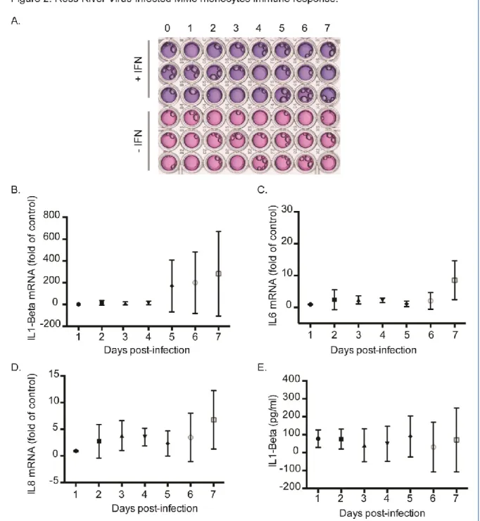
Therefore, we ask if the inability of RRV to establish a highly productive infection in MM6 cells was associated to marked production of Type-I interferons (IFNs). To determine whether RRV infection results in release of soluble IFN-, the supernatants of infected MM6 cells were collected at different time-points of infection, UV-inactivated and then analyzed on HEK-Blue IFN- cells containing the SEAP reporter gene under the control of IFN- inducible ISG54 promoter (Frumence et al., 2016) (Fig. 2A). No SEAP reporter activity was detected in supernatants of MM6 cells during the time course of infection. The addition of exogenous IFN-in the
supernatants of RRV-infected cells showed that the absence of SEAP reporter activity was not due to the inhibition of the SEAP or a block in the IFN signaling pathway. These results suggest that IFN-dependent antiviral pathway does not play a role in the restriction of RRV replication in MM6 cells. We investigated whether RRV infection of MM6 cells leads to increase of major cytokines by RT-qPCR (Fig. 2B to C). No significant difference was observed for IL-1, IL-6 or IL-8 mRNA. By using a quantitative ELISA assay (Ready-SET-Go!®, e-Bioscience), we failed to detect pro-inflammatory IL-1, IL-6, IL-8, TNF- and chemokine MCP-1 in the supernatants of MM6 cells infected with RRV-T48 (only IL-1 data shown in Fig. 2D). Altogether, these results show an unresponsiveness of MM6 cells to RRV infection with the absence of pro-inflammatory cytokine and IFN- production.
Figure 2 : RRV-infected MM6 monocytes immune response. MM6 cells were infected with RRV-GFP at MOI of 1 or mock infected. Cells or culture supernatants were collected every day from day 1 to day 7 post-infection. In (A), time course of Type 1 IFN release in response to RRV. HEK-Blue™ IFN-α/β cells were incubated 24h with UV-inactivated supernatants of RRV-infected cells and the extracellular activity of released IFN/SEAP was measured using Quanti-blue reagent. Exogenous IFN- was added to the supernatants to confirm that the absence of SEAP reporter activity was not due to the inhibition of the SEAP or to a block in the IFN signaling pathway. One representative experiment is shown. In (B) to (C), cellular mRNA encoding IL-1 beta, IL6 and IL8 were analyzed by qRT-PCR at different times post-infection. Results are the mean (± SEM) from two independent experiments performed in triplicates. In (D), ELISA assays for the measure of IL-1 beta production were performed on the supernatants of infected cells at different times post-infection. Results are the mean (± SEM) from at least three independent experiments performed in triplicates.
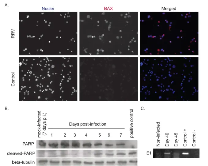
At day 5 post-infection, we observed a loss of viability of MM6 cells infected by RRV compared to control. Given that cells were displaying features of apoptotic death, the
relocalization of the pro-apoptotic mitochondrial factor Bax, a hallmark of apoptosis, was examined by indirect IF using a specific antibody. As shown in Fig. 3A, mitochondrial Bax was positively stained in MM6 cells infected one week by RRV. Immunoblot assays with a specific antibody directed against the 85 kDa-cleaved form of PARP, a hallmark of the executive phase of apoptosis, were performed on lysates of RRV-infected MM6 cells (Fig. 3B). The cleaved form of PARP was detected at 5 days post-infection and then accumulated at the later infection times. The progressive disappearance of the 115 kDa-full length form of PARP coincided with the accumulation of its cleaved form within the 5-7 days of infection. Altogether, these results showed that RRV infection results in a late apoptosis in MM6 cells although viral growth was severely restricted. Despite a very low percentage of RRV-infected cells survived to apoptosis, these cells were cultured with fresh growth medium supply every week for up to 45 days. RT-PCR analysis detected the presence of viral genome up to 45 days post-infection suggesting that RRV could potentially persist in MM6 cells on a long-time period (Fig. 3C).
Figure 3 : RRV-infected MM6 monocytes undergo apoptosis but surviving cells carry viral genome. MM6 cells were infected for seven days with RRV-GFP at MOI of 1 or mock infected. In (A), BAX signal in RRV-infected cells was analyzed by immunofluorescence assay using specific antibody. Nuclei were stained with DAPI. Results are presented individually or as merged images. In (B), cells were collected every day from day 1 to day 7 post-infection. The cleavage of PARP in infected cells was analyzed by immunoblot assay using specific antibody against the 85 kDa-cleaved form of PARP. We also visualized total PARP and beta-tubulin as loading control. In (C), Cells were collected at days 40 and 45 post-infection. Total RNAs were extracted and analyzed for the expression of viral E1 mRNA by RT-PCR.
The present study is the first demonstration that RRV has the ability to infect human monocytic MM6 cells although viral growth was poorly efficient with a very low virus progeny production. No release of IFN-β and pro-inflammatory cytokines IL-1β, IL-6 and IL-8 as well as TNF-α or MCP1 was observed in RRV infected cells indicating that host cells were not able to drive an immune response. For the first time, we demonstrated that MM6 cells underwent apoptosis in response to RRV infection as late as 5 days post infection. Relocalization of mitochondrial Bax was observed in apoptotic MM6 cells suggesting a role for mitochondrion-dependent signalling pathway in induction of apoptosis.
To date, the mechanisms by which RRV can trigger very late apoptosis in human monocytic cells remain to be investigated. In alphavirus-infected cells, apoptosis could be initiated at the level of RNA replication or thereafter by the pro-apoptotic factor Bak, which leads to cytochrome c release from mitochondria (Urban et al 2008). The production of alphaviral dsRNA molecules that are potent sensing factors for dsRNA signalling pathways, the retinoic acid inducible gene-I and the melanoma differentiation-associated gene-5 systems, could play an important role in this respect (Sanchez David et al 2016).
CHIKV was shown to infect monocytic cells in patients with a poor virus production (Her et al 2010). Macrophages were also highlighted as a possible cellular reservoir in CHIKV infected patients (Hoarau et al 2010) and in experimentally infected macaques (Labadie et al 2010). Moreover, numerous studies have pointed out the role of macrophages in the RRV pathogeny in either mice or humans (Lidbury and Mahalingam 2000, Linn et al 1996). An unexpected observation was that human monocytic cells that survived to apoptosis triggered by RRV still contained viral genetic material suggesting that such cells could be persistently infected. It is therefore urgent to determine whether the human monocytes can act as potential reservoir seed of arthralgic alphaviruses and thereby contribute to the pathogenesis of the chronic forms of RRV and CHIKV-related arthralgies.
ACKNOWLEDGEMENTS
We thank Patrick Mavingui, Etienne Frumence and Claude Giry for helpful discussions. We would like to thank R. Kuhn for providing the molecular clone T48 of RRV. This work was supported by Dim Malinf (Région Ile de France #13-521, 13.07.11), INSERM and CNRS, France. EB is a fellow of Dim Malinf (Région Ile de France #130056, 13.07.12).
REFERENCES
Aaskov JG, Mataika JU, Lawrence GW, Rabukawaqa V, Tucker MM, Miles JA et al (1981). An epidemic of Ross River virus infection in Fiji, 1979. Am J Trop Med Hyg 30: 1053-1059.
Fraser JR, Cunningham AL, Clarris BJ, Aaskov JG, Leach R (1981). Cytology of synovial effusions in epidemic polyarthritis. Aust N Z J Med 11: 168-173.
Gardner J, Anraku I, Le TT, Larcher T, Major L, Roques P et al (2010). Chikungunya virus arthritis in adult wild-type mice. J Virol 84: 8021-8032.
Hazelton RA, Hughes C, Aaskov JG (1985). The inflammatory response in the synovium of a patient with Ross River arbovirus infection. Aust N Z J Med 15: 336-339.
Heise MT, Simpson DA, Johnston RE (2000). Sindbis-group alphavirus replication in periosteum and endosteum of long bones in adult mice. J Virol 74: 9294-9299.
Henrik Gad H, Paulous S, Belarbi E, Diancourt L, Drosten C, Kummerer BM et al (2012). The E2-E166K substitution restores Chikungunya virus growth in OAS3 expressing cells by acting on viral entry. Virology 434: 27-37.
Her Z, Malleret B, Chan M, Ong EK, Wong SC, Kwek DJ et al (2010). Active infection of human blood monocytes by Chikungunya virus triggers an innate immune response. J Immunol 184: 5903-5913.
Hoarau JJ, Jaffar Bandjee MC, Krejbich Trotot P, Das T, Li-Pat-Yuen G, Dassa B et al (2010). Persistent chronic inflammation and infection by Chikungunya arthritogenic alphavirus in spite of a robust host immune response. J Immunol 184: 5914-5927.
Kummerer BM, Grywna K, Glasker S, Wieseler J, Drosten C (2012). Construction of an infectious Chikungunya virus cDNA clone and stable insertion of mCherry reporter genes at two different sites. J Gen Virol 93: 1991-1995.
Labadie K, Larcher T, Joubert C, Mannioui A, Delache B, Brochard P et al (2010). Chikungunya disease in nonhuman primates involves long-term viral persistence in macrophages. J Clin Invest 120: 894-906.
Lidbury BA, Mahalingam S (2000). Specific ablation of antiviral gene expression in macrophages by antibody-dependent enhancement of Ross River virus infection. J Virol 74: 8376-8381.
Lidbury BA, Simeonovic C, Maxwell GE, Marshall ID, Hapel AJ (2000). Macrophage-induced muscle pathology results in morbidity and mortality for Ross River virus-infected mice. J Infect Dis 181: 27-34.
Lidbury BA, Rulli NE, Musso CM, Cossetto SB, Zaid A, Suhrbier A et al (2011). Identification and characterization of a ross river virus variant that grows persistently in macrophages, shows altered disease kinetics in a mouse model, and exhibits resistance to type I interferon. J Virol 85: 5651-5663.
Linn ML, Aaskov JG, Suhrbier A (1996). Antibody-dependent enhancement and persistence in macrophages of an arbovirus associated with arthritis. J Gen Virol 77 ( Pt 3): 407-411.
Morrison TE, Whitmore AC, Shabman RS, Lidbury BA, Mahalingam S, Heise MT (2006). Characterization of Ross River virus tropism and virus-induced inflammation in a mouse model of viral arthritis and myositis. J Virol 80: 737-749. Morrison TE, Oko L, Montgomery SA, Whitmore AC, Lotstein AR, Gunn BM et al (2011). A mouse model of chikungunya virus-induced musculoskeletal inflammatory disease: evidence of arthritis, tenosynovitis, myositis, and persistence. Am J Pathol 178: 32-40.
Pohjala L, Barai V, Azhayev A, Lapinjoki S, Ahola T (2008). A luciferase-based screening method for inhibitors of alphavirus replication applied to nucleoside analogues. Antiviral Res 78: 215-222.
Sanchez David RY, Combredet C, Sismeiro O, Dillies MA, Jagla B, Coppee JY et al (2016). Comparative analysis of viral RNA signatures on different RIG-I-like receptors. Elife 5.
Soden M, Vasudevan H, Roberts B, Coelen R, Hamlin G, Vasudevan S et al (2000). Detection of viral ribonucleic acid and histologic analysis of inflamed synovium in Ross River virus infection. Arthritis Rheum 43: 365-369.
Urban C, Rheme C, Maerz S, Berg B, Pick R, Nitschke R et al (2008). Apoptosis induced by Semliki Forest virus is RNA replication dependent and mediated via Bak. Cell Death Differ 15: 1396-1407.
Way SJ, Lidbury BA, Banyer JL (2002). Persistent Ross River virus infection of murine macrophages: an in vitro model for the study of viral relapse and immune modulation during long-term infection. Virology 301: 281-292.