HAL Id: inserm-02440672
https://www.hal.inserm.fr/inserm-02440672
Submitted on 15 Jan 2020
HAL is a multi-disciplinary open access
archive for the deposit and dissemination of
sci-entific research documents, whether they are
pub-lished or not. The documents may come from
teaching and research institutions in France or
abroad, or from public or private research centers.
L’archive ouverte pluridisciplinaire HAL, est
destinée au dépôt et à la diffusion de documents
scientifiques de niveau recherche, publiés ou non,
émanant des établissements d’enseignement et de
recherche français ou étrangers, des laboratoires
publics ou privés.
Brain tumor with an ATXN1-NUTM1 fusion gene
expands the histologic spectrum of NUTM1-rearranged
neoplasia
Aurore Siegfried, Julien Masliah-Planchon, Franck-Emmanuel Roux, Delphine
Larrieu-Ciron, Gaëlle Pierron, Yvan Nicaise, Marion Gambart, Isabelle
Catalaa, Sarah Pericart, Charlotte Dubucs, et al.
To cite this version:
Aurore Siegfried, Julien Masliah-Planchon, Franck-Emmanuel Roux, Delphine Larrieu-Ciron, Gaëlle
Pierron, et al.. Brain tumor with an ATXN1-NUTM1 fusion gene expands the histologic spectrum
of NUTM1-rearranged neoplasia. Acta Neuropathologica Communications, BioMed Central part of
Springer Science, 2019, 7 (1), pp.220. �10.1186/s40478-019-0870-8�. �inserm-02440672�
L E T T E R T O T H E E D I T O R
Open Access
Brain tumor with an ATXN1-NUTM1 fusion
gene expands the histologic spectrum of
NUTM1-rearranged neoplasia
Aurore Siegfried
1,2, Julien Masliah-Planchon
3,4, Franck-Emmanuel Roux
1, Delphine Larrieu-Ciron
1, Gaelle Pierron
5,
Yvan Nicaise
2, Marion Gambart
1, Isabelle Catalaa
1, Sarah Péricart
1, Charlotte Dubucs
1,
Badreddine Mohand-Oumoussa
6, Franck Tirode
7, Franck Bourdeaut
3,4and Emmanuelle Uro-Coste
1,2*Keywords:NUTM1, ATXN1, NUTM1-rearranged neoplasia, RNA sequencing, DNA methylation-based classification, Central nervous system, Oncogenic gene fusions, CIC-ATXN1-ATXN1L axis
We report a novel ATXN1-NUTM1 gene fusion in a
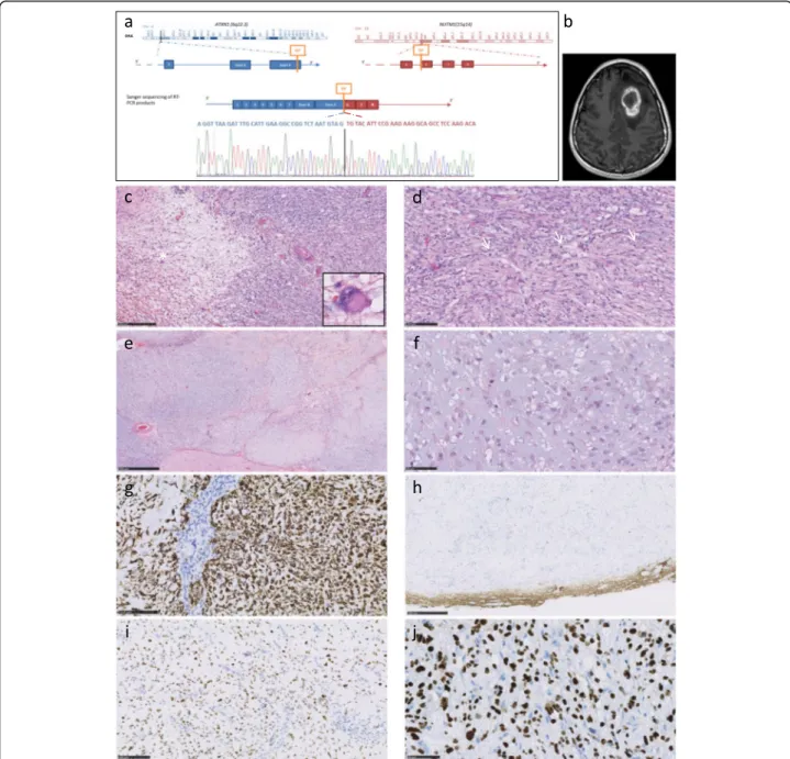
primitive brain tumor (Fig 1a). A 21-year-old woman
was seen in an emergency department for symptoms of increased intracranial pressure, visual disturbance and right hemiparesis. She reported unusual headaches for the past 3 weeks. MRI showed a frontal tumor with
intratumoral hemorrhage (Fig. 1b). The entire tumor
was surgically removed. The patient did not receive any additional treatment. 16 months after surgery, the pa-tient was symptom-free and MRI showed no recurrence of the tumor.
Histological features were characterized by a fascicular architectural pattern and chondro-myxoid areas (Fig.1c, d, e, f). Neuron-like tumor cells were apparent (Fig. 1c). Mitotic activity was overall low but increased in some foci (Fig. 1d). Strong GFAP staining led to an initial diagnosis of an unclassified glioneuronal tumor in spite of olig2 and PS100 negativity (Fig. 1g). Microscopically, the tumor was well circumscribed (Fig.1h). p53 was ac-cumulated (Fig.1i). CD56 was strongly expressed. TTF1, chromogranin, synaptophysin, CD34, p63, CK5/6 and smooth muscle actin were negative. ATRX, INI1 and BRG1 expression was maintained. Using the Heidelberg DNA methylation-based CNS tumor classifier, no class prediction was obtained with a greater than ≥0.9
confi-dence threshold [1]. The closest entity was the CNS
Ewing Family Tumor CIC group with a score of 0.235
(Additional file1: Table S1) (Case methylation data:http:// www.ncbi.nlm.nih.gov/geo; GSE138550). This tumor group is associated to the CIC-NUTM1 gene fusion [6]. We ob-served strong homogeneous nuclear staining with an
anti-NUT antibody, suggesting the presence of a CIC-NUTM1
fusion (Fig. 1j). RNA sequencing using the Illumina
TruSight RNA Fusion panel and Manta for fusion
calling revealed a novel ATXN1-NUTM1 fusion. A
CIC-NUTM1 fusion was not detected. ETV4 was over-expressed as in CIC-fused sarcomas [4, 6]. No patho-genic variants were observed in tumor DNA using a 571-gene targeted sequencing panel (Additional file 2: Table S2).
The fusion gene transcript encompassed almost all of
the ATXN1 coding sequence and the entire exon 6, 7
and 8 regions of NUTM1. The most common NUTM1
breakpoints map between exon 1 and 2, but breakpoints at the distal end of exon 5 have also been described in
someCIC-NUTM1 sarcomas [4].
Initially associated with NUT midline carcinomas, NUTM1 fusions have now been described in a broad spectrum of tumors ranging from carcinoma to sarcoma and leukemia [2,3,7]. The most common fusion partner
gene in carcinoma and sarcoma is BRD4 followed by
BRD3 and NSD3. Various new partners have been re-cently described [2,3,5]. The prognosis of these tumors is generally poor, although NUT-associated leukemias appear to be associated with a better prognosis and YAP1-NUTM1 is associated with benign skin adnexal gland tumors [3,5].
© The Author(s). 2019 Open Access This article is distributed under the terms of the Creative Commons Attribution 4.0 International License (http://creativecommons.org/licenses/by/4.0/), which permits unrestricted use, distribution, and reproduction in any medium, provided you give appropriate credit to the original author(s) and the source, provide a link to the Creative Commons license, and indicate if changes were made. The Creative Commons Public Domain Dedication waiver (http://creativecommons.org/publicdomain/zero/1.0/) applies to the data made available in this article, unless otherwise stated.
* Correspondence:uro-coste.e@chu-toulouse.fr
1
Departments of Pathology, Neurology, Neurosurgery, Radiology and Pediatric Oncology, Toulouse University Hospital, Toulouse, France 2INSERM U1037, Cancer Research Center of Toulouse (CRCT), Toulouse, France
Full list of author information is available at the end of the article
Siegfried et al. Acta Neuropathologica Communications (2019) 7:220 https://doi.org/10.1186/s40478-019-0870-8
CIC rearranged sarcomas are often fused to DUX4 and less frequently toNUTM1 [4,7]. AllCIC re-arranged tu-mors irrespective of their location or their fusion partner gene share the same transcriptomic profile defining a molecular subgroup distinct from NUT carcinoma [4,7].
Interestingly, ATXN1 codes for ataxin1 which forms a
transcriptional repressor complex with CIC. They are both part of the CIC-ATXN1-ATXN1L mitotic cell cycle
regulator axis [8]. ExcludingCIC-NUTM1 fused tumors,
only one NUTM1 rearranged brain tumor has been
previously reported, namely a cytokeratin negative BRD4-NUTM1 PNET-like parietal lobe tumor in a 3-year old boy with GFAP and synaptophysin positivity. On methylation profiling, this neoplasm did not clus-ter with tumors of the CNS Ewing Family Tumor CIC group [2].
Fig. 1ATXN1-NUTM1 gene fusion, confirmed by RT-PCR and Sanger sequencing (a). MRI identified a frontal mass. Enhancement after contrast injection (T1) (b). Representative histopathology. On the left, loose area with neuron-like tumor cells (*detail). On the right, increase in cell density (c). Fascicular architecture with three mitoses (arrows) (d). Chondroid-like, myxoid and hyalinized areas were observed (e). Undifferentiated cells with large nucleoli in a chondromyxoid background (f). Strong GFAP staining was observed. Tumor showed vascular proliferation (g).
Neurofilament staining circumscribed the tumor mass with no significant staining within the tumor (h). p53 accumulated in tumor nuclei (i). Anti-NUT antibody staining showing homogeneous intranuclear expression (j)
Myxoid and chondroid differentiation has been
re-ported in NUTM1-rearranged sarcomas but is unusual
in primary glioneuronal tumors. Whether the strong GFAP positivity of our specific case is indicative of a glial tumor or of a sarcoma with myoepithelial differenti-ation cannot be assessed due to the lack of positive staining and specificity for other markers tested. GFAP positivity has been described in 3 out of 4NUTM1 rear-ranged soft tissue or visceral sarcomas, this is in contrast
to the CNS Ewing Family Tumor CIC group which fails
to express any differentiation markers [2,6]. We recom-mend performing NUT immunohistochemistry followed
by RNA sequencing to identify any potentialNUTM1
fu-sion partner genes in GFAP+/olig2- unclassified glioma, particularly those with myxoid and/or chondroid
fea-tures. The ATXN1-NUTM1 fusion gene may define a
novel group of rare primary brain tumors. The
prognos-tic influence of NUTM1 fusion partners and the brain
localization of NUTM1-rearranged tumors warrant
further investigation.
Supplementary information
Supplementary information accompanies this paper athttps://doi.org/10. 1186/s40478-019-0870-8.
Additional file 1: Table S1. Results of the Heidelberg DNA methylation-based CNS tumor classifier (entities and scores).
Additional file 2: Table S2. List of the 517 childhood cancer genes in the dragon targeted gene sequencing panel (Illumina_TruSeq Custom Amplicon).
Acknowledgments
Samples were obtained from the CHU de Toulouse tumor bank BB-0033-00014. We thank the“Société Française des Cancers de l’Enfant” for their support.
Authors’ contributions
AS, FT, FB, EUC were major contributors in writing the manuscript. JMP, GP, YN, BMO carried out the molecular genetic studies. AS, SP, EUC characterized the histological features. YN, CD carried out the sequence alignment. FER, DLC, MG, IC contributed to the data collection. All authors read and approved the final manuscript.
Competing interests
The authors declare that they have no competing interests. Author details
1Departments of Pathology, Neurology, Neurosurgery, Radiology and Pediatric Oncology, Toulouse University Hospital, Toulouse, France.2INSERM U1037, Cancer Research Center of Toulouse (CRCT), Toulouse, France. 3Departments of Genetics and of Oncopediatry and Young Adults, Curie Institute, Paris, France.4INSERM U830, Laboratory of Translational Research in Pediatric Oncology, SIREDO pediatric oncology center, Curie Institute, Paris, France.5Department of Somatic Genetics, Curie Institute, Paris, France. 6Plateforme Post-génomique P3S, Faculté de Médecine Pierre et Marie Curie, Paris, France.7INSERM 1052, CNRS 5286, Cancer Research Center of Lyon, Centre Léon Bérard, Claude Bernard Lyon 1 University, Lyon, France.
Received: 11 December 2019 Accepted: 11 December 2019
References
1. Capper D, Jones DTW, Sill M et al (2018) DNA methylation-based classification of central nervous system tumours. Nature 555:469–474 2. Dickson BC, Sung YS, Rosenblum MK, Reuter VE, Harb M, Wunder JS,
Swanson D, Antonescu CR (2018)NUTM1 gene fusions characterize a subset of undifferentiated soft tissue and visceral tumors. Am J Surg Pathol 42:636–645
3. Hormann FM, Hoogkamer AQ, Beverloo HB et al. (2019)NUTM1 is a recurrent fusion gene partner in B cell precursor acute lymphoblastic leukemia associated with increased expression of genes on chromosome band 10p12.31–12.2. Haematologica. [ahead of print] PubMed PMID: 30872366
4. Le Loarer F, Pissaloux D, Watson S et al (2019) Clinicopathologic features of CIC-NUTM1 sarcomas, a new molecular variant of the family of CIC-fused sarcomas. Am J Surg Pathol 43:268–276
5. Sekine S, Kiyono T, Ryo E et al (2019) RecurrentMAML2 and YAP1-NUTM1 fusions in poroma and porocarcinoma. J Clin Invest 130:3827–3832 6. Sturm D, Orr BA, Toprak UH et al (2016) New brain tumor entities emerge
from molecular classification of CNS-PNETs. Cell. 164:1060–1072 7. Watson S, Perrin V, Guillemot D et al (2018) Transcriptomic definition of
molecular subgroups of small round cell sarcomas. J Pathol 245:29–40 8. Wong D, Lounsbury K, Lum A et al (2019) Transcriptomic analysis ofCIC and
ATXN1L reveal a functional relationship exploited by cancer. Oncogene 38: 273–290
Publisher’s Note
Springer Nature remains neutral with regard to jurisdictional claims in published maps and institutional affiliations.