HAL Id: inserm-02437191
https://www.hal.inserm.fr/inserm-02437191
Submitted on 13 Jan 2020
HAL is a multi-disciplinary open access
archive for the deposit and dissemination of
sci-entific research documents, whether they are
pub-lished or not. The documents may come from
teaching and research institutions in France or
abroad, or from public or private research centers.
L’archive ouverte pluridisciplinaire HAL, est
destinée au dépôt et à la diffusion de documents
scientifiques de niveau recherche, publiés ou non,
émanant des établissements d’enseignement et de
recherche français ou étrangers, des laboratoires
publics ou privés.
Rad51-Dependent Homologous Recombination
Axelle Renodon-Cornière, Pierre Weigel, Magali Le Breton, Fabrice Fleury
To cite this version:
Axelle Renodon-Cornière, Pierre Weigel, Magali Le Breton, Fabrice Fleury. New Potential Therapeutic
Approaches by Targeting Rad51-Dependent Homologous Recombination. New Research Directions in
DNA Repair, pp.467-488, 2013, 978-953-51-1114-6. �10.5772/53973�. �inserm-02437191�
New Potential Therapeutic Approaches by Targeting
Rad51-Dependent Homologous Recombination
Axelle Renodon-Cornière, Pierre Weigel,
Magali Le Breton and Fabrice Fleury
Additional information is available at the end of the chapter http://dx.doi.org/10.5772/53973
1. Introduction
Cellular DNA is constantly exposed to the effects of endogenous or environmental agents such as free radicals, radiation and chemicals. In higher organisms, these nucleic alterations are estimated at several thousands of lesions per cell [1] which can correspond to the loss of bases and also to the breaking of one or both strands of the DNA double helix. Among these DNA breaks, the double-strand break (DSB) is the most harmful because it is the most diffi‐ cult to repair. A human cell can accumulate up to 50 DSBs per cell cycle [2]. Unrepaired DSBs can have serious consequences such as permanent cell cycle arrest or cell death by apoptosis. Imperfect repair can also lead to major syndromes such as genetic disorders, pre‐ mature aging or malignant cell generation.
In response to DNA damage, the cell has developed a surveillance and DNA repair net‐ work. DSBs of DNA, which are the most severe nucleic acid alterations, are repaired mainly by either non-homologous end-joining (NHEJ) or homologous recombination (HR).
NHEJ repair leads to a direct rejoining of the separated DNA ends [3]. This pathway begins by the binding of the Ku 70/80 heterodimer to DNA ends (Figure 1] which recruits and indu‐ ces the activation of the DNA-dependent protein kinase catalytic subunit (DNA-PKc). Kin‐ ase activity is required for NHEJ since it causes the recruitment of other proteins and promotes the bringing together of DNA ends. Finally, ligase VI and XRCC4/XLF co-factors are involved in the final step of ligation and the generation of DNA repair. This process in‐ volves mainly the G0-G1 and S phases of the cell cycle. Its disadvantage is the possible loss of genetic information due to deletions or insertions of nucleic acids during the ligation of DNA ends and thus NHEJ repair is considered error-prone.
© 2013 Renodon-Cornière et al.; licensee InTech. This is an open access article distributed under the terms of the Creative Commons Attribution License (http://creativecommons.org/licenses/by/3.0), which permits unrestricted use, distribution, and reproduction in any medium, provided the original work is properly cited.
DNA repair by HR is more complex and needs a homologous sequence, which can be present in the homologous chromosome or in a gene in multicopy [4]. HR predominates in the S and G2 phases, when the sister chromatids are present and can also be a model for DNA repair [5]. In eukaryotic cells, DNA repair is supported by several protein complexes. Protein ATM (Ataxia Telangiectasia Mutated) has a role in DSB signaling via its activation induced by the MRN protein complex (MRE11-Rad50-NBS1 complex). MRE11 is a 5'-3’ exo‐ nuclease that leads to a 3' end of DNA which is required for the process [6]. This resection of single-stranded DNA is followed by the recruitment of many proteins such as RPA, BRCA1, BRCA2, Rad51, Rad52, and Rad54. Rad52 is one of the first to settle on the DSB. BRCA1 then recruits BRCA2, Rad54 and Rad51 to form the nucleoprotein filament with ssDNA, whose role is to move the blade to the homologous sequence required for HR. Rad51 protein is the main element involved in the HR process. This recombinase catalyzes the homology search and the strand exchange with a homologous sequence and thus ensures the accurate repair of the DSB. In eukaryotes, Rad51 recombinase (RecA homolog in Escherichia coli) catalyzes the essential steps of homologous recombination and interacts directly with protein sup‐ pressors of breast cancer (BRCA1, BRCA2) [8] and p53 [9] which also indicates the impor‐ tance of Rad51 in apoptosis.
HR involves a large number of protein complexes and can be divided into three main stages (Figure 1):
Formation of the HsRad51/DNA nucleofilament or pre-synaptic step. DNA DSBs are resect‐ ed by nuclease to generate 3’-protruding ends. The complex MRN (MRE11-RAD50-NSB1) contributes to DNA resection which is followed by formation of the replication protein A (RPA) complex. This ssDNA-binding factor removes secondary structures of ssDNA and is subsequently replaced by Rad51. Rad51 is recruited onto ssDNA to form the nucleofilament. Protein mediators such as Rad52 and Rad51 paralogs, Rad51B-C-D, BRCA1/2, facilitate the loading of Rad51 onto the ssDNA. The DNA binding sites of Rad51 are located in the N-terminal domain of each Rad51 monomer [8].
Homologous DNA pairing or synaptic phase. The nucleofilament of Rad51 is involved in the search for homologous DNA. Once a homologous sequence is located, the Rad51 fila‐ ment invades the duplex DNA and generates a displacement of the homologous DNA strand to form a D-loop.
Exchange and resolution of the DNA intermediate structure or post-synaptic phase. The sec‐ ond 3’ ssDNA overhang anneals to the displaced DNA strand and serves as a model strand for DNA synthesis. Two Holliday Junctions (HJ) are then formed. Their resolution is the fi‐ nal step and generates two dsDNA. HJ can be either resolved or dissolved resulting in cross‐ over or non-crossover products.
It is clear that Rad51 plays an essential role at different levels of HR and several interactions are involved such as Rad51/ssDNA, Rad51/Rad51, Rad51/dsDNA, and Rad51/nucleotide. In addition, Rad51 interacts with its partners involved in HR (e.g. Rad52, Rad54).
Many cancer treatments using chemotherapy or radiotherapy target and disrupt the func‐ tion of the DNA of tumor cells by inducing adducts or single- or double-strand breaks in DNA. However, these anticancer therapies are often faced with the emergence of radio- and chemoresistance, either induced or intrinsic to cancer cells. Since it was shown that some pathways of DNA repair can remove DNA damage induced by radio- or chemotherapy in cancer cells, these pathways have become potential therapeutic targets to sensitize tumors. This observation is especially true for the Rad51 protein. A high level of HR induced by overexpression of Rad51 is frequently described in various types of cancer cell, including breast cancer, pancreatic, nonsmall-cell lung carcinoma and leukemia (AML and CML) [9-13]. In these cancer cells, this overexpression provides a degree of cancer resistance by promoting the repair of DSBs induced by cancer treatments [14]. Moreover, it has been shown that the survival of cancer patients expressing higher levels of Rad51 is shorter and that a reduced amount of Rad51, following antisense or ribozyme treatment, increases the effectiveness of cancer treatment by radiotherapy [15-17]. Modulation of HR to potentiate treatment is an option described in many publications [18]. It can be achieved by either act‐ ing directly on the recombinase activity of Rad51 or attempting to interfere with the interac‐ tions between Rad51 and some of its partners, which are not necessarily related directly to the repair of DNA.
The purpose of this chapter is to review all the chemical modulators that can act directly or indirectly on Rad51-mediated homologous recombination. First, the different steps of Rad51 activity which can be targeted will be detailed. Secondly, the chemical molecules that inhibit Rad51 activity or affect the expression level of Rad51 will be described. Finally, their appli‐ cations in combination with anticancer treatments will be discussed in order to open up pos‐ sibilities for counteracting chemo- and radioresistance.
2. Rad51 activity in homologous recombination
Human Rad51 (Homo sapiens Rad51 or HsRad51) is composed of 339 amino acids. It is the eukaryotic homolog of the RecA protein in prokaryotes. Homologs of the human recombi‐ nase are highly conserved between species: 98.8% similarity with Mus musculus Rad51 (MmRad51] and 81% with Saccharomyces cerevisiae Rad51 (ScRad51) [19,20].
To date, no complete structure of the HsRad51 protein has been determined. The crystalline structure of ScRad51 truncated at 79 N-terminal amino acids [21], one structure of Pyrococcus
furiosus RadA [22], and five RadA of Methanococcus voltae [23,24] and Sulfolobus solfataricus
[25] have been determined.
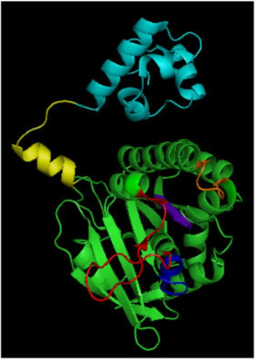
Figure 2. MvRadA subunit structure (PDB 1XU4). The N-terminal domain and the polymerization motif are colored in
cyan and yellow, respectively. The putative DNA binding L1 and L2 loops are highlighted in orange and red. The Walk‐ er A and B motifs, corresponding to the ATP binding site, are labeled in blue and purple, respectively.
RadA and Rad51 are composed of two domains: a small N-terminal domain and a C-termi‐ nal domain entitled the core domain. The structures of the two HsRad51 domains were re‐ solved by NMR [8] and crystallography [26], respectively.
The C-terminal domain of HsRad51 was crystallized in the form of a fusion protein compris‐ ing the BRC4 motif of BRCA2 protein (residues 1517-1551), a flexible linker and the central domain of HsRad51 (residues 97-339). The co-crystallization of HsRad51 with the BRC4 mo‐ tif indicated the existence of a polymerization sequence located at the subunit-subunit inter‐ face of Rad51 [26]. The C-terminal domain contains an ATPase domain comprising units of Walker A (Hs: 127-134) and Walker B (Hs: 217-222) which are essential for ATP binding while loops L1 (Hs: 230-236) and L2 (Hs: 269-287) are involved in DNA binding.
The N-terminal domain of HsRad51 interacts with double-stranded DNA by a helix-hairpin-helix structure (residues 61-69). This type of protein-DNA interaction is conserved among many proteins interacting with DNA [27]. These sites of interaction, illustrated in Figure 2, are necessary for the formation of the nucleofilament, which is the key step of the recombi‐ nase activity of Rad51 (Figure 1). Nucleofilament formation is accompanied by a stretch modification of the DNA helix. The nucleofilament can adopt several conformations, only one of which is active for DNA strand exchange. The extended conformation is the function‐ al form of the filament.
The conformation of the Rad51 filament depends on nucleotides: ATP promotes the extend‐ ed conformation whereas ADP stabilizes the compressed form. Most of the structures have been solved in the presence of ATP analogs. Thus the conversion of an extended conforma‐ tion to a compressed conformation accompanies the hydrolysis of ATP [28,29].
Several HsRad51 studies have also shown that ammonium sulfate [30], calcium [28,29] and AMPPNP [29] significantly increase the effectiveness of the strand exchange reaction in vitro by promoting the formation of an extended filament, which confirms that this structural form is the functional conformation of Rad51.
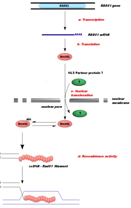
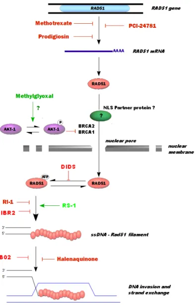
Since Rad51 protein is central to HR, all chemical molecules able to disrupt the interaction sites of Rad51 directly will also be able to modulate DNA repair by HR. Other ways of mod‐ ulating HR via Rad51 are possible. Figure 3 presents the main ways and the catalytic steps of Rad51 being targeted to modulate HR.
3. Specific molecules targeting Rad51
The great majority of compounds identified as inhibitors of Rad51 have been selected by high-throughput screening from chemical libraries.
3.1. Modulators of Rad51 recombinase activity
Figure 3. Intracellular pathways and catalytic steps of Rad51 as potential targets to modulate HR. (a) and (b) - The
modulation of transcription and translation of Rad51 leads to changes in the recombinase activity and hence to the modification of HR repair. (c) - The intracellular localization and the delivery of Rad51 to the DNA damage sites are required for the HR process. Rad51 cellular distribution has an important regulatory role in HR and any element able to modulate the nuclear translocation of Rad51 is also able to modulate HR. (d) - Finally, molecules acting on the steps of Rad51 activity will target recombinase activity and HR-mediated DNA repair.
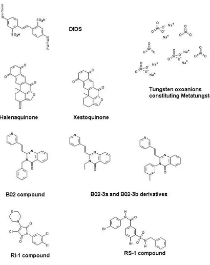
Figure 4. Structure of chemical molecules modulating the Rad51 recombinase activity. 3.1.1. 4’-Diisothiocyanostilbene-2,2’-disulfonic acid or DIDS
DIDS (Figure 4) is a molecule known since the early 1970s for its ability to inhibit ionic chan‐ nels and membrane transporters [31].
In the context of HR, Ishida T. et al. [32] have shown that DIDS can also inhibit in vitro the binding of Rad51 to DNA. In fact, strand exchange reactions are inhibited in the presence of DIDS. This process is dependent on salt concentration since the addition of 0.2 M KCl to the reaction medium changes the behavior of the inhibition. In the absence of KCl, increasing the concentration of DIDS gradually inhibits strand exchange with an IC50 (without KCl) close to 2 μM when the Rad51 concentration is 6 μM. In the pres‐ ence of 0.2 M KCl, inhibition is considered to be shifted: the addition of DIDS has no ef‐
fect up to a concentration of 1 μM. In this condition, the IC50 (with KCl) is close to 5 μM in the presence of Rad51 at 6 μM concentration. The interaction between Rad51 and DIDS probably involves electrostatic strength.
It has also been observed that the inhibition of the binding of Rad51 to ssDNA is not changed in the presence or absence of ATP. As previously described, HsRad51 contains a binding site for ATP and ATPase activity [33]. Analysis of the ATPase activity of Rad51 shows that ATP hydrolysis is greatly decreased when the protein is not bound to DNA. However, according to the results of Ishida and collaborators, Rad51 is able to hydrolyze ATP in the presence of DIDS and without DNA. The assumption is that this activation by DIDS of an asynchronous ATPase function results from the inhibition of the binding of Rad51 to DNA, which is ATP-dependent. However, Amunugama et al. [34] showed that the presence of ATP bound to Rad51 is required during strand exchange. In contrast, ATP hy‐ drolysis does not appear to be essential for recombination. Hence, inactivation of the AT‐ Pase activity by DIDS cannot alone explain the mechanism of inhibition of Rad51.
DIDS interacts physically with Rad51 and dissociates it from ssDNA by competing with ssDNA for Rad51 binding.
3.1.2. Metatungstate
Metatungstate is a polyoxometalate (POM) consisting of 12 tungsten oxoanions (Figure 4) which dissociate into monotungstates in aqueous alkaline solution. This molecule is mainly used as a catalytic agent for chemical reactions of hydrocarbons.
Li and colleagues [35] have demonstrated that the metatungstate structure can bind in vitro to MvRadA protein, a homolog of Rad51 from Archaea Methanococcus voltae (MvRadA). The main contact zones established between the protein and metatungstate concern the L1 loop region with Arg218 and Arg230 and the L2 loop region with Arg224 of RadA. Both these L1 and
L2 domains including Tyr232 and Phe203 are involved in DNA binding. It should be noted
that the same pattern is found in HsRad51 [36]. In the MvRadA filament, these locations re‐ sult in a distribution of molecules of tungstates on the longitudinal axis of rotation. It is shown that these tungsten clusters interact between the DNA-binding loops L1 and L2 stabi‐ lizing the inactive conformation of Rad51 [23,24,37].
Tungstate binding to MvRadA induces several effects on the functions of the recombinase protein activity. ATPase activity decreases by about 90% with equimolar amounts of MvRa‐ dA and metatungstate. By using gel electrophoresis in vitro, binding assays of MvRadA to ssDNA reveal that metatungstate inhibits ssDNA binding (IC50 = 0.13 μM for 1 μM MvRa‐ dA). The same in vitro assays using dsDNA also show an inhibition with a similar IC50 whereas the IC50 value of metatungstate for strand exchange activity is 0.5 μM in the pres‐ ence of KCl. These observations indicate that, in vitro, metatungstate can inhibit the ssDNA and dsDNA binding of MvRadA, thus inactivating the functions essential for HR. As men‐ tioned previously, the inactivation of ATPase activity does not alone explain the inhibition of Rad51 functions and probably those of RadA. It is therefore suggested that metatungstate acts as a competitive inhibitor of DNA binding by MvRadA.
Metatungstate is a potent inhibitor of ATPase and strand exchange activities of MvRadA and other experiments performed with HsRad51 have shown a significant increase in the IC50 of metatungstate for HsRad51 as compared with that for MvRadA (IC50RadA = 0.5 μM
and IC50Rad51 = 30 μM)[35].
3.1.3. Halenaquinone
Xestoquinone and halenaquinone molecules (Figure 4) are extracted from the marine sponge Xestospongia exigua. These molecules are similar except that xestoquinone does not contain the oxygen at the C-3 position in contrast to halenaquinone. Only halenaqui‐ none presents inhibitory properties of phosphatidylinositol 3-kinase [38] and some anti-proliferative features [39].
Takaku et al. tested 160 crude extract fractions from marine sponge and used the D-loop for‐ mation assay to detect the homologous-pairing activity of Rad51. The authors reported that the halenaquinone inhibits HR at DNA pairing and D-loop formation stages but no inhibito‐ ry effect was observed with xestoquinone [40]. By Surface Plasmon Resonance (SPR) meas‐ urement, they showed that both halenaquinone and xestoquinone are able to bind to Rad51 but the affinity between halenaquinone and Rad51 is higher than between xestoquinone and Rad51. This result can explain the efficient inhibition of Rad51-mediated homologous pair‐ ing by halenaquinone.
Takaku and collaborators then examined whether both molecules affect ssDNA and dsDNA binding by Rad51. By an electrophoretic mobility shift approach, halenaquinone was found to inhibit Rad51-dsDNA binding specifically, but not Rad51-ssDNA binding. Interestingly, the authors showed that halenaquinone inhibits the secondary dsDNA binding by the Rad51-ssDNA complex. These results suggest that halenaquinone probably interacts near the dsDNA-binding site of Rad51. It can therefore inhibit the ternary complex formation containing ssDNA, dsDNA and Rad51 which promotes the DNA homologous pairing step during the HR process. In contrast, neither ssDNA binding nor dsDNA binding by Rad51 was affected by the presence of xestoquinone.
The authors then studied the intracellular effects of halenaquinone on the Ionizing Radiation (IR)-induced formation of Rad51 foci. When human cells were exposed to IR and treated with halenaquinone, Rad51 foci formation was significantly decreased. This result indicates that halenaquinone destabilizes the Rad51 foci, probably by inhibiting the ternary complex formation. Halenaquinone may be useful in medical research as a potential inhibitor of HR.
3.1.4. Compound B02 and derivatives
By high-throughput screening based on the quenching fluorescence method, Huang and col‐ leagues have investigated the identification of specific inhibitors of the Rad51 strand ex‐ change activity [18]. From 200,000 small molecules of the NIH repository, 174 compounds were positives and, after supplementary analyses and different controls, 13 molecules were identified as potential inhibitors of Rad51 with an inhibition higher than 30%. The IC50 val‐ ues for the most potent inhibitors of Rad51-induced D-loop formation were determined.
Among these molecules, both compounds A04 and A10 were found to be inhibitors for Rad51 and RecA and their IC50 values were 5 μM and 26.6 μM, respectively. Another com‐ pound, the B02 molecule, was found to disrupt Rad51 binding to DNA and nucleoprotein filament formation. Although the B02 molecule presents an IC50 (27.4 μM) higher than A04 or A10, this molecule has a higher specificity for HsRad51.
Moreover, the study of B02 derivatives has revealed an efficient inactivation of Rad51 by both B02-3a and B02-3b, which contain an ethyl and an m-methylphenyl group, respectively (instead of the benzyl group located in the B02 molecule) (Figure 4). Modification of the pyr‐ idin radical of B02 suppresses the Rad51-induced D-loop inhibition, which demonstrates the importance of these chemical groups. The recent in vivo work of the same team has shown that B02 inhibits DSB-induced HR and increases cell sensitivity to the ICL agents, cisplatin and mitomycin C [41].
3.1.5. Compound RI-1 or 3-chloro-1-(3,4-dichlorophenyl)-4-(4-morpholinyl)-1H-pyrrole-2,5-dione
From a screening of 10,000 molecules of Chembridge DIVERSetTM, the RI-1 compound was
identified as an inhibitor of HsRad51 [42]. A first screening by fluorescence polarization (FP) enabled molecules that can bind to HsRad51 to be selected. A second screening based on the inhibition of homologous recombination in a cell line of human osteosarcoma (U20S) was used and eight molecules were identified. A final test with the human embryonic kidney cell line (HEK293) identified RI-1, whose action is the specific inactivation of HsRad51.
RI-1 is composed of a chloromaleimide moiety (Figure 4) which promotes covalent binding to the thiol group of Rad51 cysteine 319 by a Michael addition mechanism. This binding po‐ tentiates the inhibition of the polymerization of HsRad51 during nucleofilament formation. It should be noted that the binding site is located on a surface which is highly conserved among mammalian homologs of Rad51. Experiments with Saccharomyces cerevisiae Rad51 (ScRad51) also show a fixation on the corresponding cysteine target (C377). However, this site is not present in RecA and inhibition was not found. RI-1 is potentially a specific inhibi‐ tor for mammalian homologs of Rad51. The binding site is located on the interface between two monomers of HsRad51 so it inhibits the polymerization of Rad51 onto ssDNA [21]. It is known that cysteine 319 is located in an ATP-binding loop [23], therefore the binding of RI-1 may disrupt the interaction of Rad51 with ATP. Moreover, this interaction area is also in‐ volved in the binding of other HR repair proteins such as Rad52 and Rad54 [43]. The IC50 of RI-1 is from 5 to 30 μM depending on the HsRad51 intracellular concentration. A synergistic anticancer effect is also observed for the association of RI-1 with mitomycin C (MMC) in U20S, HeLa, MCF-7 and SH2038 cell lines.
3.1.6. Compound RS-1 (Rad51-Stimulatory-1) or 3-[(benzylamino)sulfonyl]-4-bromo-N-(4-bromophenyl)benzamide)
In contrast to inhibitors, few molecules stimulating HR have been reported. However, by screening 10,000 molecules (Chembridge DIVERSetTM) using the FP method described
above, Connell et al. identified a molecule, RS-1, which stimulates Rad51 binding onto ssDNA and increases the stability of the nucleofilament [44].
In the presence of RS-1 (Figure 4), the nucleofilament is in the active form characterized by the long length of the Rad51-ssDNA complex (100Å). The presence of nucleotide cofactors is also important since ATP is required for RS-1 to stimulate the formation of the active fila‐ ment. However, RS-1 does not stimulate Rad51 by inhibiting its ATPase activity since it has no effect on the Rad51-dependent hydrolysis of ATP.
The RS-1-induced extension of the Rad51-ssDNA nucleofilament stimulates the exchange step of DNA strands, which can be evaluated by estimating D-loop formation. In the pres‐ ence of non-hydrolyzable ATP or Ca2+, RS-1 increases D-loop formation by 5 to 11 times [28,44]. The stimulatory action of RS-1 is specific to HsRad51, since no effect with E. coli Re‐ cA or ScRad51 was found. This stimulation was then analyzed at the cellular level.
An analysis of cell survival (neonatal human fibroblasts) showed that the cells are more re‐ sistant to cisplatin treatment in the presence of 7.5 μM RS-1. This result is probably due to the ability of RS-1 to stimulate HR in response to DNA-damage agents like cisplatin.
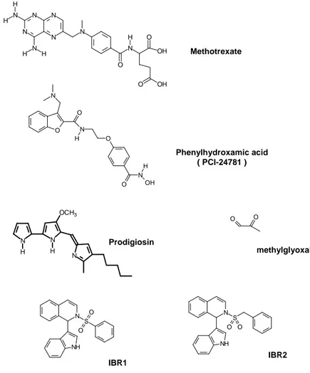
3.2. Chemical modulators of Rad51 expression 3.2.1. Methotrexate drug
The structure of methotrexate (Figure 5) is similar to that of the folate metabolic precursor of coenzyme tetrahydrofolate (FH4) involved in the synthesis of nucleic bases. Methotrexate is a molecule used as an inhibitor of dihydrofolate reductase and acts in nucleic base synthesis occurring during the S phase of the cell cycle, as well as in non-restorative homologous re‐ combination [45]. Therefore, methotrexate targets the S phase and the functions of HR by re‐ ducing the rate of repair of DNA damage, which can be shown by comet assay [46]. The study conducted by Du and colleagues [47] found that the inhibition of the formation of HsRad51 foci was effective in the presence of methotrexate in a human osteosarcoma cell line (HOS) after irradiation. This inhibition seems to be related to the Rad51 protein expres‐ sion level, which is significantly decreased by the treatment. In addition, it was observed that the expression levels of BRCA2 and Rad52 were therefore not affected by methotrexate. It induces a specific downregulation of HsRad51. It should be noted that the treatment of HOS cells with 0.1 μM methotrexate causes a decrease in the transcription of 70% and > 95% of HsRad51 after 12 and 24 hours, respectively. However, these studies cannot determine the interactions involved in decreasing the mRNA levels of HsRad51.
3.2.2. Phenylhydroxamic acid or PCI-24781
Phenylhydroxamic acid (Figure 5) belongs to the inhibitory molecules of histone deacetylas‐ es (HDACs) used for their antitumor activities [48]. In particular, PCI-24781 inhibits HDAC2, which is one of the HDAC family involved in the regulation of the factors of HR. According to the work of Adimoolam et al. [49], inhibition of HDAC by PCI-24781 reduces the expression of Rad51 in the cell line derived from human colorectal carcinoma (HTC116) and thus reduces the HR response to DSBs induced by therapy. The first observation by im‐ munofluorescence showed a complete inhibition of the formation of HsRad51 foci in the
presence of PCI-24781 after irradiation and an apoptosis rate of 7% with 0.2 μM. After 24 hours, the level of protein synthesis in the absence or presence of 0.2 mM PCI-24781 showed a decrease in transcription of 60% BRCA1 and 80% for both BRCA2 and HsRad51. BRCA2 protein is also involved in the DNA repair protein complex with Rad51 [50]. The HR inhibi‐ tion observed results in an additive effect of the reductions in expression levels of Rad51 and BRCA2. Moreover, the fall in BRCA2 in the nuclear compartment probably removes the inhibition of caspase-3 protease, which is able to cleave Rad51 and thus to inactivate HR DNA repair [51]. Therefore, PCI-24781 may indirectly activate the cleavage of Rad51 in ad‐ dition to inducing a decrease in its synthesis. Interestingly, PCI-24781 induces the expres‐ sion of the gene of the GADD45y protein which is a factor of cellular growth arrest [52]. This third effect of PCI-24781 can limit the growth of tumor cells.
Phenylhydroxamic acid ( PCI-24781 ) Methotrexate O O N O O N OH H N H Prodigiosin methylglyoxal O O N NH S O O IBR1 N NH S O O IBR2 N N OCH3 H H N N N N N N H H N H H N N O O OH O OH H
3.2.3. Prodigiosin
Prodigiosin is a tripyrrole red pigment (Figure 5) from bacteria Serratia marcescens. Recently, immunosuppressive and anticancer properties have been identified for this molecule [53-55]. The first action of prodigiosin is an increase in DNA DSBs, which probably results from the inhibition of both topoisomerase I and II [56]. Lu and collaborators described a sig‐ nificant reduction in the level of HsRad51 protein and mRNA in breast tumor cell lines (MCF-7, MDA-MB-231, T- 47D, A549, HCT116) in the presence of 50 nM prodigiosin [57]. The downregulation of Rad51 is mainly induced by lowering mRNA expression and not by proteasome-mediated Rad51 degradation. Although the tumor suppressor protein p53 is known as a repressor of Rad51 [58], prodigiosin downregulates Rad51 in a p53-independent manner. This result is an advantage for the prodigiosin-mediated therapy of cancer in which p53 is deficient compared to molecules such as flavopiridole or roscovitine, which need an activation of this protein. On the other hand, prodigiosin activates JNK and p38-MAPK sig‐ naling pathways, which are known to mediate the pro-apoptotic effect of numerous anti‐ cancer drugs [59]. By using specific inhibitors of both these signaling pathways, Lu and collaborators have shown that the level of Rad51 mRNA is restored. This result confirms the involvement of JNK and p38-MAPK signaling pathways in the prodigiosin-induced Rad51 downregulation. However, this is in contradiction with the work of Chuang et al. [60], which showed that the activation of p53-MAPK could increase the level of Rad51 protein, improving its stability and not significantly altering the level of HsRad51 mRNA. Similarly, Ko and colleagues [61] have shown a decrease in mRNA levels of HsRad51 with curcumin treatment and an inactive ERK/p38-MAPK signaling pathway. Although the Rad51 downre‐ gulation mechanism is not fully understood, prodigiosin seems to be a potent suppressor of Rad51 which may be used to overcome HR-mediated drug resistance in cancer.
4. Modulation of Rad51 by inactivation of nuclear translocation
4.1. Nuclear Localization Signal
Eukaryotic proteins are expressed in the cytoplasm. If their functions are carried out in the nucleus, they have to pass through the nuclear membrane. In contrast to small biomolecules, numerous proteins larger than 20 Kda require active transport via a signal peptide of recog‐ nition: a Nuclear Localization Signal (NLS) [62]. This signal peptide may be recognized by karyopherins [63] to form a nuclear protein complex (NPC) [64,65]. Proteins involved in HR are not exempt from this obligation for nuclear translocation. Thus, it is useful to analyze the possibility of blocking the passage of Rad51 through the nuclear membrane and thereby in‐ hibit HR. Rad51 protein does not contain an NLS so its nuclear translocation requires an as‐ sociation with another protein. Interestingly, among the Rad51 paralogs, Rad51C has an NLS [66] as do Rad54 [67] and Rad52 [68] proteins. Other proteins related to DNA repair such as BRCA2 have been reported as being involved in Rad51 transport [69]. The mecha‐ nism of Rad51 transport is not clearly understood although Rad51C seems to be an interest‐ ing candidate. In particular, Gildemeister and colleagues have found that Rad51C deficiency
significantly reduces the amount of Rad51 in the nucleus before and after DNA damage [69]. Another option is protein kinase B or AKT-1 protein kinase which is involved in the cyto‐ plasmic sequestration of Rad51 [70]. The modulation of Rad51 transport offers an excellent tool to potentiate anticancer therapy through inhibition of Rad51 nuclear translocation.
4.2. AKT-1 kinase and BRCA1 proteins
Activation of AKT-1 promotes cell proliferation and the activated form is regularly found in cancer cells. In addition, to reducing malignant cell division, the inhibition of the AKT-1 sig‐ naling pathway has been investigated for the purpose of co-therapeutic approaches [71]. AKT activation occurs through a series of successive phosphorylation steps at thr-450, thr-308 and ser-473 by JNK kinases, phosphoinositide-dependent kinase 1 and by several kinases (PKD2 and others), respectively [72]. Plo and collaborators have demonstrated an‐ other aspect of the activation of AKT-1 in HR DNA repair of chemotherapy-induced DSBs [70]. This group studied the level of HsRad51 and BRCA1 in cell lines MCF7 and MDA-MB-231 and observed a decreased level in the nucleus while both these proteins accumulat‐ ed in the cytoplasm. Although the HsRad51 and BRCA1 features are not modified, AKT-1 activation induces a retention signal of these proteins in the cytoplasm. Thus, their absence in the nucleus confers a deficiency of recombinase activity. The retention mechanism is still unknown, but it seems to be related to AKT-1-mediated BRCA1 NLS phosphorylation. In fact, it has been observed that AKT-1 phosphorylates BRCA1 on two sites located in the re‐ gion of the NLS [73] and some mutations of these sites show a suppression of nuclear trans‐ location of Rad51 and BRCA1, irrespective of the activated AKT-1. In this context, an activator of AKT-1 phosphorylation, such as methylglyoxal (Figure 5) [74,75] may promote the cytoplasmic sequestration of Rad51.
4.3. Modulation of the interaction between Rad51 and BRCA2
Human BRCA2 protein is constituted of 3418 amino acids (384 kDa) and contains several in‐ teraction domains. There is an interaction site with N-terminal RPA and in the central region of BRCA2 there are 8 repeated motifs called BRC motifs [76]. BRC1, BRC4, BRC7 and BRC8 motifs are able to interact with Rad51 with different affinities [77]. Pellegrini et al. [26] have shown that the BRC4 motif interacts with HsRad51 by mimicking the motif of Rad51 which is responsible for its polymerization. These BRC motifs can bind monomeric or oligomeric forms of Rad51 in a cell cycle-dependent manner and in response to DNA damage. HsRad51 regulation is also mediated by serine 3291 of the BRCA2 C-terminal domain. In the absence of DNA damage, this serine is phosphorylated by CDK1 whereas it is in a dephosphorylated form with inactivated CDK1. The ser-3291 can bind only to the oligomeric form in the nucle‐ oprotein filament. This binding plays a role in stabilizing the Rad51-DNA complex since the phosphorylation of ser-3291 inhibits oligomerization in the absence of DSB and then syn‐ chronizes the repair mechanism [78]. It has been proposed that the BRCA2 protein is direct‐ ly involved in the nuclear transport of Rad51 [50]. The pancreatic adenocarcinoma cell line CAPAN-1 is known to be defective in BRCA2 [79]. It has a deletion of the BRCA2 domains for DNA repair and the nuclear localization signals [80]. Rad51 exhibits impaired nuclear
translocation in CAPAN-1. Therefore, it has been proposed that Rad51 requires BRCA2 for its nuclear translocation and that C-terminally trunkated BRCA2 retains Rad51 in the cyto‐ plasm. BRCA2-Rad51 interaction is also essential in the HR process and many works have described those derivative peptides of BRCA2 that are able to mimic and bind to this inter‐ action site [81-83]. Small molecules have been proposed to disrupt the interaction and two patents have been deposited [84]. By using the two-hybrid system in yeast, Lee and Chen suggested several molecules from a drug screening. Two hydrophobic molecules (phenyl‐ sulfonyl indolyl isoquinoline derivatives) IBR1 and IBR2 (Figure 5) were found to be able to disrupt the interaction and can thus potentiate anticancer treatments. The authors suggest that the benzene ring of IBR2 interacts in the hydrophobic pocket of Rad51 which is in‐ volved in the subunit-subunit interaction during filament formation and also in the interac‐ tion with the BRC4 motif of BRCA2. This phenyl moiety of IBR2 may be a competitor with the Rad51 F86 or BRC4 F1524 [85].
The authors also analyzed the effect of IBR2 at cellular level. After irradiation, breast cancer cells (MCF-7) presented a lower number of Rad51 foci than when these cells were pre-treat‐ ed with IBR2. Another result was the fast degradation of Rad51 in the treated cells where the HR was impaired. This work, which is ongoing, has led to the development and synthesis of other IBR2 analogs [85,86].
5. Conclusion
DNA repair by homologous recombination is now a potential target in cancer therapy. The induction of DNA damage is one of the means of action against uncontrolled cell prolifera‐ tion systems, while repairs are causes of resistance to radio- and chemotherapy. DNA repair is frequently found to be deregulated in tumor cells. Rad51 is the central protein of HR and its expression level is correlated with resistance to chemotherapeutic drugs. This observa‐ tion suggests that targeted inhibition of Rad51 through small chemical molecules may im‐ prove the response to drug treatment by reducing HR.
Among antitumoral strategies, several studies have proposed numerous molecules that in‐ hibit the recombinase activity; these are described in Figure 6.
DIDS and metatungstate are molecules that deregulate the ATPase activity of Rad51. DIDS thus causes a random hydrolysis of ATP without ssDNA-bound Rad51, while metatungstate inhibits the ATPase activity. The ATPase center is located at the Rad51 subunit-subunit in‐ terface which binds and hydrolyzes ATP and regulates the conformation of the DNA bind‐ ing site. Although both molecules act differently, they induce an inhibition of the binding of Rad51 onto DNA.
Inhibition of the Rad51 polymerization is also interesting since it directly affects filament formation. The compound RI-1 can bind to the thiol group of cys-319 which inhibits the in‐ teraction between monomers of Rad51.
Figure 6. Potential inhibitors of Rad51-mediated HR repair: Methotrexate (100nM), Prodigiosin (100nM) and
PCI-24781 (200nM) treatments reduce the levels of Rad51 mRNA between 20% and 50% in cancer cells. The decrease of Rad51 transcript level also induces an inhibition of Rad51 foci formation [47,49,57]. A 10µM concentration of DIDS significantly inhibits the binding of Rad51 to DNA, leading to the strand exchange inhibition [32]. RI-1 and B02 (20µM) decreases 50% of DNA binding by Rad51 and disrupts the formation of Rad51 foci after DNA damage in cells [18,42]. IBR2 (20µM) inhibits the Rad51 oligomerization in vitro by binding to Rad51 hydrophobic pocket [85]. 20µM of RS-1 promotes the binding of Rad51 to DNA [44]. Halenaquinone (30μM) inhibits the step of DNA homologous pairing mediated by Rad51 in vitro and the Rad51 foci formation after irradiation of cells [40]. It is noteworthy that in vitro IC50 values depend on the protein concentration and technical conditions, which makes difficult to compare them each other and to values obtained in cells.
Halenaquinone modulates the recombinase activity by inhibiting the binding of the Rad51-ssDNA complex with dsDNA. The hypothesis is that the presence of halenaquinone desta‐ bilizes the Rad51-ssDNA binding or the interaction between Rad51 subunits. This instability results in a disassembly of the complex, before the recognition of the homologous sequence. Compound B02 is also capable of modulating the function of Rad51 by disrupting Rad51 binding to DNA and formation of the nucleoprotein filament. Moreover, this compound in‐ creases cell sensitivity to DNA damaging agents and to PARP1 inhibitors. Thus, small mole‐ cules acting directly on the recombinase activity steps offer a potential development for new anticancer treatment associated with chemo- or radiotherapy. Another approach is to de‐ crease the expression of the RAD51 gene. For these purposes, several studies have demon‐ strated that methotrexate and molecule PCI-24781 significantly reduce the synthesis of Rad51 mRNA. Note that only the effects on mRNA levels were observed but the mechanism of transcription control remains unclear.
Prodigiosin also decreases the level of Rad51 mRNA which seems to be related to the activa‐ tion of the JNK and p58-MAPK signaling pathways. Therefore, chemical compounds that up- and downregulate Rad51 production and/or activity may be useful for the suppression of tumor progression but the therapeutic applications of this strategy are currently incon‐ ceivable and unlikely. The last mode of action focuses on the transport of Rad51 from cyto‐ plasm to nucleus. Rad51 is a protein whose nuclear functions require partners to facilitate its entry into the nucleus. It has been noted that the activation of the anti-apoptotic protein AKT-1 inactivates the nuclear translocation of Rad51 and BRCA1. The mechanism is still poorly understood but retention in the cytoplasm causes a significant decrease in HR. Sever‐ al studies also suggest that BRCA2 is involved in this transport. Molecules such as IBR, which interfere with the Rad51-BRCA2 interaction, may induce cytoplasmic sequestration of Rad51 which decreases HR. These molecules capable of inhibiting the transport of Rad51 ap‐ pear attractive candidates.
Most of these Rad51 inhibitors have been identified from screening libraries. These small molecules were tested in vitro and in cellular models but it will be necessary to quantify their efficacy, identify their toxicities and their potentially additional pharmacokinetic and pharmacological properties by animal assays. An understanding of toxicities, adverse ef‐ fects, and special dosing considerations of existing anticancer compounds is important to the design of effective drug combinations and to the interpretation of the toxicological pro‐ file of new chemical entities.
Afterwards, a major challenge is to design new analogues to these molecules that will be more selective for Rad51 so that their efficacy will be improved and their potential toxicities will be decreased. The process of identifying and selecting these analogues has undergone a sea change in the recent decades with the development of solid-state and combinatorial chemistry and computer modeling of drug–target interactions.
By sensitizing cells to DNA damage, Rad51 inhibitors open up new perspectives in the search for agents capable of suppressing homologous recombination and thereby potentiat‐ ing chemo- and radiotherapy treatments for cancer. Moreover, these molecules may be not
only instrumental in the development of combination anticancer therapies but also excellent tools to analyze Rad51 activities and cellular functions.
Acknowledgements
This work was supported by grants from the Ligue contre le Cancer Comité de Loire Atlan‐ tique et de Vendée and the Région pays de la Loire (CIMath project). We thank G. Levillain for his help.
Author details
Axelle Renodon-Cornière, Pierre Weigel, Magali Le Breton and Fabrice Fleury*
*Address all correspondence to: fleury-f@univ-nantes.fr Unité UFIP, CNRS FRE 3478, University of Nantes, France
References
[1] Jackson, S. P., and Bartek, J. (2009) Nature 461, 1071-1078
[2] Vilenchik, M. M., and Knudson, A. G. (2003) Proc Natl Acad Sci U S A 100, 12871-12876
[3] Krejci, L., Chen, L., Van Komen, S., Sung, P., and Tomkinson, A. (2003) Prog Nucleic
Acid Res Mol Biol 74, 159-201
[4] Shinohara, A., and Ogawa, T. (1995) Trends Biochem Sci 20, 387-391
[5] Takata, M., Sasaki, M. S., Sonoda, E., Morrison, C., Hashimoto, M., Utsumi, H., Ya‐ maguchi-Iwai, Y., Shinohara, A., and Takeda, S. (1998) EMBO J 17, 5497-5508
[6] Uziel, T., Lerenthal, Y., Moyal, L., Andegeko, Y., Mittelman, L., and Shiloh, Y. (2003)
EMBO J 22, 5612-5621
[7] Rass, E., Grabarz, A., Bertrand, P., and Lopez, B. S. (2012) Cancer Radiother 16, 1-10 [8] Aihara, H., Ito, Y., Kurumizaka, H., Yokoyama, S., and Shibata, T. (1999) J Mol Biol
290, 495-504
[9] Bearss, D. J., Lee, R. J., Troyer, D. A., Pestell, R. G., and Windle, J. J. (2002) Cancer Res 62, 2077-2084
[10] Raderschall, E., Stout, K., Freier, S., Suckow, V., Schweiger, S., and Haaf, T. (2002)
[11] Slupianek, A., Hoser, G., Majsterek, I., Bronisz, A., Malecki, M., Blasiak, J., Fishel, R., and Skorski, T. (2002) Mol Cell Biol 22, 4189-4201
[12] Maacke, H., Opitz, S., Jost, K., Hamdorf, W., Henning, W., Kruger, S., Feller, A. C., Lopens, A., Diedrich, K., Schwinger, E., and Sturzbecher, H. W. (2000) Int J Cancer 88, 907-913
[13] Maacke, H., Jost, K., Opitz, S., Miska, S., Yuan, Y., Hasselbach, L., Luttges, J., Kalth‐ off, H., and Sturzbecher, H. W. (2000) Oncogene 19, 2791-2795
[14] Vispe, S., Cazaux, C., Lesca, C., and Defais, M. (1998) Nucleic Acids Res 26, 2859-2864 [15] Ohnishi, T., Taki, T., Hiraga, S., Arita, N., and Morita, T. (1998) Biochem Biophys Res
Commun 245, 319-324
[16] Christodoulopoulos, G., Malapetsa, A., Schipper, H., Golub, E., Radding, C., and Panasci, L. C. (1999) Clin Cancer Res 5, 2178-2184
[17] Collis, S. J., Tighe, A., Scott, S. D., Roberts, S. A., Hendry, J. H., and Margison, G. P. (2001) Nucleic Acids Res 29, 1534-1538
[18] Huang, F., Motlekar, N. A., Burgwin, C. M., Napper, A. D., Diamond, S. L., and Ma‐ zin, A. V. (2011) ACS Chem Biol 6, 628-635
[19] Barlow, A. L., Benson, F. E., West, S. C., and Hulten, M. A. (1997) EMBO J 16, 5207-5215
[20] Haaf, T., Golub, E. I., Reddy, G., Radding, C. M., and Ward, D. C. (1995) Proc Natl
Acad Sci U S A 92, 2298-2302
[21] Conway, A. B., Lynch, T. W., Zhang, Y., Fortin, G. S., Fung, C. W., Symington, L. S., and Rice, P. A. (2004) Nat Struct Mol Biol 11, 791-796
[22] Shin, D. S., Pellegrini, L., Daniels, D. S., Yelent, B., Craig, L., Bates, D., Yu, D. S., Shiv‐ ji, M. K., Hitomi, C., Arvai, A. S., Volkmann, N., Tsuruta, H., Blundell, T. L., Venki‐ taraman, A. R., and Tainer, J. A. (2003) EMBO J 22, 4566-4576
[23] Wu, Y., Qian, X., He, Y., Moya, I. A., and Luo, Y. (2005) J Biol Chem 280, 722-728 [24] Wu, Y., He, Y., Moya, I. A., Qian, X., and Luo, Y. (2004) Mol Cell 15, 423-435
[25] Ariza, A., Richard, D. J., White, M. F., and Bond, C. S. (2005) Nucleic Acids Res 33, 1465-1473
[26] Pellegrini, L., Yu, D. S., Lo, T., Anand, S., Lee, M., Blundell, T. L., and Venkitaraman, A. R. (2002) Nature 420, 287-293
[27] Doherty, A. J., Serpell, L. C., and Ponting, C. P. (1996) Nucleic Acids Res 24, 2488-2497 [28] Bugreev, D. V., and Mazin, A. V. (2004) Proc Natl Acad Sci U S A 101, 9988-9993 [29] Ristic, D., Modesti, M., van der Heijden, T., van Noort, J., Dekker, C., Kanaar, R., and
[30] Sigurdsson, S., Trujillo, K., Song, B., Stratton, S., and Sung, P. (2001) J Biol Chem 276, 8798-8806
[31] Makara, G. B., Stark, E., Karteszi, M., Palkovits, M., and Rappay, G. (1981) Am J Phys‐
iol 240, E441-446
[32] Ishida, T., Takizawa, Y., Kainuma, T., Inoue, J., Mikawa, T., Shibata, T., Suzuki, H., Tashiro, S., and Kurumizaka, H. (2009) Nucleic Acids Res 37, 3367-3376
[33] Stark, J. M., Hu, P., Pierce, A. J., Moynahan, M. E., Ellis, N., and Jasin, M. (2002) J Biol
Chem 277, 20185-20194
[34] Amunugama, R., He, Y., Willcox, S., Forties, R. A., Shim, K. S., Bundschuh, R., Luo, Y., Griffith, J., and Fishel, R. (2011) J Biol Chem 287, 8724-8736
[35] Li, Y., He, Y., and Luo, Y. (2009) Biochemistry 48, 6805-6810
[36] Matsuo, Y., Sakane, I., Takizawa, Y., Takahashi, M., and Kurumizaka, H. (2006) FEBS
J 273, 3148-3159
[37] Qian, X., Wu, Y., He, Y., and Luo, Y. (2005) Biochemistry 44, 13753-13761
[38] Fujiwara, H., Matsunaga, K., Saito, M., Hagiya, S., Furukawa, K., Nakamura, H., and Ohizumi, Y. (2001) Eur J Pharmacol 413, 37-45
[39] Schmitz, F. J., and Bloor, S. (1988) J.Org.Chem 53, 3922-3925
[40] Takaku, M., Kainuma, T., Ishida-Takaku, T., Ishigami, S., Suzuki, H., Tashiro, S., van Soest, R. W., Nakao, Y., and Kurumizaka, H. (2011) Genes Cells 16, 427-436
[41] Huang, F., Mazina, O. M., Zentner, I. J., Cocklin, S., and Mazin, A. V. (2012) J Med
Chem 55, 3011-3020
[42] Budke, B., Logan, H. L., Kalin, J. H., Zelivianskaia, A. S., Cameron McGuire, W., Mill‐ er, L. L., Stark, J. M., Kozikowski, A. P., Bishop, D. K., and Connell, P. P. (2012) Nucle‐
ic Acids Res
[43] Krejci, L., Damborsky, J., Thomsen, B., Duno, M., and Bendixen, C. (2001) Mol Cell Bi‐
ol 21, 966-976
[44] Jayathilaka, K., Sheridan, S. D., Bold, T. D., Bochenska, K., Logan, H. L., Weichsel‐ baum, R. R., Bishop, D. K., and Connell, P. P. (2008) Proc Natl Acad Sci U S A 105, 15848-15853
[45] Mariani, B. D., Slate, D. L., and Schimke, R. T. (1981) Proc Natl Acad Sci U S A 78, 4985-4989
[46] Ostling, O., and Johanson, K. J. (1984) Biochem Biophys Res Commun 123, 291-298 [47] Du, L. Q., Du, X. Q., Bai, J. Q., Wang, Y., Yang, Q. S., Wang, X. C., Zhao, P., Wang, H.,
[48] Buggy, J. J., Cao, Z. A., Bass, K. E., Verner, E., Balasubramanian, S., Liu, L., Schultz, B. E., Young, P. R., and Dalrymple, S. A. (2006) Mol Cancer Ther 5, 1309-1317
[49] Adimoolam, S., Sirisawad, M., Chen, J., Thiemann, P., Ford, J. M., and Buggy, J. J. (2007) Proc Natl Acad Sci U S A 104, 19482-19487
[50] Davies, A. A., Masson, J. Y., McIlwraith, M. J., Stasiak, A. Z., Stasiak, A., Venkitara‐ man, A. R., and West, S. C. (2001) Mol Cell 7, 273-282
[51] Brown, E. T., Robinson-Benion, C., and Holt, J. T. (2008) Radiat Res 169, 595-601 [52] Zhang, X., Sun, H., Danila, D. C., Johnson, S. R., Zhou, Y., Swearingen, B., and Kli‐
banski, A. (2002) J Clin Endocrinol Metab 87, 1262-1267
[53] Chang, C. C., Chen, W. C., Ho, T. F., Wu, H. S., and Wei, Y. H. (2011) J Biosci Bioeng 111, 501-511
[54] Pandey, R., Chander, R., and Sainis, K. B. (2009) Curr Pharm Des 15, 732-741
[55] Perez-Tomas, R., Montaner, B., Llagostera, E., and Soto-Cerrato, V. (2003) Biochem
Pharmacol 66, 1447-1452
[56] Montaner, B., Castillo-Avila, W., Martinell, M., Ollinger, R., Aymami, J., Giralt, E., and Perez-Tomas, R. (2005) Toxicol Sci 85, 870-879
[57] Lu, C. H., Lin, S. C., Yang, S. Y., Pan, M. Y., Lin, Y. W., Hsu, C. Y., Wei, Y. H., Chang, J. S., and Chang, C. C. (2012) Toxicol Lett 212, 83-89
[58] Lazaro-Trueba, I., Arias, C., and Silva, A. (2006) Cell Cycle 5, 1062-1065 [59] Fan, M., and Chambers, T. C. (2001) Drug Resist Updat 4, 253-267
[60] Chuang, S. M., Wang, L. H., Hong, J. H., and Lin, Y. W. (2008) Toxicol Appl Pharmacol 230, 290-297
[61] Ko, J. C., Tsai, M. S., Weng, S. H., Kuo, Y. H., Chiu, Y. F., and Lin, Y. W. (2011) Toxicol
Appl Pharmacol 255, 327-338
[62] Adam, S. A., Marr, R. S., and Gerace, L. (1990) J Cell Biol 111, 807-816
[63] Radu, A., Blobel, G., and Moore, M. S. (1995) Proc Natl Acad Sci U S A 92, 1769-1773 [64] Ryan, K. J., and Wente, S. R. (2000) Curr Opin Cell Biol 12, 361-371
[65] Gorlich, D., and Kutay, U. (1999) Annu Rev Cell Dev Biol 15, 607-660
[66] French, C. A., Tambini, C. E., and Thacker, J. (2003) J Biol Chem 278, 45445-45450 [67] Golub, E. I., Kovalenko, O. V., Gupta, R. C., Ward, D. C., and Radding, C. M. (1997)
Nucleic Acids Res 25, 4106-4110
[68] Shen, Z., Cloud, K. G., Chen, D. J., and Park, M. S. (1996) J Biol Chem 271, 148-152 [69] Gildemeister, O. S., Sage, J. M., and Knight, K. L. (2009) J Biol Chem 284, 31945-31952
[70] Plo, I., Laulier, C., Gauthier, L., Lebrun, F., Calvo, F., and Lopez, B. S. (2008) Cancer
Res 68, 9404-9412
[71] Martelli, A. M., Evangelisti, C., Chiarini, F., Grimaldi, C., Manzoli, L., and McCubrey, J. A. (2009) Expert Opin Investig Drugs 18, 1333-1349
[72] Xiao, L., Gong, L. L., Yuan, D., Deng, M., Zeng, X. M., Chen, L. L., Zhang, L., Yan, Q., Liu, J. P., Hu, X. H., Sun, S. M., Liu, J., Ma, H. L., Zheng, C. B., Fu, H., Chen, P. C., Zhao, J. Q., Xie, S. S., Zou, L. J., Xiao, Y. M., Liu, W. B., Zhang, J., Liu, Y., and Li, D. W. (2010) Cell Death Differ 17, 1448-1462
[73] Altiok, S., Batt, D., Altiok, N., Papautsky, A., Downward, J., Roberts, T. M., and Av‐ raham, H. (1999) J Biol Chem 274, 32274-32278
[74] Chang, T., Wang, R., Olson, D. J., Mousseau, D. D., Ross, A. R., and Wu, L. (2011)
FASEB J 25, 1746-1757
[75] Jia, X., Chang, T., Wilson, T. W., and Wu, L. (2012) PLoS One 7, e36610 [76] Bork, P., Blomberg, N., and Nilges, M. (1996) Nat Genet 13, 22-23
[77] Wong, A. K., Pero, R., Ormonde, P. A., Tavtigian, S. V., and Bartel, P. L. (1997) J Biol
Chem 272, 31941-31944
[78] Davies, O. R., and Pellegrini, L. (2007) Nat Struct Mol Biol 14, 475-483 [79] Jasin, M. (2002) Oncogene 21, 8981-8993
[80] Holt, J. T., Toole, W. P., Patel, V. R., Hwang, H., and Brown, E. T. (2008) Cancer Genet
Cytogenet 186, 85-94
[81] Chen, C. F., Chen, P. L., Zhong, Q., Sharp, Z. D., and Lee, W. H. (1999) J Biol Chem 274, 32931-32935
[82] Nomme, J., Renodon-Corniere, A., Asanomi, Y., Sakaguchi, K., Stasiak, A. Z., Stasiak, A., Norden, B., Tran, V., and Takahashi, M. (2011) J Med Chem 53, 5782-5791
[83] Nomme, J., Takizawa, Y., Martinez, S. F., Renodon-Corniere, A., Fleury, F., Weigel, P., Yamamoto, K., Kurumizaka, H., and Takahashi, M. (2008) Genes Cells 13, 471-481 [84] Lee, W.-H. C. L. (2006) Compositions and methods for disruption of BRCA2-Rad51
interaction. USA
[85] Lee, W.-H., Chen, P.-L., Zhou, L., and Zhu, J. (2009) Compositions and methods relat‐ ed to Rad51 inactivation in the treatment of neoplastic diseases and especially CML. the regents of the university of California, CA, US, USA
[86] Qiu, X. L., Zhu, J., Wu, G., Lee, W. H., and Chamberlin, A. R. (2009) J Org Chem 74, 2018-2027
![Figure 1. Schematic representation of the mechanism of DNA DSB repair by NHEJ and HR (figures taken from [7] ).](https://thumb-eu.123doks.com/thumbv2/123doknet/14214565.482553/3.722.98.628.453.861/figure-schematic-representation-mechanism-repair-nhej-figures-taken.webp)