Publisher’s version / Version de l'éditeur:
Journal of the American Ceramic Society, 86, 9, pp. 1638-1640, 2003
READ THESE TERMS AND CONDITIONS CAREFULLY BEFORE USING THIS WEBSITE. https://nrc-publications.canada.ca/eng/copyright
Vous avez des questions? Nous pouvons vous aider. Pour communiquer directement avec un auteur, consultez la première page de la revue dans laquelle son article a été publié afin de trouver ses coordonnées. Si vous n’arrivez pas à les repérer, communiquez avec nous à PublicationsArchive-ArchivesPublications@nrc-cnrc.gc.ca.
Questions? Contact the NRC Publications Archive team at
PublicationsArchive-ArchivesPublications@nrc-cnrc.gc.ca. If you wish to email the authors directly, please see the first page of the publication for their contact information.
NRC Publications Archive
Archives des publications du CNRC
This publication could be one of several versions: author’s original, accepted manuscript or the publisher’s version. / La version de cette publication peut être l’une des suivantes : la version prépublication de l’auteur, la version acceptée du manuscrit ou la version de l’éditeur.
Access and use of this website and the material on it are subject to the Terms and Conditions set forth at
Nanostructured silicon oxide-nickel oxide sol-gel films with enhanced
optical carbon monoxide gas sensitivity
Martucci, Alessandro; Pasquale, Mirko; Guglielmi, Massimo; Post, Mike;
Pivin, Jean Claude
https://publications-cnrc.canada.ca/fra/droits
L’accès à ce site Web et l’utilisation de son contenu sont assujettis aux conditions présentées dans le site LISEZ CES CONDITIONS ATTENTIVEMENT AVANT D’UTILISER CE SITE WEB.
NRC Publications Record / Notice d'Archives des publications de CNRC:
https://nrc-publications.canada.ca/eng/view/object/?id=d50b67cc-c8f2-431e-ab7d-c591b3f96a7a
https://publications-cnrc.canada.ca/fra/voir/objet/?id=d50b67cc-c8f2-431e-ab7d-c591b3f96a7a
Nanostructured Silicon Oxide–Nickel Oxide Sol–Gel Films with
Enhanced Optical Carbon Monoxide Gas Sensitivity
Alessandro Martucci, Mirko Pasquale, and Massimo Guglielmi
Dipartimento di Ingegneria Meccanica S. Materiali, Universita` di Padova, ItalyMike Post
ICPET, National Research Council of Canada, Ottawa, CanadaJean Claude Pivin
CSNSM, IN2P3-CNRS, Orsay Campus, FranceSol– gel derived silica (SiO2) films doped with nickel oxide
(NiO) nanocrystals were fabricated. A bifunctional ligand was used, bearing amine groups capable of coordinating the nickel ions and hydrolysable siloxane groups for anchoring the metal complex moiety to the silicate matrix. Nickel oxide nanocrys-tals precipitated at 500°C while the film was still porous. The nanocomposite films showed a reversible change in the optical transmittance in the VIS-NIR range when exposed to carbon monoxide gas. The effects of residual porosity, testing temper-ature, and carbon monoxide gas concentration on optical transmittance were studied.
I. Introduction
R
ECENTLY, nanocomposites with sensor functionality are be-coming a new area of interest for optical gas-sensing appli-cations. In fact, the optical transmittance of nanoparticles or thin films has been reported1,2to change on exposure to some gases. In particular, it was found2
that NiO, Co3O4, and Mn3O4thin films
showed a reversible decrease in the VIS-NIR absorption when exposed to CO at temperatures ⬃250°–350°C. The CO-sensitive decrease of the absorbance of NiO films can be ascribed2
to a decrease of positive hole density in the NiO caused by a decrease in oxygen anion density on the NiO surface during the catalytic oxidation of CO. As the gas-sensing mechanism is a surface-related phenomenon, the increase of the specific surface area of the functional oxide is expected to enhance the CO-sensing effect. In this work we synthesized, by sol– gel methods, porous SiO2films
doped with homogenously dispersed NiO nanocrystals. The nano-porosity of the sol– gel matrix provides a path for the gas molecules to reach the functional ultrafine particles embedded in the glass matrix, and the nanocomposite films showed a reversible change in optical transmittance when exposed to CO gas.
II. Experimental Procedure
The nanocomposites were obtained by mixing a matrix solution of tetraethylorthosilicate (TEOS; Aldrich Chemical Co., Milwau-kee, WI) and methyltriethoxysilane (MTES; Aldrich) as SiO2
precursors, with a doping solution containing NiCl2as precursor
for the NiO particles. 3-(2-Aminoethylamino)propyltrimethoxysilane (AEAPTMS; Aldrich) was used as the bifunctional ligand. This ligand bears both amine groups capable of coordinating the nickel ions, and hydrolysable silyl groups for anchoring the metal complex moiety to the silicate matrix. The matrix solution was obtained by using the following molar ratios: MTES:TEOS:H2O:HCl ⫽
1:1:1:0.005. In the doping solution the molar ratio between AE-APTMS and NiCl2was 1. The solvent was ethanol and the nominal
oxide concentration in the deposition solution was 50 g/L.
Films of composition 60SiO2䡠40NiO were deposited by dipping
at 35% RH on silicon or SiO2glass substrates and heat-treated for
1 h in air at 500°, 700°, 800°, or 900°C.
The films were characterized by X-ray diffraction (XRD; Philips, Eindhoven, The Netherlands) using a diffractometer equipped with glancing-incidence X-ray optics. The analysis was performed using CuK␣nickel-filtered radiation at 40 kV and 50
mA. The average crystallite size was calculated from the Sherrer equation after fitting the experimental profiles.
Transmission electron microscope (TEM; Philips) characteriza-tion was conducted at 200 kV. Scratched fragments of the film deposited on a holey carbon copper grid were used for the analysis. The average diameter of the NiO particles was measured from TEM images by evaluating about 200 particles.
Rutherford backscattering spectrometry (RBS; 2.2 MeV 4
He⫹
) was used to evaluate the concentration profiles of silicon, nickel, oxygen, and carbon in the film and to measure the composition of the film with respect to the different elements by simulating the RBS experimental spectra. The hydrogen content of the films was mea-sured by elastic recoil detection analysis (ERDA). The film density (mol䡠cm⫺3
) was calculated by combining thickness data (cm), ob-tained from profilometer measurements, and the film dose (mol䡠cm⫺2
) from RBS and ERDA data.
The sensing properties of the nanocomposite films were evaluated by measuring the variation of the optical transmittance of the film when exposed to CO in air. The films were mounted on a custom-fabricated heater in a gas flow chamber, with a design to permit unimpeded radiation transmission through the whole assembly. Transmission data in the 200 nm ⬍ ⬍ 900 nm range were recorded using a spectrophotometer (Model Cary1E, Varian, Palo Alto, CA) with films heated at temperatures between room temperature and 350°C and with CO concentrations of 1, 0.1, and 0.01 vol% CO in air. Details of the experimental setup are reported in Ref. 3.
III. Results and Discussion
TEOS and MTES were used as sol– gel precursors to obtain thicker films. In fact it has been shown4
that by using MTES it is possible to obtain single-layer films ⬃2 m thick at 500°C. It is P. Gouma—contributing editor
Manuscript No. 186451. Received January 6, 2003; approved March 26, 2003. 1638
journal
also reported5
that the porosity of the films can be tailored using different MTES/TEOS ratios.
In the doping solution, NiCl2 was used because of its good
solubility in ethanol. Moreover, to improve the homogenous distribu-tion of the NiO particles, a bifuncdistribu-tional ligand, AEAPTMS, was used. The amine groups present in the AEAPTMS are capable of coordi-nating the nickel ions through the formation of metal complexes that can be anchored to the silicate matrix by the hydrolysable silyl groups.6
As reported by Breitscheidel et al.,6
the metal complexes form in solution through the reaction between the AEAPTMS and the nickel salt, and NiO precipitates at 500°C after removal of the organic part. As reported by Piccaluga et al.,7
metal oxide nanoparticles in the sol– gel matrix probably form in the cavities of the matrix, substituting for a part of the adsorbed water.
TEM measurement of films heated at 500°C showed the presence of round-shaped nanocrystals with a mean diameter of 3.8 nm and a standard deviation of 1.2 nm. Figure 1 shows the XRD spectra of films heated at different temperatures. Peaks belonging to NiO cubic crystals (Powder Diffraction File No.78 – 0643, International Centre for Diffraction Data, Newtown Square, PA) have been observed in films heated at 500°C or at higher temperatures, whereas in films heated at lower temperatures no diffraction peaks were detected. The mean diameter of NiO crystallites, estimated from the linewidth of the most intense diffraction peak, was ⬃6 nm for both films heated at 800° and 900°C, whereas in films heated at lower temperatures the broadness of the diffraction peaks did not allow a reliable estimation of the mean crystallite size. Comparing the values of the mean diameter of NiO nanoparticles in films heated at 500°C, estimated from TEM measurements, with those in films heated at 800° and 900°C, estimated from XRD measurements, there is an increase in crystal size with the heat-treatment temperature, even though the mean diameter remains in the nanometer range.
The silicon/nickel ratio evaluated from RBS and ERDA analy-sis was in good agreement with the nominal one (nominal silicon/nickel ⫽ 1.5, see Table I). Moreover, the profile concen-tration was uniform throughout the film thickness. The hydrogen content decreases with the heat-treatment temperature whereas the carbon content remains constant. The density of the films increases with the heat-treatment temperature because of the progress of the sol– gel film densification.
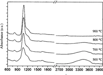
Figure 2 shows the FTIR spectra of films treated at different temperatures. In all the spectra two dominant peaks of the Si–O bond8
at 1070 and 800 cm⫺1
are evident. In the spectra of films heated at 500° and 700°C there are more bands, one very broad at 3400 –3500 cm⫺1
, and one around 1630 cm⫺2
caused by OH vibrations, and one between 910 and 940 cm⫺1
, attributed to stretching vibrations of Si–OH or SiO–
groups.9
These bands are absent in films heated at 800° and 900°C, indicating a higher degree of densification, in agreement with the variation of the hydrogen content measured by RBS-ERDA. All the spectra also
have a shoulder at ⬃1200 cm⫺1
, which, as reported by Almeida and Pantano,10
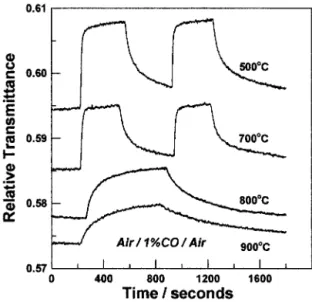
is related to the total porosity of the films. The optical transmittance over the 200 nm ⬍ ⬍ 900 nm range for films heat-treated at 500°C or higher temperatures increased revers-ibly on exposure to CO for film testing temperatures ⬎200°C. Transmission data at ⫽ 650 nm for a series of four different films, all at testing temperatures of 330°C during gas exposure, obtained as a function of exposure time to 1% CO in air is shown in Fig. 3. The films were heat-treated at 500°, 700°, 800°, and 900°C, and data of this type are reproducible over many cycles of CO/air exposure. In the series of measurements reported in Fig. 3, the two films that were heat-treated at the lower temperatures were exposed to two sequential pulses of 1% CO with air exposure in between, whereas the other two films experienced only one CO pulse. It is clear that both the relative increase in transmittance and the rate of response decrease with the film heat-treatment temperature. The films densify to a greater extent when heat-treated at the higher temperatures, as shown by RBS-ERDA and FTIR measurements, so there exists a correlation between film density and the magnitude and speed of sensor response. With increased densification, the porosity of the films decreases, and this constricts and limits the availability of paths of the gas molecules for reaching the surface of the NiO nanocrystals in the SiO2 matrix.
However, other factors, such as NiO particle size and matrix stoichi-ometry, may play a role in these observed effects, and the relative importance of these factors on sensor functionality has not yet been determined conclusively. Figure 4 shows the effect of film testing temperature on the transmittance response at ⫽ 650 nm. For this data, a film heat-treated at 500°C was used, and the exposure gas was 1% CO/air, but the film test temperature was varied in four steps from 220° to 330°C. It is clear that both the magnitude of response (i.e., change in transmittance) and the reversibility are enhanced at the higher testing temperatures. As the proposed detection mechanism is thermally activated,2
the relative variation in optical transmittance would be expected to decrease as the testing temperature decreases. Even so, at 220°C transmittance changes are still easily detectable although the response characteristics are poorer than at the higher temperatures.
Fig. 1. XRD spectra of 60SiO2䡠40NiO films heat-treated at different temperatures.
Fig. 2. FTIR spectra of 60SiO2䡠40NiO films heat-treated at different temperatures.
Table I. Results of RBS-ERDA Analysis on 60SiO2䡠40NiO
Films Heated at Different Temperatures
Temperature (°C) Relative composition (%) Density (g䡠cm⫺3) Si Ni O C H 500 17 13.5 39.5 13 17 1.91 700 16 14 44 13 13 2.12 800 18 15 42 13 12 2.45 900 18.5 15 46.5 13 7 2.51
The transmittance dependence of the films on CO concentration was also measured. Figure 5 shows transmittance data obtained at ⫽650 nm for a film at a test temperature of 280°C. Three CO concentrations in air have been used (0.01%, 0.1%, and 1% CO in air). Clearly a CO concentration dependence is evident, and transmittance changes are easily observable even at concentrations of 0.01% CO. The relative transmittance changes, based on the initial (air) base line are ⬃1.3%, 2.0%, and 2.7% for 0.01%, 0.1%, and 1% CO in air, respectively, indicating a log concentration dependence on transmittance response. Other features of the response characteristics in this data need to be noted. For example, reversibility is generally good except at the final stage where a longer tail out is present, although an eventual return to the original base line is achieved over time. An explanation of this observation is that a portion of the response at the lower CO concentrations is accompanied by the CO or its reaction products being more strongly chemisorbed or adsorbed in the NiO/SiO2 matrix. This
results in less lability with a consequential deterioration to revers-ibility. In general, for the data reported, the achievement of good return to base line before and after CO exposure is improved with
increasing testing temperature. This would be predicted for a mechanism that is thermally activated and involves desorbtion of strongly bound adsorbed or chemisorbed species.
IV. Conclusions
Nanoporous SiO2 glass films doped with monodispersed NiO
nanocrystals were obtained by the sol– gel technique. Films heat-treated between 500° and 900°C showed the presence of NiO nanocrystals and a residual porosity that allowed the reaction between the surface of the NiO particles dispersed in the SiO2
glass matrix and CO gas. The optical transmittance of 40NiO䡠60SiO2films increases on exposure to CO in air over the
300 – 800 nm wavelength range. For films that have been heat-treated at T ⬍ 700°C and are exposed to CO in air at T ⬎ 250°C, a rapid and reversible change in transmittance occurs. The change in transmittance is clearly demonstrated, even at concentrations at the 0.01% CO level. The transmittance response of the films indicates a logarithmic proportionality to CO concentration. The CO-gas-sensing properties depend on heat-treatment conditions during sol– gel preparation, the residual porosity, and the testing temperature during CO exposure.
References
1
K. Yasumoto, N. Koshizaki, and K. Suga, “Changes in Optical Property of Ultrafine Particle Doped Glass by Gas Atmosphere”; p. 604 in Sol–Gel Optics II, SPIE Proceed-ings, Vol. 1758. Edited by J. D. Mackenzie. SPIE, Bellingham, WA, 1992.
2
M. Ando, T. Kobayashi, and M. Haruta, “Combined Effects of Small Gold Particles on Optical Gas Sensing by Transition Metal Oxide Films,” Catal. Today, 36, 135 (1997).
3
M. Post and J. Yao, “Optical Transmittance of Thin Films of SrFeO2.5⫹xat
Elevated Temperatures and Applications to Gas Sensing,” Mater. Res. Soc. Symp. Proc., 403, 533 (1996).
4
P. Innocenzi, M. O. Abdirashid, and M. Guglielmi, “Structure and Properties of Sol–Gel Coatings from Methyltriethoxysilane and Tetraethoxysilane,” J. Sol-Gel Sci. Technol., 3, 47 (1994).
5
C. Mc Donagh, B. D. Mac Craith, and A. K. Mc Evoy, “Tailoring of Sol–Gel films for Optical Sensing of Oxygen in Gas and Aqueous Phase,” Anal. Chem., 70, 45 (1998).
6
B. Breitscheidel, J. Zieder, and U. Schubert, “Metal Complexes in Inorganic Matrices. 7. Nanometer-Sized, Uniform Metal Particles in a SiO2Matrix by Sol–Gel
Processing of Metal Complexes,” Chem. Mater., 3, 559 (1991).
7
G. Piccaluga, A. Corrias, G. Ennas, and A. Musinu, “Sol–Gel Preparation and Characterization of Metal–Silica and Metal Oxide–Silica Nanocomposites”; pp. 1– 65 in Materials Science Foundations. TransTech Publications, Aedermannsdorf, Swit-zerland, 2000.
8
F. G. Galeener, “Band Limits and the Vibrational Spectra of Tetrahedral Glasses,” Phys. Rev. B: Condens. Matter, 19, 4292 (1979).
9
R. M. Almeida, T. A. Guitton, and G. A. Pantano, “Characterization of Silica Gels by Infrared Reflection Spectroscopy,” J. Non. Cryst. Solids, 121, 193 (1990).
10
R. M Almeida and C. G. Pantano, “Structural Investigation of Silica-Gel Films by
Infrared Spectroscopy,” J. Appl. Phys., 68, 4225 (1990). 䡺
Fig. 3. Variation of the optical transmittance of 60SiO2䡠40NiO at ⫽ 650 nm and T ⫽ 330°C for films heat-treated at different temperatures exposed to 1 vol% CO in air.
Fig. 4. Variation of the optical transmittance of 60SiO2䡠40NiO at ⫽ 650 nm for films heat-treated at 500°C and exposed to 1 vol% CO in air at four different testing temperatures.
Fig. 5. Variation of the optical transmittance of 60SiO2䡠40NiO at ⫽ 650 nm and T ⫽ 280°C for films heat-treated at 700°C and exposed to three different CO concentrations.