HAL Id: hal-03220819
https://hal.sorbonne-universite.fr/hal-03220819
Submitted on 7 May 2021
HAL is a multi-disciplinary open access
archive for the deposit and dissemination of
sci-entific research documents, whether they are
pub-lished or not. The documents may come from
teaching and research institutions in France or
L’archive ouverte pluridisciplinaire HAL, est
destinée au dépôt et à la diffusion de documents
scientifiques de niveau recherche, publiés ou non,
émanant des établissements d’enseignement et de
recherche français ou étrangers, des laboratoires
Integrative Neuroscience of Paramecium, a “Swimming
Neuron”
Romain Brette
To cite this version:
Romain Brette. Integrative Neuroscience of Paramecium, a “Swimming Neuron”. eNeuro, Society for
Neuroscience, In press, �10.1523/ENEURO.0018-21.2021�. �hal-03220819�
Copyright © 2021 Brette Review | Integrative Systems
Integrative Neuroscience of Paramecium, a
“Swimming Neuron”
https://doi.org/10.1523/ENEURO.0018-21.2021
Cite as: eNeuro 2021; 10.1523/ENEURO.0018-21.2021
Received: 15 January 2021 Revised: 17 March 2021 Accepted: 18 March 2021
This Early Release article has been peer-reviewed and accepted, but has not been through the composition and copyediting processes. The final version may differ slightly in style or formatting and will contain links to any extended data.
Alerts: Sign up at www.eneuro.org/alerts to receive customized email alerts when the fully formatted version of this article is published.
1. Manuscript title: Integrative neuroscience of Paramecium, a “swimming neuron”
1
2
2. Abbreviated title: Integrative neuroscience of Paramecium
3
4
3. Author Names and Affiliations
5
Romain Brette
6
Sorbonne Universités, UPMC Univ Paris 06, INSERM, CNRS, Institut de la Vision, 17 rue Moreau,
7
75012 Paris, France8
9
4. Author Contributions10
RB wrote the paper, designed and performed research.
11
12
5. Correspondence should be addressed to Romain Brette, Institut de la Vision, 17, rue Moreau,
13
75012 Paris, France14
romain.brette@inserm.fr15
16
6. Number of figures: 1417
7. Number of Tables: 018
8. Number of Multimedia: 019
9. Number of words for Abstract : 67
20
10. Number of words for Significance Statement : 67
21
11. Number of words for Introduction : 916
22
12. Number of words for Discussion : 1035
23
24
13. Acknowledgements25
None.26
27
14. Conflict of Interest28
The author reports no conflict of interest.
29
30
15. Funding sources
31
This work was supported by Agence Nationale de la Recherche (ANR-20-CE30-0025-01), by the
32
Programme Investissements d’Avenir IHU FOReSIGHT (ANR-18-IAHU-01), by Sorbonne Université
33
(Programme Emergence, grant NEUROSWIM) and by Fondation Pour l’Audition (FPA RD-2017-2).
34
36
Integrative neuroscience of Paramecium, a “swimming neuron”
37
38
Abstract
39
Paramecium is a unicellular organism that swims in fresh water by beating thousands of cilia. When
40
it is stimulated (mechanically, chemically, optically, thermally…), it often swims backward then turns
41
and swims forward again. This “avoiding reaction” is triggered by a calcium-based action potential.
42
For this reason, some authors have called Paramecium a “swimming neuron”. This review
43
summarizes current knowledge about the physiological basis of behavior of Paramecium.
44
45
Significance statement
46
Paramecium is a unicellular organism that swims in fresh water by beating thousands of cilia. When
47
it is stimulated (mechanically, chemically, optically, thermally…), it often swims backward then turns
48
and swims forward again. This “avoiding reaction” is triggered by a calcium-based action potential.
49
For this reason, some authors have called Paramecium a “swimming neuron”. This review
50
summarizes current knowledge about the physiological basis of behavior of Paramecium.
51
52
Introduction
53
Even the simplest behavior must engage at least a sensory organ, a large part of the nervous system,
54
the body (muscles, skeleton), and the environment. Thus, understanding the biological basis of
55
behavior requires an integrative approach, which remains highly challenging given the complexity of
56
both the nervous and musculoskeletal systems of vertebrates. A fruitful research strategy is to study
57
model organisms that are structurally simpler and have experimental advantages. For example, the
58
biophysical basis of excitability was studied in the giant axon of the squid (Hodgkin, 1964); the
59
molecular basis of learning and memory was studied in Aplysia (Kandel, 2009). A recent model
60
organism to develop integrative approaches to behavior is C. Elegans, with its 302 neurons and a
61
known connectome (Schafer, 2018). In C. Elegans, modeling the entire organism and its interaction
62
with the body and environment seems more feasible in principle (Cohen and Denham, 2019; Cohen
63
and Sanders, 2014). Nevertheless, even in this more favorable situation, developing functional and
64
empirically valid neuromechanical models of C. Elegans remains very challenging. Two other recently
65
introduced model organisms for this type of integrative work are Hydra, which has a few thousand
66
neurons and the advantage of being transparent (Dupre and Yuste, 2017; Wang et al., 2020), and
67
jellyfish Aurelia aurita (Pallasdies et al., 2019). Here I will present a model organism that is
68
significantly simpler as it consists of a single “neuron”.
69
70
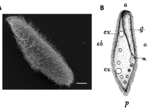
Figure 1. Paramecium morphology. A, Scanning electron microscopy image of Paramecium tetraurelia (scale
71
bar: 10 µm) (Valentine et al., 2012). B, Paramecium caudatum (Herbert S. Jennings, 1899), a large species
72
(about 200 µm) with a pointed posterior end. a, anterior end; p, posterior end; g, oral groove; m, mouth; o,
73
oral side; ab, aboral side; cv, contractile vacuole. The drawing also shows food vacuoles and cilia.
74
Paramecium is a single-cell eukaryote, 100-300 µm long depending on species (Fokin, 2010) (Fig. 1),
75
which has long been a model organism for many aspects of eukaryotic biology (Görtz, 1988;
76
Wichterman, 1986). It is a ciliate that has been living in ponds and lakes all over the world for
77
hundreds of millions of years (Parfrey et al., 2011) - a fossil has been discovered in a 200 million
78
years old piece of amber (Schönborn et al., 1999). Its abundance and large size made it a popular
79
subject of behavioral study in the late 19th century; Jennings described his culture method as follows
80
(Jennings, 1897): “A handful of hay or grass is placed in a jar and covered with hydrant water. In a few
81
weeks the solution of decaying vegetable matter swarms with Paramecia.”.
82
Paramecium swims in fresh water by beating its thousands of cilia, and feeds on smaller
83
microorganisms such as bacteria and algae. It is a prey for other microorganisms such as Didinium.
84
As beautifully described by Jennings more than a century ago (1906), Paramecium lives in a rich
85
sensory environment: it finds food by detecting and following chemicals produced by decaying plants
86
and fellow paramecia; it moves towards the water surface by gravitaxis; it avoids obstacles thanks to
87
its mechanosensitivity; it resists water currents by rheotaxis; it avoids bright light; it avoids hot and
88
cold waters; it even communicates chemically. It typically swims in helicoidal paths interrupted by
89
abrupt changes in direction called avoiding reactions, which form the “trial-and-error” basis of its
90
behavior. When an unfavorable condition is met (obstacle, unwanted chemical), the avoiding
91
reaction is triggered (Fig. 2A): Paramecium swims backward for a brief time, then turns and swims
92
forward in a new direction. By this simple mechanism, Paramecium can navigate in crowded
93
multisensory environments.
94
95
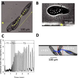
Figure 2. The avoiding reaction triggered by an action potential. A, Avoiding reaction against an obstacle, as
96
illustrated by Jennings (Jennings, 1906). B, Action potential in response to a 2 ms current pulse (top),
97
recorded with the hanging droplet method (bottom) (Naitoh et al., 1972).
98
This avoiding reaction is triggered by an action potential produced by voltage-gated calcium
99
channels located in the cilia (Fig. 2B) (Eckert, 1972). These are L-type calcium channels related to the
100
CaV1 family found in neurons, heart and muscles of mammals (Lodh et al., 2016). A number of other
101
ionic currents have been identified (Eckert and Brehm, 1979), and genes for many more ionic
102
channels have been found in the genome, often homologs of mammalian channels (Martinac et al.,
103
2008). Sensitivity to various sensory signals is provided by transduction into ionic currents, which
104
may then trigger action potentials. Piezo channels, which convey mechanosensitivity in many species
105
including mammals (Coste et al., 2010) have been identified in the genome. A rhodopsin-like protein
106
has been identified in Paramecium bursaria, a photosensitive species (Nakaoka et al., 1991). In fact,
107
many signaling pathways of neurons have been found in Paramecium, in particular calcium signaling
108
pathways (Plattner and Verkhratsky, 2018) – calcium release channels, pumps, calmodulin, centrin,
109
calcineurin, SNARE proteins, cAMP and cGMP-dependent kinases, etc. For this reason, some authors
110
have called Paramecium a “swimming neuron” (Kung and Saimi, 1985).
111
Many other motile unicellular organisms have rich behavior (Wan and Jékely, 2020) and produce
112
action potentials, including microalgae (Eckert and Sibaoka, 1968; Harz and Hegemann, 1991;
113
Taylor, 2009), other ciliates such as Stentor (Wood, 1991), other protists such as Actinocoryne
114
contractilis (Febvre-Chevalier et al., 1986) and even bacteria (Kralj et al., 2011; Masi et al., 2015). One
115
advantage of Paramecium is its large size, allowing relatively simple electrophysiological recordings
116
(Kulkarni et al., 2020; Naitoh and Eckert, 1972). For this reason, there is a rich literature on
117
Paramecium electrophysiology, mostly from the 1960-80s (Eckert, 1972; Eckert and Brehm, 1979).
118
In addition, Paramecium is still an active model organism in genetics, and benefits from many tools
119
such as RNA interference (Galvani and Sperling, 2002); its genome has also been sequenced (Aury et
120
al., 2006; McGrath et al., 2014).
121
I will first give an overview of the behavior of Paramecium, then I will explain how it moves with its
122
body and cilia, and finally I will describe the physiological basis of behavior, with a special focus on
123
the avoiding reaction. Most studies cited in this review were done on two species, P. caudatum and P.
124
aurelia.
125
126
1. The life of Paramecium
127
1.1. Swimming, feeding, reproducing
128
Behavior has been described in detail in articles and books by Jennings and a few contemporary
129
scientists, in the late 19th and early 20th century (Jennings, 1906, 1897; Jensen, 1893; Ludloff, 1895;
130
Mendelssohn, 1895) - these observations would benefit from precise and systematic measurements
131
with modern techniques. Paramecium lives in fresh water in various kinds of habitats, differing in
132
temperature and composition. It swims in spiral paths at about 1 mm/s by beating its thousands of
133
cilia, revolving around its long axis at about one cycle per second, the oral groove facing the spiral
134
axis (Fig. 3A) (Bullington, 1930; Herbert S. Jennings, 1899). These paths are occasionally interrupted
135
by abrupt changes in direction, which can be preceded by a short period of backward swimming.
136
It is often found near the water surface, as it tends to swim against gravity (Jensen, 1893, p. 18).
137
When it hits a solid surface such as glass or wood, it gives the avoiding reaction (Fig. 2A). But when it
138
encounters some fibrous material such as a decaying plant or a piece of cloth, it may stall (Jennings,
139
1897). This behavior has been termed contact reaction or thigmotaxis (Fig. 3B). It can also occur on
140
properly coated glass (Iwatsuki et al., 1996). The cilia in contact with the object are immobilized, and
141
all the other cilia are quiet or quivering except the oral cilia, which beat strongly. In this situation,
142
Paramecium may feed, for example on bacteria, yeast or algae. Food is brought into its oral groove by
143
powerful cilia, which have different properties from locomotor cilia (Aubusson-Fleury et al., 2015;
144
Jung et al., 2014).
145
146
Figure 3. Swimming, feeding and reproducing. A, Spiral swimming, with the oral groove facing the spiral axis
147
(Herbert S. Jennings, 1899). B, Thigmotactic Paramecium resting against a fiber (Jennings, 1897). Arrows
148
show water currents produced by oral cilia. C, Two paramecia in conjugation (sexual reproduction)
149
(Jennings, 1904).
150
A well-fed Paramecium can reproduce by fission every 6 hours (Beisson et al., 2010a), depending on
151
temperature (Krenek et al., 2011). Without food, Paramecium can survive for several weeks (Jackson
152
and Berger, 1984). Starvation triggers sexual reproduction, where two individuals of opposite mating
153
types attach to each other by the oral side and exchange genetic material (Fig. 3C). In P. aurelia,
154
sexual reproduction can also occur by autogamy (with itself) (Beisson et al., 2010b).
155
156
1.2. Navigating
157
When Paramecium encounters a solid obstacle, it swims backward for a fraction of second, still
158
revolving around its long axis, then the anterior end turns while the posterior end is still (Fig. 2A).
159
This is called the avoiding reaction; it forms the basis of much of its behavior. According to Jennings,
160
the organism always turns toward the same structurally defined side, the “aboral” side (away from
161
the oral groove) (Herbert S. Jennings, 1899) - although systematic measurements are lacking. But
162
since it also revolves along its long axis, from a fixed viewpoint the change in direction may alternate
163
between left and right. Thus, the change in direction may be considered as pseudo-random.
164
165
Figure 4. The avoiding reaction is graded (Jennings, 1904): swinging of the anterior end in a weak reaction
166
(A), a strong reaction (B) and a very strong reaction (C).
167
The avoiding reaction is graded (Fig. 4). A weak stimulus may only trigger a gentle reorientation with
168
no backward swimming (Fig. 4A), while a stronger stimulus induces backward swimming and
169
reorientation (Fig. 4B). A very strong stimulus may trigger long backward swimming followed by
170
turning a complete circle (Fig. 4C). This graded reaction parallels the graded action potential: the
171
duration of backward swimming correlates with the stimulus-induced depolarization (Machemer
172
and Eckert, 1973).
173
Paramecium also reacts when the rear is touched, but in a different way (Fig. 5A): it swims forward
174
faster, by beating its cilia up to twice faster (Machemer, 1974). This speed increase is accompanied
175
by a contraction along the longitudinal axis (Nakaoka and Machemer, 1990). This is called the escape
176
reaction, first described by Roesle in 1903 (1903), then by Jennings (Jennings, 1904). Non-localized
177
mechanical stimulation, as when shaking a tube of Paramecium culture, also induces an increase in
178
swimming speed that can last for several minutes.
179
When stimulated by a strong heat using a laser (5-10 °C increase), Paramecium can jump away from
180
the stimulus (possibly sideways) within just 5 ms, at about 10 mm/s (Hamel et al., 2011) (Fig. 5B). To
181
perform this feat, Paramecium throws trichocysts, which are sorts of needles docked near the
182
membrane, thereby projecting itself in the opposite direction. The same behavior occurs in reaction
183
to an appropriate chemical stimulus and in encounters with the predator Dileptus (Knoll et al., 1991).
184
185
Figure 5. Paramecium navigation. A, Escape reaction triggered by a heat stimulus (laser) near the posterior
186
end (star) (Hamel et al., 2011). B, Sideways jumping from a strong heat stimulus (star) by throwing
187
trichocysts (Hamel et al., 2011). C, Trajectory of Paramecium in a 5 mm capillary, showing an increase in
188
backward swimming after 1 min, corresponding to about 40 avoiding reactions (Kunita et al., 2014). D,
189
Bending of P. caudatum in a 160 µm channel (Jana et al., 2015).
190
When Paramecium swims in a narrow channel that does not allow it to turn, it may be trapped into a
191
dead end, where it will give the avoiding reaction repeatedly, alternatively moving backward and
192
forward against the wall (Kunita et al., 2014). But after a minute, the avoiding reaction suddenly
193
becomes much longer (several millimeters), potentially allowing the organism to escape (Fig. 5C).
194
When the channel is very narrow, Paramecium may also bend itself to move forward (Jana et al.,
195
2015; Smith, 1908) (Fig. 5D). The posterior end anchors onto the wall, presumably because tail cilia
196
do not beat (Ishikawa and Hota, 2006; Machemer and Machemer-Röhnisch, 1984), while the anterior
197
end slides along the other wall, causing the cell to bend until it can swim freely. Under some
198
conditions, Paramecium can also slide along surfaces (Li and Ardekani, 2014a; Nishigami et al., 2018;
199
Ohmura et al., 2018). Some of this behavior is due not to physiological responses but to
200
hydrodynamic interactions with surfaces (Berke et al., 2008; Lauga and Powers, 2009; Li and
201
Ardekani, 2014b; Ohmura et al., 2018).
202
Finally, in a water current, Paramecium orients itself with its anterior end directed up stream, a
203
behavior called rheotaxis. According to Jennings (1906), rheotaxis derives from the avoiding reaction.
204
When Paramecium swims along the water current, its cilia beat backwards and the water current
205
opposes that movement. This acts as a mechanical stimulus which triggers the avoidance reaction. By
206
trial and error, Paramecium turns until it faces the current. However, this remains an untested
207
hypothesis. In a few other microorganisms, rheotaxis has been attributed to hydrodynamic effects
208
(Bretherton F. P. and Rothschild Nathaniel Mayer Victor, 1961; Marcos et al., 2012).
209
210
1.3. Chemical sensing and social behavior
211
212
Figure 6. Chemotaxis and social behavior. A, Gathering of paramecia in a drop of weakly acid solution
213
(Herbert S. Jennings, 1899). B, Path followed by Paramecium in a drop of acid (Jennings, 1906). C, Paramecia
214
avoiding a drop of sodium carbonate (Herbert S. Jennings, 1899). D, Paramecia gathering in a cloud of
215
carbon dioxide generated by their respiration (Herbert S. Jennings, 1899).
216
Paramecium is sensitive to a variety of chemical compounds (Dryl, 1973; H. S. Jennings, 1899;
217
NAKATANI, 1968; Valentine et al., 2008). It is attracted by some substances, in particular bacterial
218
metabolites (folate, acetate, glutamate, cyclic AMP, biotin, ammonium, etc), weak acids, carbon
219
dioxide, colloidal solutions. These substances may indicate the distal presence of food, possibly
220
components of the “phycosphere”, the rich interface between phytoplankton and bacteria (Seymour
221
et al., 2017).
222
Other substances are repellent (e.g. alkaline solutions, quinine, ATP, GTP, GDP, NBT, Alcian Blue,
223
Cibacron blue, Cytochrome c) (Francis and Hennessey, 1995). Some of these molecules may signal
224
the distal presence of a noxious condition. For example, Hennessey speculated that ATP and GTP are
225
strong repellents because they are “blood-in-the-water signals” (Hennessey, 2005): these molecules
226
are present at high concentrations in cells, and so their presence signals cell lysis, and whatever
227
dangerous condition might have caused it.
228
Some substances only produce a reaction when Paramecium is subject to toxic doses (cane sugar,
229
dextrose, urea), effectively killing it (H. S. Jennings, 1899). For example, after some time, cane sugar
230
induces plasmolysis, and then Paramecium begins to swim backward and forward repeatedly,
231
possibly because of the induced depolarization. But many substances are toxic at doses much larger
232
than the sensitivity threshold. In a number of cases, this sensitivity is conferred by specific
233
membrane receptors, which can depolarize or hyperpolarize the cell (Van Houten, 1998) and
234
possibly modulate ionic channels (Oami, 1996a, 1996b).
235
In the 19th century, Jennings described the behavior of paramecia gathering in a drop of weak acid
236
(Fig. 6A). He linked this behavior to the avoiding reaction. When Paramecium enters a drop of acid,
237
its course is unchanged; but when it reaches the border of the drop, it gives the avoiding reaction and
238
therefore remains in the drop (Fig. 6B). On the contrary, alkaline solutions are repellent: an avoiding
239
reaction is triggered as soon as the alkaline solution is reached (Fig. 6C). More recently, various
240
substances have been characterized as attractant or repellent based on the accumulation of
241
paramecia in a test solution relative to a control solution, using different behavioral assays (Leick
242
and Helle, 1983; Levandowsky et al., 1984; Nakazato and Naitoh, 1993; Valentine and Van Houten,
243
2016; Van Houten et al., 1975).
244
As previously mentioned, when stimulated, Paramecium turns to a structurally defined side (the
245
aboral side, away from the mouth). Therefore, Paramecium is not attracted to a substance because it
246
turns towards it. Rather, its behavior seems to result from trial and error: if attractant concentration
247
increases, then Paramecium keeps on swimming in the same direction; if it decreases, then
248
Paramecium changes direction. Jennings reported that the reaction of the organism is independent of
249
where the chemical substance is applied - however, this may well depend on the compound because
250
some chemoreceptors are spatially organized (Oami, 1996b; Preston and Van Houten, 1987).
251
For this reason, this behavior is sometimes named chemokinesis (changes in motility with chemical
252
signals), which is more general than chemotaxis (movements towards a chemical stimulus) (Houten,
253
1979). In particular, chemokinesis can result not only from modulation of the avoiding reaction
254
(named klinokinesis), but also of swimming speed (named orthokinesis) (Houten, 1978).
255
Nonetheless, the chemical modulation of this apparently random motion can lead to motion towards
256
the chemical source, and presumably to a preferred orientation of the body in the direction of the
257
source (since the organism spends more time in the favored direction). There is some similarity with
258
run-and-tumble chemotaxis in bacteria for which there is a dense literature (Berg, 2008; Sourjik and
259
Wingreen, 2012), including theoretical (Berg and Purcell, 1977; Celani and Vergassola, 2010;
260
Kollmann et al., 2005; Tu, 2013; Tu et al., 2008), and with pirouettes in C. Elegans (Pierce-Shimomura
261
et al., 1999).
262
A consequence of Paramecium attraction to weak acids is social behavior, as observed by Jennings
263
(Jennings, 1897). By their respiration, Paramecium produces CO2, which is acid in solutions. At low
264
concentration, Paramecium is attracted to CO2. It follows that paramecia tend to attract each other
265
(Fig. 6D). This explains why gatherings can be observed at the bottom of a watch glass or at random
266
positions in a tube. This may play an important role in feeding behavior, as it allows paramecia to
267
collectively search for food.
268
Finally, Paramecium also has GABAA and GABAB receptors that can influence its behavior (Bucci et al.,
269
2005; Ramoino et al., 2004, 2003). For example, the activation of GABAB receptors inhibits the
270
avoiding reaction. In addition, Paramecium releases GABAupon stimulation. This release might act as
271
a signal for other paramecia, or perhaps as an externalized spatial memory for exploration (as
272
observed in slime mold (Reid et al., 2012)) - making the organism take a different action when it
273
comes back to the same location. NMDA-like receptors have also been identified (Ramoino et al.,
274
2014).
275
276
1.4. The logic of Paramecium behavior
277
Many aspects of Paramecium behavior can be described as “trial and error” (1906). If its path is
278
blocked by an obstacle, Paramecium withdraws then tries a new direction. If it encounters an
279
undesirable chemical signal, it changes direction. If it leaves a desirable region, it withdraws and tries
280
a new direction. This logic also applies to other sensory modalities. For example, when placed in a
281
gradient of temperature, Paramecium accumulates in regions with temperature close to their culture
282
temperature (Jennings, 1906; Mendelssohn, 1895). Again, this occurs by temperature-triggered
283
avoiding reactions. When temperature changes away from culture temperature (whether this
284
corresponds to a decrease or an increase), the avoiding reaction rate transiently increases;
285
conversely, the avoiding reaction rate decreases when temperature gets closer to culture
286
temperature (Nakaoka and Oosawa, 1977). This behavior is mediated by membrane potential
287
changes (Tominaga and Naitoh, 1992) produced by cold- and heat-sensitive thermoreceptors (Kuriu
288
et al., 1998, 1996; Tominaga and Naitoh, 1994).
289
Paramecium also shows photophobic responses to large changes in the intensity of visible light
290
(mainly green, and red) (Hinrichsen and Peters, 2013; Iwatsuki and Naitoh, 1982; Kenji Iwatsuki and
291
Naitoh, 1983; Kenju Iwatsuki and Naitoh, 1983). When Paramecium is kept in the dark and a bright
292
light is turned on, it displays the avoiding reaction with a latency of around a second, then adapts
293
over about 15 s. As a result, Paramecium tends to accumulate in shaded regions. A related species,
294
Paramecium bursaria, is naturally highly sensitive to light and accumulates in lighted regions (Saji
295
and Oosawa, 1974). This species harbors a symbiotic green alga named Chlorella: the alga provides
296
photosynthetic products to its host while the host brings the alga in suitable light conditions. A
297
moderate decrease in light intensity triggers an avoiding reaction, which makes P. bursaria seek light.
298
This trial-and-error behavior shares some similarity with the run-and-tumble behavior of bacteria
299
(Berg, 1975). Macroscopically, trajectories of E. Coli resemble Paramecium trajectories, with
300
helicoidal “runs” interrupted by “tumbles” where the cell changes direction randomly. Bacterial
301
chemotaxis is enabled by concentration-dependent modulation of the tumbling rate: the tumbling
302
rate decreases when concentration increases, while it is unchanged when concentration decreases.
303
Thus, tumbling is not an avoiding reaction (it is not triggered by a concentration decrease). In
304
Paramecium, the new direction is somewhat (pseudo-)random, but the turning event seems more
305
deterministically related to environmental conditions than in bacteria. In other words, the avoiding
306
reaction of Paramecium is more akin to a decision based on sensory inputs, than to a modulation of
307
the spontaneous turning rate. This difference with bacteria may be due to a difference in scale:
308
compared to bacteria, the membrane surface of Paramecium is at least two orders of magnitude
309
larger, so that the signal-to-noise ratio is at least one order of magnitude larger – membrane
310
potential fluctuations are about 1-3 mV (Moolenaar et al., 1976; Nakaoka et al., 2009).
311
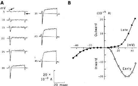
This simple logic of behavior calls for a couple of remarks in the context of neuroscience. First, it is
312
somewhat surprising that a single spiking “neuron” can control relatively complex navigation in
313
crowded multisensory environments, social behavior, and perhaps spatial memory. In terms of
314
connectionism (Seung, 2012), Paramecium is a zero-connectome organism, and yet it can accomplish
315
a variety of ecologically relevant tasks. This arises not from the complexity of the cell, which is
316
electrically much simpler than a single pyramidal cortical neuron (it is isopotential), but rather from
317
the interaction between this spiking cell and the environment, together with the exploratory
318
properties conferred by the pseudo-random nature of the effect of a spike. This highlights the
319
importance of embodiment and coupling with the environment, which are increasingly appreciated
320
in cognitive science and philosophy of mind (Ahissar and Assa, 2016; Bickhard and Terveen, 1996;
321
Brette, 2019a; Brooks, 1991; Gibson, 1979; Hurley, 2001; Maturana and Varela, 1973; O’Regan and
322
Noë, 2001; Pezzulo and Cisek, 2016; Powers, 1973).
323
Second, “control” may not be the right term to describe the relation between spiking and behavior.
324
Motor control is classically described as feedforward or feedback (Wolpert and Ghahramani, 2000).
325
In feedforward control, internal models are used to plan movements, and specific sets of neurons are
326
recruited to trigger the appropriate movements. In Paramecium, spiking produces a single type of
327
movement, regardless of the goal or stimulus: it does not move by planning specific movements. In
328
feedback control, actions are taken that reduce the difference between the observed state and a
329
desired state. In Paramecium, an action is also taken when the observed state is undesirable, but that
330
action is not directed towards the goal – rather, it is (pseudo-)random. Thus, Paramecium
331
movements are based neither on feedforward nor on feedback control, but rather on exploration and
332
selection (“trial and error”). This is reminiscent of the Darwinian insight that an apparently
goal-333
directed process can occur through random exploration and elimination of unsuccessful choices,
334
rather than by either planning or steering.
335
336
1.5. Adaptation
337
Paramecium lives in habitats of diverse ionic composition. Changes in ionic composition directly
338
affect ionic currents and reversal potentials, and therefore can potentially interfere with behavior.
339
For example, moderate changes in cation concentration can alter swimming velocity (Machemer,
340
1989; Nakaoka et al., 1983). More critically, an increase in potassium concentration can inhibit the
341
avoiding reaction through depolarization-induced inactivation of the ciliary calcium channels (Oka
342
and Nakaoka, 1989), making the organism unresponsive to stimulation. Remarkably, after a couple of
343
hours, behavior returns to its normal state before the medium changed (Oka et al., 1986) (Fig. 7A). In
344
parallel, the resting potential changes after a medium change then decays back to its original value
345
(Fig. 7B). This homeostatic regulation appears to be mediated by changes in channel permeability.
346
With a more prolonged (48 h) exposure to a high potassium solution, more complex changes in
347
excitability can occur, with enhanced responses to Mg2+ and Na+ (Preston and Hammond, 1998).
349
Figure 7. Adaptation. A, Change in swimming velocity when Paramecium adapted to a solution of 0.25 mM
350
CaCl2 and 4 mM KCl is transferred to a solution of 0.25 mM CaCl2 and 1 (open circles), 2 (closed circles), 4
351
(squares) or 16 (triangles) mM KCl (Oka et al., 1986). B, Resting potential vs. KCl concentration for cells
352
adapted to 2, 4, 8 and 16 mM KCl (top to bottom) (Oka et al., 1986). Arrows indicate the adapted state. C,
353
Accumulation of Paramecium in a warm region (Herbert S. Jennings, 1899). The top of the slide is placed on a
354
40°C bath while the bottom rests on ice. D, Change in avoiding reaction rate after paramecia cultured at 25°C
355
are transferred to 30°C (Nakaoka et al., 1982). Note the change in time scale.
356
Temperature also affects ionic channel properties and the entire metabolism of the organism, as well
357
as hydrodynamic properties (viscosity of water). For example, when temperature is lowered, the
358
ciliary calcium current is smaller and slower, action potentials are smaller and broader, cilia reverse
359
with longer latency and for a longer time (Machemer, 1974). As previously discussed, Paramecium
360
has a thermoregulation mechanism based on movement: using the avoiding reaction, it navigates
361
towards waters of a preferred temperature (Fig. 7C). However, this mechanism is insufficient if the
362
medium changes temperature globally. Remarkably, in this case, Paramecium adapts over a couple of
363
hours: behavior returns to normal and the new temperature becomes the preferred temperature
364
(Nakaoka et al., 1982) (Fig. 7D). This behavioral adaptation correlates with changes in
365
electrophysiological properties, in particular of the ciliary calcium conductance (Martinac and
366
Machemer, 1984).367
368
1.6. Learning369
Beyond adaptation, there is an important literature on learning in Paramecium and other ciliates.
370
Unfortunately, as reviewed by Applewhite (1979), many of those studies are difficult to interpret as
371
they lack appropriate controls or observations. In a series of papers (Gelber, 1962a, 1962b, 1958,
372
1957, 1956, 1952), Gelber showed an apparent reinforcement of behavior with a food reward (see
373
(Gershman et al., 2021) for a recent commentary). A platinum wire is lowered repeatedly into a
374
depression slide with paramecia. If the wire is intermittently baited with bacteria, then more and
375
more paramecia cling to the wire, even when a clean wire is finally lowered into the slide. What
376
might be the stimulus? Gelder (1956) noted that the behavior was not observed when paramecia
377
were tested in the dark, suggesting that perhaps paramecia developed an attraction to a reflection or
378
shadow cast by the wire.
379
These observations were controversial, because it was objected that lowering the baited wire
380
introduces bacteria in the fluid, to which paramecia are then attracted even when the wire is
381
removed or cleaned (Jensen, 1957). In support of this interpretation, Katz and Deterline (Katz and
382
Deterline, 1958) replicated Gelber’s main findings but found that stirring before the final test
383
destroyed the observed behavior. Naturally, this could be interpreted as an erasure of learning due to
384
the mechanical disturbance, but perhaps more crucially, they found that Gelber’s observations could
385
be reproduced when the entire experiment (not just the test) was done in the dark, effectively
386
removing any distal sensory stimulus by which paramecia may be able to recognize the wire. A
387
plausible explanation, in line with informal observations reported in this set of studies, is that
388
feeding reduces the activity of paramecia so that they tend to stay near the wire, and promotes
389
thigmotaxis so that they tend to adhere more easily to the wire. In this case, the procedure would
390
indeed reinforce a behavior, namely the feeding behavior, but not a stimulus-specific behavior. More
391
detailed observations seem necessary to understand the phenomenon.
392
Another phenomenon that has attracted some attention is tube escape learning, first described by
393
French in 1940 (French, 1940). A single Paramecium is placed in a drop and a thin tube is lowered
394
into it. The organism is drawn into the tube by capillarity. It then escapes from the bottom after
395
about 30 s. When the experiment is repeated, escape time decreases to around 15 s after a few trials.
396
French states that after the initial trials, paramecia go and back and forth in the tube only a few times
397
then take “one long dive to the bottom”. The faster escape persists for at least two hours (Huber et al.,
398
1974), which seems to rule out the possibility that Paramecium simply adapts to the mechanical
399
stimulus of capillary suction. This phenomenon has been robustly reproduced by several authors
400
(Applewhite and Gardner, 1973; Hanzel and Rucker, 1972), but its basis is unclear. Applewhite and
401
Gardner (1973) proposed that Paramecium released some substance in the tube that then influences
402
future behavior, but this hypothesis contradicts earlier results by Hanzel and Rucker (Hanzel and
403
Rucker, 1972) showing the same performance improvement in multiple paramecia with the same
404
tube. Studies of tube escape learning in Stentor, another ciliate, suggest that the phenomenon is
405
related to gravitaxis (Bennett and Francis, 1972; Hinkle and Wood, 1994). Performance
406
improvement is seen only when the tube is vertical, not when it is horizontal, where escape is fast
407
from the first trial. This suggests the following (speculative) explanation: in a vertical tube,
408
paramecia are trapped near the top because of negative gravitaxis, then prolonged confinement
409
(perhaps signaled by frequent avoiding reactions) inhibits the normal gravitactic behavior, so that
410
the organism can escape to the bottom.
411
Finally, Hennessey et al. (1979) managed to train Paramecium to react to sounds. When a tone is
412
played by a speaker below the slide, Paramecium shows no reaction. However, when the tone is
413
paired with electrical stimulation triggered in the middle of the tone, Paramecium reacts to the
414
stimulus with an avoiding reaction, then after a few trials gives an avoiding reaction at the onset of
415
the tone, in anticipation of the electrical stimulus. The authors demonstrate extinction (reaction
416
disappears when sound is presented alone), retention and specificity (reacting specifically to a 300
417
Hz tone or to a 500 Hz tone). The physiological basis is not known.
418
Armus et al. (Armus et al., 2006a, 2006b; Mingee and Armus, 2009) trained paramecia to go to a
419
lighted region. The bath is split into two compartments, one in the dark, the other one in light.
420
Initially, Paramecium spends more time in the dark compartment, because of photophobia. Training
421
consists in electrically stimulating the cell when it enters the compartment of the cathode. After
422
training, Paramecium spends more time than before in the cathodal half, which now only differs by
423
its lighting. However, if stimulation is triggered in the anodal half, then after training Paramecium
424
spends less time in that half. Therefore, the phenomenon does not seem to be based on an
425
association between the electrical stimulus and the light stimulus. A plausible interpretation is the
426
following. As is known from studies of galvanotaxis (Dale, 1901; Ludloff, 1895), electrical stimulation
427
makes Paramecium move towards the cathode. Stimulation in the lighted cathodal compartment then
428
makes Paramecium spend more time in light, which results in adaptation of the photophobic
429
behavior. Thus, after training, Paramecium spends more time than before in the lighted
430
compartment. This interpretation is supported by the observation that the “trained” behavior only
431
occurs when the cathodal compartment is lighted during training (Alipour et al., 2018), and by the
432
absence of retention (Mingee, 2013).
433
In summary, although the existing literature is complex, there is clear evidence of behavioral
434
plasticity in Paramecium. Some can be categorized as adaptation, and there is at least one
435
documented case of learning (Hennessey et al., 1979), understood as a persistent stimulus-specific
436
change in behavior.
437
438
2. The motor system of Paramecium
439
2.1. How Paramecium swims
440
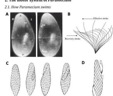
441
Figure 8. Spiral swimming. A, Organization of ciliary basal bodies on the oral (ventral) and aboral (dorsal)
442
side (Iftode et al., 1989). B, Ciliary beat cycle: power stroke (or effective stroke) and recovery stroke (Omori et
443
al., 2020). C, Water currents produced by cilia for different orientations of Paramecium (Jennings, 1904). In
444
the oral groove, currents are oriented towards the mouth. D, Metachronal waves represented by parallel
445
lines, progressing transversally, with cilia’s power stroke oriented towards the right and rear (Machemer,
446
1972). Cilia on parallel lines are at the same phase of the beat cycle. The curved arrow shows the direction of
447
movement.
448
In the absence of any stimulus, Paramecium swims in spirals. Paramecium is covered by several
449
thousand cilia (Fig. 8A) (about 4000 cilia in P. tetraurelia (Aubusson-Fleury et al., 2015); see Iftode et
450
al. (1989) for precise counts and spatial pattern), each about 10 µm long and 0.2 µm thick (Eckert
451
and Naitoh, 1970), similar to other motile cilia of eukaryotes, including mammals (Ishikawa, 2017).
452
In forward swimming, each cilium beats at 10-20 Hz (Fig. 8B), with a power stroke toward the right
453
and rear on the visible surface (Fig. 8C). Thus, on the hidden surface (further from the observer), cilia
454
beat towards the left and rear. This results in a forward movement with a rotation around the
455
longitudinal axis, as in unscrewing (over to the left) (Fig. 8D). The typical velocity is about 1 mm/s
456
(Machemer et al., 1991).
457
The spiral is wider than the cell’s width, as first described by Jennings (1901) and later by Bullington
458
(1930). A possible reason is that cilia in the oral groove beat in a specific direction, towards the
459
mouth, which counters the movement produced by the other cilia. A recent study has shown indeed
460
that properties of oral cilia differ from other cilia (Jung et al., 2014). This may explain why the
461
trajectory describes a wide spiral, with the oral side always facing its axis (Fig. 3A) (Párducz, 1967a).
462
Properties of spiral swimming can vary, in particular its speed and width. Paramecium can also swim
463
backward, with an effective stroke towards the front and slightly to the right. Thus, in backward
464
swimming, the movement is not the symmetrical of forward swimming: the cell still rotates in the
465
same direction.
466
Cilia beating is coordinated over the cell in the form of metachronal waves, which progress over the
467
surface at about 1 µm/ms (Párducz, 1967b) (Fig. 8D). These waves encircle the body in spirals
468
(Machemer, 1969; Párducz, 1967a). Cilia beat against the direction of the wave, but not at 180°, a
469
pattern called “dexio-antiplectic”. This particular kind of motor coordination is functionally
470
important. A key characteristic of swimming microorganisms is they live at low Reynolds number (R
471
0.1 for Paramecium) (Purcell, 1977), that is, inertial forces are small compared to viscous forces (as
472
if a human were trying to swim in honey). As a consequence, the swimmer stops as soon as cilia stop
473
beating. Therefore, if cilia beating were synchronized over the entire body, then the swimmer would
474
move forward in regular discontinuous steps. In fact, this can happen in the escape reaction: a strong
475
heat stimulus near the posterior end induces a synchronous power stroke of the cilia (as in the
476
butterfly stroke) (Hamel et al., 2011), which results in a transient speed increase immediately
477
followed by an almost complete stop, before the metachronal pattern is reestablished. If on the
478
contrary cilia beating were completely disorganized (which can happen transiently in the avoiding
479
reaction), then neighboring cilia might beat in inconsistent directions and this is not an efficient way
480
of swimming. In fact, it has been shown that the metachronal pattern optimizes the energetic
481
efficiency of swimming (Gueron and Levit-Gurevich, 1999; Osterman and Vilfan, 2011).
482
It was once postulated that ciliary coordination might be electrically controlled by the cell, but
483
Paramecium is essentially isopotential (Eckert and Naitoh, 1970). Instead, cilia coordination is
484
mediated by hydrodynamic interactions (Guirao and Joanny, 2007; Machemer, 1972) and mechanical
485
coupling through the compliant body (Narematsu et al., 2015), in the absence of any central agency.
486
This illustrates the concept of embodiment in motor neuroscience: part of the problem of efficient
487
coordination is solved not by manipulating body representations, but by direct physical interaction
488
of the body with its immediate environment (Tytell et al., 2011). In the case of microorganisms such
489
as Paramecium, the results of this physical interaction can be understood precisely, thanks to an
490
abundant literature on the mechanics of cilia and flagella (Blake and Sleigh, 1974; Sartori et al., 2016;
491
Wan, 2018) including mathematical models (Dillon et al., 2007; Yang et al., 2008), as well as on the
492
hydrodynamics of swimming microorganisms (Jung et al., 2014; Keller and Wu, 1977; Lauga and
493
Powers, 2009).
494
495
2.2. How Paramecium moves upward
496
As many other microorganisms (Häder and Hemmersbach, 2018), Paramecium tends to aggregate
497
near the water surface, despite the fact that it is slightly heavier than water (about 4%), a puzzling
498
phenomenon which has attracted an abundant literature, first described in detail by Jensen in 1893
499
(Jensen, 1893). When observed in a vertical plane, trajectories are curved upward (Roberts, 2010)
500
(Fig. 9A). The earliest explanation, the gravity-buoyancy model, postulates a mismatch between the
501
buoyancy center and the gravity center (Verworn, 1889): this could generate a torque making the
502
body align with gravity. Roberts (Roberts, 2010, 1970) argued that density inhomogeneities are
503
unlikely to be sufficient to account for the observations, and instead proposed a drag-gravity model:
504
as the posterior end is larger than the anterior end, the viscous drag differs and the posterior end
505
falls more rapidly than the anterior end; thus, the cell turns upward. However, Jensen (Jensen, 1893)
506
and later Kuznicki (Kuznicki, 1968) observed that dead or immobilized cells fall with no preferred
507
orientation – although this is questioned by Roberts (Roberts, 1970). This would discard both
508
passive orientation mechanisms. The propulsion-gravity model (Winet and Jahn, 1974) is a more
509
complex proposition, which links gravitaxis with ciliary beating: sedimentation introduces viscous
510
resistance to beating that is stronger in the up phase of the helicoidal cycle than in the down phase,
511
resulting in velocity-dependent reorientation.
512
In addition to these hydrodynamic mechanisms, physiological mechanisms have been postulated. It
513
has been observed that Paramecium swims slightly faster upwards than downwards, once
514
sedimentation has been subtracted (Machemer et al., 1991; Ooya et al., 1992) (Fig. 8B), and the
515
avoiding reaction is triggered more often when it swims backwards than upwards, although this bias
516
tends to disappear after some time (Nagel and Machemer, 2000) (Fig. 8C). Although spurious
517
correlations should be ruled out (e.g., cells that swim more slowly may tend to fall), Machemer and
518
coll. have proposed that this is due to pressure differences between the top and bottom ends of the
519
cell, which are sensed by mechanoreceptors. As there is a spatial gradient of mechanosensitivity
520
between the front and rear, the transduced current would be hyperpolarizing when the anterior end
521
is upward (increased pressure on the rear end) and depolarizing when the anterior end is
522
downward. In support of this hypothesis, a cell vertically immobilized between two horizontal
523
electrodes can spontaneously turn upward or downward, and small membrane potential changes
524
with the expected sign are observed, although with long latency (on the order of 20 s) (Gebauer et al.,
525
1999). These physiologically induced changes in mean velocity and avoiding reaction rate likely
526
represent a small contribution to gravitaxis, compared to the reorientation of the cell (Roberts,
527
2010), but it is conceivable that reorientation itself occurs by physiological modulation of velocity
528
within the helicoidal cycle (Mogami and Baba, 1998).
529
530
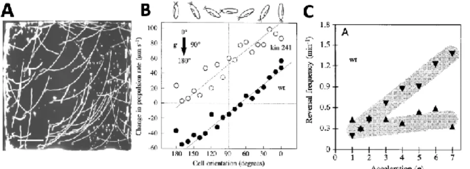
Figure 9. Gravitactic behavior of Paramecium. A, Upwardly curved trajectories of Paramecium in a vertical
531
chamber (Roberts, 2010). B, Velocity change (corrected for sedimentation) as a function of cell orientation
532
(Nagel and Machemer, 2000) - open circles correspond to a morphological mutant. C, Avoiding reaction
533
frequency as a function of acceleration in a centrifuge microscope, after 4 h of equilibration (Nagel and
534
Machemer, 2000). Triangles indicate cell direction.
535
536
2.3. How Paramecium turns
537
In the avoiding reaction, Paramecium swims backward (if the reaction is strong) then turns before it
538
swims forward again. Backward swimming occurs because cilia reorient, with the power stroke
539
oriented towards the anterior end instead of the posterior end, but how can Paramecium turn?
540
Turning requires some inhomogeneity in the ciliary beating pattern.
541
First, anterior and posterior cilia do not revert synchronously during the avoiding reaction (Fig. 10A)
542
(Machemer, 1969; Párducz, 1967a). When the avoiding reaction is initiated, all cilia simultaneously
543
strike forward, which moves the cell backward (2). The beating pattern then progressively
544
reorganizes into the metachronal pattern as the cell swims backward (3-5). Reorientation of the cell
545
starts when the anterior end reverts to the forward metachronal pattern (6-8). Thus, anterior and
546
posterior ends show different metachronal patterns, respectively of forward and backward
547
swimming.
548
549
Figure 10. Details of the avoiding reaction. A, Reorganization of the ciliary beating pattern during the
550
avoiding reaction (after (Machemer, 1969)). B, Cross-section of Paramecium seen from the anterior end,
551
during forward swimming (a, corresponding to step 1) and during reorientation (b, corresponding to step 6),
552
according to Jennings (Jennings, 1904). The arrows correspond to the induced movement of the body
553
(opposite to the beating direction).
554
It is not obvious, however, how this asynchronous pattern would make the cell turn. If the beating
555
pattern were axisymmetric, then the net force produced by either group of cilia (anterior or
556
posterior) should be directed along the main axis. Jennings claims that cilia in the oral groove may
557
also reverse, i.e., they expel fluid from the mouth (1899) (Fig. 8C). This could make Paramecium turn
558
towards its aboral (dorsal) side, as observed, but Jennings and Jamieson observed that when
559
Paramecium was cut in two pieces below the oral groove, both pieces could turn in a similar way
560
(Jennings and Jamieson, 1902). Jennings also mentions that cilia of the anterior end do not all strike
561
to the right: instead, they strike towards the oral groove (Jennings, 1904) (Fig. 10B). As a result, the
562
cell turns towards the aboral side. This is supported by more recent observations in a flattened
563
ciliary sheet from Paramecium (Noguchi et al., 1991). Thus, turning likely results from
564
inhomogeneity in the response of different groups of cilia, but details are still lacking.
565
566
3. The physiological basis of behavior
567
3.1. The action potential
568
When Paramecium touches an obstacle, mechanosensitive channels open, depolarize the membrane
569
and trigger a calcium-based action potential (Eckert, 1972). The entry of calcium then triggers the
570
reorientation of cilia, so that Paramecium swims backwards. Then calcium is buffered or pumped out
571
(Plattner et al., 2006; Yano et al., 2015) and the cilia reorient in the original direction.
572
Historically, Paramecium electrophysiology has been studied by placing the cell in a tiny droplet,
573
letting the fluid evaporate until the cell is captured by surface tension, then inserting sharp
574
microelectrodes and covering with extracellular medium (Naitoh and Eckert, 1972). A recent method
575
immobilizes the cell by suction against a filter (Kulkarni et al., 2020).
576
Paramecium is an isopotential cell, as demonstrated with two-electrode measurements (Dunlap,
577
1977; Eckert and Naitoh, 1970; Satow and Kung, 1979), which is a particularly favorable situation for
578
electrophysiological modeling. This can be sensed from an estimation of the electrotonic length
579
𝜆 = √𝑑𝑟𝑚
4𝑅𝑖, where d is diameter, rm is specific membrane resistance and Ri is intracellular resistivity.
580
For P. tetraurelia, cell width is 34 µm (Nagel and Machemer, 2000); with rm = 64 000 Ω.cm2 (Dunlap,
581
1977) and Ri = 500 Ω.cm (conservative estimate based on the ~5 lower intracellular ionic content
582
compared to mammals), we obtain 𝜆 ≈ 330 mm, much larger than the cell’s length (115 µm). In the
583
same way, for a 200 nm wide cilium, we obtain 𝜆 ≈ 260 µm, much larger than its 10 µm length.
584
Paramecium has a resting potential of about -30 to -20 mV (more depolarized than neurons),
585
depending on the extracellular medium (Naitoh and Eckert, 1968a). P. Caudatum has a capacitance of
586
about 700 pF, half of which is due to the cilia (Machemer and Ogura, 1979), and a resistance of about
587
65 M𝛺 (again depending on the extracellular medium), giving a membrane time constant of about 45
588
ms. P. tetraurelia, which is smaller, has a resistance of about 45-60 M𝛺 (Nagel and Machemer, 2000;
589
Satow and Kung, 1976). Capacitance is not documented, but a simple scaling based on membrane
590
area (Machemer and Ogura, 1979; Nagel and Machemer, 2000) gives about 300 pF. These values are
591
consistent with the surfacic capacitance of other cells including neurons (about 1 µF/cm2).
592
The negative resting potential is due to a high intracellular concentration of K+ ions (18-34 mM
593
depending on studies (Hansma, 1974; Naitoh and Eckert, 1973, 1969; Oertel et al., 1978; Ogura and
594
Machemer, 1980; Oka et al., 1986)), much larger than the extracellular concentration (typically about
595
1-4 mM KCl in experiments) (Machemer, 1998; Machemer and Ogura, 1979). Conversely, there is a
596
low intracellular concentration of Ca2+ ions at rest (50-200 nM (Iwadate, 2003; Klauke and Plattner,
597
1997)) while the extracellular concentration is orders of magnitude higher (the minimal viable
598
concentration is about 0.1 mM (Naitoh and Eckert, 1968a)). At rest, the membrane is permeable to
599
many cations (Naitoh and Eckert, 1968a). Thus, the ionic content of the cytosol is ~5 times lower
600
than metazoan cells (where intracellular K+ concentration is about 150 mM). One reason might be
601
that the extracellular medium (fresh water) typically has very low ionic content, so that the cytosolic
602
ions exert a large osmotic pressure on the membrane. In Paramecium and other protozoa, this
603
osmotic imbalance is regulated by specialized organelles, the contractile vacuoles, which expel water
604
that invades the cell by osmosis (Allen and Naitoh, 2002).
605
606
Figure 11. Membrane potential responses to mechanical stimulation with a glass stylus on the front (A) and
607
on the rear (B) (Naitoh and Eckert, 1969) (top traces: voltage command to the piezoelectric actuator).
608
When Paramecium is mechanically stimulated on the front, or a current is injected, the membrane is
609
depolarized (Fig. 11). If the stimulus is strong enough, this depolarization triggers a graded action
610
potential, with a stimulus-dependent amplitude (all-or-none spikes can occur if extracellular calcium
611
is partially replaced by barium (Naitoh and Eckert, 1968b)). This action potential is due to calcium
612
voltage-gated channels distributed over the cilia and delayed rectifier potassium channels located in
613
the somatic membrane; this can be demonstrated by removing the cilia with ethanol and shaking
614
(Machemer and Ogura, 1979). In response to a voltage step, the cell produces a current consisting of
615
two phases: a fast inward current carried by Ca2+, and a slower outward current carried by K+ (Fig.
616
12), which have been separated using behavioral mutants (Saimi and Kung, 1987).
617
618
619
Figure 12. Action potential currents in P. caudatum (Brehm and Eckert, 1978a). A, Current recorded in
620
voltage-clamp with different depolarization steps above resting potential. The first and last peaks are
621
capacitive transients. The early negative transient is mediated by calcium; the late positive current is
622
mediated by potassium. B, Early and late currents vs. membrane potential (relative to rest).
623
The Ca2+ current inactivates quickly (a few ms) by a calcium-dependent mechanism: the entry of
624
calcium (rather than voltage) inactivates the channels (Brehm et al., 1980; Eckert and Brehm, 1979;
625
Eckert and Chad, 1984) - there is also a slower voltage-gated inactivation acting over tens of seconds
626
(Hennessey and Kung, 1985). Recovery from inactivation takes a few tens to a hundred of ms (Brehm
627
et al., 1980; Naitoh et al., 1972). This is a common form of inactivation of calcium channels in
628
neurons, which has been discovered first in Paramecium (Brehm and Eckert, 1978a). It involves
629
calmodulin, a highly conserved calcium sensor that is found across all species (Ben-Johny and Yue,