HAL Id: hal-01602910
https://hal.archives-ouvertes.fr/hal-01602910
Submitted on 27 May 2020
HAL is a multi-disciplinary open access
archive for the deposit and dissemination of
sci-entific research documents, whether they are
pub-lished or not. The documents may come from
teaching and research institutions in France or
abroad, or from public or private research centers.
L’archive ouverte pluridisciplinaire HAL, est
destinée au dépôt et à la diffusion de documents
scientifiques de niveau recherche, publiés ou non,
émanant des établissements d’enseignement et de
recherche français ou étrangers, des laboratoires
publics ou privés.
Nod2: The intestinal gate keeper
Ziad Al Nabhani, Gilles Dietrich, Jean-Pierre Hugot, Frederick Barreau
To cite this version:
Ziad Al Nabhani, Gilles Dietrich, Jean-Pierre Hugot, Frederick Barreau. Nod2: The intestinal gate
keeper. PLoS Pathogens, Public Library of Science, 2017, 13 (3), �10.1371/journal.ppat.1006177�.
�hal-01602910�
REVIEW
Nod2: The intestinal gate keeper
Ziad Al Nabhani
1,2, Gilles Dietrich
3, Jean-Pierre Hugot
1,2,4*
, Frederick Barreau
3*
1 Laboratoire Inflamex, Universite´ Paris-Diderot Sorbonne Paris-Cite´, Paris, France, 2 INSERM, UMR 1149,
Paris, France, 3 IRSD, Universite´ de Toulouse, INSERM, INRA, ENVT, UPS, Toulouse, France,
4 Assistance Publique Hoˆpitaux de Paris, Hoˆpital Robert Debre´, Paris, France
*[email protected](JPH);[email protected](FB)
Abstract
Nucleotide-binding oligomerization domain 2 (NOD2) is an intracellular pattern recognition
receptor that senses bacterial peptidoglycan (PGN)-conserved motifs in cytosol and
stimu-lates host immune response. The association of NOD2 mutations with a number of
inflam-matory pathologies, including Crohn disease (CD), Graft-versus-host disease (GVHD), and
Blau syndrome, highlights its pivotal role in host–pathogen interactions and inflammatory
response. Stimulation of NOD2 by its ligand (muramyl dipeptide) activates pro-inflammatory
pathways such as nuclear factor-
κ
B (NF-
κ
B), mitogen-activated protein kinases (MAPKs),
and Caspase-1. A loss of NOD2 function may result in a failure in the control of microbial
infection, thereby initiating systemic responses and aberrant inflammation. Because the
ligand of Nod2 is conserved in both gram-positive and gram-negative bacteria, NOD2
detects a wide variety of microorganisms. Furthermore, current literature evidences that
NOD2 is also able to control viruses’ and parasites’ infections. In this review, we present
and discuss recent developments about the role of NOD2 in shaping the gut commensal
microbiota and pathogens, including bacteria, viruses, and parasites, and the mechanisms
by which Nod2 mutations participate in disease occurrence.
Introduction
The mammalian intestinal tract harbors a community of trillions of bacteria, archaea, fungi,
and viruses, which are collectively referred to as the microbiome. It is now well accepted that a
mutualistic relationship between host and microbiome is essential for immune homeostasis
[
1
]. The microbiome is required for the development [
2
] and regulation of intestinal immune
responses against commensals and pathogens, thereby maintaining the intestinal homeostasis.
Initiation of the immune response depends on the recognition of microbial-associated
molecular patterns (MAMPs) through special cell receptors called pattern recognition
recep-tors (PRRs). PRRs are classified into five distinct genetic and functional clades (for review, see
[
3
]). Most of our knowledge concerning PRRs comes from studies on toll-like receptors
(TLRs), which are localized either at the cell surface or within endosomes [
4
,
5
]. By contrast,
the nucleotide oligomerization domains (Nod)-like receptors (NLRs) are intracellular sensors,
including 22 members in humans and 34 members in mice [
6
]. The activation of multiple
PRRs in response to a pathogen triggers nuclear factor-κB (NF-κB), mitogen-activated protein
a1111111111
a1111111111
a1111111111
a1111111111
a1111111111
OPEN ACCESSCitation: Al Nabhani Z, Dietrich G, Hugot J-P,
Barreau F (2017) Nod2: The intestinal gate keeper. PLoS Pathog 13(3): e1006177. doi:10.1371/ journal.ppat.1006177
Editor: James B. Bliska, Stony Brook University,
UNITED STATES
Published: March 2, 2017
Copyright:© 2017 Al Nabhani et al. This is an open access article distributed under the terms of the
Creative Commons Attribution License, which permits unrestricted use, distribution, and reproduction in any medium, provided the original author and source are credited.
Funding: Financial support was provided by
INSERM, Universite´ Paris Diderot, Assistance Publique Hopitaux de Paris, and Association Franc¸ois Aupetit. We acknowledge the financial support of the Investissements d’Avenir programme ANR-11-IDEX-0005-02, Sorbonne Paris Cite, Laboratoire d’excellence INFLAMEX. The funders had no role in study design, data collection and analysis, decision to publish, or preparation of the manuscript.
Competing interests: The authors have declared
kinases (MAPKs), Caspase-1 activation, and both interleukin 1 (IL-1) and type I interferon
(IFN) secretion, inducing inflammation [
3
].
NOD2, also known as NLRC2, belongs to the NLR family and functions as an intracellular
PRR for muramyl dipeptide (MDP) derived from peptidoglycan (PGN) of both gram-positive
and gram-negative bacteria [
7
]. Since its identification in 2001 [
8
] and its association with
Crohn disease (CD) [
9
,
10
], the role of NOD2 in both innate and adaptive immune responses
gained increasing interest.
NOD2 mutations confer highest risks for CD, but also for
Graft-versus-host disease (GVHD) [
11
] and Blau syndrome [
12
]. Dysregulation of Nod2 signaling
causes or contributes to increased infection risks in human and animal models. This review
focuses on the role of NOD2 in the recognition and elimination of commensal and pathogenic
bacteria, viruses, and parasites in the gut.
NOD2 expression, activation, structure, and signaling
In the intestine, NOD2 is expressed by numerous cell types, including hematopoietic cells [
13
]
(such as T cells [
14
], B cells [
15
], macrophages [
16
], dendritic cells [
17
], and mast cells [
18
])
and nonhematopoietic cells (such as Paneth cells [
19
], stem cells [
20
], goblet cells [
21
], and
enterocytes [
22
,
23
]). NOD2 senses MDP, which is derived from partial degradation of PGN
[
7
]. MDP directly binds to the nucleotide-binding domain of NOD2 [
24
,
25
] from amino acids
216 to 821 [
25
] with an optimal efficiency within a pH ranging from 5.0 to 6.5 [
24
]. NOD2 is
able to detect many types of PGN; however, its level of activation is dependent on the PGN’s
origin [
26
]. Following activation, NOD2 activates NF-κB and MAPK signaling [
27
,
28
], thereby
contributing to host defense via the production of inflammatory cytokines, antimicrobial
mol-ecules [
29
], and mucins [
21
].
The mechanisms by which PGN enters eukaryotic cells and activates NOD2 remain poorly
understood, but several routes of entry have been proposed. Host cells can internalize MDP
through either phagocytosis of whole bacteria, endocytosis, uptaking of PGN fragments from
outer membrane vesicle (OMVs) [
30
,
31
], or transmembrane channels such as hPepT1 [
32
,
33
].
A new way of Nod2 activation involving the entry of MDP via the apparatus secretion system
of bacteria has recently been reported [
34
]. NOD2 activation requires its location to be in the
vicinity of the site of MDP delivery, close to the plasma membrane or endosomes in which two
peptide transporters, SLC15A3 and SLC15A4, may transport MDP toward the cytosolic
com-partment [
32
] (
Fig 1
).
NOD2 protein exhibits three domains, including caspase activation and recruitment
domains (CARDs), nucleotide-binding oligomerization domain (NOD), and leucine-rich
repeat (LRR). The NOD module contains a nucleotide-binding domain (NBD), a winged helix
(WH), and two helix domains (HD1 and HD2). The interaction between NBD and WH,
important to stabilize Nod2 in an inactive form, is maintained by ADP-mediated packed
con-formation [
35
]. In the absence of MDP binding, the LRR domain prevents NOD2
dimeriza-tion. Upon ligand binding, HD2 mediates conformational changes of the NBD, WH, and HD1
to allow ADP-ATP exchange, self-oligomerization, and downstream signaling [
36
]. The
effec-tor CARDs mediate intracellular signaling after interaction between the LRR domain and
MDP (
Fig 1
). NOD2 oligomerization induces a signaling complex named nodosome [
37
]. The
nodosome may be formed at the plasma cell membrane, where bacteria are taken in charge
[
37
]. Among the recruited interactants, NOD2 firstly attracts RIP2 via a CARD–CARD
homo-typic interaction [
8
], followed by TAK1 and TAB2 and TAB3 [
38
]. The kinase activity of
TAK1 induces the activation of MAPKs and NF-κB pathways [
38
]. The interaction of NOD2
with other partners, including Caspase-1 [
39
] and ATG16L1 [
40
], results in IL-1β secretion
and autophagy, respectively (
Fig 1
).
NOD2 and the intestinal microbiota
Humans are colonized by a collection of microbes, the largest numbers of which reside in
the distal gut. The human gut contains between 500 and 1,000 bacterial species. There is a
gradual increase in bacterial populations all along the small bowel, from approximately 10
4col-ony forming units (CFUs) per gram of luminal content in jejunum to 10
7in the ileum, with a
preponderance of gram-negative aerobes. By contrast, the human colon is highly colonized
with anaerobic bacteria, with about 10
14per gram of luminal content. The intestinal
micro-biota species belong to only eight of the 55 known bacteria phyla (Firmicutes, Bacteroidetes,
Fig 1. Mechanisms by which MDP enters into cells to trigger Nod2 signaling. Several routes of MDP entry have been evidenced. Host cells can
internalize MDP through either phagocytosis of whole bacteria, endocytosis, uptaking of PGN fragments from OMVs, or transmembrane channels such as hPepT1. A new way of Nod2 activation involving the entry of MDP via the apparatus secretion system of bacteria has recently been described. NOD2 activation requires its location to be in the vicinity of the site of MDP delivery. Two peptide transporters (SLC15A3 and SLC15A4) are able to translocate MDP toward the cytosolic compartment. NOD2 protein exhibits three domains, including caspase activation and recruitment domains (CARDs), nucleotide-binding oligomerization domain (NOD), and leucine-rich repeat (LRR). The NOD module contains a nucleotide-nucleotide-binding domain (NBD), a winged helix (WH), and two helix domains (HD1 and HD2). The interaction between NBD and WH, important to stabilize Nod2 in an inactive form, is maintained by adenosine diphosphate (ADP)-mediated packed conformation. Upon ligand binding, HD2 mediates conformational changes of the NBD, WH, and HD1 to allow ADP-ATP exchange, self-oligomerization, and downstream signaling. The effector CARDs mediate intracellular signaling after interaction between the LRR domain and MDP. NOD2 oligomerization induces a signaling complex named nodosome. NOD2 attracts receptor-interacting serine/threonine-protein kinase 2 (RIP2) via a CARD–CARD homotypic interaction, followed by transforming growth factor beta-activated kinase 1 (TAK1) and TAK1 binding proteins 2 and 3 (TAB2 and TAB3). This complex induces the activation of both MAPKs and NF-κB pathways. The interaction of NOD2 with other partners, including Caspase-1 and ATGCaspase-16LCaspase-1, results in IL-Caspase-1βsecretion and autophagy, respectively.
Actinobacteria, and Proteobacteria phyla being the most widely represented). The gut
micro-biota acts as a “metabolic organ” through breakdown of indigestible dietary carbohydrates and
proteins and generation of fermentation end-products and vitamins. The microbiota
contrib-utes also to the intestinal barrier function, which constitcontrib-utes an obstacle to pathogen invasion
of the intestinal mucosa. Commensal bacterial flora is known to be affected by numerous
fac-tors, including antibiotics, genetic background, diet, parents, and siblings. Moreover, several
human diseases, including inflammatory bowel diseases (IBDs), obesity and metabolic
disor-ders, and infectious and neurological diseases, are linked to a so-called microbiota dysbiosis.
Abnormal interactions between host and microbes (either pathogen or commensal) are
involved in IBDs, including CD and ulcerative colitis (UC). IBD physiopathology is associated
with significant shifts in the composition of the enteric microbiota (i.e., dysbiosis), notably via
an increased richness of the Bacteroidetes, Actinobacteria, and Proteobacteria phyla and a
depletion of the Firmicutes phylum [
41
,
42
]. The loss of Firmicutes is mostly due to the
reduc-tion of species that belong to the bacterial order Clostridiales, particularly members of the
Clostridium clusters XIVa and IV [
43
–
45
]. One member of this Clostridiales order that is
dras-tically reduced in the ileum of patients with CD is
Faecalibacterium prausnitzii [
46
].
Since Nod2 is an intracellular microbial sensor for gram-positive and gram-negative
bacte-ria, it has been proposed that
Nod2 deficiency or mutations can contribute to the modification
of microbial composition, and then disease development. In humans, an increased load of
Bac-teriodetes was observed in the ileal mucosa of CD patients with homozygosity in
NOD2
muta-tions [
47
].
NOD2 mutations have also been associated with an increased load of Escherichia
coli (Proteobacteria) and a reduced load of F. prausnitzii (Firmicutes) [
45
,
47
–
49
]. In mice,
numerous studies have reported the key role played by Nod2 in the maintenance of the gut
microbiota [
21
,
47
,
50
–
55
]. Compared to control mice,
Nod2
KOmice display an increased
fre-quency of the Bacteriodetes phylum and a decrease in the Firmicutes phylum in intestine and
feces [
21
,
47
,
50
–
55
]. As the modifications of the microbiota linked to
Nod2 deficiency at genus
level is dependent on the conditions of animal housing, the identification of bacterial species
impacted by Nod2 remains difficult to establish. Although microbial dysbiosis in
Nod2
KOmice
have been reported by several groups, two studies failed to show significant differences in the
gut microbiota when
Nod2
KOand wild-type (WT) mice were cohoused [
56
,
57
]. Indeed, if the
cage effect, drift in independent lines, coprophagia, and genetic background have not all been
taken into consideration, studies investigating microbiota communities in genetically altered
mice are often misleading. Cohousing seems to be a very rigorous strategy, but the absence of
any difference between WT and
Nod2
KOmice [
56
,
57
] may result from coprophagia and the
subsequent homogenization of mouse microbiota [
57
]. Indeed,
Nod2
KOmice obtained by
embryo transfer into WT mice exhibit an intestinal microbiota different from their mothers
but similar to that of single-housed
Nod2
KOmice [
53
]. Thus, the use of embryo transfer
strat-egy, which reduces the impact of environmental and mother parameters, points out the role of
Nod2 deficiency in the active acquisition of dysbiosis [
53
]. WT and
Nod2
KOmice obtained by
embryo transfer into WT mother mice exhibit the same microbiota when housed in the same
cage, confirming the homogenization of the gut microbiota between cohoused mice (likely
through coprophagia). Moreover, the difference in intestinal flora between WT and
Nod2
KOoffspring and their WT mothers shows that microbial dysbiosis linked to
Nod2 deletion is
transmissible and dominant [
53
]. Moreover, microbiota dysbiosis, which occurs in
Nod2
KObut also in
RIP2
KOmice, may enhance sensitivity to both colitis and colonic adenocarcinoma.
Sensitivity to colitis is transmissible to WT mice via the microbiota after cohousing. Since diet
dominates host genotype in shaping the gut microbiota [
58
], a common dysbiosis shared by
people in close contact might explain development of CD in spouses of CD patients and the
nonrandom distribution of CD within multiplex sibships [
59
].
The mechanisms by which
Nod2 regulates microbiota communities in the gut are still
unclear, even though it is commonly admitted that Nod2 in intestinal epithelial cells plays a
major role by promoting the production of antibacterial compounds, including defensins, by
Paneth cells [
19
,
29
,
48
,
54
,
60
–
62
]. The impact of the genetic background in the effect of Nod2
deficiency on the expression of defensins is, however, matter of debate [
29
,
57
]. Goblet cell
abnormalities, including decrease in number and mucins secretion [
21
], have also been
reported to be linked to
Nod2 deficiency. The failure in goblet cell function was associated
with an overproduction of IFN-γ by intraepithelial lymphocytes and the expansion of
Bacter-oides vulgatus.
Nod2 not only regulates the bacterial load and microbiota composition but also plays a key
role in shaping bacterial translocation and attachment on gut epithelium. Indeed,
Nod2
KOmice exhibit an increased bacterial translocation of both gram-positive and gram-negative
bac-teria and the yeast
Saccharomyces cerevisiae. This barrier defect is specifically located at Peyer’s
patches in the ileum [
63
]. Although commensal
E. coli may attach at all intestinal segments
[
64
], adherent-invasive
E. coli (AIEC), known to be associated with CD, has an excessive
capacity to attach at the surface of Peyer’s patches in
Nod2
KOmice [
65
]. The infiltration of T
helper type 1 (Th1) lymphocytes (secreting TNF-α and IFN-γ) resulting in an overexpression
of the myosin light chain kinase (MLCK) in epithelial cells was proposed as a mechanism for
bacteria translocation across the Peyer’s patches [
66
]. Similarly, a bacteria-induced
overactiva-tion of the MLCK may increase the number of TGF-β-producing regulatory CD4
+T cells in
the colonic lamina propria of
Nod2
KOmice through the induction of an excessive permeability
[
67
]. This reciprocal link between immune cells, intestinal permeability, and microbiota is
fur-ther evidenced by the fact that endocytosis of commensal bacteria in epithelial cells is
depen-dent on MLCK-activated brush border fanning triggered by IFNγ [
68
,
69
]. Thus, Nod2, by
regulating the load and the composition of the microbiota, the passage of the intestinal barrier,
and the immune response against the intestinal flora (including innate but also Th1, Th2, and
Th17 adaptive immunity), acts as a primordial barrier guard [
70
–
73
].
NOD2 and pathogens
In addition to its role in the regulation of gut microbiota in normal conditions, NOD2 is
involved in the host response against infectious pathogens, including bacteria, viruses, and
parasites. A large literature reported that TLR stimulation, required to initiate innate and
adaptive immunity upon infection, is modulated by NOD2 [
74
]. However, as pathogens are
sensed by multiple PRRs,
Nod2 deficiency has only modest effects on pathogen clearance in
vivo [
75
]. In addition, as exemplified in
Brucella abortus infection, Nod2 may also induce
inflammation via endoplasmic reticulum stress/Nod2/RIP2 pathway [
34
].
Bacteria
Since NOD2 is expressed in hematopoietic and nonhematopoietic cells and is able to recognize
a fragment of PGN from gram-positive and gram-negative bacteria, it is involved in the control
of a large panel of pathogenic bacteria. Over the last 10 years, Nod2 has emerged as a key player
in the control of pathogenic bacteria like
Campylobacter, Citrobacter, Escherichia, Helicobacter,
Listeria, Mycobacteria, Pseudomonas, Staphylococcus, Yersinia, and other species. The variety
of the cellular and animal models, as well as the large spectrum of bacterial strains, has led to
the identification of many signaling pathways involving Nod2, which sometimes may be
con-tradictory for the same pathogenic bacteria genus. However, the recruitment of RIP2/TAK1
complexes by Nod2 is consistently required to control bacterial infection and related
inflam-mation (
Table 1
).
Yersinia
Yersinia genus, a gram-negative rod-shaped bacteria, contains about ten species. Three species
are pathogenic for humans and rodents:
Y. enterocolitica, Y. pestis, and Y. pseudotuberculosis.
Y. enterocolitica and Y. pseudotuberculosis are enteropathogens, able to invade the host through
Peyer’s patches [
76
–
78
].
Y. pestis is the causative agent of the systemic invasive infectious
disease known as plague [
79
]. All of them cause a wide range of symptoms and pathologies,
including diarrhea, gastroenteritis, and mesenteric adenolymphitis, in both humans and
rodents [
80
,
81
]. These infections are usually acquired by ingestion of contaminated food or
water. In mice, oral inoculation with enteropathogenic
Yersinia results in translocation of
bac-teria from the intestine to the spleen and liver and leads to animal death [
82
]. In some cases,
especially in patients with a compromised immune system, enteric
Yersinia may disseminate
systemically [
83
,
84
].
Initial reports on humans suggested that Nod2 is involved in the recognition of pathogenic
Yersinia species [
85
,
86
]. Peripheral blood mononuclear cells (PBMCs) from homozygous
car-riers of the
NOD2
3020insCmutation display lower production of anti-inflammatory cytokines in
response to
Y. enterocolitica, Y. pestis, or Y. pseudotuberculosis [
85
]. IL-6 production induced
by
Y. enterocolitica was also impaired in PBMCs from a patient with NOD2 mutations and
chronic yersiniosis [
86
]. When orally inoculated,
Y. pseudotuberculosis induces an ileal
inflam-mation associated with an altered permeability of the intestinal barrier mediated by TLR2 [
87
]
and Nod2 signaling [
39
,
88
].
Yersinia virulent factor YopJ exacerbates this effect by blocking
the NOD2/RIP2/TAK1 signaling pathway and thus facilitating Nod2/Caspase-1 interaction
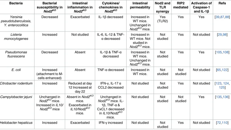
Table 1. Role of Nod2 in the host response toward pathogenic bacteria.
Bacteria Bacterial susceptibility in Nod2KO Intestinal inflammation in Nod2KO Cytokines/ chemokines in Nod2KO Intestinal permeability Nod2 and TLR synergy RIP2 mediated Activation of Caspase-1 and IL-1β Refs Yersinia pseudotuberculosis, Y. enterocolitica
Decreased Exacerbated IL-1βdecreased Increased in WT mice. Unchanged in Nod2KOmice. Yes (TLR2) Yes Yes [39,87,88] Listeria monocytogenes
Increased Not studied IL-6, IL-12 &
TNF-αdecreased Increased in WT mice. Not studied in Nod2KOmice. Not studied
Yes Not studied [29,98]
Pseudomonas fluorescens
Decreased Absent IL-1β& TNF-α
decreased Increased in WT mice. Unchanged in Nod2KOmice. Not studied Yes Yes [105,108] E. coli Increased (attachment to M-cells enhanced)
Absent TNF-αdecreased Increased in WT mice. Not studied Not studied Not studied [65,122]
Citrobacter rodentium Increased Reduced at day 12 Increased at
day 22
IFN-γ, IL-17α
CCL2 decreased
Not studied Not studied
Yes Not studied [123,124,
125]
Campylobacter jejuni Unchanged in Nod2KOmice. Increased in IL10/ Nod2KOmice Absent in Nod2KO mice. Exacerbated in IL10/Nod2KO mice. Unchanged in Nod2KOmice.
IL-1β, TNF-α& CxCL1 decreased
in IL10/Nod2KO mice.
Not studied Not studied
Not studied
Yes [135,136]
Heliobacter hepaticus Increased Exacerbated IFN-γincreased Not studied Not studied
Yes Not studied [72,110]
with a subsequent production of IL-1β. In case of Nod2 deficiency, YopJ is no more able to
activate the Nod2-dependant Caspase-1 signaling pathway, limiting the ileal inflammation at
the beginning of enteral infection [
39
]. This effect is sufficient to reduce the mortality rate of
Nod2
KOmice orally inoculated with
Y. pseudotuberculosis. By contrast, in naive
bone-marrow-derived macrophages (BMDMs), NOD2 [
89
] and RIP2 [
90
,
91
] are dispensable for innate
immune response against
Y. enterocolitica. The production of cytokines and nitric oxide, the
activation of NF-κB and MAPK, and the phagocytic activity remain unchanged in
Yersinia-infected BMDMs from
Nod2
KOmice [
91
]. In agreement, Meinzer et al. showed that Nod2 was
critical in case of infection by
Y. pseudotuberculosis via the oral (but not systemic) route in
mice [
88
].
Listeria monocytogenes
Listeria monocytogenes is a causative agent for human listeriosis, a potentially fatal foodborne
infection.
L. monocytogenes is an intracellular pathogen phagocytosed by
monocytes/macro-phages that escape from the phagosome into the host cell cytosol via its pore-forming toxin
listeriolysin O (LLO) [
92
].
L. monocytogenes also invades nonphagocytic cells, such as
entero-cytes and M cells. This process is critical for bacterial translocation through the intestinal
epi-thelium [
93
–
95
]. The role of Nod2 in the response against
L. monocytogenes is controversial.
In an earlier study, Kobayashi and collaborators reported that
Nod2
KOmice challenged with
L.
monocytogenes via the intragastrical route are more susceptible to infection, with higher
trans-location rates from the intestine to the liver and spleen [
29
,
96
]. This phenotype is lost in the
case of systemic infection. In a later study,
Rip2
KOmice were shown to be highly susceptible to
systemic
Listeria infection [
97
]. In infected
Nod2
KOmice, the number of
L. monocytogenes was
not increased in Peyer’s patches, suggesting an M cell-independent route of bacterial invasion
[
29
]. To explain the hypersensitivity to
Listeria infection, the authors reported a decrease in
the production of defensin-related cryptdin 4 (Defcr4) and Defcr-related sequence 10
(Defcr-rs10) by Paneth cells in
Nod2
KOmice [
29
]. However, the Sartor group recently reported that
WT and
Nod2
KOmice produced similar levels of a large number of cryptins/α-defensins but
do not express Defcr4 [
57
].
Contradictory results about the role of
NOD2 in the induction of pro-inflammatory
cyto-kines by macrophages in response to infection by
L. monocytogenes were also reported in vitro
[
98
,
99
]. RNA interference and other Nod2 inhibition experiments in human PBMCs, as well
as experiments using BMDMs from NLRP3 or RIP2
KOmice, demonstrated that
Listeria-induced IL-1β release was dependent on apoptosis-associated speck-like protein containing a
CARD (ASC), Caspase-1, and NLRP3, whereas NOD2, RIP2, NLRP1, NLRP6, NLRP12,
NLRC4, and absent in melanoma 2 (AIM2) appeared to be dispensable [
100
]. Furthermore, in
murine BMDMs, Nod1 and Nod2 seem to have redundant functions with regards to
Listeria
infection. Nod1 or Nod2 deficiency alone does not result in a significant alteration in cytokine
response to
Listeria infection, while cytokine production is downregulated in Rip2
KOand
Nod1-Nod2
DKOmacrophages [
101
]. Attachment of bacteria to the cell surface is sufficient to
activate macrophages [
102
]. This finding is consistent with the observation that Nod2 and
RIP2 cooperate with TLR signaling for optimal responses to TLR ligands [
101
].
P. fluorescens
P. fluorescens is present at low numbers in the intestinal lumen and in many ecological niches,
including soil, water, and refrigerated food [
103
]. Although
P. fluorescens has long been
con-sidered a psychotrophic microorganism, some clinical strains have been able to adapt at a
growth temperature of 37 ˚C [
104
]. Clinical strains of
P. fluorescens were shown to increase the
paracellular permeability, cell cytotoxicity, and cytokine response in human enterocyte cells
lines [
105
–
107
]. In vivo,
P. fluorescens increases the paracellular permeability of the intestinal
mucosa via the release of IL-1β by immune cells and the activation of MLCK in the epithelial
cells in a Nod2-dependent way [
108
].
H. hepaticus
H. hepaticus is the best studied member of the enterohepatic Helicobacter species. This
gram-negative microaerophilic bacterium is an opportunistic pathogen [
109
] that induces colitis in
immunodeficient mice. In both
Nod2
KOand
Rip2
KOmice,
Helicobacter has been associated
with the development of colitis (resembling human IBD) and cancer [
110
].
Nod2
KOand
Rip2
KOmice were reported to be unable to regulate the
H. hepaticus load in ileum [
72
]. Both of
them develop a granulomatous ileitis and enlarged Peyer’s patches and mesenteric lymph
nodes, with an expansion of IFNγ-producing CD4 and CD8 T cells [
72
]. Inflammatory Th1
response is associated with Nod2 expression in the crypts of the small intestine, suggesting a
role for Paneth cells [
72
].
Mycobacteria
Mycobacteria are an important group of pathological microorganisms. Worldwide,
2,000,000,000 people are infected with
M. tuberculosis, and 2 million people die from
tubercu-losis each year [
111
]. Other mycobacterial species, such as
M. leprae, are endemic in
develop-ing countries and are responsible for high morbidity and disability rates [
112
]. In patients with
a compromised immune system, nonpathogenic mycobacteria may also cause disease.
M.
avium paratuberculosis (MAP) has been suggested to be associated with CD. This suggestion is
controversial, but some findings support a causative role of MAP in the pathogenesis of CD
[
113
]. In cattle, MAP causes Johne disease, which clinically resembles CD [
114
]. Furthermore,
MAP has been identified by PCR and sometimes by culture in gut biopsies from CD patients
[
115
].
As
M. paratuberculosis and NOD2 have been involved in CD, the role of NOD2 in the
regu-lation of host susceptibility to
M. paratuberculosis has been investigated [
116
]. NF-κB
activa-tion in NOD2-transfected HEK293 cells was found to be dose-dependent on MAP exposure
[
116
]. Moreover, MAP-infected PBMCs from CD patients synthetize less inflammatory
cyto-kines in case of
NOD2 mutations [
116
]. Of note, genomewide association studies have
evi-denced an association between
NOD2 and RIP2 polymorphisms and leprosy caused by M.
leprae [
117
]. Recently, synthesis of characteristic
Mycobacterium PGN fragments has been
shown to modulate the innate immune responses of Nod1 and Nod2 [
118
].
E. coli
E. coli is widely spread in many ecological systems, including the human gut, where most
bac-teria are friendly commensal but a few strains are well-known pathogens [
119
]. Pathogenic
E.
coli strains are divided into two major groups: extra-intestinal pathogenic E. coli (ExPEC) and
intestinal pathogenic
E. coli (InPEC). Among the InPEC strains causing diarrheagenic
infec-tions, several well-defined pathotypes have been identified, including enteropathogenic
E. coli
(EPEC), enterotoxigenic
E. coli (ETEC), enterohemorrhagic E. coli (EHEC), enteroaggregative
E. coli (EAEC), enteroinvasive E. coli (EIEC), and AIEC [
119
]. AIEC interact with mouse and
human Peyer’s patches via long polar fimbriae (LPF) and translocate across the M cells at the
surface of Peyer’s patches[
65
]. AIEC are abnormally present in chronic ileal lesions of CD
[
120
,
121
], and they frequently exhibit the LPF operon [
65
]. Although
Nod2
KOmice do not
develop macroscopic lesions of colitis, gut colonization by AIEC does not require antibiotics
as for WT mice [
65
,
122
].
C. rodentium
C. rodentium is a mouse-restricted pathogen. It colonizes intestinal mucosa and shares several
pathogenic mechanisms with EPEC and EHEC, which are two clinically important human
gas-trointestinal pathogens [
123
].
C. rodentium induces a marked infiltration of inflammatory cells
ten days after infection, and the colonization is resolved three weeks later [
124
]. The
develop-ment of a humoral response against
C. rodentium is required for this clearance [
125
]. Nod2
regulates the bacterial clearance by controlling the production of CCL2 and the subsequent
influx of circulating inflammatory monocytes at the site of infection [
126
]. The regulation of
CCL2 by Nod2 is mediated by hematopoietic and nonhematopoietic cells [
126
]. Colonic
stro-mal cells producing CCL2 and pro-inflammatory CCR2-expressing Ly6C
himonocytes are
required for the clearance of
C. rodentium [
126
]. Signaling pathways involved in
Nod2-me-diated clearance of
C. rodentium include activation of NF-κB, MAPKs, and inflammasome, as
well as autophagy [
127
–
130
].
C. jejuni
C. jejuni is a gram-negative spiral-shaped bacteria that colonizes and survives as a commensal
in the gastrointestinal tract of many animals and humans [
128
]. It is the foremost cause of
bac-terial foodborne diarrheal diseases worldwide, with up to 2.4 million cases annually in the
United States alone. The main sources of transmission to humans are the consumption and
handling of contaminated poultry. The “invasive” nature of
C. jejuni led to investigation of
the contribution of cytoplasmic PRRs as Nod1 and Nod2 in initiating the host response.
Zil-bauer et al. suggested that NOD1 (but not NOD2) is a potential PRR for
C. jejuni in intestinal
epithelial cells in vitro [
131
]. In agreement, although
C. jejuni products elicit an inflammatory
response from intestinal epithelial cells through the activation of NF-κB and the release of
CXCL8 [
132
,
133
],
Nod2
KOmice failed to develop colitis [
134
,
135
]. However, NOD2 signaling
seems critical to control campylobacteriosis in
IL-10
KOmice by improving
nitric-oxide-depen-dent bactericidal activity [
135
].
Viruses
During infection with viruses, TLR activation induces the production of type I IFN, which
plays an important role in antiviral defense [
136
,
137
]. TLR-recognizing viral motifs include
TLR3 for viral double stranded RNA [
138
], TLR7 and TLR8 for viral single stranded RNA
[
139
], TLR9 for DNA containing unmethylated CpG motifs present in numerous viral
patho-gens, and TLR13 for bacterial ribosomal RNA. The regulatory role of Nod2 in viral infections
is related to its capacity to sense microbiota-derived MDP and to modulate the TLR pathways
activated by RNA and DNA viruses, including respiratory syncytial virus (RSV), influenza A
virus (IAV), human immunodeficiency virus type-1 (HIV-1), norovirus (NV), and human
en-terovirus species B (HEV-B) (
Table 2
). MDP upregulates the production of IFN-β in PBMCs
infected by RSV [
140
], a response that is lost when NOD2 is mutated [
140
]. In agreement with
the role of Nod2 in antiviral response,
Nod2
KOand
RIP2
KOmice are hypersensitive to infection
with RSV. This hypersensitivity is associated with a failure in mitochondria autophagy and
superoxide overproduction, resulting in mitochondrial damage and activation of the NLRP3
inflammasome and subsequent IL-18 release [
141
]. Nod2 also regulates the innate anti-RSV
response via its interaction with the adaptor protein MAVS (mitochondrial antiviral signaling)
[
142
].
Although the innate immune system is able to trigger an inflammatory response to viruses,
efficient clearance requires the combined efforts of both innate and adaptive immunity.
Indeed,
Nod2
KOmice infected IAV exhibit reduced IFN-β levels, fewer activated dendritic
cells, and virus-specific CD8
+T cells that produce low levels of IFN-γ. Nod2
KOdendritic cells
have a lower costimulatory capacity and are more prone to cell death [
143
]. Similarly, some
RNA viruses, such as HIV-1, may impact adaptive T cell response via the activation of
dectin-1/TLR2 and NOD2 in dendritic cells [
144
]. Moreover, infection by RNA viruses, including
RSV, NV, and HIV-1, is commonly associated with Nod2 upregulation, which results in the
overproduction of TNF-α [
145
].
Nod2 is also involved in the control of the replication or reactivation of DNA viruses,
including HCMV and HVs. Similar to RNA viruses, HCMV upregulates
NOD2 as early as two
hours post-infection and for up to 24 hours afterward [
146
]. As shown in HCMV-infected
cells, the overexpression of
NOD2 or its downstream kinase RIP2 leads to the production of
both IFN-β and pro-inflammatory cytokines/chemokines [
146
]. Conversely,
NOD2 deficiency,
as well as
NOD2
3020insCmutation, downregulates both IFN-β and CXCL8, thereby favoring
HCMV replication [
146
]. In contrast to HCMV, HV is not able to upregulate NOD2.
How-ever, the NOD2 mutation SNP8 (2104C>T) has been associated with HV reactivation and
bacteremia, with both occurring after allogeneic hematopoietic stem cell transplantation [
147
].
Parasites and yeasts
Little is known about the role of NOD2 in parasitic or fungal infections. Over the last ten
years, growing evidence has reported that Nod2 could be instrumental in controlling
Toxo-plasma gondii infection, while its role in Leishmania, Trypanosoma cruzia, and Candida
albi-cans infections remains minor (
Table 3
).
T. gondii is an obligate intracellular protozoan
pathogen able to infect various animal species, leading to severe diseases, including pneumonia
and encephalitis, in immunocompromised hosts. The outcome of
T. gondii infection is
depen-dent on the ability of the host to elicit a robust cellular immune response, particularly the
pro-duction of IFN-γ by natural killer cells and Th1 lymphocytes [
148
]. The role of Nod2 in the
protection of the host is supported by the demonstration that the administration of
T. gondii
orally induces a more severe ileitis in
Nod2
KOmice than in WT mice [
149
]. Infected
Nod2
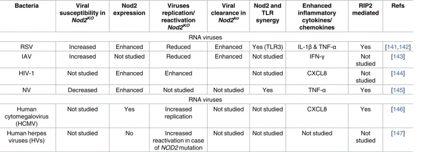
KO Table 2. Role of Nod2 in the host response toward viruses.Bacteria Viral susceptibility in Nod2KO Nod2 expression Viruses replication/ reactivation Nod2KO Viral clearance in Nod2ko Nod2 and TLR synergy Enhanced inflammatory cytokines/ chemokines RIP2 mediated Refs RNA viruses
RSV Increased Enhanced Reduced Enhanced Yes (TLR3) IL-1β& TNF-α Yes [141,142]
IAV Increased Not studied Reduced Enhanced Not studied IFN-γ Not
studied
[143]
HIV-1 Not studied Enhanced Enhanced Not studied CXCL8 Not
studied
[144]
NV Decreased Enhanced Not studied Not studied Yes TNF-α Yes [145]
RNA viruses Human
cytomegalovirus (HCMV)
Not studied Yes Increased
replication
Not studied Not studied CXCL8 Yes [146]
Human herpes viruses (HVs)
Not studied No Increased
reactivation in case of NOD2 mutation
Not studied Not studied Not studied Not studied
[147]
mice display an increase in the parasitic load in the small intestine and the brain and a higher
translocation of bacteria from the gut to the liver, spleen, and kidneys [
149
]. Reconstitution of
T cell-deficient mice with
Nod2
KOT cells followed by
T. gondii infection demonstrated an
intrinsic defect of
Nod2
KOT lymphocytes to produce IL-2 and differentiate into Th1
lympho-cytes [
14
]. Based on an inverse correlation between
Nod2 transcript levels and the intracellular
survival of
Leishmania infantum in macrophages [
150
], it has been proposed that Nod2 might
also play a role in host defense against
Leishmania. By contrast, Nod2 has virtually no impact
on the outcome of the infections with
T. cruzi [
151
] and
C. albicans [
152
], although chitin
par-ticles from the commensal yeast
C. albicans induce IL-10 through Nod2 and TLR9 pathways
[
153
]. Finally, a positive association between NOD2 mutations linked to CD and elevated
lev-els of anti-
Saccharomyces cerevisiae antibodies in the serum of CD patients has been described
[
154
].
Concluding remarks
The mucosal surfaces of the intestinal tract are constantly exposed to complex microbial
com-munities containing commensal microorganisms and sometimes pathogens. Hosts harbor
multiple mechanisms to maintain intestinal barrier integrity and immune tolerance toward
commensal bacteria while reacting against pathogens. In this context, NOD2 plays a key role
in gut–microbe homeostasis by sensing both commensal and pathogenic microbes and
modu-lating TLR signaling pathways.
CD and UC result from a chronic, uncontrolled immune response against components of
the intestinal microbiome in genetically susceptible hosts. Initiation and/or relapse of IBDs are
often associated with pathogenic microbes, including bacteria, viruses, and parasites. In
geneti-cally predisposed individuals, IBDs occur due to an alteration of the subtle interplay between
resident microbiota and the immune system, which often originates from intestinal barrier
dysfunction. Over the last 20 years, a large number of studies reported that pathogens (such as
Y. pseudotuberculosis, Y. enterocolitica, P. fluorescence, AEIC, and L. monocytogenes) and/or an
altered microbiota are often involved in the physiopathology of IBDs. All these bacterial strains
may alter paracellular permeability and favor bacterial translocation, but their detrimental
effects on host intestinal mucosa are downmodulated by Nod2. CD has also been associated
with CMV infection and, to a lesser degree, HV, rotavirus, NV, and adenovirus, all of which
alter intestinal permeability. Replication and/or reactivation of most of these viruses, as well as
the cycles of parasites and/or yeasts (known to alter intestinal permeability), are regulated by
NOD2. Furthermore,
Nod2 deficiency is often associated with exacerbated immune responses
against pathogens as diverse as bacteria (
Y. pseudotuberculosis, H. hepaticus), parasites (T
gon-dii), and viruses (Norovirus). Although the regulatory role of NOD2 in the response of the host
against pathogens is largely admitted, its impact on microbiota composition is still a matter of
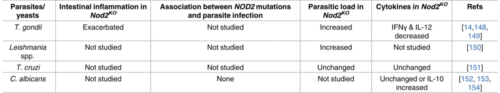
Table 3. Role of Nod2 in the host response toward parasites and yeasts. Parasites/
yeasts
Intestinal inflammation in Nod2KO
Association between NOD2 mutations and parasite infection
Parasitic load in Nod2KO
Cytokines in Nod2KO Refs
T. gondii Exacerbated Not studied Increased IFNγ& IL-12 decreased
[14,148,
149] Leishmania
spp.
Not studied Not studied Increased Not studied [150]
T. cruzi Not studied Not studied Unchanged Unchanged [151]
C. albicans Not studied None Not studied Unchanged or IL-10 increased
[152,153,
154]
debate. The main difficulty is controlling the environmental parameters known to influence
microbiota composition, such as coprophagia, which homogenizes microbiota upon
cohous-ing. The transfer of
Nod2
KOembryos into WT mothers, which represents an experimental
alternative to overcome misleading results, has shown that
Nod2 deficiency results in
domi-nant and transmissible microbial dysbiosis. The mechanisms involved and the role of bacterial
dysbiosis in the development and/or aggravation of IBD remain unclear, however. Indeed,
Nod2
KOmice display high numbers of CD4
+T cells in Peyer’s patches and an increased
intesti-nal permeability, but the transfer of microbiota from
Nod2
KOmice to WT mice alters neither
CD4
+T lymphocyte count nor permeability. By contrast, the decreased production of both
anti-microbial peptides and mucins by nonhematopoietic cells, as well as susceptibility to colitis,
may be acquired by transferring
Nod2
KO-associated dysbiosis. However, the impact of bacterial
dysbiosis on pathogen implantation and vice versa, as well as the contribution of pathogens to
the effects of dysbiosis on intestinal inflammation, still remains to be determined.
References
1. Hooper L V, Littman DR, Macpherson AJ. Interactions between the microbiota and the immune sys-tem. Science. 2012; 336: 1268–73. doi:10.1126/science.1223490PMID:22674334
2. Min YW, Rhee P-L. The Role of Microbiota on the Gut Immunology. Clin Ther. 2015; 37: 968–75. doi:
10.1016/j.clinthera.2015.03.009PMID:25846321
3. Sellge G, Kufer TA. PRR-signaling pathways–Learning from microbial tactics. Semin Immunol. 2015; 27: 75–84. doi:10.1016/j.smim.2015.03.009PMID:25911384
4. Uematsu S, Akira S. Toll-like receptors and Type I interferons. J Biol Chem. 2007; 282: 15319–23. doi:
10.1074/jbc.R700009200PMID:17395581
5. Takeda K, Akira S. Toll-like receptors in innate immunity. Int Immunol. 2005; 17: 1–14. doi:10.1093/ intimm/dxh186PMID:15585605
6. Motta V, Soares F, Sun T, Philpott DJ. NOD-like receptors: versatile cytosolic sentinels. Physiol Rev. 2015; 95: 149–78. doi:10.1152/physrev.00009.2014PMID:25540141
7. Girardin SE, Boneca IG, Viala J, Chamaillard M, Labigne A, Thomas G, et al. Nod2 is a general sensor of peptidoglycan through muramyl dipeptide (MDP) detection. J Biol Chem. 2003; 278: 8869–72. doi:
10.1074/jbc.C200651200PMID:12527755
8. Ogura Y, Inohara N, Benito A, Chen FF, Yamaoka S, Nunez G. Nod2, a Nod1/Apaf-1 family member that is restricted to monocytes and activates NF-kappaB. J Biol Chem. 2001; 276: 4812–8. doi:10. 1074/jbc.M008072200PMID:11087742
9. Hugot JP, Chamaillard M, Zouali H, Lesage S, Ce´zard JP, Belaiche J, et al. Association of NOD2 leu-cine-rich repeat variants with susceptibility to Crohn’s disease. Nature. 2001; 411: 599–603. doi:10. 1038/35079107PMID:11385576
10. Ogura Y, Bonen DK, Inohara N, Nicolae DL, Chen FF, Ramos R, et al. A frameshift mutation in NOD2 associated with susceptibility to Crohn’s disease. Nature. 2001; 411: 603–6. doi:10.1038/35079114
PMID:11385577
11. Holler E, Rogler G, Herfarth H, Brenmoehl J, Wild PJ, Hahn J, et al. Both donor and recipient NOD2/ CARD15 mutations associate with transplant-related mortality and GvHD following allogeneic stem cell transplantation. Blood. 2004; 104: 889–94. doi:10.1182/blood-2003-10-3543PMID:15090455 12. Miceli-Richard C, Lesage S, Rybojad M, Prieur AM, Manouvrier-Hanu S, Ha¨fner R, et al. CARD15
mutations in Blau syndrome. Nat Genet. 2001; 29: 19–20. doi:10.1038/ng720PMID:11528384 13. Penack O, Smith OM, Cunningham-Bussel A, Liu X, Rao U, Yim N, et al. NOD2 regulates
hematopoi-etic cell function during graft-versus-host disease. J Exp Med. 2009; 206: 2101–10. doi:10.1084/jem. 20090623PMID:19737867
14. Shaw MH, Reimer T, Sa´nchez-Valdepeñas C, Warner N, Kim Y-G, Fresno M, et al. T cell-intrinsic role of Nod2 in promoting type 1 immunity to Toxoplasma gondii. Nat Immunol. 2009; 10: 1267–74. doi:10. 1038/ni.1816PMID:19881508
15. Petterson T, Jendholm J, Månsson A, Bjartell A, Riesbeck K, Cardell L-O. Effects of NOD-like recep-tors in human B lymphocytes and crosstalk between NOD1/NOD2 and Toll-like receprecep-tors. J Leukoc Biol. 2011; 89: 177–87. doi:10.1189/jlb.0210061PMID:20844241
16. Hedl M, Li J, Cho JH, Abraham C. Chronic stimulation of Nod2 mediates tolerance to bacterial prod-ucts. Proc Natl Acad Sci U S A. 2007; 104: 19440–5. doi:10.1073/pnas.0706097104PMID:18032608
17. Cooney R, Baker J, Brain O, Danis B, Pichulik T, Allan P, et al. NOD2 stimulation induces autophagy in dendritic cells influencing bacterial handling and antigen presentation. Nat Med. 2010; 16: 90–7. doi:
10.1038/nm.2069PMID:19966812
18. Okumura S, Yuki K, Kobayashi R, Okamura S, Ohmori K, Saito H, et al. Hyperexpression of NOD2 in intestinal mast cells of Crohn’s disease patients: preferential expression of inflammatory cell-recruiting molecules via NOD2 in mast cells. Clin Immunol. 2009; 130: 175–85. doi:10.1016/j.clim.2008.08.027
PMID:18938111
19. Ogura Y, Lala S, Xin W, Smith E, Dowds TA, Chen FF, et al. Expression of NOD2 in Paneth cells: a possible link to Crohn’s ileitis. Gut. 2003; 52: 1591–7. PMID:14570728
20. Nigro G, Rossi R, Commere P-H, Jay P, Sansonetti PJ. The cytosolic bacterial peptidoglycan sensor Nod2 affords stem cell protection and links microbes to gut epithelial regeneration. Cell Host Microbe. 2014; 15: 792–8. doi:10.1016/j.chom.2014.05.003PMID:24882705
21. Ramanan D, Tang MS, Bowcutt R, Loke P, Cadwell K. Bacterial sensor Nod2 prevents inflammation of the small intestine by restricting the expansion of the commensal Bacteroides vulgatus. Immunity. 2014; 41: 311–24. doi:10.1016/j.immuni.2014.06.015PMID:25088769
22. Hisamatsu T, Suzuki M, Reinecker H-C, Nadeau WJ, McCormick BA, Podolsky DK. CARD15/NOD2 functions as an antibacterial factor in human intestinal epithelial cells. Gastroenterology. 2003; 124: 993–1000. doi:10.1053/gast.2003.50153PMID:12671896
23. Rosenstiel P, Fantini M, Bra¨utigam K, Ku¨hbacher T, Waetzig GH, Seegert D, et al. TNF-alpha and IFN-gamma regulate the expression of the NOD2 (CARD15) gene in human intestinal epithelial cells. Gastroenterology. 2003; 124: 1001–9. doi:10.1053/gast.2003.50157PMID:12671897
24. Grimes CL, Ariyananda LDZ, Melnyk JE, O’Shea EK. The Innate Immune Protein Nod2 Binds Directly to MDP, a Bacterial Cell Wall Fragment. J Am Chem Soc. 2012; 134: 13535–13537. doi:10.1021/ ja303883cPMID:22857257
25. Mo J, Boyle JP, Howard CB, Monie TP, Davis BK, Duncan JA. Pathogen Sensing by Nucleotide-bind-ing Oligomerization Domain-containNucleotide-bind-ing Protein 2 (NOD2) Is Mediated by Direct BindNucleotide-bind-ing to Muramyl Dipeptide and ATP. J Biol Chem. 2012; 287: 23057–23067. doi:10.1074/jbc.M112.344283PMID:
22549783
26. Hasegawa M, Yang K, Hashimoto M, Park J-H, Kim Y-G, Fujimoto Y, et al. Differential release and dis-tribution of Nod1 and Nod2 immunostimulatory molecules among bacterial species and environments. J Biol Chem. 2006; 281: 29054–63. doi:10.1074/jbc.M602638200PMID:16870615
27. Opitz B, Pu¨schel A, Schmeck B, Hocke AC, Rosseau S, Hammerschmidt S, et al. Nucleotide-binding oligomerization domain proteins are innate immune receptors for internalized Streptococcus pneumo-niae. J Biol Chem. 2004; 279: 36426–32. doi:10.1074/jbc.M403861200PMID:15215247
28. Theivanthiran B, Batra S, Balamayooran G, Cai S, Kobayashi K, Flavell RA, et al. NOD2 signaling con-tributes to host defense in the lungs against Escherichia coli infection. Infect Immun. 2012; 80: 2558– 69. doi:10.1128/IAI.06230-11PMID:22547547
29. Kobayashi KS, Chamaillard M, Ogura Y, Henegariu O, Inohara N, Nuñez G, et al. Nod2-dependent regulation of innate and adaptive immunity in the intestinal tract. Science. 2005; 307: 731–4. doi:10. 1126/science.1104911PMID:15692051
30. Thay B, Damm A, Kufer TA, Wai SN, Oscarsson J. Aggregatibacter actinomycetemcomitans outer membrane vesicles are internalized in human host cells and trigger NOD1- and NOD2-dependent
NF-κB activation. Infect Immun. 2014; 82: 4034–46. doi:10.1128/IAI.01980-14PMID:25024364 31. Chu H, Khosravi A, Kusumawardhani IP, Kwon AHK, Vasconcelos AC, Cunha LD, et al.
Gene-micro-biota interactions contribute to the pathogenesis of inflammatory bowel disease. Science (80-). 2016; 352: 1116–1120. doi:10.1126/science.aad9948PMID:27230380
32. Nakamura N, Lill JR, Phung Q, Jiang Z, Bakalarski C, de Mazière A, et al. Endosomes are specialized platforms for bacterial sensing and NOD2 signalling. Nature. 2014; 509: 240–4. doi:10.1038/ nature13133PMID:24695226
33. Vavricka SR, Musch MW, Chang JE, Nakagawa Y, Phanvijhitsiri K, Waypa TS, et al. hPepT1 trans-ports muramyl dipeptide, activating NF-kappaB and stimulating IL-8 secretion in human colonic Caco2/bbe cells. Gastroenterology. 2004; 127: 1401–9. PMID:15521010
34. Keestra-Gounder AM, Byndloss MX, Seyffert N, Young BM, Cha´vez-Arroyo A, Tsai AY, et al. NOD1 and NOD2 signalling links ER stress with inflammation. Nature. 2016; 532: 394–397. doi:10.1038/ nature17631PMID:27007849
35. Maekawa S, Ohto U, Shibata T, Miyake K, Shimizu T. Crystal structure of NOD2 and its implications in human disease. Nat Commun. 2016; 7: 11813. doi:10.1038/ncomms11813PMID:27283905 36. Lechtenberg BC, Mace PD, Riedl SJ. Structural mechanisms in NLR inflammasome signaling. Curr
37. Tattoli I, Travassos LH, Carneiro LA, Magalhaes JG, Girardin SE. The Nodosome: Nod1 and Nod2 control bacterial infections and inflammation. Semin Immunopathol. 2007; 29: 289–301. doi:10.1007/ s00281-007-0083-2PMID:17690884
38. Zhong Y, Kinio A, Saleh M. Functions of NOD-Like Receptors in Human Diseases. Front Immunol. 2013; 4: 333. doi:10.3389/fimmu.2013.00333PMID:24137163
39. Meinzer U, Barreau F, Esmiol-Welterlin S, Jung C, Villard C, Le´ger T, et al. Yersinia pseudotuberculo-sis effector YopJ subverts the Nod2/RICK/TAK1 pathway and activates caspase-1 to induce intestinal barrier dysfunction. Cell Host Microbe. 2012; 11: 337–51. doi:10.1016/j.chom.2012.02.009PMID:
22520462
40. Travassos LH, Carneiro LAM, Ramjeet M, Hussey S, Kim Y-G, Magalhães JG, et al. Nod1 and Nod2 direct autophagy by recruiting ATG16L1 to the plasma membrane at the site of bacterial entry. Nat Immunol. 2010; 11: 55–62. doi:10.1038/ni.1823PMID:19898471
41. Øyri SF, Műzes G, Sipos F. Dysbiotic gut microbiome: A key element of Crohn’s disease. Comp Immu-nol Microbiol Infect Dis. 2015; 43: 36–49. doi:10.1016/j.cimid.2015.10.005PMID:26616659
42. Ohkusa T, Koido S. Intestinal microbiota and ulcerative colitis. J Infect Chemother. 2015; 21: 761– 768. doi:10.1016/j.jiac.2015.07.010PMID:26346678
43. Frank DN, St. Amand AL, Feldman RA, Boedeker EC, Harpaz N, Pace NR. Molecular-phylogenetic characterization of microbial community imbalances in human inflammatory bowel diseases. Proc Natl Acad Sci. 2007; 104: 13780–13785. doi:10.1073/pnas.0706625104PMID:17699621
44. Collins MD, Lawson PA, Willems A, Cordoba JJ, Fernandez-Garayzabal J, Garcia P, et al. The phylog-eny of the genus Clostridium: proposal of five new genera and eleven new species combinations. Int J Syst Bacteriol. 1994; 44: 812–26. doi:10.1099/00207713-44-4-812PMID:7981107
45. Frank DN, Robertson CE, Hamm CM, Kpadeh Z, Zhang T, Chen H, et al. Disease phenotype and genotype are associated with shifts in intestinal-associated microbiota in inflammatory bowel dis-eases. Inflamm Bowel Dis. 2011; 17: 179–84. doi:10.1002/ibd.21339PMID:20839241
46. Sokol H, Pigneur B, Watterlot L, Lakhdari O, Bermudez-Humaran LG, Gratadoux J-J, et al. Faecali-bacterium prausnitzii is an anti-inflammatory commensal Faecali-bacterium identified by gut microbiota analy-sis of Crohn disease patients. Proc Natl Acad Sci. 2008; 105: 16731–16736. doi:10.1073/pnas. 0804812105PMID:18936492
47. Rehman A, Sina C, Gavrilova O, Ha¨sler R, Ott S, Baines JF, et al. Nod2 is essential for temporal devel-opment of intestinal microbial communities. Gut. 2011; 60: 1354–62. doi:10.1136/gut.2010.216259
PMID:21421666
48. Li E, Hamm CM, Gulati AS, Sartor RB, Chen H, Wu X, et al. Inflammatory Bowel Diseases Phenotype, C. difficile and NOD2 Genotype Are Associated with Shifts in Human Ileum Associated Microbial Com-position. Bereswill S, editor. PLoS ONE. 2012; 7: e26284. doi:10.1371/journal.pone.0026284PMID:
22719818
49. Knights D, Silverberg MS, Weersma RK, Gevers D, Dijkstra G, Huang H, et al. Complex host genetics influence the microbiome in inflammatory bowel disease. Genome Med. 2014; 6: 107. doi:10.1186/ s13073-014-0107-1PMID:25587358
50. Petnicki-Ocwieja T, Hrncir T, Liu Y-J, Biswas A, Hudcovic T, Tlaskalova-Hogenova H, et al. Nod2 is required for the regulation of commensal microbiota in the intestine. Proc Natl Acad Sci. 2009; 106: 15813–15818. doi:10.1073/pnas.0907722106PMID:19805227
51. Mondot S, Barreau F, Al Nabhani Z, Dussaillant M, Le Roux K, Dore´ J, et al. Altered gut microbiota composition in immune-impaired Nod2(-/-) mice. Gut. 2012; 61: 634–5. doi: 10.1136/gutjnl-2011-300478PMID:21868489
52. Couturier-Maillard A, Secher T, Rehman A, Normand S, De Arcangelis A, Haesler R, et al. NOD2-mediated dysbiosis predisposes mice to transmissible colitis and colorectal cancer. J Clin Invest. 2013; 123: 700–11. doi:10.1172/JCI62236PMID:23281400
53. Al Nabhani Z, Lepage P, Mauny P, Montcuquet N, Roy M, Le Roux K, et al. Nod2 deficiency leads to a specific and transmissible mucosa-associated microbial dysbiosis which is independent of the muco-sal barrier defect. J Crohns Colitis. 2016;
54. Alnabhani Z, Hugot J-P, Montcuquet N, Le Roux K, Dussaillant M, Roy M, et al. Respective Roles of Hematopoietic and Nonhematopoietic Nod2 on the Gut Microbiota and Mucosal Homeostasis. Inflamm Bowel Dis. 2016; 22: 763–73. doi:10.1097/MIB.0000000000000749PMID:26963567 55. Ramanan D, Bowcutt R, Lee SC, Tang MS, Kurtz ZD, Ding Y, et al. Helminth infection promotes
colo-nization resistance via type 2 immunity. Science (80-). 2016; 352: 608–612. doi:10.1126/science. aaf3229PMID:27080105
56. Robertson SJ, Zhou JY, Geddes K, Rubino SJ, Cho JH, Girardin SE, et al. Nod1 and Nod2 signaling does not alter the composition of intestinal bacterial communities at homeostasis. Gut Microbes. 2013; 4: 222–31. doi:10.4161/gmic.24373PMID:23549220
57. Shanahan MT, Carroll IM, Grossniklaus E, White A, von Furstenberg RJ, Barner R, et al. Mouse Paneth cell antimicrobial function is independent of Nod2. Gut. 2014; 63: 903–10. doi: 10.1136/gutjnl-2012-304190PMID:23512834
58. Carmody RN, Gerber GK, Luevano JM, Gatti DM, Somes L, Svenson KL, et al. Diet Dominates Host Genotype in Shaping the Murine Gut Microbiota. Cell Host Microbe. 2015; 17: 72–84. doi:10.1016/j. chom.2014.11.010PMID:25532804
59. Hugot J-P, Zouali H, Lesage S. Lessons to be learned from the NOD2 gene in Crohn’s disease. Eur J Gastroenterol Hepatol. 2003; 15: 593–7. doi:10.1097/01.meg.0000059147.68845.baPMID:
12840668
60. Bevins CL, Stange EF, Wehkamp J. Decreased Paneth cell defensin expression in ileal Crohn’s dis-ease is independent of inflammation, but linked to the NOD2 1007fs genotype. Gut. 2009; 58: 882-3-4.
61. Wehkamp J, Stange EF. NOD2 mutation and mice: no Crohn’s disease but many lessons to learn. Trends Mol Med. 2005; 11: 307–309. doi:10.1016/j.molmed.2005.06.003PMID:15955743 62. Wehkamp J, Wang G, Kubler I, Nuding S, Gregorieff A, Schnabel A, et al. The Paneth Cell -Defensin
Deficiency of Ileal Crohn’s Disease Is Linked to Wnt/Tcf-4. J Immunol. 2007; 179: 3109–3118. PMID:
17709525
63. Barreau F, Meinzer U, Chareyre F, Berrebi D, Niwa-Kawakita M, Dussaillant M, et al. CARD15/NOD2 is required for Peyer’s patches homeostasis in mice. PLoS ONE. 2007; 2: e523. doi:10.1371/journal. pone.0000523PMID:17565376
64. Denou E, Lolmède K, Garidou L, Pomie C, Chabo C, Lau TC, et al. Defective NOD2 peptidoglycan sensing promotes diet-induced inflammation, dysbiosis, and insulin resistance. EMBO Mol Med. 2015; 7: 259–74. doi:10.15252/emmm.201404169PMID:25666722
65. Chassaing B, Rolhion N, de Valle´e A, Salim SY, Prorok-Hamon M, Neut C, et al. Crohn disease— associated adherent-invasive E. coli bacteria target mouse and human Peyer’s patches via long polar fimbriae. J Clin Invest. 2011; 121: 966–75. doi:10.1172/JCI44632PMID:21339647
66. Barreau F, Madre C, Meinzer U, Berrebi D, Dussaillant M, Merlin F, et al. Nod2 regulates the host response towards microflora by modulating T cell function and epithelial permeability in mouse Peyer’s patches. Gut. 2010; 59: 207–17. doi:10.1136/gut.2008.171546PMID:19837677
67. Amendola A, Butera A, Sanchez M, Strober W, Boirivant M. Nod2 deficiency is associated with an increased mucosal immunoregulatory response to commensal microorganisms. Mucosal Immunol. 2014; 7: 391–404. doi:10.1038/mi.2013.58PMID:23962873
68. Wu L-L, Peng W-H, Kuo W-T, Huang C-Y, Ni Y-H, Lu K-S, et al. Commensal Bacterial Endocytosis in Epithelial Cells Is Dependent on Myosin Light Chain Kinase–Activated Brush Border Fanning by Inter-feron-γ. Am J Pathol. 2014; 184: 2260–2274. doi:10.1016/j.ajpath.2014.05.003PMID:24911373 69. Kim D, Kim Y-G, Seo S-U, Kim D-J, Kamada N, Prescott D, et al. Corrigendum: Nod2-mediated
recog-nition of the microbiota is critical for mucosal adjuvant activity of cholera toxin. Nat Med. 2016; 22: 961. doi:10.1038/nm0816-961PMID:27490437
70. Su L, Shen L, Clayburgh DR, Nalle SC, Sullivan EA, Meddings JB, et al. Targeted Epithelial Tight Junction Dysfunction Causes Immune Activation and Contributes to Development of Experimental Colitis. Gastroenterology. 2009; 136: 551–563. doi:10.1053/j.gastro.2008.10.081PMID:19027740 71. Caruso R, Warner N, Inohara N, Nu´ñez G. NOD1 and NOD2: signaling, host defense, and
inflamma-tory disease. Immunity. 2014; 41: 898–908. doi:10.1016/j.immuni.2014.12.010PMID:25526305 72. Biswas A, Liu Y-J, Hao L, Mizoguchi A, Salzman NH, Bevins CL, et al. Induction and rescue of
Nod2-dependent Th1-driven granulomatous inflammation of the ileum. Proc Natl Acad Sci U S A. 2010; 107: 14739–44. doi:10.1073/pnas.1003363107PMID:20679225
73. Geddes K, Rubino SJ, Magalhaes JG, Streutker C, Le Bourhis L, Cho JH, et al. Identification of an innate T helper type 17 response to intestinal bacterial pathogens. Nat Med. 2011; 17: 837–44. doi:
10.1038/nm.2391PMID:21666695
74. Watanabe T, Kitani A, Strober W. NOD2 regulation of Toll-like receptor responses and the pathogene-sis of Crohn’s disease. Gut. 2005; 54: 1515–8. doi:10.1136/gut.2005.071795PMID:16227353 75. Philpott DJ, Sorbara MT, Robertson SJ, Croitoru K, Girardin SE. NOD proteins: regulators of
inflam-mation in health and disease. Nat Rev Immunol. 2014; 14: 9–23. doi:10.1038/nri3565PMID:
24336102
76. Autenrieth IB, Firsching R. Penetration of M cells and destruction of Peyer’s patches by Yersinia enter-ocolitica: an ultrastructural and histological study. J Med Microbiol. 1996; 44: 285–94. doi:10.1099/ 00222615-44-4-285PMID:8606357