HAL Id: hal-01307484
https://hal-amu.archives-ouvertes.fr/hal-01307484
Submitted on 26 Apr 2016
HAL is a multi-disciplinary open access archive for the deposit and dissemination of sci-entific research documents, whether they are pub-lished or not. The documents may come from teaching and research institutions in France or abroad, or from public or private research centers.
L’archive ouverte pluridisciplinaire HAL, est destinée au dépôt et à la diffusion de documents scientifiques de niveau recherche, publiés ou non, émanant des établissements d’enseignement et de recherche français ou étrangers, des laboratoires publics ou privés.
Distributed under a Creative Commons Attribution - NonCommercial - NoDerivatives| 4.0 International License
Trigeminal neuralgia related to megadolichobasilar
artery compression: a prospective series of twenty-nine
patients treated with gamma knife surgery, with more
than one year of follow-up.
Constantin Tuleasca, Romain Carron, Noémie Resseguier, Anne Donnet, P
Roussel, Jean Gaudart, Marc Levivier, Jean Regis
To cite this version:
Constantin Tuleasca, Romain Carron, Noémie Resseguier, Anne Donnet, P Roussel, et al.. Trigeminal neuralgia related to megadolichobasilar artery compression: a prospective series of twenty-nine patients treated with gamma knife surgery, with more than one year of follow-up.. Stereotactic and Functional Neurosurgery, Karger, 2014, �10.1159/000362172�. �hal-01307484�
See discussions, stats, and author profiles for this publication at: https://www.researchgate.net/publication/263293257
Trigeminal Neuralgia Related to
Megadolichobasilar Artery Compression: A
Prospective Series of Twenty-Nine Patients
Treated with Gamma Knife Surgery, with More
Than One Year of Fol...
Article in Stereotactic and Functional Neurosurgery · June 2014 Impact Factor: 2.02 · DOI: 10.1159/000362172 · Source: PubMed CITATIONS7
READS57
8 authors, including: Romain Carron Assistance Publique Hôpitaux de Marseille 69 PUBLICATIONS 347 CITATIONS SEE PROFILE Noemie Resseguier Aix-Marseille Université 59 PUBLICATIONS 120 CITATIONS SEE PROFILE Jean Gaudart Aix-Marseille Université 185 PUBLICATIONS 1,568 CITATIONS SEE PROFILE Jean Régis Assistance Publique Hôpitaux de Marseille 389 PUBLICATIONS 10,021 CITATIONS SEE PROFILE Available from: Constantin Tuleasca Retrieved on: 26 April 2016E-Mail karger@karger.com
Clinical Study
Stereotact Funct Neurosurg 2014;92:170–177 DOI: 10.1159/000362172
Trigeminal Neuralgia Related to
Megadolichobasilar Artery Compression:
A Prospective Series of Twenty-Nine Patients
Treated with Gamma Knife Surgery, with More
Than One Year of Follow-Up
Constantin Tuleasca
a, d–g
Romain Carron
a
Noémie Resseguier
b
Anne Donnet
c
Philippe Roussel
c
Jean Gaudart
b
Marc Levivier
f, g
Jean Régis
a
a Functional and Stereotactic Neurosurgery Unit, INSERM U751, b Department of Public Health and Medical
Information, UMR 912 (INSERM-IRD-Université de la Méditerranée), and c Department of Neurology, Clinical
Neuroscience Federation, Centre Hospitalier Universitaire La Timone Assistance Publique-Hôpitaux de Marseille, Marseille , France; d Signal Processing Laboratory (LTS5), Swiss Federal Institute of Technology (EPFL), e Medical
Image Analysis Laboratory and f Neurosurgery Service and Gamma Knife Center, Centre Hospitalier Universitaire
Vaudois, and g Faculty of Biology and Medicine, University of Lausanne, Lausanne , Switzerland
all patients (100%) were pain free; the average time to com-plete pain relief was 13.5 days (range: 0–240 days). Their ac-tuarial probability of remaining pain free without medica-tion at 0.5, 1 and 2 years was 93.1, 79.3 and 75.7%, respec-tively, and remained stable until 13 years after treatment. The actuarial probability of hypoesthesia onset at 6 months was 4.3%; at 1 year it reached 13% and remained stable until 13 years after treatment. Conclusions: GKS proved to be rea-sonably safe and effective on a long-term basis as a first- and/ or second-line surgical treatment for TN due to MBA
com-pression. © 2014 S. Karger AG, Basel
Introduction
Trigeminal neuralgia (TN) – also known as ‘tic dou-loureux’, as it was named by the French surgeon Nicholas André [1] – is a serious health problem, with a prevalence Key Words
Megadolichobasilar artery · Trigeminal neuralgia · Gamma Knife surgery
Abstract
Background: Trigeminal neuralgia (TN) secondary to mega-dolichobasilar artery (MBA) compression is considerably dif-ficult to manage surgically. Objective: This study aims to evaluate the safety/efficacy of Gamma Knife surgery (GKS) in this special group of patients. Methods: Between July 1992 and November 2010, 29 patients with >1 year of follow-up presenting with MBA compression were treated with GKS at Timone University Hospital. Radiosurgery was performed us-ing a Gamma Knife (model B, C or Perfexion). A sus-ingle 4-mm isocenter was positioned in the cisternal portion of the tri-geminal nerve at a median distance of 9.1 mm (range: 6–18.2 mm) from the emergence. Results: The median follow-up period was 46.1 months (range: 12.9–157.9 months). Initially,
Received: December 16, 2013 Accepted: March 11, 2014 Published online: June 12, 2014
Jean Régis
Stereotactic and Functional Neurosurgery Department, Hôpital de la Timone 264, Rue Saint Pierre
FR–13385 Marseille (France)
E-Mail jregis @ ap-hm.fr
© 2014 S. Karger AG, Basel 1011–6125/14/0923–0170$39.50/0 www.karger.com/sfn
© FOR PERMITTED USE ONLY
ANY FURTHER DISTRIBUTION OF THIS ARTICLE REQUIRES WRITTEN PERMISSION FROM S. KARGER AG.
Radiosurgery for TN Related to MBA Compression
Stereotact Funct Neurosurg 2014;92:170–177
DOI: 10.1159/000362172 171
rate of 4–5 per 100,000 people [2] . Several causes were identified as origins of TN, and vascular compression is considered to be one of the major pathogenic factors [3– 5] . Usually, the vessels involved in the neurovascular con-flict are arteries such as the superior cerebellar or anterior inferior cerebellar artery. Less frequently, prominent veins such as the petrosal veins or draining veins of the brainstem are also involved [6] .
Microvascular decompression (MVD) of the trigemi-nal nerve is the most effective operation for treating pa-tients with TN, with long-term cure rates between 69 and 96% of the cases [7–13] . Sindou et al. [13, 14] proposed a surgical classification of the neurovascular compression and its severity, and demonstrated that this is a predictive factor for the results [14] . This classification included the following grading: 1 – simple contact; 2 – distortion, and 3 – marked indentation [14] . In the case of compression of the trigeminal nerve by an ecstatic basilar artery, it is rather difficult to grade the conflict. Megadolichobasilar artery (MBA) compression is usually creating a marked indentation on the nerve and/or the adjacent brainstem, making it difficult to visualize in some patients.
MBA compression has rarely been described in the lit-erature; Jannetta [15] did not refer to this condition in a series of 60 cases with TN due to vascular compression. Many case reports [12, 16–29] , and recently a series [30] , have described the topic of MBA-related TN, but only a few of them have included the results of decompressive vascu-lar surgery [12, 31] . There are several problems with MVD in MBA-related TN which render this type of intervention particularly difficult, such as a considerable risk of mobili-zation of the trigeminal nerve because of the high internal turgor of these large vessels as well as a tendency for the patients to be older and to develop cranial nerve deficits more often than the MVD series as a whole [12] . Beside MVD, other types of treatment have been reported in the literature, such as foramen magnum decompression [17] .
Gamma Knife surgery (GKS) is a minimally invasive surgical approach in the armamentarium for refractory TN [32, 33] . Lars Leksell first introduced the concept of stereotactic radiosurgery in 1951, when he treated a pa-tient suffering from essential TN using a prototype-guiding device linked to a dental X-ray machine [15, 34– 36] . Nowadays, the role of GKS in TN has been con-firmed by several studies in terms of its safety and efficacy [17, 37–42] , although until one year ago, none of them had specifically evaluated TN related to MBA compression. Recently, Park et al. [30] reported on a first series of 20 patients and the results of GKS for TN due to vertebrobasilar ectasia.
Our prospective study evaluates the safety and efficacy of GKS, using three parameters widely accepted in the literature: initial pain cessation, hypoesthesia and the probability of maintaining pain relief.
Subjects and Methods
Patient Population and Selection
Between July 1992 and November 2010, 737 patients present-ing with intractable TN were treated with GKS and followed up prospectively at Timone University Hospital in Marseille, France. Only patients fulfilling the criteria of the International Headache Society (2003) [43] were accepted for treatment.
An evaluation of the type of trigeminal pain was made accord-ing to the classification proposed by Eller et al. [44] into idiopath-ic TN1 and TN2. While TN1 is typidiopath-ically described as sharp, shoot-ing and electric-shock-like pain with pain-free intervals between the attacks, TN2 is described as aching, throbbing or burning pain for >50% of the time which is constant in nature (a constant back-ground pain being the most significant attribute).
From our global series, thirty-three patients presented with TN related to MBA compression, of which 29 had >1 year of follow-up; their cases were further analyzed in this study.
Radiosurgical Technique
During the 18 years of the study, various models of the Gamma Knife were used (models B, C, 4C and Perfexion; Elekta Instru-ment AB, Sweden). After application of the Leksell Model G ste-reotactic frame (Elekta Instrument AB) under local anesthesia, all patients underwent stereotactic magnetic resonance imaging (MRI) and computed tomography (CT) scanning to identify the trigeminal nerve. The MRI sequences used were T2 type CISS without contrast (Siemens) and contrast-enhanced T1-weighted imaging. CT scanning routinely supplements the neuroradiologi-cal investigation in order to correct any distortion errors on the MR images.
In the Gamma Unit in Marseille, France, we use the anterior target, which means placing a unique 4-mm isocenter on the cis-ternal portion of the trigeminal nerve using a very anterior target, located immediately posterior to the gasserian ganglion, as previ-ously described [33, 41] , at a median distance of approximately 7.5 mm from the entry point of the trigeminal nerve into the brain-stem, if the anatomical conditions allow. We initially give a dose of 90 Gy at the 100% isodose point. Beam channel blocking is used depending on the dose received by 10 mm 3 of the brainstem;
should this dose be >15 Gy, we diminish the dose and then start plugging so as to be able to avoid increasing the length of the treat-ed nerve, which could amount for more toxicity (the so-calltreat-ed Flickinger effect) [34] .
In the current series, a single 4-mm isocenter was used for all patients and was positioned in the anterior cisternal portion of the trigeminal nerve at a median distance of 9.1 mm (range: 6–18.2 mm) anteriorly to the emergence of the nerve. Radiosur-gical targeting can be very challenging in this special indication. Difficulties were sometimes encountered in visualizing the tri-geminal nerve as the compression by the ecstatic MBA, depend-ing on the grade of indentation in the nerve, made the targetdepend-ing challenging. Figure 1 presents the retrogasserian target, also
Tuleasca /Carron /Resseguier /Donnet / Roussel /Gaudart /Levivier /Régis
Stereotact Funct Neurosurg 2014;92:170–177 DOI: 10.1159/000362172
172
known as the Marseille target [41] , adapted to this special ana-tomical condition.
Plugging was used in 8 patients (27.6%). The doses were rela-tively high, with a median maximum dose of 90 Gy (range: 80–90 Gy). No patient received <80 Gy at the 100% isodose point.
Follow-Up and Assessment of Outcome
The Marseille Mediterranean University as well as the Direc-tion and Ethics Committee of Timone University Hospital (CPPRB1) approved our study. Follow-up information was ob-tained in two ways: direct clinical evaluation or telephone inter-view by the first author (C.T.), who was not involved in the selec-tion of cases for treatment.
We evaluated the probability of initial pain cessation, the onset of sensory disturbance and the rate of recurrence. We analyzed data regarding latency periods until becoming pain free, having a recurrence or developing a sensory disturbance, paying attention to date every event, the use of medication and the need for further surgical procedures so as to accurately assess all the available in-formation.
Pain was scored using 3 different scales: the Barrow Neuro-logical Institute (BNI) pain scale (class I: ‘no trigeminal pain, no medication’; class II: ‘occasional pain, not requiring medication’; class IIIa: ‘no pain, continued medication’; class IIIb: ‘controlled with medication’; class IV: ‘some pain, not adequately controlled with medication’; class V: ‘severe pain, no pain relief’), the Burchiel scale (class I: ‘pain free, no medication’; class II: ‘pain free, on med-ication’; class IIIa: ‘pain improved, no medmed-ication’; class IIIb: ‘pain improved, on medication’; class IV: ‘pain not improved’) and the Regis scale (class I: ‘no trigeminal pain, no medication’; class II: ‘no pain, with medication’; class III: ‘pain frequency reduction >90%’; class IV: ‘pain frequency reduction 50–90%’; class V: ‘no pain re-duction’; class VI: ‘pain worsening’) [45–47] . For hypoesthesia we used the BNI facial hypoesthesia scale (class I: ‘no facial numb-ness’; class II: ‘mild facial numbness, not bothersome’; class III: ‘facial numbness, somewhat bothersome’; class IV: ‘facial numb-ness, very bothersome’) [47] . For patients presenting facial sen-sory dysfunction, we also inquired about their quality of life
re-lated to TN and whether this sensory problem was bothering them or not. We asked whether or not they had difficulties in mastica-tion.
The patients and referring doctor were instructed to continue and not change the medication for at least 1 month and then were asked to diminish the drug doses progressively if the patients were pain free. A case report form was created and filled out prospec-tively since the first patient. The initial follow-up was based on a clinical evaluation – all patients being seen in person – for a prop-er evaluation of the safety and efficacy of GKS, followed by a neu-rological examination including facial sensibility and motility and corneal reflex at 3 months, 6 months and 1 year after the treat-ment, and then regularly once a year. Every clinical evaluation made by our medical team during follow-up was prospectively noted in the database so as to have continuous up-to-date infor-mation.
Statistical Analysis
Data were recorded using Microsoft Excel 2000. All statistical analyses were performed using R version 2.12.0 (R Foundation for Statistical Computing, Vienna, Austria). The R survival package was used for survival analysis.
First, a descriptive analysis of the recorded data was carried out among the MBA population. For the evaluation of outcomes such as initial pain cessation, hypoesthesia and recurrence, the time to the event was estimated by using the Kaplan-Meier method. A bi-variate analysis was then performed to identify predictive factors among the collected variables. For qualitative variables, Kaplan-Meier curves were used to graphically represent survival among the different groups, and they were compared using the univariate log rank test. For all variables, the effects were estimated and test-ed by fitting univariate Cox proportional hazards regression mod-els. Proportionality of hazards was assessed graphically by log cu-mulative hazard plots.
For qualitative variables, the χ 2 test was performed if valid,
oth-erwise Fisher’s exact test was used. For quantitative variables, the Mann-Whitney test was performed given the number of patients. All tests were two-sided, and p < 0.05 was judged to be significant.
Fig. 1. Retrogasserian target (‘Marseille tar-get’) in the special anatomical condition of ipsilateral trigeminal nerve compression by the MBA.
Radiosurgery for TN Related to MBA Compression
Stereotact Funct Neurosurg 2014;92:170–177
DOI: 10.1159/000362172 173
Results
The preoperative clinical demographic data are shown in table 1 . In this series, 13 patients (44.8%) were men and 16 (55.2%) were women. The median age was 74.9 years (range: 51–90 years). The median follow-up period was 46.1 months (range: 12–157 months). Trigeminal pain
was more frequently encountered on the left side in 19 patients (65.5%) compared with the right side in 10 pa-tients (34.5%). Only 1 patient (3.4%) had bilateral pain. Pain was predominantly distributed in the V2 and V3 dermatomes of the trigeminal nerve (41.4%), followed by only V2 (20.7%), only V3 (17.2%), V1 and V2 (13.8%), V1–V3 (3.4%), only V1 (3.4%), and V1 and V3 (0%).
Two patients (6.9%) died from an unrelated cause but were not excluded from the study as they had had at least 1 year of follow-up. Twelve patients (41.4%) were treated between July 1992 and December 2000 and 17 patients (58.6%) between January 2001 and March 2010. Six pa-tients (20.7%) had undergone a prior surgical procedure, of which 2 (6.9%) had only had 1 previous intervention, 2 (6.9%) had undergone 2 previous surgeries, and 2 patients
(6.9%) ≥ 3 previous surgeries. The previous surgical
mo-dality used was radiofrequency lesioning in 5 patients (17.2%), balloon microcompression in 3 patients (10.3%) and MVD in 1 patient (3.4%). Six patients (20.7%) had sensory disturbance preoperatively, which in all cases was a slight hypoesthesia with no severe hypoesthesia or anes-thesia caused by earlier (nerve ablation) procedure(s) (for a general overview of the previous surgeries and previous side effects related to other interventions, see table 2 ). GKS was the first surgical procedure in 23 patients (79.31%).
The general postoperative outcome is shown in ta-ble 3 . Table 4 shows the safety and efficacy of GKS over time, using the following parameters: initial pain cessa-tion, hypoesthesia and the probability of recurrence. Table 1. Preoperative clinical demographic data
Patients with MBA-related TN Gender Male Female 29 13 (44.8) 16 (55.2) Median age, years 74.9 (51–90) Median duration of follow-up, months 46.1 (12.9–157) Side of pain Right Left Bilateral 10 (34.5) 19 (65.5) 1 (3.4) Pain distribution V2 and V3 V2 V3 V1 and V2 V1–V3 V1 V1 and V3 12 (41.4) 6 (20.7) 5 (17.2) 4 (13.8) 1 (3.4) 1 (3.4) 0 Figures in parentheses are percentages or ranges.
Table 2. General overview of previous surgeries and side effects related to other interventions
Patients with MBA-related TN No prior surgery 29 23 (79.3) Prior surgery 1 surgery 2 surgeries ≥3 surgeries 6 (20.7) 2 (6.9) 2 (6.9) 2 (6.9) Type of prior surgery
Radiofrequency lesion Balloon microcompression MVD Glycerol rhizotomy 6 (20.7) 5 (17.2) 3 (10.3) 1 (3.4) 0 Side effects from prior surgery 6 (20.7) Facial sensibility before GKS
Normal Slight hypoesthesia Severe hypoesthesia Anesthesia 23 (79.3) 6 (20.7) 0 0 Figures in parentheses are percentages.
Table 3. Postoperative clinical GKS-related characteristics Patients with MBA-related TN 29 Post-GKS sensory dysfunction
Mild Severe
3 (13.04) 3 (100) 0 BNI facial hypoesthesia scale (related to GKS treatment)
No facial numbness Mild facial numbness
Facial numbness, somewhat bothersome Facial numbness, very bothersome
26 (89.7) 3 (10.3) 0 0 Additional treatment(s) after GKS
1 treatment 2 treatments ≥3 treatments Balloon microcompression Radiofrequency lesion MVD Cortical stimulation Glycerol rhizotomy 3 (10.3) 3 (10.3) 0 0 2 (6.9) 1 (3.4) 0 0 0 Figures in parentheses are percentages.
Tuleasca /Carron /Resseguier /Donnet / Roussel /Gaudart /Levivier /Régis
Stereotact Funct Neurosurg 2014;92:170–177 DOI: 10.1159/000362172
174
Initial Pain Cessation
All 29 patients (100%) were initially pain free; the aver-age time to complete pain relief was 13.5 days (range: 0–240 days). The actuarial rate of initial pain cessation at 0.5, 1, 2, 3, 4, 5 and 6 months was 62.1, 79.3, 93.1, 93.1, 93.1, 96.5 and 96.5%, respectively. The pain distribution (in single vs. multiple territories) prior to GKS treatment was statistically significant for becoming pain free with p = 0.01 (hazard ratio, HR = 0.37; confidence interval, CI, ranged between 0.17 and 0.8): 12 patients presented with trigeminal pain in 1 dermatome (log rank p = 0.03), 16 patients in 2 dermatomes with a lower probability of be-ing pain free (p = 0.02; HR = 0.37; CI ranged between 0.15 and 0.89) and 1 patient in 3 dermatomes (p = 0.06; HR = 0.12; CI ranged between 0.01 and 1.10). Neither age (p = 0.15) nor the duration of symptoms prior to treatment (p = 0.90) nor the presence of atypical pain (p = 0.94) nor previous surgeries (p = 0.73) nor sex (p = 0.16) showed any statistically significant correlation. Figure 2 shows the actuarial probability of being initially pain free for the MBA population.
Sensory Dysfunction after GKS
No patient had any early postoperative complications after GKS. Of the 23 patients with no preoperative hypo-esthesia, 3 (13%) developed objective facial sensory loss, which occurred especially during the first 12 months after radiosurgery. Hypoesthesia onset was at a median of 7 months (range: 5–12 months). The patients reported mild, not bothersome hypoesthesia in all 3 cases. The ac-tuarial probability of hypoesthesia onset at 6 months was 4.3%, and at 1 year 13%, remaining stable until 13 years.
We assessed hypoesthesia according to BNI facial hy-poesthesia classes [44] (see table 3 ): class I in 20 patients (86.9%), class II in 3 patients (13.1%), class III in 0 pa-tients and class IV in 0 papa-tients. The 3 papa-tients felt that their quality of life had improved after GKS and that the sensory dysfunction was a good trade-off for pain relief. Female patients statistically had a much lower probability of developing facial numbness, with a p value of 0.03 (in
our series, no female patient with MBA-related TN had hypoesthesia after GKS). Hypoesthesia was predominant on the left side (66.7%; p = 0.912) and in the V2 derma-tome (p = 0.288). No patient developed a trigeminal motor deficit, or any other cranial nerve deficits, after GKS. No patient developed corneal sensory loss. Figure 3 shows the actuarial probability of hypoesthesia onset in the population with MBA-related TN.
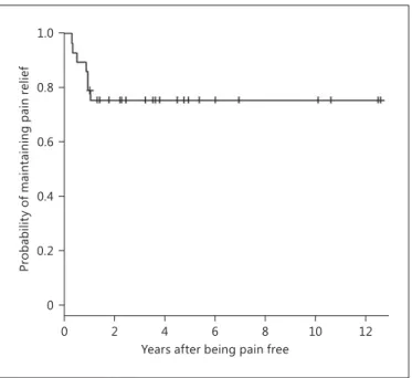
Maintaining Pain Relief after GKS and Management of Recurrence
Seven (24.13%) of the initially pain-free patients (n = 29) experienced at least 1 recurrence after GKS. The me-dian time to pain recurrence was 10.75 months (range: 3.8–12.6 months). Figure 4 shows the actuarial probabil-ity of maintaining pain relief without medication for the 29 patients with MBA-related TN. This probability at 0.5, 1 and 2 years was 93.1, 79.3 and 75.7%, respectively, and remained stable until 12 years. Recurrence was especially related to preoperative pain within the V3 territory (p = 0.023), atypical pain (p = 0.011; HR = 6.92; CI ranged be-tween 1.54 and 31.12) and age (p = 0.005; HR = 0.89; CI ranged between 0.83 and 0.97). The following parameters were not statistically significant: postoperative hypoes-thesia (p = 0.356; HR = 1), side of pain (p = 0.561), previ-ous surgical treatments (p = 0.54), sex (p = 0.15) and du-rability of the symptoms before GKS (p = 0.43).
Table 4. Main parameters evaluating outcome characteristics after GKS
Patien ts, n (%) Median period (range) Pain free 29 (100) 13.5 (0–240) days Hypoesthesia 3 (13.04) 7 (5–12) months Recurrence 7 (24.1) 10.75 (3.8–12.6) months 0.8 0.6 0.4 0.2 1.0 0 Pr ob ab ili ty o f b ei ng i ni tia lly p ai n f re e 10 8 6 4 2 0 12
Months after radiosurgery
Fig. 2. Actuarial probability of initial pain cessation free in the pop-ulation with MBA-related TN.
Radiosurgery for TN Related to MBA Compression
Stereotact Funct Neurosurg 2014;92:170–177
DOI: 10.1159/000362172 175
Because of recurrent medically refractory pain, 3 pa-tients (10.34%) required further surgery ( table 3 shows the postoperative characteristics). In our unit, the most frequent intervention after failed GKS was balloon micro-compression, practiced in 2 patients (6.9%), followed by thermocoagulation in 1 case (3.4%).
Evaluation of Outcome at the Last Follow-Up
At the last follow-up, a good outcome was observed as follows: by the BNI scale in all 29 patients (100%), by the Burchiel classification in 24 patients (82.8%), and by the Regis classification in 28 patients (96.6%; table 5 ).
Discussion
The role of GKS in essential TN has evolved in the last 20 years, and this kind of surgery nowadays represents a less invasive technique [48, 49] for the treatment of in-tractable TN, with results that vary from one study to an-other in terms of initial pain cessation as well as hypoes-thesia and recurrence rates [37–39, 41, 42, 46, 50–55] .
Regarding the etiology of TN, vascular compression is considered to be one of the major pathogenic factors [3– 5] . Walter Dandy [56] was the first to note the association between TN and vascular compression as well as their causal relationship. Since his observation, approximately
34 studies of TN caused by vertebral and/or basilar artery compression have been published, usually in the form of case reports [12, 16, 18–23, 25–29, 31, 57–59] . In most of them, the incidence of this anatomical condition ranges from 0 to 2.8% [7, 13, 57, 60–62] , with only 1 large series from Japan reporting a higher incidence (7.7%) [31] . Park et al. [30] recently reported a percentage of 2% of the glob-al population treated with GKS. In our study, the inci-dence was 4.47%.
MVD has been the accepted modality of treatment for TN caused by neurovascular compression [63] . Thus, a few articles in the literature treated the subject of MVD in the case of MBA compression causing TN. Linskey et al. [12] described a series of 31 patients of whom all were pain free (off medication) immediately after surgery and 90% were pain free (off medication) at the time of the re-port; 3 patients in that series had had pain recurrence. The overall 1-, 3- and 10-year rates of patients being pain free Table 5. Results at last follow-up by different evaluation scales
BNI Burchiel Regis Good result, n (%) 29 (100) 24 (82.8) 28 (96.6) Poor result, n (%) 0 5 (17.2) 1 (3.4) 0.8 0.6 0.4 0.2 1.0 0 Pr obabil ity o f h ypoes thesia onset 10 8 6 4 2 0 12
Years after radiosurgery
Fig. 3. Actuarial probability of hypoesthesia onset in the popula-tion with MBA-related TN.
0.8 0.6 0.4 0.2 1.0 0 Pr ob abili ty of maint ainin g p ain re lie f 10 8 6 4 2 0 12
Years after being pain free
Fig. 4. Actuarial probability of maintaining pain relief in the popu-lation with MBA-related TN.
Tuleasca /Carron /Resseguier /Donnet / Roussel /Gaudart /Levivier /Régis
Stereotact Funct Neurosurg 2014;92:170–177 DOI: 10.1159/000362172
176
(off medication) were 96, 92 and 86%, respectively. In terms of toxicity, 51.6% of the patients had minor trigem-inal hypoesthesia/hypoalgesia preoperatively, and 41.9% of the patients had newly or mildly worsened minor hy-poesthesia/hypoalgesia postoperatively, with only 1 pa-tient having masseter weakness and major hypoesthesia (a patient who underwent a complete nerve section). Oth-er deficits wOth-ere trochlear or abducens nOth-erve palsy (22.6%), hearing loss (12.9%) and aseptic meningitis (29%).
Ogawa et al. [58] reported the use of a synthetic vascu-lar graft sutured to the clival dura to move the vessel away from the cranial nerves. Stone et al. [59] used a silicone sling sutured to the petrous dura to reposition the basilar artery away from the trigeminal nerve. Takamiya et al. [27] used a fenestrated clip and decompressed the nerves. Other authors, such as Goel and Shah [17] , proposed fo-ramen magnum decompression in a particular case of ec-tatic basilar artery and basilar invagination. There was immediate relief from the neuralgic pain and no recur-rence observed at 18 months.
Park et al. [30] recently published the first series in the literature concerning MBA-related TN treated with GKS. In their population, 75% of the patients achieved initial pain relief that was adequate or better (BNI pain scale classes I–IIIb), with a probability of maintaining pain re-lief of 53, 38 and 10% at 1, 2 and 5 years, respectively. Some degree of facial sensory dysfunction occurred in 10% of the patients. Fourteen patients (70%) underwent an additional surgical procedure.
In our series, all the 29 patients (100%) had initial pain cessation without medication in a median time of 13.5 days (range: 0–240 days). The hypoesthesia rate was low, with hypoesthesia present in only 3 patients (13.04%). Re-current pain was observed in 7 patients (24.1%), of whom 3 (10.3%) needed further surgery.
Conclusions
GKS can be used as a first- and/or second-line treat-ment for TN caused by MBA compression. The rarity of complications even in the long run and the considerable probability of a long-lasting effect make GKS a pragmat-ic surgpragmat-ical first-intention treatment alternative for MBA-related TN.
Acknowledgments
This study was funded by the Centre Hospitalier Universitaire La Timone Assistance Publique-Hôpitaux de Marseille, France.
Disclosure Statement
J.R. received congress organization sponsoring from Accuray, Brainlab, Elekta and Varian. The other authors report no conflict of interest.
References
1 André N: Observations pratiques sur les ma-ladies de l’urethre: et sur plusieurs faits con-vulsifs, & la guérison de plusieurs maladies chirurgicales, avec la décomposition d’un remède propre à réprimer la dissolution gan-gréneuse & cancéreuse, & à la réparer; avec des principes qui pourront servir à employer les différents caustiques. Paris, Delaguette, 1976 (1756).
2 Alexander E, Loeffler JS, Lunsford DL: Ste-reotactic Radiosurgery. New York, McGraw-Hill, 1993, vol 1, p 254.
3 Haines SJ, Jannetta PJ, Zorub DS: Microvas-cular relations of the trigeminal nerve: an an-atomical study with clinical correlation. J Neurosurg 1980; 52: 381–386.
4 Hamlyn PJ, King TT: Neurovascular com-pression in trigeminal neuralgia: a clinical and anatomical study. J Neurosurg 1992; 76: 948– 954.
5 Jannetta PJ: Arterial compression of the geminal nerve at the pons in patients with tri-geminal neuralgia. J Neurosurg 1967; 26 (suppl):159–162.
6 Lorenzoni JG, et al: Neurovascular compres-sion anatomy and pain outcome in patients with classic trigeminal neuralgia treated by radiosurgery. Neurosurgery 2008; 62: 368– 375, discussion 375–376.
7 Bederson JB, Wilson CB: Evaluation of mi-crovascular decompression and partial sen-sory rhizotomy in 252 cases of trigeminal neuralgia. J Neurosurg 1989; 71: 359–367. 8 Burchiel KJ, et al: Comparison of
percutane-ous radiofrequency gangliolysis and micro-vascular decompression for the surgical man-agement of tic douloureux. Neurosurgery 1981; 9: 111–119.
9 Goya T, Wakisaka S, Kinoshita K: Microvas-cular decompression for trigeminal neuralgia with special reference to delayed recurrence. Neurol Med Chir (Tokyo) 1990; 30: 462–467.
10 Jannetta PJ: Microvascular decompression of the trigeminal root entry zone: theoretical considerations, operative anatomy, surgical technique, and results; in Rovit RL, Murali R, Janetta PJ (eds): Trigeminal Neuralgia. Balti-more, Williams & Wilkins, 1990, pp 201–222. 11 Kolluri S, Heros RC: Microvascular
decom-pression for trigeminal neuralgia: a five-year follow-up study. Surg Neurol 1984; 22: 235– 240.
12 Linskey ME, Jho HD, Jannetta PJ: Microvas-cular decompression for trigeminal neuralgia caused by vertebrobasilar compression. J Neurosurg 1994; 81: 1–9.
13 Sindou M, Amrani F, Mertens P: Décompres-sion vasculaire microchirurgicale pour névralgie du trijumeau: comparaison de deux modalités techniques et déductions physio-pathologiques. Etude sur 120 cas. Neuro-chirurgie 1990; 36: 16–25, discussion 25–26.
Radiosurgery for TN Related to MBA Compression
Stereotact Funct Neurosurg 2014;92:170–177
DOI: 10.1159/000362172 177
14 Sindou M: Microvascular decompression for trigeminal neuralgia; in Sindou M (ed): Prac-tical Handbook of Neurosurgery. Vienna, Springer, 2009, pp 1448–1462.
15 Jannetta PJ: Observations on the etiology of trigeminal neuralgia, hemifacial spasm, acoustic nerve dysfunction and glossopha-ryngeal neuralgia: definitive microsurgical treatment and results in 117 patients. Neuro-chirurgia (Stuttg) 1977; 20: 145–154. 16 Corkill G, Sarwar M, Virapongse C: Evolution
of dolichoectasia of the vertebrobasilar sys-tem as evidenced by serial computed tomog-raphy. Surg Neurol 1982; 18: 262–266. 17 Goel A, Shah A: Trigeminal neuralgia in the
presence of ectatic basilar artery and basilar invagination: treatment by foramen magnum decompression. J Neurosurg 2009; 111: 1220– 1222.
18 Grigoryan YA, Dreval ON, Michailova SI: Painful tic convulsif caused by a contralateral vertebral artery. Surg Neurol 1991; 35: 471–474. 19 Harsh GR 4th, et al: Magnetic resonance imag-ing of vertebrobasilar ectasia in tic convulsif: case report. J Neurosurg 1991; 74: 999–1003. 20 Hashimoto K, et al: Trigeminal neuralgia
caused by a dolichoectatic vertebrobasilar ar-tery: a case report. Rinsho Hoshasen 1987; 32: 331–334.
21 Koyanagi S, et al: Bilateral fenestrations of the vertebrobasilar artery with trigeminal neural-gia: case report. Neurol Med Chir (Tokyo) 1991; 31: 995–998.
22 Lye RH: Basilar artery ectasia: an unusual cause of trigeminal neuralgia. J Neurol Neu-rosurg Psychiatry 1986; 49: 22–28.
23 Miner ME, et al: Trigeminal neuralgia due to dolichoectasia: angiographic and CT findings in a patient with the EEC syndrome. Neuro-radiology 1980; 20: 163–166.
24 Niizuma H, Ikeda S, Ohyama H: Trigeminal neuralgia and hemifacial spasm caused by the compression of tortuous vertebro-basilar sys-tem: a case report (author’s transl) (in Japa-nese). No Shinkei Geka 1981; 9: 1167–1170. 25 Petty PG, Southby R: Vascular compression
of lower cranial nerves: observations using microsurgery, with particular reference to tri-geminal neuralgia. Aust NZ J Surg 1977; 47: 314–320.
26 Suzuki S, et al: New method of MVD using a vascular tape for neurovascular compression involving the vertebrobasilar artery: report of two cases. Neurol Med Chir (Tokyo) 1990; 30: 1020–1023.
27 Takamiya Y, et al: Trigeminal neuralgia and hemifacial spasm caused by a tortuous vertebro-basilar system. Surg Neurol 1985; 24: 559–562. 28 Waga S, Morikawa A, Kojima T: Trigeminal
neuralgia: compression of the trigeminal nerve by an elongated and dilated basilar ar-tery. Surg Neurol 1979; 11: 13–16.
29 Yoshida M, Asano M: Direct compression by megadolichobasilar anomaly as a cause of tri-geminal neuralgia: a case diagnosed by MRI. Tohoku J Exp Med 1994; 172: 327–332.
30 Park KJ, et al: Outcomes of Gamma Knife sur-gery for trigeminal neuralgia secondary to vertebrobasilar ectasia. J Neurosurg 2012; 116: 73–81.
31 Miyazaki S, et al: Trigeminal neuralgia due to compression of the trigeminal root by a basi-lar artery trunk: report of 45 cases. Neurol Med Chir (Tokyo) 1987; 27: 742–748. 32 Regis J, Tuleasca C: Fifteen years of Gamma
Knife surgery for trigeminal neuralgia in the Journal of Neurosurgery : history of a revolu-tion in funcrevolu-tional neurosurgery. J Neurosurg 2011; 115(suppl):2–7.
33 Tuleasca C, et al: Patterns of pain-free re-sponse in 497 cases of classic trigeminal neu-ralgia treated with Gamma Knife surgery and followed up for at least 1 year. J Neurosurg 2012; 117(suppl):181–188.
34 Flickinger JC, et al: Does increased nerve length within the treatment volume improve trigeminal neuralgia radiosurgery? A pro-spective double-blind, randomized study. Int J Radiat Oncol Biol Phys 2001; 51: 449–454. 35 Foote KD, et al: Analysis of risk factors
associ-ated with radiosurgery for vestibular schwan-noma. J Neurosurg 2001; 95: 440–449. 36 Frazier CH: Operation for the radical cure of
trigeminal neuralgia: analysis of five hundred cases. Ann Surg 1928; 88: 534–547.
37 Brisman R, Mooij R: Gamma knife radiosur-gery for trigeminal neuralgia: dose-volume histograms of the brainstem and trigeminal nerve. J Neurosurg 2000; 93(suppl 3):155– 158.
38 Dhople AA, et al: Long-term outcomes of Gamma Knife radiosurgery for classic trigem-inal neuralgia: implications of treatment and critical review of the literature. Clinical arti-cle. J Neurosurg 2009; 111: 351–358.
39 Massager N, et al: Gamma knife surgery for idiopathic trigeminal neuralgia performed using a far-anterior cisternal target and a high dose of radiation. J Neurosurg 2004; 100: 597– 605.
40 Pollock BE: Radiosurgery for trigeminal neu-ralgia: is sensory disturbance required for pain relief? J Neurosurg 2006; 105(suppl): 103–106.
41 Regis J, et al: Prospective controlled trial of gamma knife surgery for essential trigeminal neuralgia. J Neurosurg 2006; 104: 913–924. 42 Verheul JB, et al: Gamma Knife surgery for
trigeminal neuralgia: a review of 450 consecu-tive cases. J Neurosurg 2010; 113(suppl): 160–167.
43 Headache Classification Subcommittee of the International Headache Society: The Interna-tional Classification of Headache Disorders, ed 2. Cephalalgia 2004; 24(suppl):9–160. 44 Eller JL, Raslan AM, Burchiel KJ: Trigeminal
neuralgia: definition and classification. Neu-rosurg Focus 2005; 18:E3.
45 Miller JP, et al: Predictors of long-term suc-cess after microvascular decompression for trigeminal neuralgia. J Neurosurg 2009; 110: 620–626.
46 Regis J, et al: Radiosurgery in trigeminal neu-ralgia: long-term results and influence of op-erative nuances. Neurochirurgie 2009; 55: 213–222.
47 Rogers CL, et al: Gamma knife radiosurgery for trigeminal neuralgia: the initial experience of the Barrow Neurological Institute. Int J Ra-diat Oncol Biol Phys 2000; 47: 1013–1019. 48 Cruccu G, et al: AAN-EFNS guidelines on
tri-geminal neuralgia management. Eur J Neurol 2008; 15: 1013–1028.
49 Gronseth G, et al: Practice parameter: the di-agnostic evaluation and treatment of trigemi-nal neuralgia (an evidence-based review): re-port of the Quality Standards Subcommittee of the American Academy of Neurology and the European Federation of Neurological So-cieties. Neurology 2008; 71: 1183–1190. 50 Kondziolka D, et al: Gamma Knife
stereotac-tic radiosurgery for idiopathic trigeminal neuralgia. J Neurosurg 2010; 112: 758–765. 51 Maesawa S, et al: Clinical outcomes after
ste-reotactic radiosurgery for idiopathic trigemi-nal neuralgia. J Neurosurg 2001; 94: 14–20. 52 Matsuda S, et al: Gamma knife radiosurgery
for trigeminal neuralgia: the dry-eye compli-cation. J Neurosurg 2002; 97(suppl):525–528. 53 Nicol B, et al: Gamma knife radiosurgery us-ing 90 Gy for trigeminal neuralgia. J Neuro-surg 2000; 93(suppl 3):152–154.
54 Pollock BE, et al: Results of repeated gamma knife radiosurgery for medically unrespon-sive trigeminal neuralgia. J Neurosurg 2000; 93(suppl 3):162–164.
55 Pollock BE, et al: Stereotactic radiosurgery for idiopathic trigeminal neuralgia. J Neurosurg 2002; 97: 347–353.
56 Dandy W: Concerning the cause of trigeminal neuralgia. Am J Surg 1934; 24: 447–455. 57 Dandy W: Intracranial Arterial Aneurysms.
Ithaca, Comstock, 1944.
58 Ogawa A, et al: Repositioning of the tortuous vertebrobasilar artery for trigeminal neural-gia: a technical note. Surg Neurol 1992; 38: 232–235.
59 Stone JL, Lichtor T, Crowell RM: Microvascu-lar sling decompression for trigeminal neu-ralgia secondary to ectatic vertebrobasilar compression: case report. J Neurosurg 1993; 79: 943–945.
60 Apfelbaum RI: Surgery for tic douloureux. Clin Neurosurg 1983; 31: 351–368.
61 Klun B: Microvascular decompression and partial sensory rhizotomy in the treatment of trigeminal neuralgia: personal experience with 220 patients. Neurosurgery 1992; 30: 49– 52.
62 Piatt JH Jr, Wilkins RH: Treatment of tic dou-loureux and hemifacial spasm by posterior fossa exploration: therapeutic implications of various neurovascular relationships. Neuro-surgery 1984; 14: 462–471.
63 Jannetta PJ: Microsurgical management of trigeminal neuralgia. Arch Neurol 1985; 42: 800.