Annals of Oncology 12 (Suppl. I): S9-S13, 2001.
© 2001 Kluwer Academic Publishers. Printed in the Netherlands.
Symposium article
The role of overexpressed HER2 in transformation
R. M. Neve, H. A. Lane & N. E. Hynes
Fhedrich Miescher Institute, Basel, SwitzerlandSummary
The HER family of receptors has an important role in the network of cell signals controlling cell growth and differentia-tion. Although the activity of the HER receptor is strictly controlled in normal cells, HER2 receptor overexpression plays a pivotal role in transformation and tumorigenesis. HER2 gene amplification and/or overexpression of the receptor has been detected in subsets of a wide range of human cancers including breast cancer, and is an indicator of poor prognosis. It is proposed that overexpressed HER2 in combination with HER3 causes high activity of cell-signaling networks, thereby resulting in tumor cell proliferation. Thus, the HER2 receptor is an attractive target for new anti-cancer treatments.
Mono-clonal antibodies directed against the receptor are the most promising of these, and the humanized anti-HER2 monoclonal antibody trastuzumab (Herceptin) has shown significant clin-ical efficacy in clinclin-ical trials. The anti-tumor mechanisms of anti-HER2 monoclonal antibodies are not completely under-stood. However, some tumor types are not sensitive to trastu-zumab, suggesting that the response of a tumor to trastuzumab may not only be dependent on overexpressed HER2, but may also be influenced by other members of the HER receptor family expressed in the tumor cell.
Key words: breast cancer, erbB-2, HER2, neu, receptor
over-expression, tyrosine kinase activity
The HER2 receptor as a target for cancer treatment
The HER family of human epidermal growth factor receptors consists of four members: HER1, HER2, HER3 and HER4. The transmembrane HER receptors have important roles in the network of cell signals con-trolling cell growth and differentiation. In a normal cell, the activity of the receptors is strictly controlled, most significantly through the HER2 receptor [1].
In vitro and animal studies have clearly indicated that HER2 protein overexpression plays a pivotal role in oncogenic transformation and tumorigenesis. Studies using NIH 3T3 cells implicate HER2 overexpression in
malignant transformation and tumorigenesis [2-4]. Transfection of the HER2 gene into human breast and ovarian tumor cell lines produced more aggressive growth characteristics, such as increased DNA synthesis, cell growth, growth in soft agar in vitro, and tumori-genicity and metastatic potential in mice [5, 6].
HER2 gene amplification and/or overexpression of the receptor has been detected in subsets of a wide range of human cancers, including breast cancer [7—11]. The significance of HER2 amplification and HER2 over-expression have been studied most widely in breast cancer; around 20% of breast tumors overexpress HER2. Slamon et al. [12] first observed that HER2-gene amplifi-cation independently predicted overall (OS) and disease-free survival (DFS), and many studies have confirmed that HER2 overexpression is an indicator of poor prog-nosis in women with breast cancer. These patients have
poor response rates and short DFS and OS compared with women whose tumors do not overexpress this receptor.
The HER2 receptor is being investigated as a target for new more effective anti-cancer treatments. Its acces-sible location on the cell surface makes it particularly attractive. A number of approaches are being devel-oped, including tyrosine kinase inhibitors, anti-sense approaches which downregulate expression of the HER2 gene, intracellular expression of single-chain antibodies (ScFvs) to functionally inactivate the receptor, and immunization to boost anti-HER2 responses. The growth of tumors and human breast cancer cell lines over-expressing the HER2 receptor is also inhibited by monoclonal antibodies directed at the extracellular domain of the receptor [13, 14]. The humanized anti-HER2 monoclonal antibody trastuzumab (Herceptin) has shown significant clinical efficacy in trials of women with breast cancer who have not responded to or who have relapsed after previous treatment with chemo-therapy.
Our laboratory has tried to identify how overexpressed HER2 is involved in the transformation of cells in tumors. Understanding this mechanism will help clarify the mechanism of action of the monoclonal antibodies and allow this promising new treatment to be opti-mized.
have been identified in human cancers [16].
The usual sequence of oncogenic transformation appears to result from initial HER2 gene amplification, which generates more than the two normal gene copies in the epithelial cell; more than 10 gene copies are usually seen in the amplified state [17-19]. The specific factors that induce amplification of the HER2 gene have yet to be fully elucidated [20]. HER2 gene amplification leads to increased transcription of the HER2 gene, which gives rise to increased HER2 mRNA levels and con-comitant increased synthesis of HER2 protein. HER2 protein is then consequently overexpressed on the cell surface. HER2 protein levels are about 10- to 100-fold greater on the cell surface of HER2-positive breast cancer cells compared with adjacent normal breast epi-thelial cells [21].
Overexpression of HER2 protein on the cell surface probably leads to constitutive activation of HER2 homo-dimer receptors without the need for ligand binding. This results in unregulated cell growth and oncogenic transformation, in some instances resulting in viable cancer. As a result of spontaneous formation of receptor dimers, HER2 has a high basal activity in tumors in which it is overexpressed, which may affect the activity and/or expression of other HER family members. The finding that HER3 has elevated levels of phosphotyro-sine in HER2-positive tumors implies that HER2 contributes to malignant growth by recruiting other HER receptors, particularly HER3.
HER2 and cell transformation
In order to elucidate the role of overexpressed HER2 in transformation of cells, HER2 in_SjU3r3_t>reast tumor cells was functionally inactivated via inducible expres-sion of a HER2-directed ScFv5R [22, 23]. The HER2 receptor is normally expressed on the cell membrane. In cells expressing the ScFv5R, the HER2 receptor binds the single chain antibody in the endoplasmic reticulum leading to its retention in that compartment and functional inactivation of the receptor. In
HER2-over-Myc Myc/cyclin D degradation Cdk2 Active »• Cdk2 Inactive Degradation
Figure 2. Cell cycle control in the normal cell (for explanation see text).
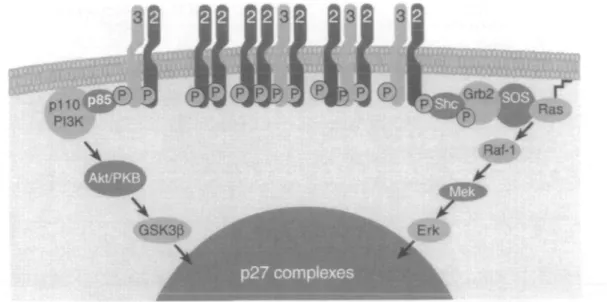
expressing SKBr3 cells induction of ScFv5R expression leads to loss of plasma membrane localized HER2. Simultaneously, the signaling activity of HER3, as measured by phosphotyrosine content, MAP kinase and PKB, decreased dramatically suggesting that active HER2/HER3 dimers are necessary for signal transduc-tion. HER3 is one of the most important cytoplasmic effectors of HER2, but is 'kinase dead' due to substitu-tions in important residues in the kinase domain. Thus, HER3 requires heterodimerization with another member of the HER family, usually HER2, to exert its cell-signaling action. It is proposed that overexpressed HER2 in combination with HER3 causes high activity of sig-naling pathways which results in uncontrolled cyclin E/Cdk2 activity. Thus, HER2 and HER3 function together to stimulate signaling networks that result in tumor cell proliferation (Figure 1) [24].
Loss of HER2 from the plasma membrane caused the SKBr3 cells to accumulate in the Gl phase of the cell cycle and the cells ceased proliferating after three days of ScFv5R induction [24]. Cell cycling and homeostasis in normal cells is maintained via an interacting network of cytoplasmic signaling molecules and nuclear cell cycle regulators (Figure 2). These regulators include the nuclear cyclins and their associated kinases, the cyclin-dependent kinases (Cdks) which control normal pro-gression through the cell cycle. An important regulator of Gl is Cdk2, which is positively regulated by associa-tion with cyclin E and is negatively regulated by various
11
p27 complexes
Figure 3. Model of p27 regulation. X, one of a range of molecules with which p27 can form complexes (for explanation see text).
mechanisms including binding with the cyclin kinase inhibitor p27. Regulation of p27 involves a number of steps (Figure 3). p27 exists in numerous complexes; in particular it is sequestered from cyclin E/Cdk2 by asso-ciation with the D type cyclin complexes. Downregula-tion and funcDownregula-tional inactivaDownregula-tion of HER2 after 72 hours of ScFv5R induction dramatically decreased the activity of Cdk2 to 20% of that observed in control cells. The level of c-Myc and the D-cyclins, proteins involved in p27 sequestration, also decreased in the absence of func-tional HER2, freeing a pool of uncomplexed p27, which is able to bind and inhibit cyclin E Cdk2 [24].
Reversal of malignant growth by anti-HER2 antibodies
The underlying mechanisms that mediate the anti-tumor effects of anti-HER2 monoclonal antibodies are not completely understood, but there are several proposed mechanisms. Treatment of cancer cells overexpressing HER2 with monoclonal antibodies results in a marked downregulation of HER2 expression [25, 26]. This anti-body-induced downregulation of HER2 has been shown to induce reversion of the transformed phenotype in «ew-transformed cells (neu is the rat HER2 equivalent) [13]. A relationship between accelerated receptor endo-cytosis and degradation, and tumor effects of anti-HER2 monoclonal antibodies is supported by correlative in vitro studies with a range of monoclonal antibodies [27]. Another property of some of these antibodies is their partial ability to disrupt the formation of HER2-HER3 and HER2-HER4 heterodimers [27, 28]. Possible additional in vivo mechanisms of action may be caused by an anti-angiogenic activity with downregulation of vascular endothelial growth factor and other angiogenic factors [29]. Furthermore, results from our laboratory suggest that monoclonal antibodies inhibit the signaling potential of overexpressed, oncogenic HER2 [30].
We have been studying the molecular effects of
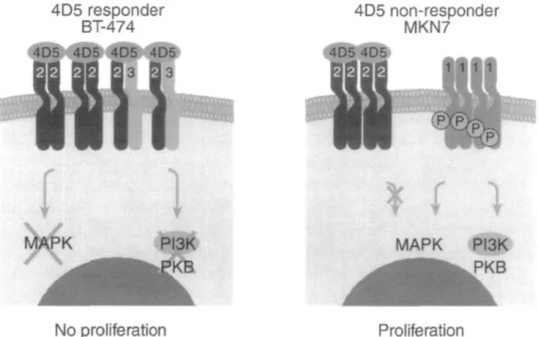
monoclonal antibody 4D5, the murine precursor to trastuzumab, on the BT-474 HER2-positive breast tumor cells in order to understand the mechanism of action of this growth inhibitory antibody. Furthermore, despite the fact that trastuzumab has shown clinical success, not all patients whose tumors overexpress HER2 respond to this treatment. In order to elucidate the cause of non-response, we have included in our studies the MKN7 gastric carcinoma tumor cells which overexpress HER2 but are not growth inhibited by 4D5.
Both BT-474 and MKN7 cells were treated with 4D5 and its effects on HER2 activity and the activity of intracellular signaling proteins were measured. Surpris-ingly, 4D5 treatment led to a rapid (10-minute) down-regulation of the phosphotyrosine content and activity of HER2 in both the responder and the non-responder cell line. However, in the BT-474 cells, but not in the MKN7 cells, a concomitant decrease in the activity of the PI3K pathway, as measured by PKB activity, was observed [30]. Furthermore, an examination of the HER receptor profile in these two cell lines revealed that only the BT-474 cells have high levels of active HER3. In contrast, the MKN7 cells have high levels of active HER 1/EGF receptor.
A model has been developed to demonstrate the difference between these two cell lines (Figure 4). In BT-474 cells, the HER2-HER3 heterodimer is very important for maintaining high signaling activity. Treat-ment with 4D5 affects activity of both HER2 and HER3 receptors because HER3 is dependent upon HER2 for its activity. There is a dramatic decrease in PKB and MAP kinase activity and the cells no longer proliferate. In MKN7 cells, 4D5 treatment leads to a decrease in phosphotyrosine content of HER2, but has no effect on signaling pathways because these cells have a high level of EGF receptor (HER1), which is unaffected by 4D5 treatment. This suggests that the response of a patient to trastuzumab may not only be dependent on over-expressed HER2, but may also be influenced by other
MAPK PI3K
PKB
No proliferation Proliferation
Figure 4. Reversal of malignant growth by 4D5 (for explanation see text).
members of the HER receptor family, which are ex-pressed in the tumor cell.
Conclusions
Amplification of the HER2 gene which leads to over-expression of the HER2 receptor has a crucial role in the transformation of cells. Overexpressed HER2 receptor, combined preferentially with the HER3 receptor, sustains overactivity of cell-signaling networks leading to high levels of cell cycle regulators. This causes uncontrolled cell growth, proliferation and tumorigenesis. In contrast, functional inactivation of HER2 restrains cell growth and proliferation.
Its crucial role in the development of tumors makes HER2 a valid target for new anti-cancer treatments. Monoclonal antibodies directed against the HER2 re-ceptor are currently the most promising approach. The recombinant human anti-HER2 monoclonal antibody, trastuzumab, has been effective in phase II and III clinical trials of women with HER2-positive metastatic breast cancer either as first-line therapy or in patients who had failed or relapsed after previous extensive treat-ment with chemotherapy [31-33]. The mechanism of action of growth inhibitory monoclonal antibodies tar-geted to HER2 is not fully understood and seems to be more complex than a simple downregulation of HER2 levels, as proposed from earlier studies. Some patients with HER2-positive tumors do not respond to mono-clonal antibodies, suggesting the involvement of factors additional to HER2 overexpression. Studies in different cell lines indicate that the response of a patient to trastuzumab may not only be dependent on overex-pressed HER2, but may also be influenced by expression
of other members of the HER receptor family in the tumor cell.
The incidence of breast cancer has been increasing steadily and is now the most common malignancy and the second most common cause of cancer-related death in European and North American women [34]. How-ever, marked progress has been made in treating this disease. The biologic behavior of this cancer, risk factors and prognostic factors have been better characterized. Several genetic abnormalities that are associated with the development of breast cancer and/or correlated with prognosis and response to treatment have been identified. Recognition and understanding of the role of HER2 in tumor development is allowing the development of new and more effective treatments for this disease.
Note
The authors have not reported any financial relationships with compa-nies whose products are mentioned in the text.
References
1. Rubin I, Yarden Y. The basic biology of HER2. Ann Oncol 2001; 12 (Suppl 1): S3-S8 (this supplement).
2. Di Fiore PP, Pierce JH, Fleming TP et al. Overexpression of the human EGF receptor confers an EGF-dependent transformed phenotype to NIH 3T3 cells. Cell 1987; 51: 1063-70.
3. Di Fiore PP, Pierce JH, Kraus MH et al. ErbB-2 is a potent oncogene when overexpressed in NIH/3T3 cells. Science 1987;
237:178-82.
4. Hudziak RM, Schlessinger J, Ulrich A. Increased expression of the putative growth factor receptor pl85H E R 2 causes
transforma-tion and tumorigenesis of NIH 3T3 cells. Proc Natl Acad Sci USA
13
5. Benz CC, Scott GK, Sarup JC et al Estrogen-dependent, tamoxifen-resistant tumorigenic growth of MCF-7 cells transfected with HEK2/neu. Breast Cancer Res Treat 1992; 24: 8 5 -95.
6. Chazin VR, Kaleko M, Miller A D et al. Transformation mediated by the human HER-2 gene independent of epidermal growth factor receptor. Oncogene 1992; 7: 1859-66.
7. Berns EMJJ, KJijn JG, van Staveren IL et al. Prevalence of amplification of the oncogenes c-myc, HER2lneu, and mt-2 in one thousand human breast tumours: Correlation with steroid receptors. Eur J Cancer 1992; 28: 697-700.
8. Borg A, Baldetorp B, Ferno M et al. ERBB2 amplification in breast cancer with a high rate of proliferation. Oncogene 1991; 6: 137-43.
9. Clark GM, McGuire WL. Follow up study of HER-2//ieu ampli-fication in primary breast cancer. Cancer Res 1991; 51: 944-8. 10. Slamon D, Godolphin W, Jones LA et al. Studies of the HER-2/
neu protooncogene in human breast and ovarian cancer. Science
1989; 244: 707-12.
11. Berger MS, Locher GW, Saurer S et al. Correlation of c-erbB-2 gene amplification and protein expression in human breast carci-noma with nodal status and nuclear grading. Cancer Res 1988; 48: 1238-43.
12. Slamon DJ, Clark GM, Wong SG et al. Human breast cancer: Correlation of relapse and survival with amplification of the
HER-2/neu oncogene. Science 1987; 235: 177-82.
13. Drebin JA, Link VC, Stern DF et al. Down-modulation of an oncogene protein product and reversion of the transformed phenotype by monoclonal antibodies. Cell 1985: 41: 697-706. 14. McKenzie SJ, Marks PJ, L a m T e t al. Generation and
character-ization of monoclonal antibodies specific for the human neu oncogene product, pl85. Oncogene 1989; 4: 543-8.
15. Hynes NE, Stern DF. The biology of erbB-2/neu/HER-2 and its role in cancer. Biochem Biophys Acta Rev Cancer 1994; 1198: 165-84.
16. Aaronson SA. Growth factors and cancer. Science 1991, 254: 1146-53.
17. Liu E, Thor A, He M et al. The //£7?2(c-erbB-2) oncogene is frequently amplified in m situ carcinomas of the breast. Oncogene 1992; 7: 1027-1032.
18. Seshadri R, Matthews C, Bobrovic A, Horsfall DJ. The signifi-cance of oncogene amplification in primary breast signifi-cancer. Int J Cancer 1989; 43: 270-2.
19. Szollosi J, Balazs M, Feuerstein BG et al. ERBB-2 (HER2/«eu) gene copy number, pl85HER"2 overexpression, and its intratumor
heterogeneity in human breast cancer. Cancer Res 1995; 55: 5400-7.
20. Pietras RJ, Pegram MD, Finn RS et al. Remission of human breast cancer xenografts on therapy with humanized monoclonal antibody to HER-2 receptor and DNA-reactive drugs. Oncogene 1998; 17: 2235-49.
21. Venter DJ, Tuzi NL, Kumar S, Gullick WJ. Overexpression of the c-erbB-2 oncoprotein in human breast carcinomas: Immuno-histological assessment correlates with gene amplification. Lancet 1987; ii: 69-72.
22. Beerli RR, Wels W, Hynes NE. Intracellular expression of single chain antibodies reverts ErbB-2 transformation. J Biol Chem
1994; 269: 23931-6.
23. Graus-Porta D, Beerli RR, Hynes NE. Single-chain antibody-mediated intracellular retention of ErbB-2 impairs Neu differentia-tion factor and epidermal growth factor signaling. Mol Cell Biol 1995; 15: 1182-91.
24. Neve RM, Sutterliity H, Pullen N et al. Effects of oncogenic ErbB2 on Gl cell cycle regulators in breast tumour cells. Oncogene 2000; 19: 1647-56.
25. Hudziak RM, Lewis GD, Winget M et al. P185HER2 monoclonal
antibody has antiproliferative effects in vitro and sensitises human breast tumor cell to tumor necrosis factor. Mol Cell Biol 1989; 9: 1165-72.
26. Kumar R, Shepard HM, Mendelsohn J. Regulation of phos-phorylation of the c-erbB-2/HER2 gene product by a mono-clonal antibody and serum growth factor(s) in human mammary carcinoma cells. Mol Cell Biol 1991; 11: 979-86.
27. Klapper LN, Vaisman N, Hurwitz E et al. A subclass of tumor-inhibitory monoclonal antibodies to ErbB-2/HER2 blocks cross-talk with growth factor receptors. Oncogene 1997; 14. 2099-109. 28. Reese D, Arboleda J,Twaddell J et al. Effects of the 4D5 antibody
on WERllneu heterodimerization with other class I receptors in human breast cancer cells. Proc Am Assoc Cancer Res Annu Meet 1996; 37: 51 (Abstr 353).
29. Petit AM, Rak J, Hung MC et al. Neutralizing antibodies against epidermal growth factor and erbB-2/neu receptor tyrosine kinases down-regulate vascular endothelial growth factor production by tumor cells in vitro and in vivo' Angiogenic implications for signal transduction therapy of solid tumors. Am J Pathol 1997; 151:
1523-30.
30. Lane HA, Beuvink I, Motoyama AB et al. ErbB2 potentiates breast tumor proliferation through modulation of p27Klpl-Cdk2
complex formation: Receptor overexpression does not determine growth factor dependency. Mol Cell Biol 2000; 20: 3210-23. 31. Baselga J, Tripathy D, Mendelsohn J et al. Phase II study of
weekly intravenous recombinant humanized anti-pl85H E R 2
monoclonal antibody in patients with HER2/«eu-overexpressing metastatic breast cancer. J Clin Oncol 1996; 14: 737-44. 32. Baselga J, Norton L, Albanell J et al. Recombinant humanized
anti-HER2 antibody (Herceptin®) enhances the antitumour activ-ity of paclitaxel and doxorubicin against HER2lneu overexpressing human breast cancer xenografts. Cancer Res 1998, 58: 2825-31. 33. Pegram MD, Lipton A, Hayes DF et al. Phase III study of
receptor-enhanced chemosensitivity using recombinant human-ized anti-pl85H E R 2 / n e" monoclonal antibody plus cisplatin in
patients with HER2/«eu-overexpressing metastatic breast cancer refractory to chemotherapy treatment. J Clin Oncol 1998; 16: 2659-71.
34. Wingo P, Tong T, Bolden S. Cancer statistics. Cancer J Clin 1995; 45: 8-30.
Correspondence to.
N. E. Hynes, PhD
Friedrich Miescher Institute P.O. Box 2543
4002 Basel Switzerland