Prophylactic Insertion of Optional Vena Cava
Filters in High-Risk Trauma Patients
Christoph Meier
1, Roger Pfiffner
2, Ludwig Labler
1, Andreas Platz
1, Thomas Pfammatter
2,
Otmar Trentz
1Ab stract
Background: Vena cava interruption is a form of
pulmonary embolism prophylaxis that is being used in high-risk patients who do not tolerate pharmacologic prophylaxis. Retrievable prophylactic vena cava filters (VCFs) are of particular interest for severely injured patients where the necessity for VCF is often only temporary.
Methods: In a single-institution case series of
consecu-tive patients who received prophylactic VCFs after polytrauma, between 04/1998 and 07/2004, the demographic data, injury pattern and complications were analysed.
Results: Ninety-five prophylactic VCFs were placed in
polytrauma patients (median ISS of 38). Median age was 38 years (range 16–80 years). Median delay between trauma and filter placement was 1 day (range 0–31 days). No complication was seen related to filter insertion or retrieval. Sixty-five VCFs (68.4%) were retrieved after 4–25 days (median 13 days). One filter migration (1.1%) was observed. Retrieval failed in two patients (3.0%). A total of 30 VCFs (31.6%) were left permanently. One non-fatal PE (1.1%) occurred 21 days after filter retrieval despite prophylaxis with LMWH. DVT developed in two patients (2.1%) including one vena caval occlusion (1.1%). Overall mortality was 7.4%.
Conclusions: Early prophylactic placement of VCF in a
high-risk trauma patient should be considered when anticoagulation is contraindicated. Filter insertion and retrieval is safe with a low complication rate.
Key Words
Prophylactic vena cava filters · Optional · Retrievable · High-risk trauma patients
1 Division of Trauma Surgery, University Hospital Zurich, Switzerland, 2 Institute of Diagnostic Radiology, University Hospital Zurich,
Switzerland.
Received: July 26, 2005; revision accepted: December 20, 2005.
Eur J Trau ma 2006;32:37–43 DOI 10.1007/s00068-006-0086-z
Introduction
Deep venous thrombosis (DVT) and pulmonary embolism (PE) play a major role in trauma. The inci-dence for venous thromboembolic events (VTE) varies widely in the literature from 5 to 58% [1–3]. In some series, PE represents the third major cause of death after trauma in those patients who survive longer than 24 h after injury [4]. Mortality associated with PE was 18.7% in an analysis of 1,602 episodes of VTE from the National Trauma Data Bank [5]. Since most PE are clinically silent, the incidence of occult PE is surely underappreciated. Schultz et al. [6] documented a 24% incidence of asymptomatic PE in a study of 90 moder-ately to severely injured trauma patients undergoing surveillance contrast-enhanced helical CT scanning. Thirty percent of patients receiving pharmacologic prophylaxis had a PE in this study.
Unfractionated heparin followed by oral anticoagu-lation for 3 months prevents PE in 95% of patients with proximal DVT [7, 8]. However, low-dose heparin (LDH) has very little proven efficiency in the preven-tion of VTE after trauma [9, 10]. Several studies have indicated that low-molecular-weight heparin (LMWH) is superior to LDH for prophylaxis with the same or less bleeding risk [10–12]. The use of pneumatic compres-sion devices and arteriovenous foot pumps in trauma has shown no benefit over no prophylaxis [11–14]. Most DVT develop in the first 2 weeks after injury [15]. In a retrospective case series Owings et al. [16] demon-strated that 6% of all PE in the trauma setting occurred on day 1 following injury. This finding demonstrates the importance of efficient early VTE prophylaxis. Severely
injured patients are often at higher bleeding risks in the first days after trauma. As a secondary cerebral or ab-dominal hemorrhage may have fatal consequences, se-lection of VTE prophylaxis can be a challenging balance between VTE risk and bleeding risk.
Vena cava interruption became common after vena cava filters (VCFs) became available in the late 1960s [17]. Inferior VCFs are effective in preventing PE [17– 19], but they do not prevent DVT or postthrombotic syndrome. In high-risk trauma patients without DVT, prophylactic insertion of a VCF has shown to decrease the rate of PE in multiple studies [20–22]. Interruption of the inferior vena cava (IVC) for the prevention of PE has become popular in trauma patients. Rogers et al. [20] considered 4.3% of all trauma patients admitted to one-centre appropriate candidates for prophylactic in-sertion of VCFs.
The first VCFs were designed for permanent place-ment. Retrievable filters have been developed to avoid potential long-term device-related complications, such as IVC perforation or occlusion, thrombosis and filter migration [23, 24]. Optional filters are devices which can remain in place, acting as a permanent VCF, or be re-moved percutaneously as a retrievable filter [25]. This advantage is of particular interest in trauma patients. For many of these patients anticoagulation is only con-traindicated for a short period after trauma and the ne-cessity for vena cava interruption in trauma patients is often time-limited. For these cases an effective filter, which can be safely retrieved, is an attractive alternative to permanent filters.
Patients and Methods
The trauma registry and interventional radiology data-base were reviewed for all VCFs placed in trauma patients from April 1998 to July 2004. Clinical and radiological records were reviewed for age and gender of the patient, injury pattern, indication for VCF, type of filter, interval between trauma and VCF insertion, time of retrieval, complications and long-term follow-up for permanent vena cava filtration. Only patients with polytrauma and prophylactic insertion of a VCF entered the study. Polytrauma was defined, according to the guidelines of the German Society for Trauma Surgery, as a life-threatening injury to several physical regions/organ systems with an Injury Severity Score (ISS) ≥ 16.
All patients without contraindication to pharmaco-logical prophylaxis initially received intravenous LDH
administered continuously over 24 h. This regimen was changed to either LMWH or to wafarin derivates ac-cording to the injury pattern later on. Prophylactic VCF placement was evaluated according to the guidelines of the Eastern Association for the Surgery of Trauma (EAST) [11]. In addition, the patient’s risk for VTE was assessed using the RAPT score developed by Green-field and co-workers [3] (Table 1). High-risk trauma pa-tients (RAPT score ≥ 5) were evaluated for prophylactic
VCF when pharmacologic prophylaxis for VTE was contraindicated. All patients received high-thigh anti-embolic stockings. Patients unable to wear these stock-ings, because of fractures or open wounds of the lower extremities, had them applied unilaterally whenever possible. Insertion of the VCF was performed as early as possible regarding the patient’s condition. For pa-tients with cardiovascular instability, high intracranial pressure and other physical conditions forbidding trans-port to the angiography suite and the stress of an inter-ventional procedure filter insertion was delayed.
All filters were inserted and retrieved by a senior interventional radiologist under fluoroscopic guidance in the angiography suite. The VCFs were exclusively placed infrarenally. Two different optional filter models
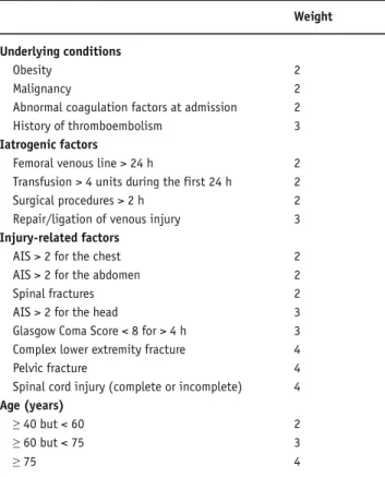
Table 1. VTE risk factor assessment in adult trauma patients (RAPT score).
Weight
Underlying conditions
Obesity 2
Malignancy 2
Abnormal coagulation factors at admission 2 History of thromboembolism 3 Iatrogenic factors
Femoral venous line > 24 h 2 Transfusion > 4 units during the first 24 h 2 Surgical procedures > 2 h 2 Repair/ligation of venous injury 3 Injury-related factors
AIS > 2 for the chest 2 AIS > 2 for the abdomen 2 Spinal fractures 2 AIS > 2 for the head 3 Glasgow Coma Score < 8 for > 4 h 3 Complex lower extremity fracture 4
Pelvic fracture 4
Spinal cord injury (complete or incomplete) 4 Age (years)
≥ 40 but < 60 2 ≥ 60 but < 75 3
were used in the time studied. In total, 65 Günther Tulip devices (William Cook, Bjaekerskov, Denmark) and 30 OptEase filters (Cordis Endovascular, J&J, Roden, The Netherlands) were placed. The former was inserted from April 1998 to August 2003 and the latter from August 2003 to July 2004.
The Günther Tulip filter consists of four stainless-steel legs forming a cone. The clot-trapping areas are formed by thinner wires which are shaped like tulip leaves. They extend from the apex to the distal ends of each leg. Small barbed hooks at the caudal ends of the legs provide fixation in the IVC. A hook at the proximal end of the device facilitates retrieval.
In contrast, the OptEase device has a double-basket design with six straight nitinol struts connecting the proximal and distal baskets. Six fixation barbs at the cranial end of the filter prevent migration. A hook located at the caudal base of the device allows retrieval.
Both filter types can be delivered through either a jugular or femoral venous approach. Access for retriev-al depends on the particular filter designs. The Günther Tulip device must be retrieved through the jugular vein whereas retrieval of the OptEase filter is from the femoral vein access.
Since contraindication for anticoagulation is often only temporary in trauma patients, filter retrieval was routinely evaluated. All patients considered candidates for retrieval underwent inferior cavography prior to the planned procedure. The presence of only minor throm-boemboli at the filter struts, defined as less than 25% of the diameter of the IVC, was no contraindication for re-trieval. For larger thromboemboli the filter was left in situ and a therapeutic anticoagulation with intravenous heparin was initiated with follow-up cavography between 7 and 14 days later. Filter retrieval was per-formed in the same session when a reduction of the thromboembolic mass to the aforementioned dimen-sion was evident on cavography. For persistent filter thrombosis the filter was left in place and the patient was started on a long-term anticoagulation.
Patients with permanent VCF were asked to attend follow-up examination which was performed by a fellow interventional radiologist. Follow-up included a clinical examination, color-flow duplex scan and a plain radio-graph of the abdomen.
Results
A total of 107 trauma patients underwent vena cava in-terruption at our department. Ninety-five patients
(88.8%) with polytrauma and prophylactic insertion of a VCF entered the study. Five polytrauma patients (4.7%) with therapeutic VCF insertion were excluded from further evaluation. Seven patients (6.5%) with single injuries or non-life-threatening multiple injuries were excluded from the study as well.
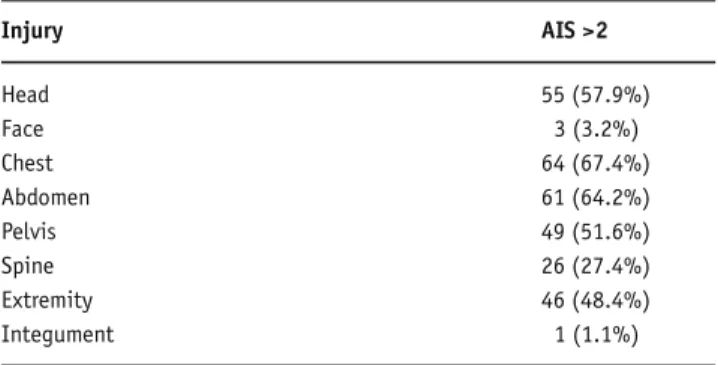
Median age was 38 years (range 16–80 years). Sixty-seven patients were male (70.5%), 28 female (29.5%). The ISS ranged from 17 to 66 (median 38). Injuries to chest, abdomen and head were among the most frequent injury patterns (Table 2). Median hospital stay was 26 days (range 6–159 days) including 11 days (range 1–50 days) at the ICU. Patients were ventilated for a median time of 7 days (range 1–36 days).
Indication for prophylactic VCF placement was based on the EAST guidelines [11]. Ninety-three pa-tients (97.9%) fulfilled the high-risk criteria of the RAPT score. Two patients (2.1%) with rib fractures in combination with a ruptured spleen and liver, respec-tively, received a VCF despite a RAPT score < 5. Both patients were treated nonoperatively. Decision making for temporary vena cava interruption in these patients was based on individual judgement by the treating phy-sicians and was not in accordance with our concept. Both patients were included in the study, and filter retrieval in these patients was assessed and performed similar to the patients with a RAPT score > 5.
The interval between trauma and filter placement averaged 2.4 days (median 1 day, range 0–31 days). For the last year of our observation period this interval de-creased to 1.8 days (median 1 day, range 0–7 days). Fif-ty-four patients (56.8%) had their VCFs placed within 1 day after admittance. All filters were placed infrare-nally. Both filter types were predominantly delivered through a femoral venous approach (n = 91, 95.8%); the jugular vein was accessed in 4.6% (n = 4). Thirty-six Günther Tulip filters (94.7%) were retrieved from the
Table 2. Injury pattern.
Injury AIS >2 Head 55 (57.9%) Face 3 (3.2%) Chest 64 (67.4%) Abdomen 61 (64.2%) Pelvis 49 (51.6%) Spine 26 (27.4%) Extremity 46 (48.4%) Integument 1 (1.1%)
jugular vein access; in two cases (5.3%) a combined femoro-jugular access was necessary. Only the femoral vein was accessed for retrieval of the OptEase device (n = 27).
Overall, 65 VCFs were placed temporarily (68.4%) and 30 filters (31.6%) were permanent. VCF retrieval was successful in 65 out of 67 cases (97.0%). Two retrieval procedures (Günther Tulip filter) had to be abandoned due to technical difficulties (3.0%). Both fil-ters presented with a tilt of > 10° within the vessel
struc-ture, making it technically impossible to grab the filter hook. The median duration between placement and re-trieval was 13 days (range 4–25 days). One patient decided to be transferred to another hospital for further treatment, 5 days after trauma. As VCF retrieval was not available in the other hospital, it was decided to re-move the filter exceptionally early 4 days after insertion. All other patients had their filters placed for at least 7 days.
A total of 21 filters (22.1%) showed strands of or-ganised thrombus on the filter struts on follow-up ca-vography before potential retrieval. Thirteen VCFs with a thrombotic mass # 25% were retrieved as planned without delay or additional antithrombotic therapy. No complication related to retrieval of these filters was ob-served. Preretrieval cavography demonstrated partial filter thrombosis (50–75%) in two cases which was com-bined with caudal filter migration towards the right common iliac vein in one patient (1.1%). Retrieval was delayed and anticoagulation therapy was initiated for both patients 12 days after trauma as they had no ongo-ing contraindication to anticoagulation. On follow-up cavography 22 and 25 days after trauma no residual fil-ter thrombosis was seen. Uneventful retrieval of both devices was performed in the same session. VCF re-trieval was cancelled in six cases due to critical sizes of filter thrombosis (> 25%) and ongoing contraindication to anticoagulation. One of these patients had his filter retrieved and successfully replaced by a second device in order to prevent impending IVC occlusion in the same session 13 days after initial placement. Uneventful retrieval of the second VCF was performed 12 days later.
No hematoma or thrombosis occurred at the access site neither for insertion nor retrieval. One nonfatal PE (1%) was diagnosed 21 days after filter retrieval despite adequate prophylaxis with LMWH. This 30-year-old woman was involved in a road traffic accident, sustaining an unstable lumbar spine fracture and a blunt trauma to chest and abdomen (ISS 32, RAPT score 10). Damage
control laparotomy with perihepatic packing and inser-tion of bilateral chest tubes were performed. Two sec-ond-look laparotomies followed within 4 days. A VCF was placed less than 48 h after trauma. The spine frac-ture was stabilised by an internal fixator 4 days later. VCF removal took place 13 days after trauma. Symp-tomatic PE occurred 21 days later. Helical CT angiogra-phy showed multiple segmental pulmonary emboli and therapeutic anticoagulation was initiated. DVT was ob-served in two patients. One IVC occlusion with DVT was detected on inferior cavography 12 days after filter placement. The filter was not retrieved and long-term anticoagulant therapy was initiated. Thirty-seven days after filter placement duplex scanning revealed persis-tent DVT, but a recanalised IVC. At follow-up (73 months after VCF insertion) duplex scanning showed no residual thrombosis either in the IVC or the deep venous system of the pelvis and lower extremities. Mor-tality was 7.4%; no death was related to VTE or the VCF. Median interval between trauma and death was 13 days (range 7–99 days). Five patients died after the decision was made to leave the filters permanently; in two cases death occurred before filter retrieval was considered.
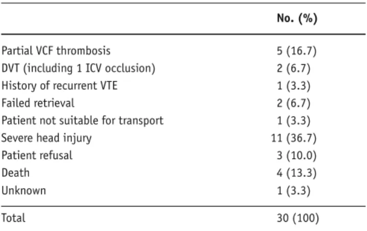
The ratio of retrieved filters compared to perma-nent placements was more favourable for the OptEase device (90.0 vs. 58.5%) which replaced the Günther Tulip filter in August 2003. This difference in the re-trieval rate between the two devices may be partly due to increasing lenience towards the presence of clot in the filter as a contraindication for retrieval as the oper-ator’s experience grew. Furthermore, changing the practice concerning contraindication to anticoagulation for different injury patterns over the last years has led to increasing numbers of filter retrieval. Head injury, for example, is not a strict contraindication to pharmaco-logic prophylaxis anymore. Indication and timing for anticoagulation is determined for each head trauma pa-tient individually by neurosurgical consultation. The same practice is pursued for blunt abdominal trauma and other injuries. This approach in combination with extended interval for filter retrieval has gained increas-ing acceptance in our unit. Filter retrieval is now the standard practice for trauma patients, leaving perma-nent filters for special situations only (Table 3).
Of the 23 survivors with permanent VCF follow-up, imaging was possible in 14 patients (60%; 12 Günther Tulip, 2 OptEase). Mean time of follow-up was 33.7 months (range 0.9–76.7 months). All patients
under-went colour-flow duplex scanning. A plain radiograph of the abdomen was obtained for nine patients. In one patient filter position was assessed on thoracoabdomi-nal computed tomography which was performed for other reasons. Abdominal X-ray was refused by four patients. Twelve patients with Günther Tulip VCFs (57.1%) were available for follow-up (mean 38 months, range 1–77 months). No filter migration was found. Fil-ter tilt was seen in two patients (16,7 %). All VCFs were patent and no signs of chronic venous insufficiency were recorded. OptEase devices were permanent in only three patients. One patient died from his severe head trauma; the other two patients presented for follow-up (mean 8 months, range 3–12 months). Both patients showed a patent IVC, no signs of filter migration or tilt and no DVT.
Discussion
Prophylactic filter placement for high-risk trauma pa-tients without documented VTE is still controversial. Current recommendation is based on Level III data ac-cording to the EAST Practice Management Guidelines Work Group [11]. There is no question about filter ef-ficiency. In most series a decrease in the incidence of PE is reported when prophylactic VCFs were inserted. Greenfield and Proctor [26] reported a success rate of 98% in preventing PE from lower extremity DVT. In contrast only one retrospective study was published demonstrating a significantly higher rate of PE in a time period when more filters were placed compared to a time period with more restrictive filter indications [27]. The authors did not give any explanation for this finding.
Defining who should receive a filter remains a ma-jor issue. Level I data supported only spinal cord inju-ries and spinal fractures, both being high-risk factors for
venous thromboembolism. Age was an increased risk factor, but the analysis failed to determine the exact age at which the risk increased substantially. All other tradi-tional risk factors such as long bone fractures, pelvic fractures or head injuries were not found to be powerful risk factors on meta-analysis [11].
However the authors outlined the need for addi-tional adequately sized prospective studies for re-evalu-ating the role of different possible risk factors. In the same article the EAST Practice Management Guide-lines Work Group had suggested that prophylactic VCF should be considered in patients with contraindication to anticoagulation and high-risk criteria, such as pro-longed immobilisation including severe head trauma and spinal cord injury, complex pelvic fractures and multiple long bone fractures.
Beside the EAST guidelines we routinely applied the RAPT score to identify patients with a high risk for thromboembolism [3]. Vena cava interruption was con-sidered for all patients with a score ≥ 5 and contraindica-tion for pharmacologic prophylaxis. According to the literature, patients with a RAPT score of ≥ 5 are three times more likely to develop VTE than patients with a lower score [3, 28].
An optional VCF is an attractive alternative to per-manent or temporary filter systems for polytrauma pa-tients. It can be left permanently if long-term vena caval interruption is indicated. Patients who subsequently un-dergo anticoagulant therapy after initial contraindica-tion benefit by having the filter safely removed. Our experience with optional filter systems is comparable with the literature [29–32]. We had a retrieval technical success of 97.0% with no retrieval-related morbidity. Compared to the literature we had a high retrieval rate of 68.4% and only 31.6% of permanent placements. In the last year of observation this rate increased to 90.0% displaying the consequent implementation of our concept.
Recommended interval for retrieval of most option-al filters is within 14 days. Our longest intervoption-al was 25 days with uneventful retrieval. Intervals for Günther Tulip filters up to 126 days are reported in the literature [33]. This suggests that retrievable filters can potentially provide caval interruption for a longer time period without the risk of long-term complications when- ever retrieved.
Decousus et al. [18] showed that VCFs only prevent PE in the short term after placement. They also found that medical patients receiving a VCF had significantly
Table 3. Reasons for permanent filter placement.
No. (%)
Partial VCF thrombosis 5 (16.7) DVT (including 1 ICV occlusion) 2 (6.7) History of recurrent VTE 1 (3.3) Failed retrieval 2 (6.7) Patient not suitable for transport 1 (3.3) Severe head injury 11 (36.7) Patient refusal 3 (10.0)
Death 4 (13.3)
Unknown 1 (3.3)
higher rates of DVT recurrence than those treated with anticoagulation alone. They suggested that long-term anticoagulant therapy should be considered after place-ment of a permanent filter to counterbalance this pos-sible effect. However, a higher DVT rate was not seen in trauma patients with prophylactic filter placement com-pared with non-filter patients [34].
On a long-term follow-up (4–42 months), Patton et al. [35] found that 26% of the patients had physical find-ings and duplex evidence of postphlebitic syndrome. Wojcik et al. [36] reported an early DVT rate of 44% after prophylactic VCF placement in trauma patients who could not initially receive anticoagulation. Diag-nosed by routine duplex scanning only 39% of the pa-tients were symptomatic. Greenfield et al. [37] reported a DVT rate of 15.6% after prophylactic filter placement during hospitalisation. At follow-up (mean 2.4 years) another 10.8% presented with new DVT, a long-term PE rate of 1.5% and no late filter-related complications. They emphasised the importance of continued DVT prophylaxis after filter placement. There is no question about the necessity of adequate DVT prophylaxis at the earliest possible time in high-risk trauma patients. Whether insertion of prophylactic VCF makes patients more prone to subsequent DVT is not known. Very high-risk patients as classified by Geerts et al. [1] had a DVT rate of 40–50% without VCF. In what way the use of retrievable filters may result in a lower incidence of late DVT remains unclear at present.
In our series filter thrombosis was seen in 22.1%. In most cases the mass was measured to be <25% of the diameter of the IVC. This raises the question of the ori-gin of these thrombus formations. Adherent thrombotic material, usually containing fibrinous material, on the filter struts is commonly seen [31]. This material proba-bly does not represent embolisation, but rather in situ thrombus formation [29]. The other possible origin is from trapped small thromboemboli which otherwise would have caused asymptomatic PE. This can clearly not be considered a complication as VCFs are designed to trap thromboemboli.
DVT and PE can develop shortly after trauma. Schultz et al. [6] studied 90 patients with an ISS $ 9 with-out symptoms suggestive of VTE undergoing contrast-enhanced helical CT scanning between 3 and 7 days af-ter trauma. A 24% incidence of asymptomatic PE with mainly a minor clot burden was found. This report and others emphasize the timing of VTE prophylaxis. In a retrospective case series Owings et al. [16]
demonstrat-ed that 6% of all PE in the trauma setting occurrdemonstrat-ed on day 1 following injury. Fifty-six percent of our patients had their VCFs placed within 1 day and 77.9% within 3 days after admittance, providing early protection from fatal PE.
Wicky et al. [38] published the only study, so far, reporting long-term outcome of nine non-retrieved Günther Tulip filters with a mean follow-up of 30 months (range 12–55 months). All presented with pat-ent IVC. Proximal filter migration of 20–25 mm was observed in two patients (22%). Another two filters showed a tilting of 10°. Duplex scan showed no venous thrombosis. In our study 12 patients with Günther Tulip filters were available for follow-up (mean 38 months). We did not see any filter migration and a comparable long-term tilt rate of 16.7%. Like Wicky et al. [38] we did not find any correlation between tilt and occurrence of VTE.
In conclusion, early prophylactic placement of VCFs in a high-risk trauma patient should be considered when anticoagulation is contraindicated. Optional VCFs can act as permanent filters but also allow retrieval. Patients who subsequently undergo anticoagulant therapy after initial contraindication benefit by having the filter safely removed, avoiding potential long-term complications.
References
1. Geerts WH, Code KI, Jay RM, Chen E, Szalai JP. A prospective study of venous thromboembolism after major trauma. N Engl J Med 1994;331:1601–6.
2. Spain DA, Richardson JD, Polk HC Jr, Bergamini TM, Wilson MA, Miller FB. Venous thromboembolism in the high-risk trauma patient: do risks justify aggressive screening and prophylaxis? J Trauma 1997;42:463–7.
3. Greenfield LJ, Proctor MC, Rodriguez JL, Luchette FA, Cipolle MD, Cho J. Posttrauma thromboembolism prophylaxis. J Trauma 1997;42:100–3. 4. Knudson MM, Ikossi DG. Venous thromboembolism after trauma.
Curr Opin Crit Care 2004;10:539–48.
5. Knudson MM, Ikossi DG, Khaw L, Morabito D, Speetzen LS. Throm-boembolism after trauma. An analysis of 1602 episodes from the American College of Surgeons National Trauma Data Bank. Ann Surg 2004;240:490–8.
6. Schultz DJ, Brasel KJ, Washington L, Goodman LR, Quickel RR, Lipchik RJ, Clever T, Weigelt J. Incidence of asymptomatic pulmo-nary embolism in moderately to severely injured trauma patients. J Trauma 2004;56:727–33.
7. Hyers TM, Hull RD, Weg JG. Antithrombotic therapy for venous thromboembolic disease. Chest 1995;108:Suppl:335S–51S. 8. Leizorovicz A. Comparison of the efficacy and safety of low
mo-lecular weight heparins and unfractionated heparin in the initial treatment of deep venous thrombosis: un updated meta-analysis. Drugs 1996;52:Suppl 7:30–7 .
9. Upchurch GR Jr, Demling RH, Davies J, Gates JD, Knox JB. Efficacy of subcutaneous heparin in prevention of venous thromboembolic events in trauma patients. Am Surg 1995;61:749–55.
10. Geerts WH, Jay RM, Code KI, Chen E, Szalai JP, Saibil EA, Hamilton PA. A comparison of low-dose heparin with low-molecular weight heparin as prophylaxis against venous thromboembolism after major trauma. N Engl J Med 1996;335:701–7.
11. Rogers FB, Cipolle MD, Velmahos G, Rozycki G, Luchette FA. Practice management guidelines for the prevention of venous thrombo-embolism in trauma patients; the EAST practice management guidelines work group. J Trauma 2002;53:142–64.
12. Knudson MM, Morabito D, Paiement GD, Shackleford S. Use of low molecular weight heparin in preventing thromboembolism in trauma patients. J Trauma 1996;41:446–59.
13. Velmahos GC, Kern J, Chan LS, Oder D, Murray JA, Shekelle P. Pre-vention of venous thromboembolism after injury: an evidence-based report – Part I: analysis of risk factors and evaluation of the role of vena cava filters. J Trauma 2000;49:132–9.
14. Spain DA, Bergamini TM, Hoffmann JF, Carrillo EH, Richardson JD. Comparison of sequential compression devices and foot pumps for prophylaxis of deep venous thrombosis in high-risk trauma patients. Am Surg 1998;64:522–6.
15. Velmahos GC, Nigro J, Tatevossian R, Murray JA, Cornwell EE III, Belzberg H, Asensio JA, Berne TV, Demetriades D. Inability of an ag-gressive policy of thromboprophylaxis to prevent deep venous thrombosis (DVT) in critically injured patients: are current meth-ods of DVT prophylaxis insufficient? J Am Coll Surg 1998;187: 529–33.
16. Owings JT, Kraut E, Battistella F, Cornelius JT, O’Malley R. Timing of the occurrence of pulmonary embolism in trauma patients. Arch Surg 1997;132:862–6.
17. Greenfield LJ, Michna BA. Twelve-year clinical experience with the Greenfield vena cava filter. Surgery 1988;104:706–12.
18. Decousus H, Leizorovicz A, Parent F, Page Y, Trdy B, Girard P, Laporte S, Faivre R, Charbonnier B, Barral FG, Huet Y, Simmoneau G. A clinical trial of vena cava filters in the prevention of pulmonary embolism in patients with proximal deep-vein thrombosis. N Engl J Med 1998;338:409–15.
19. Greenfield LJ, Proctor MC, Cho KJ, Cutler BS, Ferris EJ, McFarland D, Sobel M, Tisnado J. Extended evaluation of the titanium Green-field vena cava filter. J Vasc Surg 1994;20:458–64.
20. Rogers FB, Shackford SR, Wilson J, Ricci MA, Morris CS. Prophylactic vena cava filter insertion in severely injured trauma patients: indi-cations and preliminary results. J Trauma 1993;35:637–41. 21. Rogers FB, Strindberg G, Shackford SR, Osler TM, Morris CS, Ricci
MA, Najarian KE, D’Agostino R, Pilcher DB. Five-year follow-up of prophylactic vena cava filters in high-risk trauma patients. Arch Surg 1998;133:406–12.
22. Khansarinia S, Dennis JW, Veldenz HC, Butcher JL, Hartland L. Pro-phylactic Greenfield filter placement in selected high-risk trauma patients. J Vasc Surg 1995;22:231–6.
23. Ray CE, Kaufman JA. Complications of inferior vena cava filters. Ab-dom Imaging 1996;21:368–74.
24. Kinney TB. Update on inferior vena cava filters. J Vasc Interv Radiol 2003;14:425–40.
25. Millward SF, Oliva VL, Bell SD, Valentini DA, Rasuli P, Ach M, Hadzi-omerovic A, Kachura JR. Günther Tulip retrievable vena cava filter: results from the registry of the Canadian interventional radiology association. J Vasc Interv Radiol 2001;12:1053–8.
26. Greenfield LJ, Proctor MC. Recurrent thromboembolism in patients with vena cava filters. J Vasc Surg 2001;33:510–4.
27. McMurtry AL, Owings JT, Anderson JT, Battistella FD, Gosselin R. Increased use of prophylactic vena cava filters in trauma patients failed to decrease overall incidence of pulmonary embolism. Am Coll Surg 1999;189:314–20.
28. Gaerhart MM, Luchette FA, Proctor MC, Lutomski DM, Witsken C, James L, Davies K Jr, Johannigman JA, Hurst JM, Frame SB. The risk assessment profile score identifies trauma patients at risk for deep venous thrombosis. Surgery 2000;128;631–40.
29. Morris CS, Rogers FB, Najarian KE, Bhave AD, Shackford SR. Current trends in vena cava filtration with the introduction of a retrievable filter at a level I trauma center. J Trauma 2004;57:32–6.
30. Stein PD, Alnas M, Skaf E, Kayali F, Siddiqui T, Olson RE, Kiritkumar P. Outcome and complications of retrievable inferior vena cava fil-ters. Am J Cardiol 2004;94:1090–3.
31. Millward SF, Bhargava A, Aquino J Jr, Peterson R, Veinot JP, Bor- manis J, Wells PS. Günther Tulip filter: preliminary clinical experi-ence with retrieval. J Vasc Interv Radiol 2000;11:75–82.
32. Offner PJ, Hawkes A, Madayag R, Seale F, Mines C. The role of tem-porary inferior vena cava filters in critically ill surgical patients. Arch Surg 2003;138:591–5.
33. Terhaar OA, Lyon SM, Given MF, Foster AE, McGrath F, Lee MJ. Ex-tended interval for retrieval of Günther Tulip filters. J Vasc Interv Radiol 2004;15:1257–62.
34. Rodriguez JL, Lopez JM, Proctor MC, Conley JL, Gerndt SJ, Marx MV, Taheri PA, Greenfield LJ. Early placement of prophylactic vena cava filters in injured patients at high-risk for pulmonary embolism. J Trauma 1996;40:797–804.
35. Patton JH, Fabian TC, Croce MA, Minard G, Rritchard FE, Kudsk KA. Prophylactic Greenfield filters: acute complications and long-term follow-up. J Trauma 1996;41:231–6.
36. Wojcik R, Cipolle MD, Fearen I, Jaffe J, Newcomb J, Pasquale MD. Long-term follow-up of trauma patients with a vena cava filter. J Trauma 2000;49;839–43.
37. Greenfield LJ, Proctor MC, Michaels AJ, Taheri PA. Prophylactic vena cava filters in trauma: the rest of the story. J Vasc Surg 2000;32: 490–7.
38. Wicky S, Doenz F, Meuwly JY, Portier F, Schnyder P, Denys A. Clinical experience with retrievable Günther Tulip vena cava filters. J Endo-vasc Ther 2003;10:994–1000.
Address for Correspondence
Christoph Meier, MD Division of Trauma Surgery Department of Surgery University Hospital Zürich Rämistrasse 100
8091 Zürich Switzerland
Phone (+41/1) 255-1111, Fax -4406 e-mail: [email protected]