Research Article
Membrane translocation and oligomerization of hBok are
triggered in response to apoptotic stimuli and Bnip3
S. Gaoa, W. Fub, M. Dürrenbergerc, C. De Geytera,* and H. Zhanga,*
aDepartment of Research and University Women’s Hospital, University of Basel, Schanzenstrasse 46,
4031 Basel (Switzerland), Fax: + 41612659191, e-mail: [email protected] or [email protected]
bCenter of Medical Research,University Hospital Zürich, 8091 Zürich (Switzerland) cBiocenter, University of Basel, 4031 Basel (Switzerland)
Received 7 December 2004; received after revision 23 February 2005; accepted 4 March 2005
Abstract. hBok is a human pro-apoptotic member of the
Bcl-2 family. By fluorescence in situ hybridization and in silico analysis, hBok was found to be located on chromo-some 2q37.3. Its expression was detected in various or-gans and several hormonally regulated cancer cells. Ex-pression of hBok was shown to be upregulated in estro-gen-dependent breast cancer by estrogen deprivation and in myocardial cells during hypoxia. Confocal laser scan-ning microscopy examinations and subcellular fractiona-tion studies showed that hBok was distributed in both the
DOI 10.1007/s00018-005-4543-3 © Birkhäuser Verlag, Basel, 2005
CMLS
Cellular and Molecular Life Sciencescytosol and intracellular membranes of healthy cells. Upon overexpression of hBok or stimulation of apopto-sis, hBok became integrated into the membrane. Further-more, apoptosis and oligomerization were promoted by BH3-only proteins, such as Bid, Bnip3 and p53, but pre-vented by BFL-1. hBok was found to interact with Bnip3. Our findings suggest that functional BH3-only proteins facilite the oligomerization and insertion of hBok into the membrane to activate it.
Key words. hBok; apoptosis; Bnip3; subcellular localization; oligomerization.
Introduction
The Bcl-2 family proteins are crucial regulators of apop-tosis, in which they exert direct control through the release of apoptogenic factors, such as cytochrome c, to activate the proteolytic caspase cascade [1]. In mammalian cells, at least 20 different Bcl-2-related proteins have already been identified [2, 3]. Based on functional studies and the retention of BH (‘Bcl-2 homology’) domains, the Bcl-2 family is divided into three subgroups: (i) the Bcl-2 sub-group, containing more than one BH domain, including all anti-apoptotic proteins, such as Bcl-2, Bcl-xL, A1/Bfl-1
*Corresponding authors.
and Mcl-1; (ii) the Bax subgroup consisting of multi-BH domain pro-apoptosis members, such as Bax, Bak, Bok and Bcl-rambo [4–6]; (iii) the third subgroup containing BH3-only proteins, such Bid and Bim, which can interact with either anti-apoptotic proteins or pro-apoptosis mem-bers and promote apoptosis [4 ].
BH3-only proteins are important in facilitating oligomer-ization of Bax and Bak leading to pore formation in the mitochondrial membrane allowing the release of cytochrome c [7–9]. Bax has also been reported to kill cells by a caspase-independent mechanism [10]. Although knockout studies in mice revealed that inactivation of either bax or bak alone has little consequence, while elimination of both genes dramatically impairs develop-mental apoptosis in many tissues [11], recent studies have shown that in some tissues, the activation of Bak requires
Bax [12]. These results suggest that the regulation of Bax and Bak interaction may not be simply redundant [13]. The existence of other members of the Bax subfamily further implies that modulation of apoptosis may be con-trolled by more than one mechanism [14 ]. Indeed, recent studies revealed that oligomerization of both Bax and Bak could be stimulated by p53, but the latter can only interact with Bak [15] and not with Bax [16]. Therefore, p53 functions also as a BH3-only-like protein.
The mechanisms of action of other multi-domain proteins, such as Bok, and their contribution to the developmental susceptibility of apoptosis have not yet been established. The human Bok gene (hBok) has been identified together with its rat and mouse counterparts, rBok and Mtd [4, 5, 17]. Rat Bok was shown to be expressed exclusively in reproductive tissues [4]. In this study, we show that hBok originally identified in human granulosa cells is expressed more widely in other tissues. We have further characterized the expression and regulation of human Bok. We also investigated in detail both the activation of human Bok and its mode of action.
Materials and methods
Chromosomal localization of hBok by fluorescence in situ hybridization and partial sequencing
The molecular cloning of hBok was described previously with the GenBank accession number AF174487 [5]. A P1 DNA was isolated by PCR screening of P1 Human Libraries (Genome Systems, St. Louis, M.) with
primers 5¢-CCTCGCGGGTCTGAATGGAAGG-3¢ and
5¢-CGAGCGGTCAAAGGCGTCCAT-3¢ according to the
manufacturer’s recommended protocol and sequenced partially. Fluorescence in situ hybridization (FISH) was performed on human metaphase chromosomes. The bok PAC DNA probe was labeled with digoxigenin-11-dUTP using a nick translation kit (Roche, Basel, Switzerland), and ethanol-precipitated with salmon sperm DNA and
Escherichia coli tRNA as described previously [18].
Hybridization and detection of fluorescence signals were performed accordingly. Fluorescence images were ob-served by an Olympus BX-60 epifluorescence micro-scope with Olympus filter (Olympus, Tokyo, Japan) set U-MWIB (excitation at 460–490 nm), U-MSWG (480–550 nm) and U-MWU (330–385 nm). The gene was located to human chromosome 2q37.2 using a query of the sequence to the NCBI STS database and the Human, Mouse Genome Browser program. PCP was used to determine the sizes of introns.
Plasmid constructions
The original PCR products amplified from human gran-ulosa cDNA were cloned in the EcoRI/NotI site of vector pZero-1 (Invitrogen, Groningen, The Netherlands) and sequenced [5]. An EST clone (IMAGE 32090) was also obtained from the collection of the IMAGE Consortium. Full-length hBok cDNA was further subcloned into the pLEGFP-c1 vector (Clontech, Palo Atlo, LA, USA) in the
HindIII/BamHI site. Forward and reverse primers are listed
in table 1. The constructs were confirmed by sequencing.
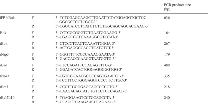
Table 1. PCR primers used to generate fragments for hBok constructs and to detect mouse Bcl2-related gene expression PCR product size (bp) GFP-hBok F 5¢-TCTCGAGCAAGCTTGAATTCTATGGAGGTGCTGC 636 GGCGCTCCTCGGT-3¢ R 5¢-CGGGATCCTCATCTCTCTGGCAGCAGCACGAAG-3¢ hBok F 5¢-CCTCGCGGGTCTGAATGGAAGG-3¢ 164 R 5¢-CGAGCGGTCAAAGGCGTCCAT-3¢ mBok F 5¢-CTCCCTCACTCAAATTGGGA-3¢ 267 R 5¢-ACTGAGGCCAGCTCATGTCT-3¢ mNip3 F 5¢-GGGTTTTCCCCAAAGGAATA-3¢ 179 R 5¢-GACCACCCAAGGTAATGGTG-3¢ mBad F 5¢-TTCCAGATCCCAGAGTTTG-3¢ 488 R 5¢-GGAGATCACTGGGAGGGGGTGG-3¢ mNoxa F 5¢-CGTCGGAACGCGCCAGTGAACCC-3¢ 335 R 5¢-TCCTTCCTGGGAGGTCCCTTCTTGC-3¢ mBmf F 5¢-CCCTTGGGGAGCAGCCCCCTG-3¢ 218 R 5¢-CAAGACAGTATCTGTCCTCCCAGAC-3¢ mBcl2L10 F 5¢-TGAGGAAGTCCTCCAGCCTA-3¢ 240 R 5¢-GCAGCTCAAGAACCCAGAAC-3¢
Cell culture, treatment and cell viability assay
The MCF-7, HEK293 or Hela cells were grown in full medium (RPMI 1640 or DMDM, Gibco, Basel, Switzer-land) supplemented with 10% heat-inactivated fetal bovine serum (Gibco) in 5% CO2, at 37 ° C, according to
the ATCC recommendation. For estrogen deprivation, MCF-7 cells were washed three times with PBS then further cultured in DMEM with 10% charcoal/dextran-treated FBS (Hyclone, Logan, UT, USA) for 48 h. This time point served as a 0 hour time point control. Cells were either further cultured for 10 h or 50 nM 17
b-estra-diol (E2) was added for various times before harverst. To mimic the hypoxia condition, 500 mM CoCl2 was applied to 2¥105MCF-7 or HEK293 cells for the indicated times.
To study apoptosis, HEK293 cells or the transfectants were incubated with 100 nM staurosporine for 12 h. Via-bility was determined at various time points by trypan blue exclusion, counting at least 100 cells from each in-dividual culture. The percentage of cell survival was cal-culated as the number of surviving cells per total cell count [19].
Tissue expression pattern analysis by RT-PCR
To determine the expression patterns of Bok mRNA in different human tissues, PCR was performed using the primers listed in table 1 on a panel of first-strand cDNAs from various human tissues (Origene, Rockville, Md.). The PCR conditions were as described in the manufac-turer’s protocol with 30 cycles performed on the b-actin
control employing Taq polymerase. To examine the ex-pression of members of the Bcl-2 family in mouse, mouse hearts were dissected from two animals. Each dissected mouse heart was divided into two parts and incubated in either 21% O2 (normoxic condition) or 1% O2 (hypoxic
condition) for 24 h. Total RNA was purified and normal-ized by determination of concentration at 260 nm. On mi-crogram of RNA was used for reverse transcription. PCR was performed using the primers listed in table 1 with 28 cycles. b-Actin served as a control.
Immunohistochemical investigations
Biopsies were taken following a study protocol approved by the Ethics Committee of the University Hospital in Basel. Briefly, cryostat-frozen sections were fixed in 3% paraformaldehyde followed by application of normal rabbit serum (Vector, Burlingame, Calif.). The slides were then incubated with the primary antibody (1:50 polyclonal rabbit anti-Bok, Sc11424; Santa Cruz, Calif.) overnight at 4 °C. After one wash, specimens were blocked with avidin (Zymed, San Francisco, Calif.) then with biotin (Zymed). The sections were incubated with biotinylated goat anti-rabbit IgG antibody (Vector). They were then washed again and incubated with a peroxidase block (Zymed), and reacted with the ABC Elite Kit (Vector). 3,30-Diaminobenzidine tetrahydrochloride tablets
(Sigma, Deisenhofen, Germany) were added under light protection. The reaction was then stopped with PBS. Sections were counterstained with hemalum, transferred to tap water, dehydrated and mounted in Pertex (Medite, Nunningen, Switzerland).
Cellular fractionation and Western blotting
Cells were pelleted and suspended in 100 ml of an
enzy-matic reaction buffer [19], supplemented with protease inhibitors and lysed by sonication for 15 s in an ice bath. The lysate was centrifuged for 10 min at 1000 g for removal of unlysed cells and nuclei. The supernatant was then centrifuged for 1 h at 100,000 g (Kontron Instruments, Munich, Germany). The final supernatant served as a cytosolic fraction, the pellet as a crude membrane frac-tion which was resuspended in one volume of the lysis buffer with 10% Triton X-100. The protein concen-tration of both cytosolic and membrane fractions was determined by a BioRad protein assay (BioRad, Hercules, CA, USA).
Alkali extraction of membrane proteins was as decribed previously [20]. Briefly, the membrane fraction was resuspended (1 mg protein/ml) in freshly prepared 0.1 M Na2CO3(pH 11.5) and incubated for 20 min on ice. The
membranes were then pelleted by centrifugation. Mem-brane proteins corresponding to 20 µg protein were sepa-rated by 15% SDS-PAGE (BioRad) and verified using anti-Bok antibody Sc11424 (Santa Cruz).
To determine oligomerization of hBok, the mitochondrial-enriched fraction was suspended in isotonic HIM buffer [21]. Briefly, bismaleim-idohexane (Pierce, Rockford IL, USA) was added to the membrane fraction from a tenfold stock solution (in dimethyl sulfoxide) to a final concen-tration of 10 mM. After incubation for 30 min at room temperature, the cross-linker was quenched by the addi-tion of 1 M Tris-HCl, pH 7.5, to a final concentraaddi-tion of 20 mM. After quenching, the samples were lysed and an-alyzed by Western blot with an anti-Bok antibody Sc11424 (Santa Cruz).
Fractionated cell lysates were boiled for 10 min. Samples (20 mg) were separated on 15% SDS-PAGE,
electrotrans-ferred to an Immobilon-P membrane (Millipore, Biller-ica, MA, USA), immunoblotted with a rabbit polyclonal anti-His antibody (Milan Analytica, Milan, Italy) or anti-Bok antibody (Santa Cruz), and developed with NBT/BCIP (Sigma, St. Louis, Mo.).
Subcellular localization of hBok proteins
To examine the subcellular localization of endogenous hBok, 105MCF-7 cells were seeded on sterile glass
cover-slips. After 12 h culture with or without 100 nM stau-rosporine or 50 nM epothilone B (Novartis, Basel, Switzerland), cells were washed with PBS and fixed with 4% paraformaldehyde (in 10 mM PIPES, 2 mM MgCl2,
were further quenched with 20 mM NH4Cl and
perme-abilized in 0.25% Triton X-100. A rabbit anti-Bok antibody (Sc-11424; Santa Cruz) was used as primary antibody at 1:200 dilution. The secondary antibody was Alexa-488-conjugated goat anti-rabbit IgG at 1:1000 dilution. To examine the subcellular localization of transfected hBok, HEK293 or Hela cells were transfected with 0.3 mg of
expression plasmids encoding GFP-hBok using Effectene reagent (Qiagen, Hilden, Germany). Cells were incubated with 100 nM Mitotracker Red 580 (Molecular Probes,
Eugene, Ore.) and then fixed in 3.7% formaldehyde 24 h post-transfection. The results were further confirmed by a rabbit polyclonal antibody against Bok BH3 domain (AP1310a; Abgent, San Diego, Calif.). Images were ac-quired with a confocal microscope (Leica NT SP1; Leica, Heidelberg, Germany) with oil immersion using a ¥63
lens on a Leica DM RxE. The images were acquired with Leica confocal software with the image format of 1024 ¥
1024 pixels ¥ 8 bit, and transferred to a graphics program
for printing.
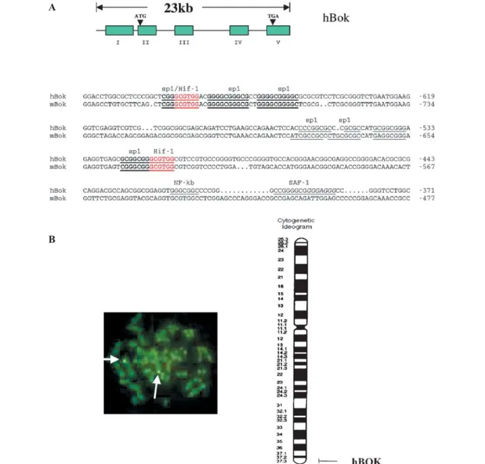
Figure 1. Genomic organization and chromosomal localization of the human Bok gene (hBok). (A) Schematic representation of the hBok gene structure. The boxes represent the exons. The exon numbers are indicated below the boxes. The thin lines denote introns and flanking regions. The lower panel shows the alignment of the conserved 5 ¢-UTR sequences of human (–371to –709) and mouse (–477 to –821) Bok
genes. Underlines represent transcriptional factor-binding sites. (B) Left panel shows that in situ hybridization of a biotin-labeled human
Bok probe to human metaphase cells resulted in specific labeling on chromosome 2 (arrow). Right panel shows results of FISH mapping.
The detailed position of the Bok gene, on human chromosome 2q37.3, was determined from careful analysis of 40 metaphase cells and compared to the NCBI STS database.
A
Co-immunoprecipitation analysis
HEK293 cells (106 ) were transfected with 1 mg of the
expression plasmids pcDNA3-his-tBid, pcDNA3-his-Bnip3 and pc53-SN3 encoding Bid, Bnip3 and p53, respectively. Twenty-four hours after transfection, cells were lysed in M-PER protein extraction reagent (Pierce). His-Bind resin (50 ml; Novagen, San Diego, CA, USA) or 5 mg of
anti-p53 (Ab-6) antibody (Oncogene, Cambridge, MA, USA) were incubated with 100 ml cell lysate. Protein
G-fast Flow Sepharose (25 ml; Amersham, Uppsala,
Swe-den) was added to collect the anti-p53 antibody. The gels were collected after washing and boiled in SDS sample buffer followed by SDS-PAGE and Western blot with anti-Bok antibody (Sc-11424, Santa Cruz).
Results
Isolation and characterization of the human Bok gene (hBok)
To identify the chromosomal location of the Bok gene, we isolated and identified one positive PAC clone, which covers all coding regions of the human Bok gene from a human P1 genomic library (see Materials and methods). Sequence analysis revealed that it contains four coding exons in which the exon-intron organization is conserved between human, mouse and rat genes, but the intron sizes are different between these species (fig. 1A). The complete
Bok gene spanned about 23 kb of genomic DNA. We
then used this PAC clone as a probe for FISH studies. We examined chromosomes in metaphase from a normal male for fluorescence signals. Out of 40 metaphases examined, 32 possessed a specific signal in the region 2q37 (fig. 1B). No background signals were observed. Localization to 2q37 is consistent with in silico analysis of the draft human DNA sequence at the National Center for Biotechnology Information, which mapped the genomic locus of hBok in human chromosome 2 between the markers D2S125 and chr2_qTEL. Together with all other data, the Bok gene was localized on human chromosome 2q37.3 (fig. 1B, right panel).
Differential expression of hBok in human tissues
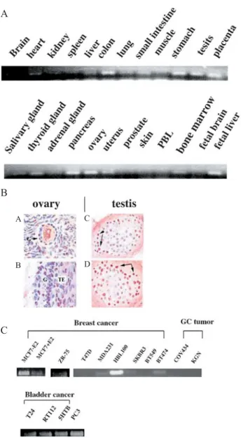
Semiquantitative RT-PCR analysis (semi-Q-RT-PCR) was performed using a pair of primers corresponding to sequences flanking the first intron (Materials and meth-ods). As shown in figure 2A, human Bok was expressed mainly in colon, stomach, testis, placenta, pancreas, ovary and uterus. The expression in fetal liver exhibited a higher level than that in adult tissue. The expression of the human Bok protein in various human tissues was also examined with immunohistochemistry. Figure 2B shows the localization of hBok protein in ovarian and testicular tissue samples. Human Bok was expressed predominantly
in the granulosa cell compartment of ovarian follicles, consistent with the observation for rBok [4], and in the spermatocytes of testicular seminiferous tubules. The expression was further examined in various hormon-ally regulated cancer cell lines using semi-Q-RT-PCR. All seven breast cancer cell lines displayed much lower Bok expression than HBL100, which is considered a non-metastatic breast epithelial cell line. In contrast, granulosa cell tumors lacked mRNA expression. However, clearcut expression of Bok mRNA was detected in some other
Figure 2. Expression of hBok mRNA species varies in different tissues. (A) PCR was performed on a panel of first-strand cDNAs prepared from different human tissues (Origene). The PCR products obtained from each reaction following 30 cycles were separated on a 1.2% agarose gel and visualized by SYBR green I. (B) The local-ization of hBok protein was examined with immunohistochemistry in ovarian follicles (A, B) and testicular seminiferous tubules (C, D). G, granulosa cells; TE, theca cells; arrows, spermatocytes. (C).
hBok expression was examined in various cancer cell lines using
semi-Q-RT-PCR. A B C A B C D
cancer cells, such as bladder and prostate cancer cells (fig. 2C).
Differential regulation of hBok expression
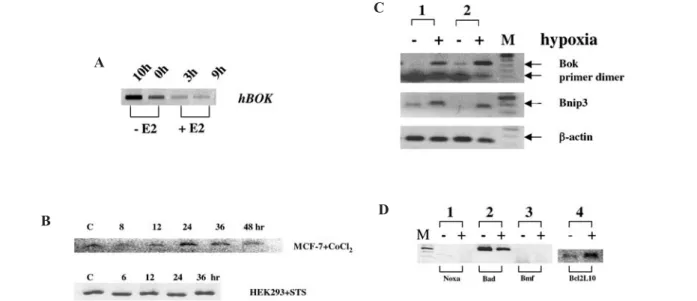
During our initial studies on the regulaton of apoptosis in hormonally regulated breast cancer cell lines, we observed that hBok expression was enhanced in MCF-7 cells both by E2 withdrawal and by hypoxia. MCF-7 cells showed low but detectable levels of hBok mRNA. Cells cultured in an estrogen-free, serum-free medium for 2 days or treated with the steroidal anti-estrogen, ICI 182780, gener-ated increased levels of hBok mRNA, whereas E2 treat-ment induced the downregulation of hBok mRNA (fig. 3A). In contrast, c-Myc expression was increased by addition of E2 (data not shown). Furthermore, the expres-sion in MCF-7 cells treated with various concentrations of CoCl2, a condition which was chosen to mimic
hy-poxia, resulted in increased hBok expression after 24 h. However, combined treatment of ICI 182780 and hypoxia did not further enhance the expression of hBok. Similar observations were made with HEK293 cells (data not shown). Although staurosporine induced apoptosis in both cell lines, expression of hBok was not enhanced (fig. 3B), indicating that expression of hBok is regulated dis-tinctively upon apoptotic stimuli.
Hypoxia has been shown previously to induce brain injury and apoptosis of cardiomyocytes, resulting in ischemia, and this process is, at least in part, preventable by estrogen [22, 23]. We therefore further examined the expression of
Bok in mouse hearts. As demonstrated in figure 3C, Bok
was expressed at very low levels in primary cells, but hypoxia caused a significant increment in Bok mRNA level after 24 h. The expression level of Bnip3 was also observed to be increased, as Bnip3 has already been reported as a response gene during hypoxia-induced apoptosis [24]. The expression of three other BH3-only genes was also examined under the same con-dition: the expression of Bad was decreased, whereas that of Noxa [25] and Bmf [26] remained unexpressed and that of Bcl2L10 [27, 28] increased (fig. 3D). Thus, hypoxia appears to regulate expression of Bok both in the human and mouse. Examination of the hBok promoter region revealed the existence of two conserved Hif-1a binding
sites (fig. 1A). These results suggest that the expression of Bok may be specifically hypoxia regulated in certain organs.
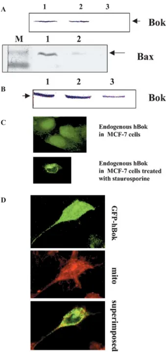
Death signals alter the subcellular localization of hBok
We first assayed localization of hBok by subcellular frac-tionation studies. Cells were lysed and fractionated into a cytosolic and a membrane fraction after removal of the low-speed nuclear pellet. As shown in figure 4A, the endogenous hBok protein could be detected in both cytosolic and membrane fractions upon subcellular frac-tionation of healthy MCF-7, wherease Bax was detected mainly in the cytosolic fraction [29]. Similar results were obtained in HEK293 and Hela cells lysed in a buffer with
Figure 3. Regulation of hBok mRNA expression in various conditions studied by semi-Q-RT-PCR. (A) MCF-7 cells were cultured in an estrogen (E2)-free, serum-free medium for 48 h. 10 h indicates further culture for 10 h in the estrogen-free medium (-E2) or addition of 50 nM E2 for 3 or 9 h. (B) Expression of hBok mRNA was examined in 2¥105MCF7 cells treated with 500 mM CoCl
2 for the indicated
times. Expression of hBok mRNA was also examined in HEK293 cells after treatment with 100 nM staurosporine (STS). (C) Examination of Bok expression in dissected mouse hearts from two animals. Each dissected mouse heart was divided into two parts and incubated in either 21% O2 condition (–, normaxia) or 1% O2 (+, hypoxia). Total RNA was normalized by determination of concentration at 260 nm.
One microgram of RNA was used for reverse transcription. As controls, the expression of Bnip3 and the housekeeping gene b-actin were examined in parallel. (D) Expression of four members of the Bcl2 family, Bad, Noxa, Bmf and Bcl2L10 were examined in the same condition as in C.
A
B
C
or without detergent (data not shown). Moreover, an alkali treatment was effective in extracting hBok from the membrane, indicating that in healthy cells, Bok appears to associate only weakly with the membranes (fig. 4A). However, after treatment of the cells with various apoptotic stimuli, the membrane-bound hBok could not be released by alkali treatment, indicating that hBok had become intergrated into the membrane (fig. 4B). Furthermore, we assessed the subcellular localization of hBok by confocal laser scanning microscopy. In MCF-7 cells, the expression of endogenous hBok was rather diffuse thoughout the cell, examined using two different anti-Bok antibodies (fig. 4C). However, the distribution of hBok became localized together with the membrane when cells were treated with staurosporine (fig. 4C) or epothilone B (not shown). Moreover, the full-length hBok was fused to a GFP expression vector. As shown in figure 4D, the experiment of transient overexpression of hBok in Hela cells revealed that hBok was mainly associated with mitochondria, as it was localized very strongly with the mitochrondial marker. Similar results were found in HEK293 cells. These results suggest that in healthy cells, hBok is associated loosely with the membrane in a hit-and-run mode. The insertion and accumulation of hBok on membranes is then enhanced through the activity of death signals, resulting in the integration of the membrane-bound hBok protein into the membrane.
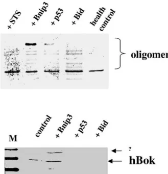
Oligomerization of hBok is promoted by apoptotic stimuli
Bnip3 and p53 were induced or accumulated together with hBok during hypoxia (fig. 3) [24] and because Bnip3, p53 and hBok share several properties, such as their common cytoplasmic and mitochondrial localization and their pro-apoptotic function, we explored the possibil-ity of a functional co-operation of these proteins during apoptosis. Similar to the staurosporine treatment, co-expression of Bid [20] and Bnip3 further sensitized cells to apoptosis, which could be counteracted by expression of BFL-1 (data not shown). Furthermore, oligomerization of hBok was detected in all transfectants with Bnip3, Bid and p53, indicating that these BH3-only proteins were able to induce oligomerization of hBok, as seen with staurosporine treatment (fig. 5A). However, p53 was not able to further increase the level of apoptosis under hBok overexpression (data not shown). Next, we explored the mode of action of Bnip3, Bid and p53 by examining their interaction with the endogenous Bok by co-immunopre-cipitation. HEK293 cells were transfected with His-Bnip3, p53 or His-Bid (Materials and methods). Cellular lysates of transfectants were incubated with anti-His or anti-p53 antibodies for pull-down assay. hBok was then detected by anti-Bok antibody. As shown in figure 5B, hBok could only be detected in Bnip3 interaction. However, p53 and Bid could not be detected. These results indicate that hBok
Figure 4. Intracellular localization of hBok. (A) Subcellular localiza-tion of Bok by cell fraclocaliza-tionalocaliza-tion. 5¥106MCF-7 cells were collected,
lysed and fractionated for high-speed cytosolic and membrane frac-tions as described in Materials and methods. The protein samples were separated by 15% SDS-PAGE and analyzed by Western blot with a polyclonal antibody against human Bok (Santa Cruz) or Bax (BD/Pharmingen). The proteins were fractionated into cytosolic (1) and membrane (2) fractions. 3 indicates alkaline-treated membrane fractions. (B) MCF-7 cells were treated with 100 nM staurosporine for 12 h. Cell lysates were prepared and analysed as in A. (C) Immunolocalization of endogenous hBok in MCF-7 cells treated with or without 100 nM staurosporine. Micrograph showing a single, confocal image plane of MCF-7 cells labeled with a rabbit anti-Bok antibody (Sc-11424) followed by an Alexa-Fluor-488-conjugated secondary antibody (green). (D) Subcellular localization of GFP-Bok by confocal microscopy with a magnification of¥63. Hela cells
were transiently transfected at GFP-Bok as described in Materials and methods. MitoTracker was used to label mitochondria (mito).
A
B
C
interacts with Bnip3 but not with p53 or Bid directly, suggesting that regulation of hBok activation is mediated, at least in part, by two distinct mechanisms.
Discussion
The present studies demonstrate that the function of Bok during apoptosis is regulated both at the transcriptional and at the post-transcriptional level. In both rat and mouse, the expression of Bok is almost entirely restricted to reproductive tissues [4]. Although human Bok is expressed in human ovarian granulosa cells and has been reported to be expressed in pronucleate oocytes [30], expression of human hBok mRNA was detected outside the reproductive organs and found in many tissues, suggesting that human Bok may play a more important role in homeostasis and development than in the rat or mouse. Indeed, expression is higher in fetal than in adult liver, suggesting a functional role for hBok in the function of that organ during fetal development.
We also demonstrated that the expression of hBok is regu-lated by only certain apoptotic stimuli, such as estrogen withdrawal and hypoxia. In accordance with our
observa-tions, a recent study showed that DNA damage induces expression of Bok in neuroblastoma cells [31]. However, the treatment of breast cancer cells with staurosporine was not able to stimulate the expression of Bok, indicating that regulation of Bok expression may vary according to the type of apoptotic signal. Sequence analysis on promoter regions of the human, mouse and rat Bok gene revealed a highly conserved region which contains two Hif-1a-binding sites. Further experiments are underway
to investigate the function of Hif-1a in the regulation of
Bok. How does the induction of Bok during estrogen de-privation and hypoxia relate to the initiation of apoptosis? Bik, a BH3-only protein, could be induced by estrogen deprivation [32]. Bnip3, another BH3-only protein, is also induced by hypoxia [24]. One possibility for the initiation of apoptosis is that activation of Bok is triggered by distinct BH3-only proteins, such as Bnip3 or Bik. Knock-down experiments by short interfering RNAs (siRNA) to Bok will facilitate analysis of the role Bok plays in the apop-totic pathway triggered by estrogen deprivation and hy-poxia.
Like many members of the Bcl-2 family, hBok possesses a hydrophobic COOH-terminal domain. hBok can be readily isolated from both the membrane and the cytosolic fraction in healthy cells and it could also be released by alkali treatment, indicating that it is not an integral mem-brane protein, which distinguishes it from Bcl-xL and
Bak, which are [33], or Bax, which resides primarily in the cytoplasm of healthy cells [25, 34]. The hydrophobic COOH-terminal residues of Bax and Bcl-w form a helix that creates a hydrophobic groove, into which a BH3 domain may bind [35, 36]. A domain that encompasses the hydrophobic helices H6 and H7 of Bid (a cytosolic protein that lacks the hydrophobic C-terminal domain) is the minimal region necessary for strong mitochondrial-binding activity [37, 38]. Furthermore, the acidic phos-pholipid cardiolipin is the receptor for Bid [39]. All these observations lead us to speculate that there is a domain beside the COOH terminus responsible for the weak membrane association of hBok. Further study should investigate this issue.
The weak association of hBok with the mitochondrial membranes of healthy cells is dramatically altered in dying cells, where hBok becomes firmly integrated into the membrane. Several studies have shown that damage signals induce the expression of a BH3 domain-only protein, which then replaces the hydrophobic COOH-terminal helix, causing the translocation of Bax from the cytosol to mitochondrial membranes, where it forms oligomers [8, 11, 40, 41].
Interestingly, our co-immunoprecipitation studies could only detect the interaction of hBok with His-Bnip3 but not with p53 and His-Bid, suggesting that the regulation of hBok activation is mediated, at least in part, by two different mechanisms: oligomerization of hBok can be
Figure 5. Oligomerization of hBok by BH3-like proteins and inter-action of hBok with Bnip3. (A) Oligomerization of hBok. The membrane fraction of cell lysates was incubated with 10 mM BMH cross-linker for 30 min at room temperature. Mitochondrial proteins were extracted with a Chaps buffer and subjected to SDS-PAGE under non-denaturing conditions. Cells were transfected without (health control) or with the genes indicated or treated with 100 nM staurosporine (STS). Western blot was performed with a rabbit anti-Bok antibody (Sc-11424, Santa Cruz). (B) HEK293 cells were transfected with Bnip3, p53 or tBid with non-transfected cells as a control. Western blot was performed with a polyclonal antibody against human Bok (Sc-11424, Santa Cruz).
triggered directly by Bnip3 but oligomers formed only in the presence of p53 and Bid via a specific but still unknown element. Hypoxia-induced activation of hBok, Bnip3 and p53 and oligomerization of hBok could be triggered by Bnip3 and p53, which suggests that these factors act together. Bnip3 is a BH3-only protein, and regulates activation-induced cell death of effector cyto-toxic T lymphocytes [42]. Bnip3 has previously been demonstrated to interact with Bcl-2/Bcl-xL [43] and CD47 [44]. hBok could be another Bnip3-interacting protein; the binding activates hBok by triggering its oligomerization and is thus associated with the initiation of cell death. Overexpression of Bnip3 alone has been reported to initiate permeability transition pore opening and induce a necrosis-like cell death [45]. However, many cellular events, such as hypoxia, are accompanied by changes in the expression of more than one gene, which may work co-operatively. Whether the association of Bnip3 with hBok plays a role in mediating necrotic cell death remains to be elucidated.
p53 is a key tumor suppressor, which induces cell cycle arrest, DNA repair and apoptosis. p53 is normally thought to be localized in the nucleus and to regulate numerous p53-responsive genes [46, 47]. However, recent studies revealed that p53 also has a role in the cytoplasm and functions as a BH3-like protein during apoptosis [15, 16]. Our studies showed that although p53 was unable to interact directly with hBok, it was able to activate hBok by triggering oligomerization. Many pro-apoptotic Bcl-2-related genes, such as Bax, DR5, Bid, Puma and Noxa, are p53-dependent target genes [48–51]. HBok has not been shown to be a direct target of p53, and we could not exclude some function of nuclear p53 in this study. Unlike direct p53-Bak interaction [15], p53 could not in-teract with hBok directly. p53 may sequester Bcl-2/Bcl-xL to release pro-apoptotic Bcl-2 proteins or BH3-only proteins, such as Bnip3, as suggested with Bax [16]. At present, our data suggest that the weak binding to the mitochondrial membrane represents a primary contact in preparation for activation of apoptosis. Interaction of BH3 domain proteins, such as Bnip3, with hBok induces a secondary membrane insertion step resulting in integration of hBok into the membrane.
Acknowledgements. We are grateful to Prof. Martinou at the
Departement of Cell Biology, University of Geneva, for providing us with pcDNA3-His-Bid employed in this study. We are also grateful to Prof. Gudat of the Institute of Pathology for assistance and discussion of immunohistochemical studies.
1 Green D. R. and Kroemer G. (2004) The pathophysiology of mitochondrial cell death. Science 305: 626–629
2 Cory S. and Adams J. M. (2002) The Bcl2 family: regulators of the cellular life-or-death switch. Nat. Rev. Cancer 2: 647–656 3 Gross A., McDonnell J. M. and Korsmeyer S. J. (1999) BCL-2
family members and the mitochondria in apoptosis. Genes Dev.
13: 1899–1911
4 Hsu S. Y., Kaipia A., McGee E., Lomeli M. and Hsueh A. J. (1997) Bok is a pro-apoptotic Bcl-2 protein with restricted expression in reproductive tissues and heterodimerizes with selective anti-apoptotic Bcl-2 family members. Proc. Natl. Acad. Sci. USA 94: 12401–12406
5 Zhang H., Holzgreve W. and De Geyter C. (2000) Evolutionarily conserved Bok proteins in the Bcl-2 family. FEBS Lett. 480: 311–313
6 Kataoka T., Holler N., Micheau O., Martinon F., Tinel A., Hofmann K. et al. (2001 ) Bcl-rambo, a novel Bcl-2 homologue that induces apoptosis via its unique C-terminal extension. J. Biol. Chem. 276: 19548–19554
7 Kuwana T., Mackey M. R., Perkins G., Ellisman M. H., Latterich M., Schneiter R. et al. (2002) Bid, Bax, and lipids cooperate to form supramolecular openings in the outer mitochondrial membrane. Cell 111: 331–342
8 Antonsson B., Montessuit S., Sanchez B. and Martinou J. C. (2001) Bax is present as a high molecular weight oligomer/ complex in the mitochondrial membrane of apoptotic cells. J. Biol. Chem. 276: 11615–11623
9 Roucou X., Montessuit S., Antonsson B. and Martinou J. C. (2002) Bax oligomerization in mitochondrial membranes requires tBid (caspase-8-cleaved Bid) and a mitochondrial protein. Biochem. J. 368: 915–921
10 Knudson C. M., Tung K. S., Tourtellotte W. G., Brown G. A. and Korsmeyer S. J. (1995) Bax-deficient mice with lymphoid hyperplasia and male germ cell death. Science. 270: 96–99 11 Lindsten T., Ross A. J., King A., Zong W. X., Rathmell J. C.,
Shiels H. A. et al. ( 2000 ) The combined functions of proapoptotic Bcl-2 family members bak and bax are essential for normal development of multiple tissues. Mol. Cell. 6: 1389–1399
12 Mikhailov V., Mikhailova M., Degenhardt K., Venkatachalam M. A., White E. and Saikumar P. (2003) Association of Bax and Bak homo-oligomers in mitochondria: Bax requirement for Bak reorganization and cytochrome c release. J. Biol. Chem.
278: 5367–5376.
13 Gillissen B., Essmann F., Graupner V., Starck L., Radetzki S., Dorken B. et al. (2003) Induction of cell death by the BH3-only Bcl-2 homolog Nbk/Bik is mediated by an entirely Bax-dependent mitochondrial pathway. EMBO J. 22: 3580–3590 14 Xiang J., Zhao D. T. and Korsmeyer S. J. (1996) Bax-induced
cell death may not require interleukin 1b-converting enzyme-like proteases. Proc. Natl. Acad. Sci. USA 93: 14559–14563 15 Leu J. I., Dumont P., Hafey M., Murphy M. E. and George D. L.
(2004) Mitochondrial p53 activates Bak and causes disruption of a Bak-Mcl1 complex. Nat. Cell Biol. 5: 443–450
16 Chipuk J. E., Kuwana T., Bouchier-Hayes L., Droin N. M., Newmeyer D. D., Schuler M. et al. (2004) Direct activation of Bax by p53 mediates mitochondrial membrane permeabilization and apoptosis. Science 303: 1010–1014
17 Inohara N., Ekhterae D., Garcia I., Carrio R., Merino J., Merry A. et al. (1998) Mtd, a novel Bcl-2 family member activates apoptosis in the absence of heterodimerization with Bcl-2 and Bcl-XL. J. Biol. Chem. 273: 8705–8710
18 Sauter G., Moch H., Carroll P., Kerschmann R., Mihatsch M. J. and Waldman F. M. (1995) Chromosome-9 loss detected by fluorescence in situ hybridization in bladder cancer. Int. J. Cancer 64: 99–103
19 Zhang H., Heim J. and Meyhack B. (1998) Redistribution of Bax from cytosol to membranes is induced by apoptotic stimuli and is an early step in the apoptotic pathway. Biochem. Biophys. Res. Commun. 251: 454–459
20 Eskes R., Desagher S., Antonsson B. and Martinou J. C. (2000) Bid induces the oligomerization and insertion of Bax into the outer mitochondrial membrane. Mol. Cell Biol. 20: 929– 935
21 Grinberg M., Sarig R., Zaltsman Y., Frumkin D., Grammatikakis N., Reuveny E. et al. (2002) tBID homooligomerizes in the
mitochondrial membrane to induce apoptosis. J. Biol. Chem.
277: 12237–12245
22 Harms C., Lautenschlager M., Bergk A., Katchanov J., Freyer D., Kapinya K. et al. (2001) Differential mechanisms of neuro-protection by 17 beta-estradiol in apoptotic versus necrotic neurodegeneration. J. Neurosci. 21: 2600–2609
23 Kim Y. D., Chen B., Beauregard J., Kouretas P., Thomas G., Farhat M. G. et al. (1996) 17 beta-estradiol prevents dysfunc-tion of canine coronary endothelium and myocardium and reperfusion arrhythmias after brief ischemia/reperfusion. Cir-culation 94: 2901–2908
24 Bruick R. K. (2000) Expression of the gene encoding the proapoptotic Nip3 protein is induced by hypoxia. Proc. Natl. Acad. Sci. USA 97: 9082–9087
25 Oda E., Ohki R., Murasawa H., Nemoto J., Shibue T., Yamashita T. et al . ( 2000 ) Noxa, a BH3-only member of the Bcl-2 family and candidate mediator of p53-induced apoptosis. Science 288: 1053–1058
26 Puthalakath H., Villunger A., O’Reilly L. A., Beaumont J. G., Coultas L., Cheney R. E. et al (2001) Bmf: a proapoptotic BH3-only protein regulated by interaction with the myosin V actin motor complex, activated by anoikis. Science 293: 1829–1832. 27 Inohara N., Gourley T. S., Carrio R., Muniz M., Merino J.,
Garcia I. et al. (1998) Diva, a Bcl-2 homologue that binds directly to Apaf-1 and induces BH3-independent cell death. J. Biol. Chem. 273: 32479–32486
28 Zhang H., Holzgreve W. and De Geyter C. (2001) Bcl2-L-10, a novel anti-apoptotic member of the Bcl-2 family, blocks apoptosis in the mitochondria death pathway but not in the death receptor pathway. Hum. Mol. Genet. 10: 2329–2339 29 Hsu Y. T., Wolter K. G. and Youle R. J. (1997)
Cytosol-to-membrane redistribution of Bax and Bcl-X(L) during apoptosis. Proc. Natl. Acad. Sci. USA 94: 3668–3672
30 Metcalfe A. D., Helen R. H., Debra J. B., Brian A. L., Helen M. P., Henry J. L. et al ( 2004) Expression of 11 members of the BCL-2 family of apoptosis regulatory molecules during human preimplantation embryo development and fragmentation. Mo.l Reprod. Dev. 68: 35–50
31 Yakovlev A. G., Di Giovanni S., Wang G., Liu W., Stoica B. and Faden A. I. (2004) Bok and Noxa are essential mediators of p53-dependent apoptosis. J. Biol. Chem. 279: 28367–28374 32 Hur J., Chesnes J., Coser K. R., Lee R. S., Geck P., Isselbacher K.
J. et al. (2004) The Bik BH3-only protein is induced in estrogen-starved and antiestrogen-exposed breast cancer cells and provokes apoptosis. Proc. Natl. Acad. Sci. USA 101: 1829–1832 33 Kaufmann T., Schlipf S., Sanz J., Neubert K., Stein R. and Borner C. (2003) Characterization of the signal that directs Bcl-x(L), but not Bcl-2, to the mitochondrial outer membrane. J. Cell. Biol. 160: 53–64
34 Putcha G. V., Deshmukh M. and Johnson E. M. Jr. (1999) BAX translocation is a critical event in neuronal apoptosis: regulation by neuroprotectants, BCL-2, and caspases. J. Neurosci. 19: 7476–7485
35 Sattler M., Liang H., Nettesheim D., Meadows R. P., Harlan J. E., Eberstadt M. et al. (1997) Structure of Bcl-xL-Bak peptide complex: recognition between regulators of apoptosis. Science
275: 983–986
36 Petros A. M., Olejniczak E. T. and Fesik S. W. (2004) Structural biology of the Bcl-2 family of proteins. Biochim. Biophys. Acta 1644: 83–94
37 Desagher S., Osen-Sand A. and Nichols A. (1999) Bid-induced conformational change of Bax is responsible for mitochondrial cytochrome c release during apoptosis. J. Cell Biol. 144: 891–901
38 Hu X., Han Z., Wyche J. H. and Hendrickson E. A. (2003) Helix 6 of tBid is necessary but not sufficient for mitochondrial binding activity. Apoptosis 8: 277–289
39 Lutter M., Fang M., Luo X., Nishijima M., Xie X. and Wang X. (2000) Cardiolipin provides specificity for targeting of tBid to mitochondria. Nat. Cell Biol. 10: 754–761
40 Nechushtan A., Smith C. L., Hsu Y. T. and Youle R. J. (1999) Conformation of the Bax C-terminus regulates subcellular location and cell death. EMBO J. 18: 2330–2341
41 Nechushtan A., Smith C. L., Lamensdorf I., Yoon S. H. and Youle R. J. (2001) Bax and Bak coalesce into novel mitochondria-associated clusters during apoptosis. J. Cell Biol. 153: 1265– 1276
42 Wan J., Martinvalet D., Ji X., Lois C., Kaech S. M., Von Andrian U. H. et al. (2003) The Bcl-2 family pro-apoptotic molecule, BNIP3 regulates activation-induced cell death of effector cytotoxic T lymphocytes. Immunology 110: 10–17
43 Ray R., Chen G., Vande Velde C., Cizeau J., Park J. H., Reed J. C. et al. (2000) BNIP3 heterodimerizes with Bcl-2/Bcl-X(L) and induces cell death independent of a Bcl-2 homology 3 (BH3) domain at both mitochondrial and nonmitochondrial sites. J. Biol. Chem. 275: 1439–1448
44 Lamy L., Ticchioni M., Rouquette-Jazdanian A. K., Samson M., Deckert M., Greenberg A. H. et al. (2003) CD47 and the 19 kDa interacting protein-3 (BNIP3) in T cell apoptosis. J. Biol. Chem. 278: 915–921
45 Vande Velde C., Cizeau J., Dubik D., Alimonti J., Brown T., Israels S. et al. (2000) BNIP3 and genetic control of necrosis-like cell death through the mitochondrial permeability transition pore. Mol Cell Biol. 20: 5454–5468
46 Liang S. H. and Clarke M. F. (2001) Regulation of p53 local-ization. Eur. J. Biochem. 268: 2779–2783
47 Fei P., Bernhard E. J. and El-Deiry W. S. (2002) Tissue-specific induction of p53 targets in vivo. Cancer Res. 62: 7316–7327 48 Miyashita T. and Reed J. C. (1995) Tumor suppressor p53 is a
direct transcriptional activator of the human bax gene. Cell 80: 293–299
49 Sax J. K., Fei P., Murphy M. E., Bernhard E., Korsmeyer S. J. and El-Deiry W. S. (2002) BID regulation by p53 contributes to chemosensitivity. Nat. Cell Biol. 4: 842–849
50 Villunger A., Michalak E. M., Coultas L., Mullauer F., Bock G., Ausserlechner M. J. et al. (2003) p53- and drug-induced apop-totic responses mediated by BH3-only proteins puma and noxa. Science 302: 1036–1038
51 Sheikh M. S., Burns T. F., Huang Y., Wu G. S., Amundson S., Brooks K. S. et al. (1998) p53-dependent and -independent reg-ulation of the death receptor KILLER/DR5 gene expression in response to genotoxic stress and tumor necrosis factor alpha. Cancer Res. 58: 1593–1598