HAL Id: hal-00813764
https://hal.inria.fr/hal-00813764
Submitted on 17 Apr 2013
HAL is a multi-disciplinary open access
archive for the deposit and dissemination of
sci-entific research documents, whether they are
pub-lished or not. The documents may come from
teaching and research institutions in France or
abroad, or from public or private research centers.
L’archive ouverte pluridisciplinaire HAL, est
destinée au dépôt et à la diffusion de documents
scientifiques de niveau recherche, publiés ou non,
émanant des établissements d’enseignement et de
recherche français ou étrangers, des laboratoires
publics ou privés.
Horizontal Data Fusion for Integrative Modelling: the
MedINRIA Fusion Toolbox
Benoît Bleuzé, Olivier Clatz, Pierre Fillard, Maxime Sermesant, Nicolas
Toussaint, Julien Wintz
To cite this version:
Benoît Bleuzé, Olivier Clatz, Pierre Fillard, Maxime Sermesant, Nicolas Toussaint, et al.. Horizontal
Data Fusion for Integrative Modelling: the MedINRIA Fusion Toolbox. 2010. �hal-00813764�
Horizontal Data Fusion for Integrative Modelling:
the MedINRIA Fusion Toolbox
B. Bleuz´
e, O. Clatz, P. Fillard, M. Sermesant, N. Toussaint
∗, J. Wintz
INRIA, Asclepios Team, Sophia Antipolis, France
1
Introduction
The Virtual Physiological Human research aims at integrating in computational models an extensive representation of the human body. Given the nu-merous phenomena interacting in such a complex living system, several different modalities of obser-vation are needed in order to gather information on these phenomena. Moreover following up on the time evolution implies comparing data at different points in time. Finally, superposition of simulated volumes and acquired data is also a needed tool. All these needs require a so called ”Horizontal Data Fusion” which registers in the same spatial coordi-nates different datasets. The most active area of research on horizontal data fusion is medical im-age processing. Medical imim-ages come from a larger and larger variety of modalities, and the number of image types for a single modality increases just as fast. However, the problems linked to that ex-plosion of data and the need to put them together is that each registration algorithm has its own in-put/output formats, graphical user interface and visualisation. Thus, its use by a non-expert is chal-lenging and the combination or comparison of such algorithms is difficult. Therefore, there is a need for a standard in horizontal data fusion.
The aim of our project at INRIA within the Vir-tual Physiological Human Network of Excellence1
is to provide a toolbox enabling the visualisation and the aggregation of data fusion tools and algo-rithms: Medinria-fusion2
. Through the use of a core/plug-ins architecture, it explores solutions to make it easy for researchers to import their own tools, wrap them around a single application or
ex-∗N. Toussaint is now with the King’s College London,
Division of Imaging Sciences, St Thomas’ Hospital
1
http://www.vph-noe.eu/
2
http://www-sop.inria.fr/asclepios/projects/noe/
change them between applications supporting the same framework. That attempt at a universal so-lution is a step towards standardisation of the ex-change of algorithms. Standardisation calls for sus-tainability, maintainability.
The proposed framework can also help quanti-tative and qualiquanti-tative tests of functionally similar methods. Medinria-fusion is a demonstrator that implements a registration algorithm, using the API we propose as an open framework. This software allows to visualise several formats of images (among them is DICOM).
2
Architecture
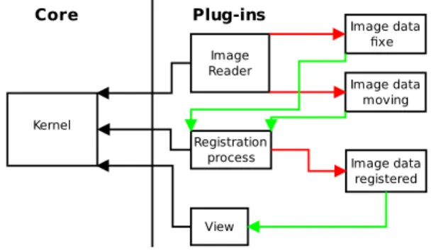
Medinria-fusion demonstrates the viability of a core/plug-in architecture developed at INRIA. The visualisation code takes advantage of the work done in earlier versions of MedINRIA 1.x [1] and vtkIN-RIA3D [2]. Kernel View Core Plug-ins Registration process Image Reader Image data moving Image data fixe Image data registered
Figure 1: General architecture for registration al-gorithms within the MedINRIA framework(inputs are green, output red).
Figure 1 describes the general architecture. The
kernel (or core) provides an abstraction layer al-lowing the interaction between data, processes and views. A plug-in is an implementation of a data reader, writer, process or view. Registration al-gorithms are typical processes. Data are inputs and outputs for processes, views consume Data as inputs as well. They use data such as ITK3
im-age objects loaded for instance from DICOM files through readers. Views can be a VTK4
view for in-stance, or any other viewer. Object factories take care of the instantiation. A typical registration al-gorithm takes two data inputs: a fixed image that will act as a reference, and a so called moving im-age on which the transformation will be applied. The output is a third data object: the estimated transformation. The plug-ins are interchangeable, and an affine transformation can be replaced with a non linear one by just switching the process objects, which are dynamic libraries, without any change to the remaining of the software.
A clear advantage of that abstraction layer deliv-ered as an open API is that open and closed source plug-ins from different institutions or vendors can live together in the same execution environment without any trouble, as long as the licences allow linking to other programmes.
3
Demonstrator
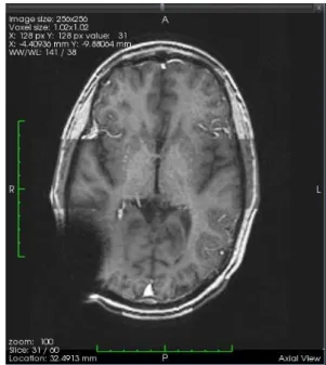
The demonstrator, named MedINRIA-fusion is a subset of a larger platform called MedINRIA, de-veloped at INRIA. MedINRIA-fusion builds on the architecture and GUI from MedINRIA and puts the framework through its paces by implementing registration tools within it. The programme im-ports and stores DICOM files. The intuitive and slick looking interface makes it easy to use, and also particularly appealing to clinicians thanks to its simplicity (see figure 2).
The algorithm used for this demonstrator is a simple rigid transformation estimation, based on the ITK registration framework. Different visuali-sation modes to evaluate the results are proposed, including the checker-board, alternating parts of each image. As an example result, figure 3 shows the multimodal registration of two MRIs (T1 and
3
http://www.itk.org
4
http://www.vtk.org
T2 modes). Three representations have been im-plemented so far: side by side comparison, and alpha-blending or checker-board mixing of the two images.
Figure 3: Checker-board fused MRIs (T1 and T2)
4
Perspectives
Within the VPH Toolkit, the objective of such tool-box is to aggregate the different tools existing in the community, thus it should suit the needs of other already existing algorithms to be wrapped as MedINRIA-fusion plug-ins. Moreover, by release a simple API and the software, the objective is also to attract more developers to the framework, so that any researcher can create his own plug-in.
Regarding the functionalities, the horizontal data fusion is also including other data supports, like meshes, so we plan to integrate fusion between meshes or between meshes and images in the next release.
5
Conclusion
Still in its infancy, this versatile framework showed an ease to import an existing algorithm, as well as an easy-to-use interface. The open API is our first
Figure 2: Medinria-fusion: viewer interface, while using the registration tools
attempt to federate researchers or developers under the same common language. More demonstrators are however needed to harden the concept.
References
[1] N. Toussaint, J.C. Souplet, and P. Fillard. Med-inria: Medical image navigation and research tool by inria. In Proc. of MICCAI’07 Work-shop on Interaction in medical image analysis and visualization, Brisbane, Australia, 2007. [2] N. Toussaint, M. Sermesant, and P. Fillard.
vtkinria3d: A vtk extension for spatiotemporal data synchronization, visualization and man-agement. In Proc. of Workshop on Open Source and Open Data for MICCAI, Brisbane, Aus-tralia, October 2007.