HAL Id: hal-02432440
https://hal.uca.fr/hal-02432440
Submitted on 8 Jan 2020
HAL is a multi-disciplinary open access
archive for the deposit and dissemination of sci-entific research documents, whether they are pub-lished or not. The documents may come from teaching and research institutions in France or abroad, or from public or private research centers.
L’archive ouverte pluridisciplinaire HAL, est destinée au dépôt et à la diffusion de documents scientifiques de niveau recherche, publiés ou non, émanant des établissements d’enseignement et de recherche français ou étrangers, des laboratoires publics ou privés.
for preventing Candida albicans infection in the
invertebrate model Caenorhabditis elegans: First
mechanistic insights
Cyril Poupet, Taous Saraoui, Philippe Veisseire, Muriel Bonnet, Caroline
Dausset, Marylise Gachinat, Christophe Chassard, Adrien Nivoliez, Stéphanie
Bornes
To cite this version:
Cyril Poupet, Taous Saraoui, Philippe Veisseire, Muriel Bonnet, Caroline Dausset, et al.. Lactobacillus rhamnosus Lcr35 as an effective treatment for preventing Candida albicans infection in the invertebrate model Caenorhabditis elegans: First mechanistic insights. PLoS ONE, Public Library of Science, 2019, 14 (11), �10.1371/journal.pone.0216184�. �hal-02432440�
Lactobacillus rhamnosus Lcr35 as an effective
treatment for preventing Candida albicans
infection in the invertebrate model
Caenorhabditis elegans: First mechanistic
insights
Cyril PoupetID1*, Taous Saraoui1, Philippe Veisseire1, Muriel Bonnet1,
Caroline DaussetID2, Marylise Gachinat1, Olivier Camarès1, Christophe Chassard1, Adrien Nivoliez2, Ste´phanie Bornes1
1 Universite´ Clermont Auvergne, INRA, VetAgro Sup, Aurillac, France, 2 Biose Industrie, Aurillac, France
*cyril.poupet@uca.fr
Abstract
The increased recurrence of Candida albicans infections is associated with greater resis-tance to antifungal drugs. This involves the establishment of alternative therapeutic proto-cols, such as probiotic microorganisms whose antifungal potential has already been demonstrated using preclinical models (cell cultures, laboratory animals). Understanding the mechanisms of action of probiotic microorganisms has become a strategic need for the development of new therapeutics for humans. In this study, we investigated the prophylactic anti-C. albicans properties of Lactobacillus rhamnosus Lcr35®using the in vitro Caco-2 cell model and the in vivo Caenorhabditis elegans model. In Caco-2 cells, we showed that the strain Lcr35®significantly inhibited the growth (~2 log CFU.mL-1) and adhesion (150 to 6,300 times less) of the pathogen. Moreover, in addition to having a pro-longevity activity in the nematode (+42.9%, p = 3.56.10−6), Lcr35®protects the animal from the fungal infection (+267% of survival, p<2.10−16) even if the yeast is still detectable in its intestine. At the mechanistic level, we noticed the repression of genes of the p38 MAPK signalling pathway and genes involved in the antifungal response induced by Lcr35®, suggesting that the patho-gen no longer appears to be detected by the worm immune system. However, the DAF-16/ FOXO transcription factor, implicated in the longevity and antipathogenic response of C. ele-gans, is activated by Lcr35®. These results suggest that the probiotic strain acts by stimulat-ing its host via DAF-16 but also by suppressstimulat-ing the virulence of the pathogen.
1 Introduction
Candida albicans is a commensal yeast found in the gastrointestinal and urogenital tracts [1,2] and is responsible for various diseases ranging from superficial infections affecting the skin to life-threatening systemic pathologic states i.e., candidemia [3]. Its pathogenicity is based on
a1111111111 a1111111111 a1111111111 a1111111111 a1111111111 OPEN ACCESS
Citation: Poupet C, Saraoui T, Veisseire P, Bonnet
M, Dausset C, Gachinat M, et al. (2019)
Lactobacillus rhamnosus Lcr35 as an effective
treatment for preventing Candida albicans infection in the invertebrate model Caenorhabditis elegans: First mechanistic insights. PLoS ONE 14(11): e0216184.https://doi.org/10.1371/journal. pone.0216184
Editor: Ilse D. Jacobsen, Leibniz Institute for
Natural Products Research and Infection Biology-Hans Knoell Institute, GERMANY
Received: April 12, 2019 Accepted: October 22, 2019 Published: November 6, 2019
Copyright:© 2019 Poupet et al. This is an open access article distributed under the terms of the Creative Commons Attribution License, which permits unrestricted use, distribution, and reproduction in any medium, provided the original author and source are credited.
Data Availability Statement: All relevant data are
within the paper.
Funding: This work was supported by the
European funds FEDER and the Auvergne-Rhoˆne-Alpes Region in the form of grants to CP, as well as biose, as part of the doctoral thesis of CP. The funder biose provided support in the form of salaries for authors CD and AN. The specific roles
several factors, such as the formation of biofilms, thigmotropism, adhesion and invasion of host cells, secretion of hydrolytic enzymes [3] and the transition from yeast to hyphal fila-ments, which facilitates its spread [4,5].
There is an increase in the number of fungal infections, mainly due to the increaseraise in resistance to drugs [6,7] and to the limited number of available antifungals, some of which are toxic [8]. In addition, it is very common that antifungal treatments destabilize, more or less severely, the host commensal microbiota, leading to dysbiosis [9] which is favourable to the establishment of another pathogen or recurrence. In addition, because of the presence of simi-larities between yeasts and human cells (i.e., eukaryotic cells), the development of novel mole-cules combining antifungal activity and host safety is particularly complicated [8]. These different elements demonstrate the need to develop new therapeutic strategies. These aimed at effectively treating a fungal infection while limiting the health risks for the host; in particular, by preserving the integrity of its microbiota. The use of probiotics to cure candidiasis or fun-gal-infection-related dysbiosis is part of these novel strategies [10–12]. The World Health Organization (WHO) and the Food and Agriculture Organization of the United Nations (FAO) define probiotics as “live microorganisms, which, when administered in adequate amounts, confer a health benefit on the host” [13]. Under this appellation of probiotics, a wide variety of microbial species, are found within both prokaryotes and eukaryotes (yeasts, such as
Saccharomyces), although these are mainly lactic bacteria, such as the genera Lactobacillus and Bifidobacterium [14]. Currently, a new name is increasingly used to replace the term probiotic: live biotherapeutic products (LBP). These LBP are biological products containing live biother-apeutic microorganisms (LBM) used to prevent, treat or cure a disease or condition of human beings, excluding vaccines [15].
In this issue, we focused onL. rhamnosus Lcr351
, which is a well-known probiotic strain whosein vitro and in vivo characteristics are widely documented [16–23]. It is a gram-positive bacterium commercialized by biose1as a pharmaceutical product for more than 60 years for preventive and curative gastrointestinal and gynaecological indications. Nivoliezet al.
demon-strated the probiotic properties of the native strain such as resistance to gastric acidity and bile stress and lactic acid production. Under its commercial formulations, the Lcr351strain has the ability to adhere to intestinal (Caco-2, HT29-MTX) and vaginal (CRL -2616) epithelial cells, but the inhibition of pathogen adhesion to intestinal cells by Lcr351has not been investi-gated by the authors. This study has also shown that Lcr351leads to a strong inhibition of vag-inal (C. albicans, Gardnerella vaginalis) and intestinal (enterotoxigenic and enteropathogenic Escherichia coli (ETEC, EPEC), Shigella flexneri) pathogens [24]. These probiotic and antimi-crobial effects have been observed during clinical trials, but we know little about the molecular mechanisms underlying these properties. Randomized trials conducted in infants and children have shown that preventive intake of probiotics has a positive impact on the development of infectious or inflammatory bowel diseases by reducing their symptoms and maintaining the balance of the microbiota [25].In vitro and in vivo studies using preventive approaches have
revealed certain mechanisms of action of probiotics [26].
Up to now, most probiotics used in both food and health applications are selected and char-acterized on the basis of their properties obtained within vitro models [27] before being tested on complexin vivo models (murine models) and in human clinical trials. The in vitro studies
are used mainly for ethical and cost issues [28] but also allow experimentations under defined and controlled conditions. As a result, some strains meeting the criteria forin vitro selection
no longer respondin vivo and vice versa [29]. This fact reinforces the idea thatin vitro and in vivo tests are complementary and necessary for the most reliable characterization of probiotic
properties. of these authors are articulated in the ‘author
contributions’ section. The funders did not otherwise have any role in the study design, data collection and analysis, decision to publish, or preparation of the manuscript.
Competing interests: The authors have read the
journal’s policy and the authors of this manuscript have the following competing interests: Adrien Nivoliez (AN) and Caroline Dausset (CD) had an institutional affiliation with the company biose, which manufactures Lcr35 products. The doctoral thesis of Cyril Poupet (CP) is partially financed by the company biose. This does not alter our adherence to PLOS ONE policies on sharing data and materials. There are no patents, products in development or marketed products to declare.
Here, we propose to use bothin vitro Caco-2 cell culture and the invertebrate host C. ele-gans as an in vivo model to investigate microorganism-microorganism-host interactions.
Caco-2 cells are a well-characterized enterocyte-like cell line. They are a reliablein vitro system
to study the adhesion capacity of lactobacilli as well as their probiotic effects, such as protection against intestinal injury induced by pathogens [30,31]. Nevertheless, the use ofin vivo models,
which are closer to the complex environment of the human body, is inevitable in the case of a mechanistic study. Indeed, while rudimentary models such asC. elegans or Drosophila exhibit
obvious benefits for (large) screening purposes, they are also not devoid of relevance in deci-phering more universal signalling pathways, even related to mammalian innate immunity [32]. With its many genetic and protein homologies with human beings [33],C. elegans has
become the ideal laboratory tool for physiological as well as mechanistic studies. This round-worm has already been used to study the pathogenicity mechanisms ofC. albicans. The work
of Pukkila-Worley has demonstrated a rapid antifungal response inC. elegans with the
overex-pression of antimicrobials encoding genes such asabf-2, fipr-22, fipr-23, cnc-7, thn-1 and
chiti-nases (cht-1 and T19H5.1) or detoxification enzymes (oac-31, trx-3). It has also been shown
thatC. albicans hyphal formation is a key virulence factor that modifies gene expression in the C. elegans killing assay [34]. Some of these genes are notably dependent on the highly con-served p38 MAPK signalling pathway [35]. Several recent studies have established that the transition from yeast morphology to hyphal form is largely dependent on environmental parameters. It is also controlled byC. albicans genetic factors, such as eIF2 kinase Gcn2 [36] or SPT20 [37], whose mutations induce a decrease in virulence of the pathogen and an enhanced survival of the host. However, few studies have been conducted with the nematode on the use of probiotic microorganisms for the treatment ofC. albicans fungal infection [38].
In this context, the aim of this study was to evaluate the effect of theLactobacillus rhamno-sus Lcr351strain on the prevention of fungal infection due toC. albicans using the in vitro
cel-lular model Caco-2 and thein vivo model C. elegans. To overcome the experimental limits of
thein vitro model, we conducted a mechanistic study solely on the C. elegans model. The
worm survival and gene expression in response to the pathogen and/or the probiotic were evaluated.
2 Material and methods
2.1 Microbial strains and growth conditions
TheE. coli OP50 strain was provided by the Caenorhabditis Genetics Center (Minneapolis,
MN, USA) and was grown on Luria Broth (LB, Miller’s Modification) (Conda, Madrid, Spain) at 37 ˚C overnight. TheL. rhamnosus Lcr351strain was provided by biose1(Aurillac, France) and was grown in de Man, Rogosa, Sharpe (MRS) broth (bioMe´rieux, Marcy l’Etoile, France) at 37 ˚C overnight.C. albicans ATCC 10231 was grown in yeast peptone glucose (YPG) broth
pH 6.5 (per L: 10 g yeast extract, 10 g peptone, 20 g glucose) at 37 ˚C for 48 h. Microbial sus-pensions were spun down for 2 min at 1,500 rpm (Rotofix 32A, Hettich Zentrifugen, Tuttlin-gen, Germany) and washed with M9 buffer (per L: 3 g KH2PO4, 6 g Na2HPO4, 5 g NaCl, 1 mL
1 M MgSO4) to obtain a final concentration of 100 mg.mL-1.
2.2 Influence of Lcr35
1on
C. albicans growth and on C. albicans biofilm
formation on Caco-2 cell monolayers
Growth inhibition ofC. albicans by the probiotic strain Lcr351was examined using the human colorectal adenocarcinoma cell line Caco-2 [39]. Caco-2 cells were grown in Dulbec-co’s modified Eagle’s minimal essential medium (DMEM, Life Technologie,
Villebon-sur-Yvette, France) supplemented with 20% inactivated foetal calf serum (Life Technologie) at 37 ˚C with 5% CO2in air atmosphere. For the assays, the cells were seeded at a concentration
of 3.5x10⁵cells.well-1in 24-well plates (Dutscher, Brumath, France) and placed in growth con-ditions for 24 h. Microbial strains were grown according to Nivoliezet al. [24]. After growth, cell culture medium was removed and replaced by 1 mL of DMEM and 250μL of Lcr351 cul-ture (108CFU.mL-1) in each well and incubated for 24 h. Two hundred and fifty microliters of
C. albicans culture at different concentrations (102, 103, 104, 105, 106and 107CFU.mL-1) were added to each well. After incubation for 24 and 48 h, the inhibition ofC. albicans by Lcr351
was evaluated. One hundred microliters of suspension were taken from each of the wells, and the number of viable bacteria and/or yeasts was determined by plating serial dilutions of the suspensions onto MRS or Sabouraud agar plates. For the measurement ofC. albicans biofilm
formation, after incubation for 48 h, the wells were washed twice with 0.5 mL of PBS and cells were harvested with 1 mL of trypsin at 37 ˚C. For the inhibition assay, the number of viable bacteria and/or yeasts was determined by plating serial dilutions of the suspensions onto MRS or Sabouraud agar plates. The plates were incubated at 37 ˚C for 72 h (MRS) or 48 h (Sabour-aud). Each assay was performed three times independently and contained two technical replicates.
2.3
C. elegans maintenance
C. elegans N2 (wild-type) and TJ356 (daf-16p::daf-16a/b::GFP + rol-6(su1006)) strains were
acquired from theCaenorhabditis Genetics Center. The nematodes were grown and
main-tained at 20 ˚C on nematode growth medium (NGM) (per L: 3 g NaCl; 2.5 g peptone; 17 g agar; 5 mg cholesterol; 1 mM CaCl2; 1 mM MgSO4, 25 mL 1 M potassium phosphate buffer at
pH 6) plates supplemented with yeast extract (4 g.L-1) (NGMY) and seeded withE. coli OP50
[40]. For all experiments, wild-typeC. elegans N2 were used except for the study of the
localiza-tion of DAF-16 (TJ356 strain).
2.4
C. elegans synchronization
To avoid variations in results due to age differences, a worm synchronous population was required. Gravid worms were washed off using M9 buffer and spun down for 2 min at 1,500 rpm. Five millilitres of worm bleach (2.5 mL of M9 buffer, 1.5 mL of bleach, 1 mL of 5 M sodium hydroxide) was added to the pellet and vigorously shaken until adult worm body dis-ruption. The action of worm bleach was stopped by adding 20 mL of M9 buffer. The egg sus-pension was then spun down for 2 min at 1,500 rpm and washed twice with 20 mL of M9 buffer. Eggs were allowed to hatch under slow agitation at 25 ˚C for 24 h in approximately 20 mL of M9 buffer. L1 larvae were then transferred onto NGMY plates seeded withE. coli OP50
until they reached the L4/young adult stage.
2.5
C. elegans bodyb size measurement
Individual adult worms were imaged using an Evos FL microscope (Invitrogen, Eugene, USA, 10X magnification). After reaching the L4 stage, they were transferred onto NGMY plates pre-viously seeded with the probiotic strain Lcr351, and their sizes were measured daily for three days. The length of the worm body was determined using ImageJ software as described by Mo¨rck and Pilon (41) and compared toE. coli OP50-fed worms. At least 10 nematodes per
2.6
C. elegans lifespan assay
Synchronous L4 worms were transferred to NGMY with 0.12 mM 5-fluorodeoxyuridine FUdR (Sigma, Saint-Louis, USA) to avoid egg hatching and seeded with 100μL of microbes at 100 mg.mL-1microbial strain (~50 worms per plate) as previously stated. The plates were kept at 20 ˚C, and live worms were scored each day until the death of all animals. An animal was scored as dead when it did not respond to a gentle mechanical stimulation. This assay was per-formed as three independent experiments with three plates per condition.
2.7 Effects of
L. rhamnosus Lcr35
1on candidiasis in
C. elegans
Sequential feeding with Lcr351andC. albicans were induced in C. elegans in all experiments
(preventive assays). As control groups, monotypic contamination was induced inC. elegans by
inoculation with onlyC. albicans, Lcr351orE. coli OP50.
2.7.1 Preparation of plates containing probiotic bacteria or pathogenic yeasts. One
hundred microliters of Lcr351orE. coli OP50 suspension (100 mg.mL-1) was spread on NGMY + 0.12 mM FUdR plates and incubated at 37 ˚C overnight. ConcerningC. albicans
strains, 100μL of suspension was spread on Brain Heart Infusion BHI (Biokar Diagnostics, Beauvais, France) + 0.12 mM FUdR plates and incubated at 37 ˚C overnight.
2.7.2 Survival assay: Preventive treatment. The survival assay was performed according
to the work of de Barros [38], with some modifications. During a preventive treatment, young adult worms were placed on plates containing Lcr351at 20 ˚C for different times (2, 4, 6 and 24 h). Next, the worms were washed with M9 buffer to remove bacteria prior to being placed onC. albicans plates for 2 h at 20 ˚C. Infected nematodes were washed off plates using M9
buffer prior to being transferred to a 6-well microtiter plate (approximately 50 worms per well) containing 2 mL of BHI/M9 (20%/80%) + 0.12 mM FUdR liquid assay medium per well and incubated at 20 ˚C. For the control groups (i.e.,E. coli OP50 + C. albicans, E. coli OP50
only, Lcr351only andC. albicans only), worms were treated in the same way. Nematodes
were observed daily and were considered dead when they did not respond to a gentle mechani-cal stimulation. This assay was performed as three independent experiments containing three wells per condition.
2.8 Colonization of
C. elegans intestine by C. albicans
To study the colonization of the worm gut by the pathogenC. albicans, fluorescent staining of
the yeast was performed. The yeast was stained with rhodamine 123 (Yeast Mitochondrial Stain Sampler Kit, Invitrogen) according to the manufacturer’s instructions. A fresh culture of
C. albicans was performed in YPG broth as described before, 1.6 μL of rhodamine 123 at 25
mM was added to 1 mL ofC. albicans suspension and incubated at room temperature in the
dark for 15 min. The unbound dye was removed by centrifugation (14,000 rpm for 5min at 4 ˚C) (Beckman J2-MC Centrifuge, Beckman Coulter, Brea, USA) and washed with 1 mL of M9 buffer. Subsequently, the nematodes were fed withE. coli OP50 or Lcr351on NGMY plates for 4 h and then with labelledC. albicans on BHI plates for 72 h. The nematodes were then
visualized using a fluorescence microscope at 100X magnification (Evos FL, Invitrogen).
2.9 RNA isolation and RT- quantitative PCR
Approximately 10,000 worms were harvested from NGMY plates with M9 buffer. Total RNA was extracted by adding 500μL of TRIzol reagent (Ambion by Life Technologies, Carlsbad, USA). Worms were disrupted using a Precellys (Bertin Instruments, Montigny-le-Bretonneux, France) and glass beads (PowerBead Tubes Glass 0.1 mm, Mo Bio Laboratories, USA). Beads
were removed by centrifugation at 14,000 rpm for 1 min (Eppendorf15415D, Hamburg, Ger-many), and 100μL of chloroform was added to the supernatant. Tubes were vortexed for 30 seconds and incubated at room temperature for 3 minmin. The phenolic phase was removed by centrifugation at 12,000 rpm for 15 min at 4 ˚C. The aqueous phase was treated with chloro-form as previously described. RNA was precipitated by adding 250μL of isopropanol for 4 min at room temperature and spun down at 12,000 rpm for 10 min (4 ˚C). The supernatant was discarded, and the pellet was washed with 1,000μL of 70% ethanol. The supernatant was discarded after centrifugation at 14,000 rpm for 5 min (4 ˚C), and the pellet was dissolved in 20μL of RNase-free water. RNA was reverse-transcribed using a High-Capacity cDNA Archive kit (Applied Biosystems, Foster City, USA) according to the manufacturer’s instruc-tions. For real-time qPCR assay, each tube contained 2.5μL of cDNA, 6.25 μL of Rotor-Gene SYBR Green Mix (Qiagen GmbH, Hilden, Germany), 1.25μL of 10 μM primers (reported in
Table 1) (Eurogentec, Seraing, Belgium) and 1.25μL of water. All samples were run in tripli-cate. Rotor-Gene Q Series Software (Qiagen GmbH) was used for the analysis. In our study, two reference genes,cdc-42 and Y45F10D.4, were used in all the experimental groups. The
quantification of gene-of-interest expression (EGOI) was performed according to the following
formula [41] taking into account the efficiency of the PCR for each primer pair and normaliz-ing the expression of the gene of interest by two reference genes (cdc-42 and Y45F10D.4):
EGOI¼ ðGOI efficiencyÞ
DCtGOI
ffiffiffiffiffiffiffiffiffiffiffiffiffiffiffiffiffiffiffiffiffiffiffiffiffiffiffiffiffiffiffiffiffiffiffiffiffiffiffiffiffiffiffiffiffiffiffiffiffiffiffiffiffiffiffiffiffiffiffiffiffiffiffiffiffiffiffiffiffiffiffiffiffiffiffiffiffiffiffiffiffiffiffiffiffiffiffiffiffiffiffiffiffiffiffiffiffiffiffiffiffiffiffiffiffiffiffiffiffiffiffiffiffiffiffiffiffiffiffiffiffiffi ðcdc 42efficiencyÞDCtcdc 42� ðY45F10D:4 efficiencyÞDCtY45F10D:5
q
The worms fed withE. coli OP50 were used as control conditions for the gene expression
calculation.
2.10 Statistical analysis
Data are expressed as the mean± standard deviation.
TheC. elegans survival assay was examined using the Kaplan-Meier method, and
differ-ences were determined using the log-rank test with R software version 3.5.0 [45], and the sur-vival [46] andsurvminer [47] packages. ForC. albicans growth inhibition and biofilm
formation andC. elegans growth and gene expression of the genes analysed, differences
between conditions were determined by a two-way ANOVA followed by a Fisher’s Least Sig-nificant Difference (LSD) post hoc test using GraphPad Prism version 7.0a for Mac OS X (GraphPad Software, La Jolla, California, USA). Ap-value � 0.05 was considered significant. Table 1.C. elegans gene primers for qPCR analysis. GOI: Gene of interest.
Gene name Gene type Forward Primer (5’– 3’) Reverse Primer (5’– 3’) Reference
cdc-42 housekeeping ATCCACAGACCGACGTGTTT GTCTTTGAGCAATGATGCGA [42]
Y45F10D.4 housekeeping CGAGAACCCGCGAAATGTCGGA CGGTTGCCAGGGAAGATGAGGC [43]
daf-2 GOI AAAAGATTTGGCTGGTCAGAGA TTTCAGTACAAATGAGATTGTCAGC [44]
daf-16 GOI TTCAATGCAAGGAGCATTTG AGCTGGAGAAACACGAGACG [44]
sek-1 GOI GCCGATGGAAAGTGGTTTTA TAAACGGCATCGCCAATAAT [44]
pmk-1 GOI CCGACTCCACGAGAAGGATA AGCGAGTACATTCAGCAGCA [44]
abf-2 GOI TCGTCCGTTCCCTTTTCCTT CCTCTCTTAATAAGAGCACC This study
fipr-22/fipr-23 GOI CCCAATCCAGTATGAAGTTG ATTTCAGTCTTCACACCGGA This study
cnc-4 GOI ATGCTTCGCTACATTCTCGT TTACTTTCCAATGAGCATTC This study https://doi.org/10.1371/journal.pone.0216184.t001
2.11 DAF-16 nuclear localization
DAF-16 nuclear localization was followed as described elsewhere [48] using a transgenic TJ-356 worm strain constitutively expressing the DAF-16 transcription factor combined with GFP (DAF-16::GFP). Once adults, worms were exposed to a single strain:E. coli OP50,
Lcr351orC. albicans for 2, 4, 6, 24 and 76 h at 20 ˚C. A preventive approach was also
con-ducted: worms were placed in the presence ofE. coli OP50 or Lcr351for 4 h and thenC. albi-cans for 2 hh. The nematodes were subsequently imaged 2, 4, 6 and 24 h after infection. The
translocation of DAF-16::GFP was scored by assaying the presence of GFP accumulation in theC. elegans cell nuclei using a fluorescence microscope at 40X magnification (Evos FL,
Invitrogen).
3 Results
3.1 Anti-
C. albicans effects of Lcr35
1on Caco-2 cell monolayer
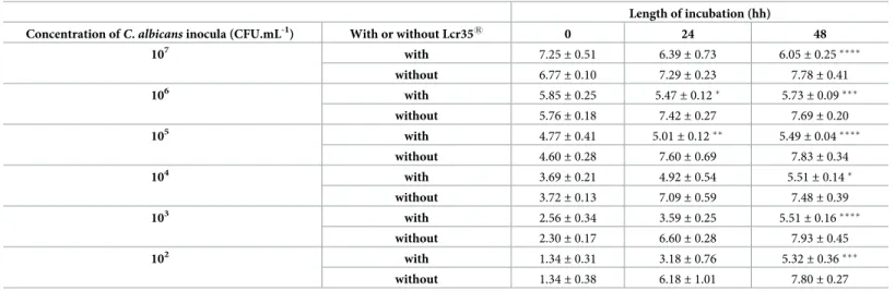
3.1.1 Growth inhibition ofCC. albicans. In the presence of Caco-2 cells, regardless of the
concentration of theC. albicans inoculum, the yeast grew to similar concentrations that ranged
from 7.48± 0.39 to 7.83 ± 0.34 log CFU.mL-1after 48 h of incubation. When prophylactic treatment was used, i.e., when the Caco-2 cells were pre-incubated with the probiotic Lcr351, we observed an inhibition ofC. albicans growth. Indeed, the bacterium induced a significant
inhibition of the yeast growth of 2 log CFU.mL-1, which then reached a concentration ranging from 5.40± 0.07 to 6.05 ± 0.25 log CFU.mL-1. Two different inhibition profiles were observed after 48 h. On the one hand, when the inoculum was highly concentrated (7 log CFU.mL-1), we observed a decrease in the yeast population, which is a sign of cell death. On the other hand, when the inoculum was less concentrated (2 to 4 log CFU.mL-1), we noticed that the yeast was able to grow, although its growth seemed to stop between 5.32± 0.36 and 5.51 ± 0.14 log CFU.mL-1(Table 2).
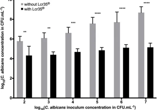
3.1.2 Inhibition ofC. albicans biofilm formation. After 48h of incubation, the C. albi-cans biofilm contained between 5.78 log CFU.mL-1(inoculum at 102CFU.mL-1) and 8.69 log
Table 2. Evolution of the concentration ofC. albicans in the presence or absence of Lcr351on Caco-2 cell monolayers.
Length of incubation (hh)
Concentration ofC. albicans inocula (CFU.mL-1) With or without Lcr351 0 24 48
107 with 7.25 ± 0.51 6.39± 0.73 6.05± 0.25���� without 6.77± 0.10 7.29± 0.23 7.78± 0.41 106 with 5.85 ± 0.25 5.47± 0.12� 5.73± 0.09��� without 5.76± 0.18 7.42± 0.27 7.69± 0.20 105 with 4.77 ± 0.41 5.01± 0.12�� 5.49± 0.04���� without 4.60± 0.28 7.60± 0.69 7.83± 0.34 104 with 3.69 ± 0.21 4.92± 0.54 5.51± 0.14� without 3.72± 0.13 7.09± 0.59 7.48± 0.39 103 with 2.56 ± 0.34 3.59± 0.25 5.51± 0.16���� without 2.30± 0.17 6.60± 0.28 7.93± 0.45 102 with 1.34 ± 0.31 3.18± 0.76 5.32± 0.36��� without 1.34± 0.38 6.18± 1.01 7.80± 0.27
The results are expressed as log10CFU.mL -1
of yeast alone (controls) or co-incubated with Lcr351(mean± standard deviation). A comparison between the conditions with and without Lcr351was performed using a two-way ANOVA followed by a Fisher’s LSD post hoc test
(p < 0.05:�; p < 0.01:��; p < 0.001:���; p < 0.0001:����)
CFU.mL-1of yeast (inoculum at 107CFU.mL-1). However, since the cells were pre-exposed to Lcr351and for the sameC. albicans inocula, we observed a significant decrease in the amount
of yeast in the biofilm: 4.32 to 5.16 log CFU.mL-1, which corresponded to an inhibition rang-ing from 1.46 to 3.53 log. The strongest inhibition was observed in the case where the inocu-lum ofC. albicans was the most concentrated (Fig 1).
3.2 Effects of Lcr35
1on
C. elegans physiology
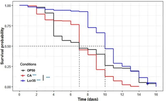
3.2.1 Lcr351extends theC. elegans lifespan. We investigated the effects on C. elegans
lifespan induced by either the pathogenic yeastC. albicans or the probiotic Lcr351. Feeding adult nematodes with the probiotic strain resulted in a significant increase in the mean life-span compared toE. coli OP50-fed worms (p = 3.56.10−6) evolving from 7 to 10 days (+ 42.9%), whereasC. albicans had no impact on the mean lifespan of C. elegans. On the
other hand, whenC. albicans was used as a feeding source, worms displayed a significantly
reduced longevity (p = 1.27.10−5), which dropped from 16 to 14 days (-12.5%). Lcr351did not increase the worm longevity compared toE. coli OP50 (Fig 2). These results showed that the probiotic strain ameliorated the mean lifespan without increasing the life expectancy of the worm.
3.2.2 Lcr351does not modifyC. elegans growth. The body size of Lcr351fed nema-todes was compared to that ofE. coli OP50-fed worms. Feeding worms with the probiotic
strain did not significantly change the growth rate or body size, as they all reached their maxi-mal length after three days (Fig 3).
Fig 1. Determination of theC. albicans concentration in the biofilm in the presence or absence of Lcr351(108 CFU.mL-1) on the Caco-2 cell monolayer (mean
± standard deviation). Different concentrations of yeast were
tested, and the amount present in the biofilm was evaluated after 48 h of incubation. Comparison between conditions with and without Lcr351was performed using a two-way ANOVA followed by a Fisher’s LSD post hoc test (p < 0.05:
�; p < 0.01:��; p < 0.001:���; p < 0.0001:����).
3.3 Effect of Lcr35
1preventive treatment on candidiasis
3.3.1 Effect of Lcr351onC. elegans survival after C. albicans exposure. When C. ele-gans was sequentially exposed to Lcr351for 2 h prior to being infected byC. albicans, the Fig 2. Influence ofLactobacillus rhamnosus Lcr351and
C. albicans on the lifespan of the C. elegans wild-type N2 strain. Worms were fedE. coli OP50 (n = 285), C. albicans ATCC 10231 (n = 242), and Lcr351(n = 278). The mean
lifespan, where half of the population was dead, is represented on the abscissa. The asterisks indicate thep-values
(log-rank test) withE. coli OP50 as a control (p < 0.05:�; p < 0.01:��; p < 0.001:���).
https://doi.org/10.1371/journal.pone.0216184.g002
Fig 3. Growth ofC. elegans (adult) on E. coli OP50 and on Lcr351. All results are represented as means +/- standard
deviations (ns: statistically not significant). https://doi.org/10.1371/journal.pone.0216184.g003
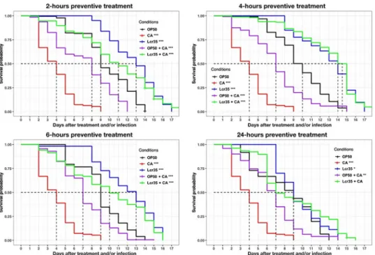
survival of the nematodes increased significantly as the mean lifespan increased from 3 to 11 days (267% increase in survival) compared with that observed withC. albicans infection alone
(p < 2.10−16). There was no significant difference in worm survival between those sequentially exposed to Lcr351andC. albicans and those exposed to Lcr351only (Fig 4) (p = 1). Similar results were obtained with the 4-hours treatment time. In that case, we observed that Lcr351 completely protectedC. elegans from infection since there was no significant difference with
the Lcr351control condition without infection (p = 0.4).
For longer treatment times (6 and 24 h), we observed a significant decrease in the mean sur-vival in the presence of Lcr351(condition 6 h: p = 0.04, condition 24 h: p <2.10−16) or Lcr351 andC. albicans (condition 6 h: p = 9.10−13, condition 24 h: p < 2.10−16) compared to the treat-ment of 4 h. Taken together, the results showed that the 4 probiotic treattreat-ment was the most protective against infection.
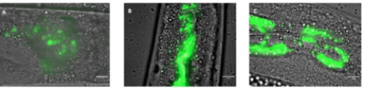
3.3.2 Influence of Lcr351onC. albicans colonization of the worm gut. To determine
whether the anti-C. albicans effects observed were due to the removal of the pathogen,
coloni-zation of the nematode intestine byC. albicans was observed by light microscopy. After three
days of incubation in the presence of the pathogen, wild-type worms exhibited notable coloni-zation of the entire digestive tract (Fig 5A). However, this strain ofC. albicans was not able to
form hyphae within the worm. We subsequently applied prophylactic treatment to the worms for 4 h before infecting them with yeast. We observed that after treatment withE. coli OP50 Fig 4. Preventive effects of Lcr351against
C. albicans ATCC 10231. Mean survival, where half of the population
was dead, is represented on the abscissa. The asterisks indicate thep-values (log-rank test) against E. coli OP50
(p < 0.05:�; p < 0.01:��; p < 0.001:���). Infection duration: 2 hour;2-hour preventive treatment (E. coli OP50
(OP50, n = 126);C. albicans ATCC 10231 (CA, n = 424); Lcr351(Lcr35, n = 93);E. coli OP50 + C. albicans (OP50 +
CA, n = 287); Lcr351+
C. albicans (Lcr35 + CA, n = 224));4-hour preventive treatment (E. coli OP50 (OP50,
n = 313);C. albicans ATCC 10231 (CA, n = 424); Lcr351(Lcr35, n = 259);
E. coli OP50 + C. albicans (OP50 + CA,
n = 120); Lcr351+C. albicans (Lcr35 + CA, n = 164)); 6-hour preventive treatment (E. coli OP50 (OP50, n = 222); C. albicans ATCC 10231 (CA, n = 424); Lcr351(Lcr35, n = 165);E. coli OP50 + C. albicans (OP50 + CA, n = 339);
Lcr351+
C. albicans (Lcr35 + CA, n = 300)); 24-hour preventive treatment (E.treatment coli OP50 (OP50, n = 248); C. albicans ATCC 10231 (CA, n = 424); Lcr351(n = 170);
E. coli OP50 + C. albicans (OP50 + CA, n = 220); Lcr351+
C. albicans (Lcr35 + CA, n = 183)).
(Fig 5B) or the probiotic Lcr351(Fig 5C) followed by infection, the yeastC. albicans was still
detected in the digestive tract of the host.
3.4 Mechanistic study
3.4.1 Modulation ofC. elegans gene expression induced by Lcr351andC. albicans. To
elucidate the mechanisms involved in the action of Lcr351againstC. albicans, we studied the
expression of sevenC. elegans genes (Table 3). We targeted three groups of genes:daf-2 and daf-16 (insulin signalling pathway), which are involved in host longevity and
anti-pathogenic-ity;sek-1 and pmk-1 (p38 MAPK signalling pathway), which concern the immune response;
andabf-2, cnc-4 and fipr-22/fipr-23, which encode antimicrobial proteins. We noted that
Lcr351tended to induce an overexpression ofdaf-16 (p = 0.1635) and had no effect on daf-2
(p = 0.2536), whileC. albicans tended to induce an upregulation of both genes (p = 0.1155 and
p = 0.2396, respectively). We did not observe any expression modulation ofdaf-2 or daf-16
using a preventive treatment withE. coli OP50 (p = 0.1258 and p = 0.1215, respectively) or
with Lcr351(p = 0.1354 and p = 0.3021, respectively).
The expression of thesek-1 and pmk-1 immunity genes was significantly downregulated in
the presence of Lcr351by 2.63-fold (p = 0.015) and 2.78-fold (p = 0.0149), respectively, while they were upregulated byC. albicans 3.21-fold (p = 0.0247) and 4.33-fold (0.1618), respectively.
In the control condition, in the presence ofE. coli OP50 and C. albicans, sek-1 was repressed
2.70 times (0.37-fold with p = 0.0204), butpmk-1 tended to be overexpressed. Preventive
treat-ment with Lcr351had the same effect onsek-1 (p = 0.0016) but induced no change in pmk-1
expression (p = 0.8205). Finally, among the 3 antimicrobials encoding the genes tested, only the expression ofcnc-4 seemed to be modulated in the presence of Lcr351, andcnc-4 was
overexpressed (p = 0.1753).C albicans also seemed to induce the overexpression of abf-2
(p = 0.2213) andcnc-4 (p = 0.3228), but interestingly, fipr-22/fipr-23 (p = 0.8225) expression
remained unchanged. Overexpression ofabf-2 (6.25-fold, p = 0.3158) and significant
Fig 5.C. albicans colonization of the C. elegans gut after 72 h (A) and after a 4-hour prophylactic treatment with E. coli OP50 (B) or Lcr351(C). The green colour represents yeast labelled with rhodamine 123. Scale bar, 10μm. https://doi.org/10.1371/journal.pone.0216184.g005
Table 3. Relative expression of theC. elegans genes of interest in the presence of Lcr351and
C. albicans in pure or sequential cultures in comparison with the con-trol conditionE. coli OP50 (alone).
Genes of interest
Insulin signalling pathway p38 MAPK signalling pathway Antimicrobials
Conditions daf-2 daf-16 sek-1 pmk-1 abf-2 cnc-4 fipr-22 /
fipr-23
Lcr351 1.35 2.18 0.38�� 0.36� 1.70 3.39 0.61
C. albicans 2.48 3.31 3.21� 4.33 11.33 22.32 1.08
E. coli OP50 + C. albicans 1.82 0.53 0.37� 3.40 4.69 0.16�� 0.78
Lcr351+C. albicans 0.69 1.74 0.31�� 1.15 1.61 0.41� 0.42�
Genes were considered differentially expressed when the p-value was lower than 0.05 (�) or 0.01 (��) according to Fisher’s LSD test and simultaneously when the
expression change was at least 2 times or 0.5 times. https://doi.org/10.1371/journal.pone.0216184.t003
repression ofcnc-4 (p = 0.0088) were observed when E. coli OP50 was added before infection
withC. albicans. Using a Lcr351preventive treatment,cnc-4 and fipr-22/fipr-23 were
signifi-cantly repressed (p = 0.0396 and p = 0.0385, respectively).
3.4.2 Influence of Lcr351andC. albicans on DAF-16 nuclear translocation. To further
investigate the mechanisms involved in the anti-C. albicans effects of Lcr351, we followed the nuclear translocation of the DAF-16/FOXO transcription factor using the DAF-16::GFP strain. Whatever the incubation time, the worms did not show any translocation of DAF-16 when fed withE. coli OP50 (Fig 6A). When Lcr351was used as food, we observed a nuclear translo-cation of the transcription factor, taking place gradually from 4 h of incubation with a maxi-mum intensity in the nuclei after 6 hh. The distribution of DAF-16 was both cytoplasmic and nuclear (Fig 6B). When the nematode was fed exclusively withC. albicans, we observed a rapid
nuclear translocation of the transcription factor after two hours of incubation in the presence of the pathogen (Fig 6C). This translocation was maintained throughout the experiment, i.e., 76 h.
3.4.3 Effect of Lcr351preventive treatment on DAF-16 nuclear translocation. We
investigated the effect of preventive treatment on the cellular localization of DAF-16 over time after infection byC. albicans using the C. elegans DAF-16::GFP mutant. When nematodes
were first fed withE. coli OP50 before being infected, DAF-16 was fully observed in the nuclei
Fig 6. DAF-16 cellular localization inC. elegans transgenic strain TJ-356 (daf-16p::daf-16a/b::GFP + rol-6 (su1006)) expressing DAF-16::GFP. Worms fed on E. coli OP50 (A), on Lcr351(B) and on
C. albicans ATCC 10231
(C). Scale bar, 100μm.
up to 4 h after infection and then gradually translocated to the cytoplasm after 24 h (Fig 7A). Conversely, the worms that were first exposed to Lcr351and then to the pathogen showed a different response, and the transcription factor was found only in the nuclei (Fig 7B).
4 Discussion
The selection of microbial strains as probiotics is based on a combination of functional probi-otic properties revealed first by classical basicin vitro testing. Beyond resistance to gastric pH
or bile salts, the ability of a strain to adhere to epithelial cells is frequently studied since this represents a prerequisite for mucosal colonization as part of the anti-pathogen activity. Adhe-sion is also a key parameter for pathogens since it allows them to release toxins and enzymes directly into the target cell, facilitating their dissemination [49]. Nivoliezet al. showed that the Fig 7. Impact of preventive Lcr351treatment on DAF-16 cellular localization in the
C. elegans transgenic strain TJ-356 expressing DAF-16::GFP. Worms fed withE. coli OP50 + C. albicans (A) and on Lcr351+C. albicans (B).
Scale bar, 100μm.
native probiotic strain Lcr351adhered rather weakly to Caco-2 intestinal cells, while the industrial formulation increased this capacity [24]. We have demonstrated here the ability of Lcr351to inhibit the growth of the pathogenC. albicans and the formation of a C. albicans
biofilm on an intestinal cell monolayerin vitro. As described by Jankowska et al., the low
adherence ofL. rhamnosus compared to C. albicans seems to reflect that competition for
mem-brane receptors is not the only mechanism. It is probably related to the synthesis of antifungal effectors by the probiotic as well [49]. Exopolysaccharides (EPS) secreted by certain lactobacilli have been shown to modify the surface properties (hydrophobicity) of microorganisms with direct consequences on their adhesion capacities [50]. EPS have antifungal effects by inhibiting
C. albicans growth and adhesion to epithelial cells. The surface polysaccharides of L. rhamno-sus GG, a strain phylogenetically close to Lcr35, appear to interfere in the binding between the
fungal lectin-like adhesins and host sugars or between the fungal cell wall carbohydrates and their epithelial adhesion receptor [51]. A recent study has shown that purified fractions of exo-polysaccharides also interfered with adhesion capacities of microorganisms [52]. It would be interesting to assay the inhibitory properties of Lcr351EPS. However, to fully understand the probiotic mechanisms,in vitro approaches are too limited. Moving to an in vivo approach is
mandatory to better understand the interactions between microorganisms (probiotics and pathogens) and the host response.
C. elegans is considered a powerful in vivo model for studying the pathogenicity of
microor-ganisms [34,35,53–55] and the antimicrobial properties of lactic acid bacteria [56,57]. The nature of the nutrient source is an important parameter that has a great influence on nematode physiology. Regarding worm growth, it appears that there is some disparity depending on the type of lactic acid bacteria used to feedC. elegans. Bifidobacterium spp. had no influence on the
size of adult worms, although their growth was slightly slowed down [58,59].Lactobacillus spp.
by contrast usually result in reduced growth rates and sizes and are sometimes even lethal to the larvae [60,61]. The mechanisms for explaining the longevity extension induced by lactic acid bacteria are not fully understood, but some authors have suggested the involvement of caloric restriction [62–64]. In our case, similar to the work of Komuraet al., it seems that
Lcr351did not induce pro-longevity effects through caloric restriction insofar as the growth of Lcr351-fed nematodes is identical compared toE. coli OP50-fed worms [65].
After demonstrating the preventive effect of Lcr35 againstC. albicans in the nematode, we
decided to better understand the protective effect at the mechanistic level. InC. elegans, the
insulin/IGF-1 signalling pathway is strongly involved in regulating the longevity and immu-nity of the animal. Signal transduction is mediated through DAF-16, a highly conserved FOXO transcription factor [66]. Using the GFP fusion protein, we have shown that Lcr351 induces translocation of DAF-16 to the nucleus, suggesting that DAF-16 is involved in the pro-biotic mechanisms of action of Lcr351. According to several studies, our data suggested that the pro-longevity effect of Lcr351implements mechanisms involving different regulatory pathways linked to DAF-16, such as the DAF-2/DAF-16 insulin pathway [67] or the c-Jun N-terminal kinase JNK-1/DAF-16 pathway [59]. The absence of modulation ofdaf-2 expression
in the presence of Lcr351suggests that the DAF-2/DAF-16 pathway is not involved and that the anti-Candida capacity of Lcr351is due to the JNK signalling pathway. The involvement of these pathways needs to be followed at proteomic and phosphoproteomic levels to validate this hypothesis.
The yeastC. albicans is capable of inducing a severe infection in C. elegans, causing a rapid
death of the host and even after a very short contact time. This infection is first manifested by the colonization of the whole intestinal lumen by yeasts and then by the formation of hyphae piercing the cuticle of the nematode leading to its death [34,68]. In addition, it has been shown that strains ofC. albicans incapable of forming hyphae, such as SPT20 mutants, have a
significantly reduced pathogenicity inC. elegans as well as in Galleria mellonella or Mus mus-culus models while still being lethal [37]. In the nematode, it seems that the distention of the intestine caused by the accumulation of yeast is one of the causes of the death of the animal [35]. Recently, de Barroset al. [38] showed thatL. paracasei 28.4 had anti-C. albicans activity
bothin vitro and in vivo by inhibiting filamentation of yeast protecting the nematode.
AlthoughC. albicans ATCC 10231 is able to form hyphae during in vitro assays, it failed to kill C. elegans by filamentation. Therefore, it is likely that Lcr351represses virulence factors in yeast other than filamentation.
From a mechanistic point of view, several hypotheses can explain the anti-C. albicans
prop-erties of Lcr351in the nematode: a direct interaction between the two microorganisms as well as an immunomodulation of the host by the probiotic. According to Nivoliezet al.
demon-strating the inhibitory capacity of Lcr351with respect to the pathogen during a co-culture experiment [24], our data showedCandida albicans inhibition on mammalian cell monolayers.
This inhibition may be due to nutrient competition (i.e., glycogen consumption) or to the pro-duction of toxic metabolites against the yeast [24]. We have shown that even after preventive treatment with the probiotic, the digestive tract of the nematode is colonized by the pathogen without showing a pathological state. This suggests that Lcr351induced repression of viru-lence factors inC. albicans, as shown by De Barros et al. [38]. Moreover, anin vitro study on
human dendritic cells revealed that Lcr351induced a large dose-dependent modulation not only in the expression of genes mainly involved in the immune response but also in the expres-sion of CD, HLA and TLR membrane proteins. Highly conserved and found inC. elegans,
TLR also plays a role in the antipathogenic response of the nematode by activating the p38 MAPK pathway [59]. A pro-inflammatory effect has also been shown through cytokine secre-tion, such as IL-1β, IL-12, TNFα. However, this immunomodulation takes place only in the presence of a high concentration of Lcr351[69]. InC. elegans, DAF-16 is closely related to
mammalian FOXO3a, a transcription factor involved the inflammatory process [70]. There-fore, nuclear translocation of DAF-16 by Lcr351can be interpreted as the establishment of an inflammatory response in the host allowing it to survive an infection. In our study, we observed that the duration of the Lcr351treatment influences the preventive anti-C. albicans
effect on nematode lifespan, suggesting that the quantity of Lcr351ingested and/or the treat-ment period of time may have an impact on the efficiency of the treattreat-ment. A thorough tran-scriptional study will be interesting to characterize the dose-dependent effect probiotics administered. We demonstrated that Lcr351induces a transcriptional response in the host by activating the transcription factor DAF-16 and repressing the p38 MAPK signalling pathway, including in the presence ofC. albicans. We also observed the repression of the genes encoding
antimicrobials when fungal infection was preceded by probiotic treatment. The work of Puk-kila-Worleyet al. [35] demonstrated thatC. albicans induced a fast antifungal response in the
host inducing the expression of antimicrobial genes such asabf-2, cnc-4, cnc-7, fipr-22 and fipr-23. With the exception of abf-2, all these genes are under the control of PMK-1, whose
inactivation makes the nematode susceptible to infection. In our study, we showed that an Lcr351preventive treatment induced a down-regulation in thecnc-4, fipr-22 and fipr-23
genes, whilepmk-1 remained unchanged compared to the control condition. Based on the
data of Pukkila-Worleyet al., the absence of overexpression of these genes in the presence of C. albicans after pre-exposure with Lcr351
suggests again that the probiotic inhibits yeast viru-lence, obviating the establishment of a defence mechanism by the host. Similar results have also been observed withSalmonella Enteritidis, where the authors hypothesize that the
probi-otics used induce immunotolerance in the nematode rather than the synthesis of antimicrobi-als [58]. The use ofC. elegans mutants or RNAi could be further considered to decipher the
5 Conclusion
This study demonstrates the preventive anti-C. albicans properties of Lcr351
using bothin vitro and in vivo models. The probiotic strain inhibits the growth of the pathogenic yeast and
its ability to form biofilms on intestinal cellsin vitro. Lcr351
allows protection of the hostC. elegans against infection despite the presence of C. albicans in its gut. Lcr351duringC. albi-cans infection seems to induce a decrease in the immune response of the nematode
(downre-gulation ofsek-1, pmk-1, abf-2, cnc-4 and fipr-22/23). Extra studies on C. elegans whole
transcriptome modulation by Lcr351would be interesting to further reveal other mechanisms involved. The study of the yeast virulence gene modulation induced by Lcr351could be very informative about the complex mechanisms of the probiotic mechanisms of action. Addition-ally, in a second phase, the realization of a comparative study between Lcr351and other Lacto-bacillus strains (L. rhamnosus, L. casei, L. paracasei) as well as between different strains of CC. albicans, including clinical strains, could be of interest to determine the degree of strain
depen-dence of our results.
Acknowledgments
Some strains were provided by the CGC, which is funded by the NIH Office of Research Infra-structure Programs (P40 OD010440).
We thank Jonathan Heuze´, Muriel The´ret and Jeanne Riom, trainees at the UMRF 0545 (UCA/INRA/VAS) for their involvement in the implementation of several experiments. We thank greatly all those who participated in the writing of this article.
Author Contributions
Conceptualization: Cyril Poupet. Formal analysis: Cyril Poupet.
Investigation: Taous Saraoui, Philippe Veisseire, Muriel Bonnet, Marylise Gachinat, Olivier
Camarès.
Methodology: Cyril Poupet.
Supervision: Adrien Nivoliez, Ste´phanie Bornes. Validation: Ste´phanie Bornes.
Writing – original draft: Cyril Poupet.
Writing – review & editing: Philippe Veisseire, Muriel Bonnet, Caroline Dausset, Christophe
Chassard, Ste´phanie Bornes.
References
1. Cauchie M, Desmet S, Lagrou K. Candida and its dual lifestyle as a commensal and a pathogen. Res Microbiol [Internet]. 2017 Nov [cited 2018 Sep 5]; 168(9–10):802–10. Available from:https://linkinghub. elsevier.com/retrieve/pii/S0923250817300402PMID:28263903
2. Neville BA, D’enfert C, Bougnoux M-E. Candida albicans commensalism in the gastrointestinal tract. FEMS Yeast Res [Internet]. 2015 [cited 2018 Sep 5]; 15:81. Available from:https://unite.ut.ee/
3. Mayer FL, Wilson D, Hube B. Candida albicans pathogenicity mechanisms. Vol. 4, Virulence. 2013. p. 119–28.
4. Kadosh D, Antonio S. Control of Candida albicans morphology and pathogenicity by post-transcriptional mechanisms. Cell Mol Life Sci. 2017; 73(22):4265–78.
5. Wa¨chtler B, Wilson D, Haedicke K, Dalle F, Hube B. From attachment to damage: Defined genes of Candida albicans mediate adhesion, invasion and damage during interaction with oral epithelial cells.
Munro C, editor. PLoS One [Internet]. 2011 Feb 23 [cited 2018 Sep 10]; 6(2):e17046. Available from:
http://www.ncbi.nlm.nih.gov/pubmed/21407800PMID:21407800
6. Farmakiotis D, Kontoyiannis DP. Epidemiology of antifungal resistance in human pathogenic yeasts: current viewpoint and practical recommendations for management. Int J Antimicrob Agents [Internet]. 2017 Sep [cited 2018 Sep 5]; 50(3):318–24. Available from:https://linkinghub.elsevier.com/retrieve/pii/ S0924857917302364PMID:28669831
7. Sanguinetti M, Posteraro B, Lass-Flo¨ rl C. Antifungal drug resistance among Candida species: Mecha-nisms and clinical impact. Mycoses [Internet]. 2015 Jun [cited 2018 Sep 5]; 58(S2):2–13. Available from:http://www.ncbi.nlm.nih.gov/pubmed/26033251
8. Scorzoni L, de Paula E Silva ACA, Marcos CM, Assato PA, de Melo WCMA, de Oliveira HC, et al. Anti-fungal Therapy: New Advances in the Understanding and Treatment of Mycosis. Front Microbiol [Inter-net]. 2017 [cited 2018 Sep 5]; 8:36. Available from:http://www.ncbi.nlm.nih.gov/pubmed/28167935
PMID:28167935
9. Wheeler ML, Limon JJ, Bar AS, Leal CA, Gargus M, Tang J, et al. Immunological Consequences of Intestinal Fungal Dysbiosis. Cell Host Microbe [Internet]. 2016; 19(6):865–73. Available from:http://dx. doi.org/10.1016/j.chom.2016.05.003PMID:27237365
10. Hu H-J, Zhang G-Q, Zhang Q, Shakya S, Li Z-Y. Probiotics Prevent Candida Colonization and Invasive Fungal Sepsis in Preterm Neonates: A Systematic Review and Meta-Analysis of Randomized Con-trolled Trials. Pediatr Neonatol [Internet]. 2017 Apr [cited 2018 Sep 5]; 58(2):103–10. Available from:
http://linkinghub.elsevier.com/retrieve/pii/S1875957216301401PMID:27793494
11. Matsubara VH, Bandara HMHN, Mayer MPA, Samaranayake LP. Probiotics as Antifungals in Mucosal Candidiasis. 2016 [cited 2018 Sep 5];https://academic.oup.com/cid/article-abstract/62/9/1143/ 1745140
12. Agrawal S, Rao S, Patole S. Probiotic supplementation for preventing invasive fungal infections in pre-term neonates—a systematic review and meta-analysis. Mycoses [Internet]. 2015 Nov 1 [cited 2018 Sep 5]; 58(11):642–51. Available from:http://doi.wiley.com/10.1111/myc.12368PMID:26468692
13. FAO, WHO. Health and Nutritional Properties of Probiotics in Food including Powder Milk with Live Lac-tic Acid Bacteria. Food Nutr Pap [Internet]. 2001 [cited 2016 Jun 14];http://www.crcnetbase.com/doi/ abs/10.1201/9781420009613.ch16
14. Fijan S. Microorganisms with Claimed Probiotic Properties: An Overview of Recent Literature. Int J Environ Res Public Heal Int J Environ Res Public Heal Int J Environ Res Public Heal [Internet]. 2014 [cited 2017 May 13]; 11:4745–67. Available from:www.mdpi.com/journal/ijerph
15. Olle B. Medicines from microbiota. Nat Biotechnol [Internet]. 2013 Apr 5 [cited 2017 Mar 31]; 31(4):309–15. Available from:http://www.nature.com/doifinder/10.1038/nbt.2548PMID:23563425
16. Coudeyras S, Jugie G, Vermerie M, Forestier C. Adhesion of human probiotic Lactobacillus rhamnosus to cervical and vaginal cells and interaction with vaginosis-associated pathogens. Infect Dis Obstet Gynecol [Internet]. 2008 Jan 27 [cited 2018 Sep 10]; 2008:549640. Available from:http://www.ncbi.nlm. nih.gov/pubmed/19190778PMID:19190778
17. Coudeyras S, Marchandin H, Fajon C, Forestier C. Taxonomic and strain-specific identification of the probiotic strain Lactobacillus rhamnosus 35 within the Lactobacillus casei group. Appl Environ Microbiol [Internet]. 2008 May [cited 2018 Sep 10]; 74(9):2679–89. Available from:http://www.ncbi.nlm.nih.gov/ pubmed/18326671PMID:18326671
18. Forestier C, De Champs C, Vatoux C, Joly B. Probiotic activities of Lactobacillus casei rhamnosus: in vitro adherence to intestinal cells and antimicrobial properties. Res Microbiol [Internet]. 2001 Mar [cited 2018 Sep 10]; 152(2):167–73. Available from:http://www.ncbi.nlm.nih.gov/pubmed/11316370PMID:
11316370
19. de Champs C, Maroncle N, Balestrino D, Rich C, Forestier C. Persistence of colonization of intestinal mucosa by a probiotic strain, Lactobacillus casei subsp. rhamnosus Lcr35, after oral consumption. J Clin Microbiol [Internet]. 2003 Mar [cited 2018 Sep 10]; 41(3):1270–3. Available from:http://www.ncbi. nlm.nih.gov/pubmed/12624065PMID:12624065
20. Petricevic L, Witt A. The role of Lactobacillus casei rhamnosus Lcr35 in restoring the normal vaginal flora after antibiotic treatment of bacterial vaginosis. BJOG An Int J Obstet Gynaecol [Internet]. 2008 Oct [cited 2016 Jun 14]; 115(11):1369–74. Available from:http://doi.wiley.com/10.1111/j.1471-0528. 2008.01882.x
21. Muller C, Mazel V, Dausset C, Busignies V, Bornes S, Nivoliez A, et al. Study of the Lactobacillus rham-nosus Lcr35®properties after compression and proposition of a model to predict tablet stability. Eur J Pharm Biopharm. 2014; 88(3):787–94.https://doi.org/10.1016/j.ejpb.2014.07.014PMID:25128853
22. Nivoliez A, Veisseire P, Alaterre E, Dausset C, Baptiste F, Camarès O, et al. Influence of manufacturing processes on cell surface properties of probiotic strain Lactobacillus rhamnosus Lcr35®. Appl Microbiol
Biotechnol [Internet]. 2015 [cited 2017 Jan 1]; 99(1):399–411. Available from:http://link.springer.com/ 10.1007/s00253-014-6110-zPMID:25280746
23. Dausset C, Patrier S, Gajer P, Thoral C, Lenglet Y, Cardot JM, et al. Comparative phase I randomized open-label pilot clinical trial of Gynophilus®(Lcr regenerans®) immediate release capsules versus slow
release muco-adhesive tablets. Eur J Clin Microbiol Infect Dis [Internet]. 2018 [cited 2019 Apr 2]; 37 (10):1869–80. Available from:https://doi.org/10.1007/s10096-018-3321-8PMID:30032443
24. Nivoliez A, Camares O, Paquet-Gachinat M, Bornes S, Forestier C, Veisseire P. Influence of
manufacturing processes on in vitro properties of the probiotic strain Lactobacillus rhamnosus Lcr35®. J
Biotechnol. 2012; 160(3–4):236–41.https://doi.org/10.1016/j.jbiotec.2012.04.005PMID:22542933
25. Isolauri E, Kirjavainen P V, Salminen S. Probiotics: a role in the treatment of intestinal infection and inflammation? Gut [Internet]. 2002; 50(Supplement 3):iii54–9. Available from:http://gut.bmj.com/cgi/ doi/10.1136/gut.50.suppl_3.iii54
26. do Carmo MS, Santos C itapary dos, Arau´jo MC, Giro´ n JA, Fernandes ES, Monteiro-Neto V. Probiotics, mechanisms of action, and clinical perspectives for diarrhea management in children. Food Funct [Inter-net]. 2018; 9(10):5074–95. Available from:http://dx.doi.org/10.1039/c8fo00376aPMID:30183037
27. Coudeyras S, Forestier C. Microbiote et probiotiques: impact en sante´ humaine. Can J Microbiol [Inter-net]. 2010 [cited 2018 Jan 30]; 56(8):611–50. Available from:http://www.nrcresearchpress.com/doi/ pdfplus/10.1139/W10-052PMID:20725126
28. Lacroix C, de Wouters T, Chassard C. Integrated multi-scale strategies to investigate nutritional com-pounds and their effect on the gut microbiota. Curr Opin Biotechnol [Internet]. 2015 [cited 2017 Apr 30]; 32:149–55. Available from:http://dx.doi.org/10.1016/j.copbio.2014.12.009PMID:25562815
29. Vinderola G, Gueimonde M, Gomez-Gallego C, Delfederico L, Salminen S. Correlation between in vitro and in vivo assays in selection of probiotics from traditional species of bacteria. Trends Food Sci Tech-nol [Internet]. 2017; 68:83–90. Available from:http://dx.doi.org/10.1016/j.tifs.2017.08.005
30. Montoro BP, Benomar N, Lerma LL, Gutie´ rrez SC, Ga´lvez A, Abriouel H. Fermented aloreña table olives as a source of potential probiotic Lactobacillus pentosus strains. Front Microbiol. 2016; 7(OCT).
31. Roselli M, Finamore A, Britti MS, Mengheri E. Probiotic bacteria Bifidobacterium animalis MB5 and Lac-tobacillus rhamnosus GG protect intestinal Caco-2 cells from the inflammation-associated response induced by enterotoxigenic Escherichia coli K88. Br J Nutr [Internet]. 2006; 95(06):1177. Available from:
http://www.journals.cambridge.org/abstract_S0007114506001589
32. Papadimitriou K, Zoumpopoulou G, Foligne´ B, Alexandraki V, Kazou M, Pot B, et al. Discovering probi-otic microorganisms: In vitro, in vivo, genetic and omics approaches. Front Microbiol. 2015; 6(FEB):1– 28.
33. Lai CH, Chou CY, Ch’ang LY, Liu CS, Lin W. Identification of novel human genes evolutionarily con-served in Caenorhabditis elegans by comparative proteomics. Genome Res [Internet]. 2000 May [cited 2018 Sep 10]; 10(5):703–13. Available from:http://www.ncbi.nlm.nih.gov/pubmed/10810093PMID:
10810093
34. Pukkila-Worley R, Peleg AY, Tampakakis E, Mylonakis E. Candida albicans hyphal formation and viru-lence assessed using a Caenorhabditis elegans infection model. Eukaryot Cell [Internet]. 2009 [cited 2018 Feb 7]; 8(11):1750–8. Available from:http://ec.asm.org/content/8/11/1750.full.pdfPMID:
19666778
35. Pukkila-Worley R, Ausubel FM, Mylonakis E. Candida albicans infection of Caenorhabditis elegans induces antifungal immune defenses. PLoS Pathog. 2011; 7(6).
36. Alves V de S, Mylonakis E. The eIF2 kinase Gcn2 modulates Candida albicans virulence to Caenorhab-ditis elegans. Clin Microbiol Infect Dis [Internet]. 2018; 3(2):1–4. Available from:https://www.oatext. com/the-eif2-kinase-gcn2-modulates-candida-albicans-virulence-to-caenorhabditis-elegans.php
37. Tan X, Fuchs BB, Wang Y, Chen W, Yuen GJ, Chen RB, et al. The role of Candida albicans SPT20 in filamentation, biofilm formation and pathogenesis. PLoS One. 2014; 9(4):1–10.
38. de Barros PP, Scorzoni L, Ribeiro F de C, Fugisaki LR de O, Fuchs BB, Mylonakis E, et al. Lactobacillus paracasei 28.4 reduces in vitro hyphae formation of Candida albicans and prevents the filamentation in an experimental model of Caenorhabditis elegans. Microb Pathog [Internet]. 2018; 117(November 2017):80–7. Available from:https://doi.org/10.1016/j.micpath.2018.02.019
39. Pinto M, Robineleon S, Appay MD, Kedinger M, Triadou N, Dussaulx E, et al. Enterocyte-like differentia-tion and polarizadifferentia-tion of the human colon carcinoma cell line Caco-2 in culture. Biol Cell [Internet]. 1983 Jan 1 [cited 2018 Oct 10]; 47:323–30. Available from:https://www.scienceopen.com/document?vid= 07f3fdcd-c23c-47d4-ad63-105346ef5453
40. Brenner S. The genetics of Caenorhabditis elegans. Genetics. 1974; 77(1):71–94. PMID:4366476
41. Hellemans J, Mortier G, De Paepe A, Speleman F, Vandesompele J. qBase relative quantification framework and software for management and automated analysis of real-time quantitative PCR data.
2007 [cited 2017 Jun 14]; 8(2). Available from:http://download.springer.com/static/pdf/804/art% 253A10.1186%252Fgb-2007-8-2-r19.pdf?originUrl=http%3A%2F%2Fgenomebiology.biomedcentral. com%2Farticle%2F10.1186%2Fgb-2007-8-2-r19&token2=exp=1497424427~acl=%2Fstatic%2Fpdf% 2F804%2Fart%25253A10.1186%25252Fgb-2
42. Semple JI, Garcia-Verdugo R, Lehner B. Rapid selection of transgenic C. elegans using antibiotic resis-tance. Nat Methods [Internet]. 2010 Sep 22 [cited 2017 Apr 13]; 7(9):725–7. Available from:http://www. ncbi.nlm.nih.gov/pubmed/20729840PMID:20729840
43. Hoogewijs D, Houthoofd K, Matthijssens F, Vandesompele J, Vanfleteren JR. Selection and validation of a set of reliable reference genes for quantitative sod gene expression analysis in C. elegans. BMC Mol Biol [Internet]. 2008 Jan 22 [cited 2017 Apr 13]; 9:9. Available from:http://www.ncbi.nlm.nih.gov/ pubmed/18211699PMID:18211699
44. Nakagawa H, Shiozaki T, Kobatake E, Hosoya T, Moriya T, Sakai F, et al. Effects and mechanisms of prolongevity induced by Lactobacillus gasseri SBT2055 in Caenorhabditis elegans. Aging Cell. 2016; 15(2):227–36.https://doi.org/10.1111/acel.12431PMID:26710940
45. R Core Team. R: A language and Environment for Statistical Computing [Internet]. Vienna, Austria: R Foundation for Statistical Computing; 2018.https://www.r-project.org/
46. Therneau TM. _A Package for Survival Analysis in S_. 2015.
47. Kassambara A, Kosinski M. survminer: Drawing Survival Curves using “ggplot2.” 2017.
48. Fatima S, Haque R, Jadiya P, Shamsuzzama, Kumar L, Nazir A. Ida-1, the Caenorhabditis elegans orthologue of mammalian diabetes autoantigen IA-2, potentially acts as a common modulator between Parkinson’s disease and diabetes: Role of Daf-2/Daf-16 insulin like signalling pathway. PLoS One. 2014; 9(12).
49. Jankowska A, Laubitz D, Antushevich H, Zabielski R, Grzesiuk E. Competition of Lactobacillus paraca-sei with Salmonella enterica for adhesion to Caco-2 cells. J Biomed Biotechnol. 2008; 2008(1). 50. Nowak A, Motyl I,Śliżewska K, Libudzisz Z, Klewicka E. Adherence of probiotic bacteria to human
colon epithelial cells and inhibitory effect against enteric pathogens–In vitro study. Int J Dairy Technol. 2016; 69(4):532–9.
51. Allonsius CN, van den Broek MFL, De Boeck I, Kiekens S, Oerlemans EFM, Kiekens F, et al. Interplay between Lactobacillus rhamnosus GG and Candida and the involvement of exopolysaccharides. Microb Biotechnol. 2017; 10(6):1753–63.https://doi.org/10.1111/1751-7915.12799PMID:28772020
52. Ruas-Madiedo P, Gueimonde M, Margolles A, de los Reyes-Gavilan CG, Salminen S. Exopolysacchar-ides Produced by Probiotic Strains Modify the Adhesion of Probiotics and Enteropathogens to Human Intestinal Mucus. J Food Prot [Internet]. 2006; 69(8):2011–5. Available from:http://jfoodprotection.org/ doi/abs/10.4315/0362-028X-69.8.2011PMID:16924934
53. Irazoqui JE, Troemel ER, Feinbaum RL, Luhachack LG, Cezairliyan BO, Ausubel FM. Distinct patho-genesis and host responses during infection of C. elegans by P. aeruginosa and S. aureus. PLoS Pathog. 2010; 6(7):1–24.
54. Wu K, Conly J, McClure JA, Elsayed S, Louie T, Zhang K. Caenorhabditis elegans as a host model for community-associated methicillin-resistant Staphylococcus aureus. Clin Microbiol Infect. 2010; 16(3):245–54.https://doi.org/10.1111/j.1469-0691.2009.02765.xPMID:19456837
55. Souza ACR, Fuchs BB, Alves V de S, Jayamani E, Colombo AL, Mylonakis E. Pathogenesis of the Can-dida parapsilosis complex in the model host Caenorhabditis elegans. Genes (Basel). 2018; 9(8). 56. Park MR, Ryu S, Maburutse BE, Oh NS, Kim SH, Oh S, et al. Probiotic Lactobacillus fermentum strain
JDFM216 stimulates the longevity and immune response of Caenorhabditis elegans through a nuclear hormone receptor. Sci Rep [Internet]. 2018 [cited 2019 Jan 3]; 8(1):7441. Available from:www.nature. com/scientificreports/PMID:29748542
57. Kim Y, Mylonakis E. Caenorhabditis elegans immune conditioning with the probiotic bacterium Lactoba-cillus acidophilus strain ncfm enhances gram-positive immune responses. Infect Immun. 2012; 80(7):2500–8.https://doi.org/10.1128/IAI.06350-11PMID:22585961
58. Ikeda T, Yasui C, Hoshino K, Arikawa K, Nishikawa Y. Influence of lactic acid bacteria on longevity of Caenorhabditis elegans and host defense against Salmonella enterica serovar Enteritidis. Appl Environ Microbiol. 2007; 73(20):6404–9.https://doi.org/10.1128/AEM.00704-07PMID:17704266
59. Zhao L, Zhao Y, Liu R, Zheng X, Zhang M, Guo H, et al. The transcription factor DAF-16 is essential for increased longevity in C. elegans Exposed to Bifidobacterium longum BB68. Sci Rep [Internet]. 2017; 7(1):7408. Available from:http://www.nature.com/articles/s41598-017-07974-3PMID:28785042
60. Zanni E, Laudenzi C, Schifano E, Palleschi C, Perozzi G, Uccelletti D, et al. Impact of a complex food microbiota on energy metabolism in the model organism Caenorhabditis elegans. Biomed Res Int. 2015; 2015.
61. Guantario B, Zinno P, Schifano E, Roselli M, Perozzi G, Palleschi C, et al. In Vitro and in Vivo selection of potentially probiotic lactobacilli from nocellara del belice table olives. Front Microbiol. 2018; 9 (MAR):595.
62. Phelan JP, Rose MR. Why dietary restriction substantially increases longevity in animal models but won’t in humans. Ageing Res Rev. 2005; 4(3):339–50.https://doi.org/10.1016/j.arr.2005.06.001PMID:
16046282
63. Smith ED, Kaeberlein TL, Lydum BT, Sager J, Welton KL, Kennedy BK, et al. Age- and calorie-indepen-dent life span extension from dietary restriction by bacterial deprivation in Caenorhabditis elegans. BMC Dev Biol. 2008; 8:1–13.
64. Heestand BN, Shen Y, Liu W, Magner DB, Storm N, Meharg C, et al. Dietary Restriction Induced Lon-gevity Is Mediated by Nuclear Receptor NHR-62 in Caenorhabditis elegans. PLoS Genet. 2013; 9(7).
65. Komura T, Ikeda T, Yasui C, Saeki S, Nishikawa Y. Mechanism underlying prolongevity induced by bifi-dobacteria in Caenorhabditis elegans. Biogerontology. 2013; 14(1):73–87.https://doi.org/10.1007/ s10522-012-9411-6PMID:23291976
66. Tullet JMA. DAF-16 target identification in C. elegans: past, present and future. Biogerontology [Inter-net]. 2015; 16(2):221–34. Available from:http://link.springer.com/10.1007/s10522-014-9527-yPMID:
25156270
67. Grompone G, Martorell P, Llopis S, Gonza´ lez N, Genove´s S, Mulet AP, et al. Anti-Inflammatory Lacto-bacillus rhamnosus CNCM I-3690 Strain Protects against Oxidative Stress and Increases Lifespan in Caenorhabditis elegans. PLoS One. 2012; 7(12).
68. Breger J, Fuchs BB, Aperis G, Moy TI, Ausubel FM, Mylonakis E. Antifungal chemical compounds iden-tified using a C. elegans pathogenicity assay. PLoS Pathog. 2007; 3(2):0168–78.
69. Evrard B, Coudeyras S, Dosgilbert A, Charbonnel N, Alame´ J, Tridon A, et al. Dose-dependent immuno-modulation of human dendritic cells by the probiotic Lactobacillus rhamnosus Lcr35. PLoS One. 2011; 6(4):1–12.
70. Singh V, Aballay A. Regulation of DAF-16-mediated Innate Immunity in Caenorhabditis elegans. J Biol Chem [Internet]. 2009 Dec 18 [cited 2018 Dec 14]; 284(51):35580–7. Available from:http://www.ncbi. nlm.nih.gov/pubmed/19858203PMID:19858203