HAL Id: hal-03016676
https://hal.sorbonne-universite.fr/hal-03016676
Submitted on 20 Nov 2020HAL is a multi-disciplinary open access archive for the deposit and dissemination of sci-entific research documents, whether they are pub-lished or not. The documents may come from teaching and research institutions in France or abroad, or from public or private research centers.
L’archive ouverte pluridisciplinaire HAL, est destinée au dépôt et à la diffusion de documents scientifiques de niveau recherche, publiés ou non, émanant des établissements d’enseignement et de recherche français ou étrangers, des laboratoires publics ou privés.
Activation of innate immunity by 14-3-3 ϵ, a new
potential alarmin in osteoarthritis
M. Millerand, L. Sudre, M. Nefla, F. Pène, C. Rousseau, A. Pons, A. Ravat,
G. André-Leroux, S. Akira, T. Satoh, et al.
To cite this version:
M. Millerand, L. Sudre, M. Nefla, F. Pène, C. Rousseau, et al.. Activation of innate immunity by 14-3-3 ϵ, a new potential alarmin in osteoarthritis. Osteoarthritis and Cartilage, Elsevier, 2020, 28 (5), pp.646-657. �10.1016/j.joca.2020.03.002�. �hal-03016676�
1
ACTIVATION OF INNATE IMMUNITY BY 14-3-3 ε, A NEW POTENTIAL 1
ALARMIN IN OSTEOARTHRITIS 2
3 4
Marion MILLERAND(1), Laure SUDRE(1), Meriam NEFLA(1), Frédéric PÈNE(2), 5
Christophe ROUSSEAU(2), Anna PONS(1), Arnaud RAVAT(1), Gwenaëlle ANDRE-6
LEROUX(3), Shizuo Akira(5), Takashi Satoh(5), Francis BERENBAUM(1,4) and Claire 7
JACQUES(1) 8
9
(1) Sorbonne Université, INSERM (UMR S938) and Labex Transimmunom, Paris, 10
France 11
(2) Institut Cochin, INSERM U1016, CNRS UMR8104, Paris, France ; Université 12
Paris Descartes, Sorbonne Paris Cité, Paris France 13
(3) MaIAGE, INRA, Université Paris-Saclay, 78350 Jouy-en-Josas, France 14
(4) Sorbonne Université, Department of Rheumatology, AP-HP, Hôpital Saint-15
Antoine, and Labex Transimmunom, Paris, France 16
(5) Laboratory of Host Defense, WPI Immunology Frontier Research Center (IFReC), 17
Osaka University, Osaka 565-0871, Japan 18
19
Address correspondence to: 20
Dr. F. Berenbaum: UMR_S938, CDR Saint-Antoine - INSERM - Sorbonne Université, 21
184 Rue du Faubourg Saint-Antoine - 75012 Paris, France. 22
Tel : +33 149-28-25-20, Fax: +33 149-28-25-13 23
E-mail: francis.berenbaum@sat.aphp.fr 24
2 Abstract 1
Objective: The innate immune system plays a central role in osteoarthritis (OA). We 2
identified 14-3-3ε as a novel mediator that guides chondrocytes toward an 3
inflammatory phenotype. 14-3-3ε shares common characteristics with alarmins. 4
These endogenous molecules, released into extracellular media, are increasingly 5
incriminated in sustaining OA inflammation. Alarmins bind mainly to TLR2 and TLR4 6
receptors and polarize macrophages in the synovium. We investigated the effects of 7
14-3-3ε in joint cells and tissues and its interactions with TLRs to define it as a new 8
alarmin involved in OA. 9
Design: Chondrocyte, synoviocyte and macrophage cultures from murine or OA 10
human samples were treated with 14-3-3ε. To inhibit TLR2/4 in chondrocytes, 11
blocking antibodies were used. Moreover, chondrocytes and bone marrow 12
macrophage (BMM) cultures from KO TLRs mice were stimulated with 14-3-3ε. Gene 13
expression and release of inflammatory mediators (IL-6, MCP-1, TNFα) were 14
evaluated via RT-qPCR and ELISA. 15
Results: In vitro, 14-3-3ε induced gene expression and release of IL6 and MCP1 in 16
the treated cells. The inflammatory effects of 14-3-3ε were significantly reduced 17
following TLRs inhibition or in TLRs KO chondrocytes and BMM. 18
Conclusions: 14-3-3ε is able to induce an inflammatory phenotype in synoviocytes, 19
macrophages and chondrocytes in addition to polarizing macrophages. These effects 20
seem to involve TLR2 or TLR4 to trigger innate immunity. Our results designate 14-3-21
3ε as a novel alarmin in OA and as a new target either for therapeutic and/or 22
prognostic purposes. 23
3
Keywords : 14-3-3 ε, alarmin, osteoarthritis, Innate immunity, TLR, synovitis 1
Running headline : 14-3-3 ε : a new alarmin in osteoarthritis 2
3
Abbreviations
4BMM: bone marrow derived macrophages 5
BSA: bovine serum albumin 6
cDNA: complementary deoxyribonucleic acid 7
DAMP: damage-associated molecular pattern 8
DMEM: Dulbecco's modified Eagle's medium 9
EDTA: Ethylenediaminetetraacetic acid 10
ELISA: enzyme linked immunosorbent assay 11
FBS: fetal bovine serum 12
FLS: Fibroblast-like synoviocyte 13
Glu: glutamine 14
H&E: Hematoxylin and eosin 15 HPRT: Hypoxanthine-guanine phosphoribosyltransferase 16 IL : interleukin 17 KO: Knockout 18 LPS: lipopolysaccharides 19
mAB: monoclonal antibody 20
4 MCP1 : monocyte chemoattractant protein-1 1
MMP: matrix metalloproteinases 2
mRNA: messenger ribonucleic acid 3
OA: osteoarthritis 4
OxPAPC: Oxidized 1-palmitoyl-2-arachidonoyl-sn-glycero-3-phosphocholine 5
PBS: phosphate buffered saline 6
PRRs: pattern-recognition receptors 7
PS: penicillin/streptomycin 8
RA: rheumatoid arthritis 9
RNA: ribonucleic acid 10
RT-PCR: real-time polymerase chain reaction 11
TLR: toll like receptor 12
TNFα: Tumor necrosis factor alpha 13
WT: wild-type 14
5
Introduction
12
Osteoarthritis (OA) is a highly complex and the most prevalent joint disorder 3
with a total of 242 million people affected worldwide1. Clinical symptoms include 4
severe pain, joint stiffness and reduced function, which seriously decrease quality of 5
life1. Unfortunately, only a few drugs are weakly effective for treating symptoms, and 6
no disease-modifying osteoarthritis drugs (DMOADs) are available to date. Initially 7
considered cartilage driven, OA is a much more complex disease with inflammatory 8
mediators released by cartilage, bone and synovium2. Increasing evidence suggests 9
that inflammation is present in OA and has raised the possibility that inflammation 10
and the innate immune system could be active players in the development and 11
progression of OA 3,4. Innate immune responses involve the activation of resident 12
leukocytes such as macrophages, production of inflammatory mediators (cytokines, 13
chemokines, and lipid metabolites), recruitment of neutrophils and 14
monocytes/macrophages, and aims to eliminate invading microorganisms and injured 15
tissues5. Histopathological studies have confirmed that immune cell infiltration is 16
extremely common in OA histological specimens6. The innate immune system 17
participates in inflammation triggered by host molecules or fragments collectively 18
called DAMPS (danger-associated molecular patterns) or alarmins 5,7,8. 19
The term “alarmin” was proposed by Oppenheim and co-workers in 2005 to 20
classify proteins that are rapidly released during infection or tissue damage, 21
activating immune cells after interaction with their specific receptors7. Alarmins are 22
now considered to be markers of destructive processes in joints9. Because of their 23
fast release as a result of cell stressor nonprogrammed cell death, alarmins are 24
among the first factors to be secreted and, as such, act as first responders to stimuli. 25
6
In addition to their role in disease initiation, alarmins also amplify and sustain 1
inflammatory processes and, thus, play a notable role in the pathogenesis of 2
inflammatory conditions10,11. A number of alarmins have been detected at high levels 3
in OA tissues and synovial fluid, including HMGB1, UA, ATP, thymosin ß4 and 4
various S100 proteins, enhancing catabolic processes and inflammatory responses 5
that contribute to disease progression8,12–14. 6
7
Synovitis, the inflammation of the synovium, can occur in early stages of OA15. 8
OA synovitis directly contributes to several clinical signs and symptoms including joint 9
swelling and effusion, and reflects the structural progression of the disease15,16. 10
Moreover, synovitis is significantly associated with OA severity17. Macrophages are 11
the main immune cell type in the healthy synovium and are likely the front-line cells 12
that sense joint damage. These cells also contribute to OA progression in response 13
to alarmins by producing MMPs and cytokines18. The main morphological 14
characteristic of synovitis is macrophage accumulation in the intimal lining19. 15
Macrophages are characterized by heterogeneity and plasticity in response to stimuli 16
from their microenvironment, leading to a continuum of phenotypes where 17
M1/M2 are the 2 extremes 20. M1 macrophages are activated by interferon-γ and 18
lipopolysaccharide (LPS) or tumor necrosis factor alpha (TNF-α), resulting in the 19
secretion of large amounts of proinflammatory cytokines and mediators such as TNF-20
α, interleukin (IL)-1 and IL-621
. M2 macrophages have been further divided into 21
specific subtypes and possess anti-inflammatory activity22. Studies have shown that 22
macrophages accumulate and become polarized (M1 or M2) in the synovium during 23
OA development 23. The classification of macrophages into M1/M2 subtype is 24
reductive. Recently, the heterogeneity of macrophage phenotypes in OA patients has 25
7
been studied and revealed a more complex classification 24. However, the role of 1
macrophages, their polarization in OA development and the underlying mechanisms 2
are still unknown. 3
Activated synovial cells (synoviocytes or macrophages) secrete several 4
degenerative enzymes and inflammatory mediators, as well as alarmins 14,25. These 5
alarmins activate pattern-recognition receptors (PRRs) including TLRs (toll-like 6
receptor) in the OA-affected cartilage and synovium 26,27, which in turn amplifies 7
inflammation and degeneration of cartilage. TLRs are transmembrane receptors that 8
display binding affinity for a variety of DAMPs. According to their ligands, location and 9
signaling pathways, ten different genes can be distinguished in humans 28. TLRs 10
activate signaling pathways that result in the production of cytokines, chemokines, 11
and various inducible molecules associated with the immune response. TLR2 and 12
TLR4 are overexpressed in OA cartilage, and their presence correlates with 13
histopathological damage 29,30. 14
Our team identified 14-3-3ε as a novel soluble mediator that is critical in the 15
communication between subchondral bone and cartilage in OA 31. The 14-3-3 16
proteins, a family of seven isoforms (β, ε, γ, η, θ, σ, ξ), are involved in a wide range 17
of vital regulatory processes by binding to more than 200 intracellular proteins 32. 18
Under normal conditions, these proteins reside intracellularly; however, some reports 19
indicate its presence extracellularly. Externalization of 14-3-3 appears to be mediated 20
by a non-classical pathway, similar to IL1-β, due to the absence of a signal peptide 21
sequence 33. 14-3-3 proteins are released into the extracellular space through an 22
exosomal pathway 34. Extracellular 14-3-3s are now thought to play an important role 23
in the pathogenesis of certain inflammatory conditions. In OA, our team showed that 24
14-3-3ε, released by osteoblasts in response to mechanical stress, skews 25
8
chondrocytes toward a pro-catabolic phenotype by strongly inducing the expression 1
of MMPs in an CD13/APN-dependent manner 31,35. 14-3-3ε seems to share common 2
characteristics with other alarmins (such as ATP and thymosin β4) in inducing 3
degradation of the cartilage matrix8. Extracellular 14-3-3ε proteins could, therefore, 4
be classified as alarmins that are derived from activated or damaged osteoblasts. 5
Our aim is to investigate the effects of 14-3-3ε on the different cell types of the 6
joint and its role in the activation of innate immunity by studying interactions with its 7
potential receptors TLR2 and TLR4. 8 9 10 11 12 13 14 15 16 17 18 19 20 21 22 23 24 25
9 1
Methods
2 3 Materials 4All reagents were purchased from Sigma-Aldrich (Lyon, France), unless stated 5
otherwise. Fetal bovine serum (FBS) was obtained from Invitrogen (Cergy-Pontoise, 6
France). Liberase TM and complete protease inhibitor mixture were from Roche 7
Diagnostics (Meylan, France). Recombinant human 14-3-3ε was from Enzo Life 8
Sciences. Anti-TLR2 antibody and OXPAPc (oxidized
1-palmitoyl-2-arachidonoyl-sn-9
glycero-3-phosphocholine) were from InvivoGen (Toulouse, France). Anti-TLR4 10
antibody was from Santa Cruz Technology (Heidelberg, Germany. 11
12
Collection of human OA synovium 13
Human OA knee explants were obtained from patients undergoing total knee 14
arthroplasty due to OA at Saint-Antoine Hospital (Paris) or at the Maussins clinic 15
(Paris) (BioJOINT, a biobank of OA human knee , legal authorization: CPP Paris Ile 16
de France V, CNIL reference: MMS/ HGT/AR177404). Informed consent for the use 17
of tissue and clinical data was obtained from each patient before surgery. 18
Experiments with human samples were approved by a French Institutional Review 19
Board (Comité de Protection des Personnes, Paris Ile de France V and Commission 20
Nationale de Informatique et des Libertés). 21
22
10 1
Mice 2
Mice on a C57BL/6 J background, 8–12 weeks old, were used in all experiments. 3
Wild-type (WT) mice were purchased from Janvier Laboratories. The animal housing 4
facility was granted approval (C 75-12-01) by the French Administration. All 5
experiments were conducted according to the European Communities Council 6
Directive (2010/63/UE) and approved by the Regional Animal Care and Use 7
Committee (Ile-de-France, Paris, no5; agreement number 00917.02 and 4625). 8
Tlr2−/−, and Tlr4−/− mice were kind gifts from Professor Shizuo Akira (Osaka
9
University, Japan) and with the collaboration of Professor F. Pene (Institut Cochin, 10
France). All knockout (KO) were maintained in the specific pathogen-free (SPF) 11
animal facility of the Cochin Institute. 12
13
Cell cultures 14
Detailed protocols are described in supplemental data and (36). 15
Synovial explants : For explants, synovium from OA patients was cut into small
16
pieces aseptically and incubate in 24-well plate before treatment. 17
Primary culture of synovial fibroblasts: Primary cultures of fibroblast like synoviocytes
18
are obtained after enzymatic digestion (collagenase and DNase) of human synovial 19
membrane samples (Biojoint biobank). The digestion solution was then placed in 20
culture flasks during an overnight incubation. The digestion solution was removed, 21
and adherent cells were washed 3 times with phosphate buffered saline (PBS) and 22
cultured in FLS growth medium at 37°C in a humidified atmosphere (5% CO2). After 23
11
reaching their confluence, FLS were counted and cultured in 12-well plates for 1
stimulations 37. 2
Primary culture of murine articular chondrocytes: Mouse primary chondrocytes were
3
isolated from the articular cartilage of 5 to 6-day-old C57Bl6 mice from Janvier (St. 4
Berthevin, France). All experiments were performed according to protocols approved 5
by the French and European ethics committees (Comité Régional d’Ethique en 6
Expérimentation Animale N°3 de la région Ile de France). Each littermate among the 7
mice was used for one experiment. After 1 week, the cells were incubated in fasting 8
medium for 24h before treatment. 9
In addition, experiments were also performed using TLR2−/− and TLR4−/− mice. 10
Primary culture of murine bone marrow mononuclear (BMM) cells: Bone marrow
11
mononuclear (BMM) phagocytic precursor cells were isolated from femurs and tibiae 12
of WT and TLR2−/− and TLR4−/− mice. These precursors were differentiated into 13
adherent mature macrophages (BMM) for 7 days in complete medium containing 10 14
ng/mL of macrophage colony stimulating factor (PeproTech, Neuilly-sur-Seine, 15
France). 16
Culture of the THP-1 cell line: Human monocytic THP1 cells (American Type Culture
17
Collection, Rockville, MD, USA) were kind gifts from Professor Rouis (Sorbonne 18
University, France). Cells were routinely grown to a primary macrophage culture 19
following differentiation from monocytes with a 24h treatment with Phorbol 12-20
Myristate 13-Acetate (50 nM) PMA, Sigma, Saint-Louis, USA). After 72h, the 21
macrophages were starved before treatment. 22
12
Treatment by recombinant 14-3-3ε: All cell cultures were stimulated with
1
recombinant 14-3-3ε at 1 µg/ml for 24h. Supernatants and total mRNA collected after 2
cell lysis were harvested and stored at -80°C. 3
For blocking antibody and pharmacological experiments, murine articular 4
chondrocytes were pretreated for 20 minutes with increasing concentrations (1 and 5 5
µg/ml) of a mouse TLR2 or TLR4 antibody or oxidized 1-palmitoyl-2-arachidonoyl-sn-6
glycero-3-phosphocholine (OxPAPC) at 0.3, 3 and 30 µg/ml before the treatment by 7
recombinant 14-3-3ε. 8
9
RNA extraction and quantitative RT-PCR 10
Total RNA was extracted from murine chondrocytes using the ReliaPrep RNA Cell 11
Miniprep System kit (Promega, Madison, WI, USA) from human synovial fibroblasts, 12
BMM and THP1 by Trizol chloroform. Concentrations were determined by 13
spectrophotometry (Eppendorf, Le Pecq, France). Reverse transcription was 14
performed with 500 ng of total RNA with the Omniscript RT kit (Qiagen). mRNA levels 15
were quantified with the Light Cycler LC480 (Roche Diagnostics, Indianapolis, IN, 16
USA). PCR amplification conditions are described in supplemental data. Product 17
formation was detected at 72°C in the fluorescein isothiocyanate channel. The mRNA 18
levels were normalized to those of murine HPRT or Human 18S. Specific primer 19
sequences are presented in Table S1. 20
21
Protein secretion quantification by ELISA 22
Total mouse and human IL6, MCP1, TNFα, MMP-3 and 14-3-3ε secretion were 23
assayed in cell-free supernatants using an enzyme-linked immunosorbent assay 24
13
(ELISA) kit (R&D Systems and Abbexa) according to the manufacturer’s instructions. 1
Concentrations were analyzed in duplicate at serial dilutions and determined by 2
comparison against a standard curve. 3
4
Endotoxin tests: 5
Protocol of the experiments is described in supplemental data 6
7
Statistical Analysis 8
The choice of the number of experiments was established by power analysis tests 9
and previous and published work from our laboratory. The small number of 10
experiments used for each part of the work can be considered as a limitation. All data 11
were showed as mean values +/- s.e.m. In Fig. 5a and b, the stimulated condition 12
(14-3-3ε) was normalized to 1 in order to study the inhibition rates and data were 13
showed as mean values with 95% confidence intervals (CI). Statistical analyses 14
were performed with the Mann Whitney test to compare mean values between 2 15
groups or by the Wilcoxon test when analyses were based on patient paired-matched 16
samples (Fig.1 and 2). One-way analysis of variance (ANOVA) and two-way ANOVA 17
with the Bonferroni multiple comparisons post-test were used to compare mean 18
values between more than 2 groups using GraphPad Prism software (GraphPad 19
Software, San Diego, CA). P < 0.05 was considered statistically significant. *P<0.05; 20
**P<0.01; ***P<0.001; ns: not significant. 21
14
Results
12
Stimulation of synovium explants from OA patients by 14-3-3ε elicits the 3
release of pro-inflammatory factors. 4
To verify whether 14-3-3ε is able to induce synovium inflammation in humans, we 5
used synovium explants from OA patients to mimic the pathophysiological 6
environment as closely as possible. Stimulation of these human synovium explants 7
with recombinant 14-3-3ε (1 µg/ml) induced the secretion of MCP1 and IL6 protein 8
(Fig. 1A, B). Indeed, mean difference of IL6 protein release showed a 5.7-fold 9
increase between control and 14-3-3ε stimulation (0.6, 95%CI [0.3;0.9] vs 3.4, 95%CI 10
[2.7;4.1] respectively) (Fig. 1A) and mean difference of MCP1 protein release showed 11
a 9.5-fold increase between control and 14-3-3ε stimulation (4.8, 95%CI [0.9;8.6] vs 12
45.6, 95%CI [9.9;81.3] respectively) (Fig. 1B). 13
14
14-3-3ε elicits a pro-inflammatory phenotype in fibroblast-like synoviocytes 15
(FLS). 16
To more precisely study the impact of 14-3-3ε on the two main cell types residing in 17
the synovium, FLS and macrophages, we stimulated primary cultures of FLS from 18
OA patients with 14-3-3ε recombinant protein (1 µg/ml) for 24h. The levels of IL-6 19
and MCP1 in controls samples are below the detection threshold. We found that 20
stimulated synoviocytes had increased mRNA expression and secretion of both IL6 21
and MCP1 (Fig. 2 A-D). Mean difference of IL6 mRNA expression showed an 22
increase of 7.5 fold between control and 14-3-3ε stimulation (0.5, 95%CI [0;1.0] vs 23
3.7, 95%CI [2.1;5.3] respectively) and MCP1 mRNA expression showed an increase 24
of 4.4 fold mean difference between control and 14-3-3ε (0.6, 95%CI [0.1;1.1] vs 2.6, 25
95%CI [0.9;4.3] respectively) (Fig. 2A, B). Mean difference of protein secretion of IL6 26
15
and MCP1 showed a fold increase of 3.7 (5.5, 95%CI [-0.6;11.6] vs 20.54 95%CI 1
[-0.7;41.7]) and 31.8 (1.7, 95%CI [1.0;2.4] vs 54.2, 95%CI [32.7;75.7]) between 2
control and 14-3-3ε stimulation respectively (Fig. 2C, D). 3
4 5
14-3-3ε skews macrophages toward a pro-inflammatory phenotype involving 6
Toll-like receptors signaling. 7
Stimulation of human macrophages derived from the THP1 cell line with 14-3-3ε 8
induced mRNA expression and secretion of both IL6 and MCP1 (Fig. 3A, B, D, E). 9
Mean difference of mRNA expression levels of IL6 and MCP1 showed a fold increase 10
of 7.2 (0.1, 95%CI [0;0.2] vs 0.7, 95%CI [0.5;0.8]) and 3.1 (0.2, 95% CI [0;0.4] vs 0.7, 11
95%CI [0.5;0.8]) between control and 14-3-3ε stimulation respectively. Similarly, the 12
release of IL6, MCP1 and TNFα protein was increased in the supernatants of 13
macrophages stimulated with 14-3-3ε, showing a fold mean difference of 17.8 (0.03, 14
95%CI [0;0.05] vs 6.2, 95%CI [1.9;10.4]), 18.9 (23.9, 95%CI [23;24.8] vs 453, 95%CI 15
[379.8;525.6]) and 15.1 (0.04, 95%CI [0;0.07] vs 0.6, 95%CI [0.2;1.1]) between 16
control and 14-3-3ε stimulation respectively (Fig. 3D, E, F). Moreover, macrophages 17
subjected to 14-3-3ε stimulation displayed increased mRNA expression of CD38, 18
another marker associated with the M1 pro-inflammatory phenotype, with a 6.6-fold 19
mean difference between control and 14-3-3ε stimulation (0.1, 95%CI [0;0.2] vs 0.9, 20
95%CI [0.7;1.1]) (Fig. 3C), 21
To assess the implications of TLR2 and TLR4 in the cellular response to 14-3-3ε, we 22
used primary cultures of bone marrow macrophages (BMM) from TLR2 or TLR4 KO 23
mice and stimulated them with recombinant 14-3-3ε. Untreated WT, TLR2 KO and 24
TLR4 KO BMM showed no protein release of IL6 and MCP1, whereas IL-6 level and 25
16
MCP1 release were increased by WT BMM cells stimulated with 14-3-3ε. IL-6 protein 1
release was significantly decreased in the supernatants of TLR2 and TLR4 KO BMM 2
stimulated with 14-3-3ε compared to the WT BMM stimulated with 14-3-3ε (64% 3
inhibition for TLR2, WT 2.5, 95%CI [0.9;4.1] vs TLR2 KO 0.9, 95%CI [0.4;1.3] and 4
84% inhibition for TLR4; TLR4 KO 0.4, 95%CI [0.2;0.6]) (Fig. 4A). MCP1 showed the 5
same tendency (22% inhibition for TLR2 WT 2.8, 95%CI [-2.0;7.7] vs TLR2 KO 2.2, 6
95%CI [-1.5;6.0] and 68% inhibition for TLR4; TLR4 KO 0.9, 95%CI [-2.1;3.8]) (Fig. 7 4B). 8 9 10 11
14-3-3ε elicits a catabolic and inflammatory phenotype in murine articular 12
chondrocytes involving TLR2 and TLR4. 13
Murine chondrocytes were sensitive to stimulation with 14-3-3ε recombinant protein 14
and showed increased mRNA expression and secretion of pro-catabolic (MMP3) and 15
pro-inflammatory (IL6) factors. 16
To confirm the involvement of TLR2 and TLR4 in 14-3-3ε signaling, murine articular 17
chondrocytes were pre-treated with specific TLR2, TLR4 blocking antibody or the 18
pharmacologic inhibitor (OxPAPC) inhibiting both receptors followed by 14-3-3ε 19
stimulation. MMP3 mRNA expression and protein secretion induced by 14-3-3ε were 20
significantly and dose-dependently reduced by TLR4 blocking antibody treatment 21
(MMP3 mRNA expression inhibition fold: 78%, (95%CI [-0.1;0.6]) (1 µg/ml) and 77% 22
(95%CI [-0.02;0.5]) (5 µg/ml) (Fig. 5A); secretion: 69%, (95%CI [-0.18;0.81]) (1 µg/ml) 23
and 84%, (95%CI [-0.09;0.42]) (5 µg/ml) (Fig. 5D)). Anti-TLR2 blocking antibody 24
reproduced the same pattern (mRNA expression: 79%, (95%CI [-0.01;0.43]) (1 25
17
µg/ml) and 83%, (95%CI [0.04;0.30]) (5 µg/ml) (Fig. 5B); secretion 73%, (95%CI [-1
0.07;0.61]) (1 µg/ml) and 74%, (95%CI [0.04;0.48]) (5 µg/ml) (Fig. 5E)). Furthermore, 2
the pharmacologic inhibitor OxPAPC, inhibiting both receptors, markedly decreased 3
MMP3 mRNA expression and protein release in a dose-dependent manner (mRNA 4
expression: from 71% (95%CI [0.04;0.54]) (0.3 µg/ml) to 98% (95%CI [-0.01;0.04]) 5
(30 µg/ml) (Fig. 5C); secretion: from 64% (95%CI [0.14;0.58]) (0.3 µg/ml) to 96% 6
(95%CI [-0.01;0.08]) (30 µg/ml) (Fig. 5F)). 7
Inhibition of TLR2 and/or TLR4 also impacted the mRNA and protein expression of 8
the pro-inflammatory cytokine IL6 by murine chondrocytes. (IL6 mRNA expression: 9
anti-TLR4 treatment: 73% (95%CI 0.15;0.68]) (1 µg/ml) and 71% (95%CI [-10
0.10;0.69]) (5 µg/ml) (Fig. 5G); secretion: 79% (95%CI [-0.12;0.55]) (1 µg/ml) and 11
81% (95%CI [-0.10;0.48]) (5 µg/ml) (Fig. 5J); anti-TLR2 treatment: IL6 mRNA 12
expression 53% (95%CI [-0.02;0.97]) (1 µg/ml) and 51% (95%CI [0.04;0.94]) (5 13
µg/ml) (Fig. 5H); secretion 69% (95%CI [-0.03;0.64]) (1 µg/ml) and 78% (95%CI 14
[0.01;0.43]) (5 µg/ml) (Fig. 5K)). Inhibiting both receptors simultaneously with 15
OxPAPC resulted in decreases IL6 expression and secretion (IL6 mRNA expression: 16
from 69% (95%CI [0.01;0.60]) (0.3 µg/ml) to 92% (95%CI [0;0.15]) (30 µg/ml) (Fig. 17
5I); secretion: from 69% (95%CI [0.05;0.58]) (0.3 µg/ml) to 96% (95%CI [-0.02;0.09]) 18
(30 µg/ml) (Fig. 5L). Stimulations of chondrocytes with the different treatments 19
showed high variability between cultures. 20
To confirm the results obtained in KO BMM, we also stimulated TLR2 or TLR4 KO 21
murine articular chondrocytes with 14-3-3ε. TLR2 and TLR4 KO murine chondrocytes 22
exhibited decreased mRNA expression and protein release of IL6 but also mRNA 23
expression of MMP3 and MMP13 compared to WT chondrocytes (Fig. 6). IL6 mRNA 24
expression was significantly decreased in TLR2 and TLR4 KO chondrocytes treated 25
18
with 14-3-3ε compared to the WT chondrocytes (75% for TLR2; WT 65, 95%CI 1
[-53.7;183.8] vs TLR2 KO 16.5, 95%CI [3.6;29.4] and 97% for TLR4; TLR4 KO 1.8, 2
95%CI [1.0;2.5]) (Fig. 6B), and its release was greatly attenuated in TLR2 (77% for 3
TLR2 (WT 21.8, 95%CI [-1.5;45.1] vs TLR2 KO 5.0, 95%CI [2.0;8.0]) and TLR4 (by 4
97% for TLR4, TLR4 KO 0.7, 95%CI [0.5;0.9]) KO chondrocytes in response to 14-3-5
3ε stimulation respectively (Fig. 6A). Similarly, MMP3 and MMP13 mRNA expression 6
levels were also decreased in TLR2 and TLR4 KO chondrocytes (For MMP3: 60% for 7
TLR2, WT 988, 95% CI [-588;2566] vs TLR2 KO 394.1, 95%CI [242.3;545.8] and 8
94% for TLR4; TLR4 KO 61.4, 95%CI [46.9;75.9]; For MMP13: 22% for TLR2, WT 9
0.5, 95% CI [0;1] vs TLR2 KO 66.4, 95%CI [48.6;84.2] and 77% for TLR4; TLR4 KO 10
20.2, 95% CI [10.4;30.1]) (Fig. 6C and 6D). Taken together, these results 11
demonstrate the involvement of both TLR2 and TLR4 receptors in 14-3-3ε signaling. 12 13 14
Discussion
15 16We have recently identified 14-3-3ε as a new soluble mediator involved in 17
deleterious biochemical interactions between bone and cartilage 30. In the 18
present study, we showed that 14-3-3ε is a new alarmin and can act particularly in 19
the synovium during OA pathogenesis. Its role in the activation of innate immunity 20
leading to synovitis could be due to interactions with its potential receptors, TLR2 and 21
TLR4. 22
It is now established that up to 50% of OA patients have synovitis, as 23
demonstrated by magnetic resonance imaging, ultrasonography and arthroscopy 38. 24
Based on these results and to study the involvement of 14-3-3ε in the synovium of 25
19
the OA joint, we analyzed the effects of 14-3-3ε on whole synovial tissue explants 1
from OA patients by measuring the release of pro-inflammatory factors. We found 2
that IL6 and MCP1 release in culture media was significantly increased after 14-3-3ε 3
stimulation compared with the control explants. In these complete synovial explants 4
from OA patients, the pathophysiological environment was conserved. There was 5
obvious heterogeneity in the degree of inflammation of the synovium between patient 6
samples, which could be due to the different pathological mechanisms leading to 7
OA among patients39,40. Although fewer synovial macrophages are present in OA 8
compared with RA, they are crucial for the production of proinflammatory cytokines 9
such as IL618. Previous studies have shown that selective depletion of synovial 10
macrophages during experimental OA largely reduces cartilage damage and 11
osteophyte formation, which are 2 major hallmarks of OA41. Thus, we would like to 12
separately analyze the role in synovitis of 2 main cell types residing in the synovium: 13
fibroblast-like synoviocytes (FLS) and macrophages. We found that 14-3-3ε was able 14
to increase the mRNA expression and protein secretion of IL6 (7.5 and 4.3-fold 15
increase respectively) and MCP1 (4.4 and 32-fold increase) in FLS. These cells were 16
able to respond to 14-3-3ε resulting in an inflammatory phenotype. Moreover, Thp1 17
cells, a human monocyte cell line, were used to address whether 14-3-3ε could 18
polarize these cells toward a pro-inflammatory macrophage phenotype. In our current 19
study, we showed that 14-3-3ε increased the mRNA expression and protein secretion 20
of IL6 (7.2 and 17.8-fold increase respectively) and MCP1 (3.1 and 19-fold increase). 21
In addition to the experiments on Thp1, we also performed stimulations of primary 22
cultures of murine macrophages (Bone marrow macrophages: BMM) by 14-3-3ε and 23
studied the expression of different markers. mRNA expressions of pro-inflammatory 24
mediators such as iNOS, IL-1β and TNFα were increased whereas mRNA 25
20
expression of anti-inflammatory marker (EGR2) was decreased in BMM (Fig. S1 1
Supplemental data). This protein subsequently appears to polarize macrophage 2
toward the M1 phenotype. In the case of knee OA, a study analyzing M1 3
macrophages and M2 macrophages in synovial fluid in normal versus OA knees 4
found a higher ratio of M1/M2 in OA versus normal knees, and the ratio was 5
significantly correlated to the Kellgren-Lawrence grade42. These results suggest that 6
macrophage polarization may indeed play a role in the control and even progression 7
of OA disease. However, it should be noted that the classification of macrophages 8
into M1/M2 subtype is reductive. This ability of macrophages to modify their 9
phenotype in response to external signals gives rise to a broad spectrum of 10
possibilities depending on their interactions24. Our study indicates that 14-3-3ε can 11
induce a proinflammatory environment in the OA synovium through stimulation of 12
macrophages which possibly contributes to the joint destruction that occurs during 13
OA. Similar results have been obtained previously for the alarmin S100A9 in the 14
synovium43. 15
Interestingly, high levels of many alarmins have been described in the synovial 16
fluid of OA patients12. Numerous studies have shown that these DAMPs stimulate 17
synovial cell proliferation, influence hypertrophic chondrocyte differentiation and 18
induce inflammatory and pro-catabolic events in vitro, and they promote synovitis and 19
cartilage degradation in vivo in murine OA models44. We hypothesized that 14-3-3ε is 20
a new alarmin involved in OA. Our results showed that this protein was able to lead 21
to an inflammatory and catabolic response in joint similarly to other alarmins. 22
Moreover, OA is mainly linked to activation of innate immunity by the binding of 23
damage-associated molecular patterns (DAMPs) to so-called pattern recognition 24
receptors (PRRs)45. Of central importance in the PRR family are the Toll-like 25
21
receptors (TLRs)45. TLR2 and TLR4 are overexpressed in OA cartilage, and their 1
presence correlates with histopathological damage29. Blockade of TLR signaling -as 2
shown in TLR2/TLR4 and in MyD88 knockout mice- downregulates cartilage 3
catabolic response in vitro, and it can protect animals from experimental OA46. 4
Moreover, synovial fluid proteins from patients with OA activate macrophages via 5
TLR2/TLR4 receptors, in turn translocating NF-κB to the nucleus9
. Synovial 6
fibroblasts are sensitive to both mechanical alterations and DAMPs due to the 7
expression of different TLRs on the cell membrane, resulting in increased synthesis 8
of pro-inflammatory mediators27. In particular, TLR-2 and TLR-4 are used by many 9
alarmins47. 10
To examine whether 14-3-3ε response was driven by TLR, we used TLR2 and TLR4 11
KO mice to study their potential involvement in macrophage and chondrocyte 12
responses. Our results showed that these two receptors were involved in the 13
inflammatory and catabolic phenotype after stimulation with 14-3-3ε, with TLR4 14
showing predominant involvement. To further investigate the involvement of TLR2 15
and TLR4, we used blocking antibodies against them and a pharmacological inhibitor 16
(OXPAPC) that is able to inhibit both TLR2 and TLR4. Our results validated the 17
results obtained in the KO mice and confirmed that 14-3-3ε could elicit a catabolic 18
and inflammatory phenotype in murine articular chondrocytes and a pro-inflammatory 19
phenotype in BMM macrophages. Interestingly, a TLR4 monoclonal antibody has 20
recently been demonstrated to have an adequate safety profile, and a phase II 21
clinical trial in patients with RA has been launched48. 22
In the present study, we used recombinant 14-3-3ε protein produced in 23
Escherichia coli, similarly to many commercially available recombinant proteins. 24
Although this expression system has many advantages, including rapid expression, 25
22
high yields, ease of culture and low cost49, the proteins recovered may be 1
contaminated with endotoxin, a highly complex lipopolysaccharide (LPS) constitutive 2
of the outer membrane of most gram-negative bacteria50. LPS is recognized by a 3
receptor complex composed of TLR4, CD14 and MD-251,52. Consequently, using 4
recombinant 14-3-3ε in this study, we wanted to be sure that the effects of 14-3-3ε on 5
joint tissues were due to the protein itself and not to endotoxin contamination. Low 6
levels of endotoxins measured by a LAL kit (14-3-3ε contained less than 0.15 ng/ml 7
of LPS, data not shown) and no significant inhibition by the PMB on 14-3-3ε 8
chondrocyte stimulation (Fig. S2 supplemental data) and 14-3-3ε macrophages 9
stimulation (data not shown) confirmed the proper effect of 14-3-3ε recombinant
10
protein. Moreover, in our previous study31, we demonstrated that immunodepletion 11
and blocking of 14-3-3ε in conditioned media of compressed osteoblasts inhibited its 12
catabolic effect on chondrocytes confirming that 14-3-3ε itself is involved in the
13
establishment of a procatabolic phenotype in chondrocytes.
14
Thus, although we cannot rule out that a small component of the effects observed
15
herein were due to endotoxin contamination, we are confident that the cellular
16
responses resulted from the activity of 14-3-3ε protein itself.
17
Taken together, our results designate 14-3-3ε as a novel alarmin for further 18
exploration in OA for either therapeutic or prognostic purposes. 19 20 21 Acknowledgements 22 23
The authors thank the Department of Orthopaedic Surgery and Traumatology of 24
Saint-Antoine Hospital for providing human OA tissues. The authors thank Dr. F. 25
Pène (Institut Cochin, INSERM U1016, CNRS UMR8104, Paris, France ; Université 26
23
Paris Descartes, Sorbonne Paris Cité, Paris France) for his kind gift of TLR2 and 1
TLR4 KO mice. The authors thank T. Ledent, L. Dinard, A. Guyomard, T. Coulais, 2
and Q. Pointout (animal housing facility, INSERM, Saint-Antoine Research Center, 3
Sorbonne University, Paris) for their excellent work. 4
5
Contributions 6
7
- Conception and design: MM, FB, CJ, FP 8
- Collection and assembly of data: MM, LS, AR, AP, MN, CR 9
- Analysis and interpretation of the data: MM, CJ, FB 10
- Drafting of the article: MM, CJ, FB, 11
- Critical revision of the article for important intellectual content: MM, CJ, FB, 12
FP, GA-L, 13
- Final approval of the article: MM, LS, MN, FP, CR, AR, AP, GA-L, FB, CJ 14
Role of the funding source 15
16
This work was supported by grants from INSERM, Sorbonne University, French 17
Society of Rheumatology (Société Française de rhumatologie) and foundation 18
Arthritis - Courtin . M.M. was supported by a doctoral fellowship from Ministère de 19
l’Enseignement Supérieur et de la Recherche. 20
Conflict of interest 21
22
No potential conflicts of interest relevant to this article were reported. All authors disclose 23
any financial and personal relationships with other people or organizations that could 24
potentially and inappropriately influence their work and conclusions. 25
24 1
25
References
12
1. Global, regional, and national incidence, prevalence, and years lived with disability for 3
301 acute and chronic diseases and injuries in 188 countries, 1990–2013: a systematic 4
analysis for the Global Burden of Disease Study 2013. Lancet. 2015;386(9995):743-800. 5
doi:10.1016/S0140-6736(15)60692-4 6
2. Felson DT. Osteoarthritis of the Knee. New England Journal of Medicine. 7
2006;354(8):841-848. doi:10.1056/NEJMcp051726 8
3. Hügle T, Geurts J. What drives osteoarthritis?—synovial versus subchondral bone 9
pathology. Rheumatology (Oxford). 2017;56(9):1461-1471. 10
doi:10.1093/rheumatology/kew389 11
4. Berenbaum F. Osteoarthritis as an inflammatory disease (osteoarthritis is not 12
osteoarthrosis!). Osteoarthritis and Cartilage. 2013;21(1):16-21. 13
doi:10.1016/j.joca.2012.11.012 14
5. Herrero-Beaumont G, Pérez-Baos S, Sánchez-Pernaute O, Roman-Blas JA, Lamuedra 15
A, Largo R. Targeting chronic innate inflammatory pathways, the main road to prevention 16
of osteoarthritis progression. Biochemical Pharmacology. 2019;165:24-32. 17
doi:10.1016/j.bcp.2019.02.030 18
6. Chen GY, Nuñez G. Sterile inflammation: sensing and reacting to damage. Nat Rev 19
Immunol. 2010;10(12):826-837. doi:10.1038/nri2873 20
7. Furman BD, Kimmerling KA, Zura RD, et al. Articular Ankle Fracture Results in Increased 21
Synovitis, Synovial Macrophage Infiltration, and Synovial Fluid Concentrations of 22
Inflammatory Cytokines and Chemokines. Arthritis Rheumatol. 2015;67(5):1234-1239. 23
doi:10.1002/art.39064 24
8. Oppenheim JJ, Yang D. Alarmins: chemotactic activators of immune responses. Current 25
Opinion in Immunology. 2005;17(4):359-365. doi:10.1016/j.coi.2005.06.002 26
9. Nefla M, Holzinger D, Berenbaum F, Jacques C. The danger from within: alarmins in 27
arthritis. Nature Reviews Rheumatology. 2016;12(11):669-683. 28
doi:10.1038/nrrheum.2016.162 29
10. Liu-Bryan R, Terkeltaub R. Emerging regulators of the inflammatory process in 30
osteoarthritis. Nat Rev Rheumatol. 2015;11(1):35-44. doi:10.1038/nrrheum.2014.162 31
11. Manfredi AA, Capobianco A, Bianchi ME, Rovere-Querini P. Regulation of dendritic- and 32
T-cell fate by injury-associated endogenous signals. Crit Rev Immunol. 2009;29(1):69-86. 33
12. Tamaki Y, Takakubo Y, Hirayama T, et al. Expression of Toll-like Receptors and Their 34
Signaling Pathways in Rheumatoid Synovitis. The Journal of Rheumatology. 35
2011;38(5):810-820. doi:10.3899/jrheum.100732 36
13. Ke X, Jin G, Yang Y, et al. Synovial Fluid HMGB-1 Levels are Associated with 37
Osteoarthritis Severity. Clin Lab. 2015;61(7):809-818. 38
14. Wang L, Zhang H, Shao L, et al. S100A12 levels in synovial fluid may reflect clinical 39
severity in patients with primary knee osteoarthritis. Biomarkers. 2013;18(3):216-220. 40
doi:10.3109/1354750X.2013.766262 41
26
15. Sunahori K, Yamamura M, Yamana J, et al. The S100A8/A9 heterodimer amplifies 1
proinflammatory cytokine production by macrophages via activation of nuclear factor 2
kappa B and p38 mitogen-activated protein kinase in rheumatoid arthritis. Arthritis Res 3
Ther. 2006;8(3):R69. doi:10.1186/ar1939 4
16. Sellam J, Berenbaum F. The role of synovitis in pathophysiology and clinical symptoms 5
of osteoarthritis. Nature Reviews Rheumatology. 2010;6(11):625-635. 6
doi:10.1038/nrrheum.2010.159 7
17. Loeser RF, Goldring SR, Scanzello CR, Goldring MB. Osteoarthritis: A Disease of the 8
Joint as an Organ. Arthritis Rheum. 2012;64(6):1697-1707. doi:10.1002/art.34453 9
18. Raghu H, Lepus CM, Wang Q, et al. CCL2/CCR2, but not CCL5/CCR5, mediates 10
monocyte recruitment, inflammation and cartilage destruction in osteoarthritis. Ann 11
Rheum Dis. 2017;76(5):914-922. doi:10.1136/annrheumdis-2016-210426 12
19. Bondeson J, Blom AB, Wainwright S, Hughes C, Caterson B, Berg WB van den. The role 13
of synovial macrophages and macrophage-produced mediators in driving inflammatory 14
and destructive responses in osteoarthritis. Arthritis & Rheumatism. 2010;62(3):647-657. 15
doi:10.1002/art.27290 16
20. Daghestani HN, Pieper CF, Kraus VB. Soluble Macrophage Biomarkers Indicate 17
Inflammatory Phenotypes in Patients With Knee Osteoarthritis. Arthritis Rheumatol. 18
2015;67(4):956-965. doi:10.1002/art.39006 19
21. Sica A, Mantovani A. Macrophage plasticity and polarization: in vivo veritas. J Clin Invest. 20
2012;122(3):787-795. doi:10.1172/JCI59643 21
22. Lopa S, Leijs MJC, Moretti M, Lubberts E, Osch GJVM van, Bastiaansen-Jenniskens 22
YM. Arthritic and non-arthritic synovial fluids modulate IL10 and IL1RA gene expression 23
in differentially activated primary human monocytes. Osteoarthritis and Cartilage. 24
2015;23(11):1853-1857. doi:10.1016/j.joca.2015.06.003 25
23. Shapouri-Moghaddam A, Mohammadian S, Vazini H, et al. Macrophage plasticity, 26
polarization, and function in health and disease. Journal of Cellular Physiology. 27
2018;233(9):6425-6440. doi:10.1002/jcp.26429 28
24. Wood MJ, Leckenby A, Reynolds G,et al. Macrophage proliferation distinguishes 2 29
subgroups of knee osteoarthritis patients. JCI Insight. 2019 Jan 24;4(2). pii: 125325. doi: 30
10.1172/ jci.insight.125325. 31
25. Manferdini C, Paolella F, Gabusi E, et al. Adipose stromal cells mediated switching of the 32
pro-inflammatory profile of M1-like macrophages is facilitated by PGE2: in vitro 33
evaluation. Osteoarthritis and Cartilage. 2017;25(7):1161-1171. 34
doi:10.1016/j.joca.2017.01.011 35
26. van Lent PLEM, Blom AB, Schelbergen RFP, et al. Active involvement of alarmins 36
S100A8 and S100A9 in the regulation of synovial activation and joint destruction during 37
mouse and human osteoarthritis. Arthritis & Rheumatism. 2012;64(5):1466-1476. 38
doi:10.1002/art.34315 39
27. Liu-Bryan R, Terkeltaub R. The Growing Array of Innate Inflammatory Ignition Switches 40
in Osteoarthritis. Arthritis Rheum. 2012;64(7):2055-2058. doi:10.1002/art.34492 41
28. Liu-Bryan R. Synovium and the Innate Inflammatory Network in Osteoarthritis 42
Progression. Curr Rheumatol Rep. 2013;15(5):323. doi:10.1007/s11926-013-0323-5 43
27
29. Bobacz K, Sunk IG, Hofstaetter JG, et al. Toll-like receptors and chondrocytes: The 1
lipopolysaccharide-induced decrease in cartilage matrix synthesis is dependent on the 2
presence of toll-like receptor 4 and antagonized by bone morphogenetic protein 7. 3
Arthritis & Rheumatism. 2007;56(6):1880-1893. doi:10.1002/art.22637 4
30. Kim HA, Cho M-L, Choi HY, et al. The catabolic pathway mediated by Toll-like receptors 5
in human osteoarthritic chondrocytes. Arthritis & Rheumatism. 2006;54(7):2152-2163. 6
doi:10.1002/art.21951 7
31. Priam S, Bougault C, Houard X, et al. Identification of Soluble 14-3-3∊ as a Novel 8
Subchondral Bone Mediator Involved in Cartilage Degradation in Osteoarthritis: Soluble 9
14-3-3∊ in Bone-Cartilage Communication. Arthritis & Rheumatism. 2013;65(7):1831-10
1842. doi:10.1002/art.37951 11
32. Sun S, Wong EWP, Li MWM, Lee WM, Cheng CY. 14-3-3 and its binding partners are 12
regulators of protein–protein interactions during spermatogenesis. J Endocrinol. 13
2009;202(3):327-336. doi:10.1677/JOE-09-0041 14
33. Nickel W. The mystery of nonclassical protein secretion. European Journal of 15
Biochemistry. 2003;270(10):2109-2119. doi:10.1046/j.1432-1033.2003.03577.x 16
34. Chavez‐Muñoz C, Morse J, Kilani R, Ghahary A. Primary human keratinocytes 17
externalize stratifin protein via exosomes. Journal of Cellular Biochemistry. 18
2008;104(6):2165-2173. doi:10.1002/jcb.21774 19
35. Nefla M, Sudre L, Denat G, et al. The pro-inflammatory cytokine 14-3-3 is a ligand of 20
CD13 in cartilage. Journal of Cell Science. 2015;128(17):3250-3262. 21
doi:10.1242/jcs.169573 22
36. Gosset M, Berenbaum F, Thirion S, Jacques C. Primary culture and phenotyping of 23
murine chondrocytes. Nature Protocols. 2008;3(8):1253-1260. doi:10.1038/nprot.2008.95 24
37. Eymard F, Pigenet A, Citadelle D, et al. Induction of an inflammatory and prodegradative 25
phenotype in autologous fibroblast-like synoviocytes by the infrapatellar fat pad from 26
patients with knee osteoarthritis. Arthritis Rheumatol. 2014 Aug;66(8):2165-74. doi: 27
10.1002/art.38657. 28
38. Bijlsma JW, Berenbaum F, Lafeber FP. Osteoarthritis: an update with relevance for 29
clinical practice. The Lancet. 2011;377(9783):2115-2126. doi:10.1016/S0140-30
6736(11)60243-2 31
39. Van Spil WE, Kubassova O, Boesen M, Bay-Jensen A-C, Mobasheri A. Osteoarthritis 32
phenotypes and novel therapeutic targets. Biochemical Pharmacology. 2019;165:41-48. 33
doi:10.1016/j.bcp.2019.02.037 34
40. Blom AB, Lent PL van, Libregts S, et al. Crucial role of macrophages in matrix 35
metalloproteinase–mediated cartilage destruction during experimental osteoarthritis : 36
Involvement of matrix metalloproteinase 3. Arthritis & Rheumatism. 2007;56(1):147-157. 37
doi:10.1002/art.22337 38
41. Ambarus CA, Krausz S, van Eijk M, et al. Systematic validation of specific phenotypic 39
markers for in vitro polarized human macrophages. Journal of Immunological Methods. 40
2012;375(1):196-206. doi:10.1016/j.jim.2011.10.013 41
28
42. Liu B, Zhang M, Zhao J, Zheng M, Yang H. Imbalance of M1/M2 macrophages is linked 1
to severity level of knee osteoarthritis. Exp Ther Med. 2018;16(6):5009-5014. 2
doi:10.3892/etm.2018.6852 3
43. Bosch MH van den, Blom AB, Schelbergen RF, et al. Alarmin S100A9 Induces 4
Proinflammatory and Catabolic Effects Predominantly in the M1 Macrophages of Human 5
Osteoarthritic Synovium. The Journal of Rheumatology. 2016;43(10):1874-1884. 6
doi:10.3899/jrheum.160270 7
44. Schelbergen RFP, Blom AB, van den Bosch MHJ, et al. Alarmins S100A8 and S100A9 8
elicit a catabolic effect in human osteoarthritic chondrocytes that is dependent on Toll-like 9
receptor 4. Arthritis & Rheumatism. 2012;64(5):1477-1487. doi:10.1002/art.3349543. 10
45. Kawai T, Akira S. The role of pattern-recognition receptors in innate immunity: update on 11
Toll-like receptors. Nature Immunology. 2010;11(5):373-384. doi:10.1038/ni.1863 12
46. Nasi S, Ea H-K, Chobaz V, et al. Dispensable role of myeloid differentiation primary 13
response gene 88 (MyD88) and MyD88-dependent toll-like receptors (TLRs) in a murine 14
model of osteoarthritis. Joint Bone Spine. 2014;81(4):320-324. 15
doi:10.1016/j.jbspin.2014.01.018 16
47. Park JS, Svetkauskaite D, He Q, et al. Involvement of Toll-like Receptors 2 and 4 in 17
Cellular Activation by High Mobility Group Box 1 Protein. J Biol Chem. 2004;279(9):7370-18
7377. doi:10.1074/jbc.M306793200 19
48. Monnet E, Lapeyre G, Poelgeest E van, et al. Evidence of NI-0101 pharmacological 20
activity, an anti-TLR4 antibody, in a randomized phase I dose escalation study in healthy 21
volunteers receiving LPS. Clinical Pharmacology & Therapeutics. 2017;101(2):200-208. 22
doi:10.1002/cpt.522 23
49. Demain AL, Vaishnav P. Production of recombinant proteins by microbes and higher 24
organisms. Biotechnology Advances. 2009;27(3):297-306. 25
doi:10.1016/j.biotechadv.2009.01.008 26
50. Heumann D, Roger T. Initial responses to endotoxins and Gram-negative bacteria. 27
Clinica Chimica Acta. 2002;323(1):59-72. doi:10.1016/S0009-8981(02)00180-8 28
51. Triantafilou M, Triantafilou K. Lipopolysaccharide recognition: CD14, TLRs and the LPS-29
activation cluster. Trends in Immunology. 2002;23(6):301-304. doi:10.1016/S1471-30
4906(02)02233-0 31
52. Pålsson-McDermott EM, O’Neill LAJ. Signal transduction by the lipopolysaccharide 32
receptor, Toll-like receptor-4. Immunology. 2004;113(2):153-162. doi:10.1111/j.1365-33 2567.2004.01976.x 34 35 36 37 38 39 40 41 42 43 44
29
Figure legends
12 3
Fig. 1: Stimulation of MCP1 and IL6 protein release by synovium explants from 4
OA patients with 14-3-3ε. 5
Synovium explants from OA patients were incubated with medium in the presence or 6
absence of 14-3-3ε (1 µg/ml) for 24 hours. A, B: Protein levels of IL6 (A) (n=5) and 7
MCP1 (B) (n=5) released by human synovium explants treated with the control or 14-8
3-3ε were measured using the enzyme-linked immunosorbent assay (ELISA). Bars 9
show the mean ± s.e.m. (Wilcoxon test) *P<0.05; **P<0.01; ***P<0.001, ns, not 10
significant. 11
12
Fig. 2: Stimulation of IL6 and MCP1 expression and protein release in response 13
to 14-3-3ε in primary cultures of human fibroblast-like synoviocytes. 14
Synovium explants from OA patients were digested and seeded in 6-well culture 15
plates until confluence. The primary cultures were then stimulated with the control 16
medium in the presence or absence of recombinant 14-3-3ε (10 µg/ml) for 24 hours. 17
A,B: Total RNA was extracted, and mRNA levels of IL6 (A) (n=6) and MCP1 (B) (n=6) 18
were determined by qRT-qPCR. Protein levels of IL6 (C) (n=6) and MCP1 (D) (n=6) 19
in cell supernatants were measured by ELISA. Bars show the mean ± s.e.m. 20
(Wilcoxon test) *P<0.05; **P<0.01; ***P<0.001; ns, not significant. 21
22
Fig. 3: Stimulation of IL6, MCP1 and CD38 mRNA expression and IL6, MCP1, 23
and TNFα protein release in human macrophages (derived from the THP1 cell 24
line). 25
30
Stimulation of macrophages (THP1 cells) was performed using control medium or 14-1
3-3ε (1 µg/ml) treatment. A,B,C: Total RNA was extracted, and mRNA levels of IL6 2
(A) (n=5), MCP1 (B) (n=5), and CD38 (C) (n=5) were determined by qRT-PCR. D, E, 3
F: Protein levels of IL6 (D) (n=5), MCP1 (E) (n=5) and TNFα (F) (n=5) in cell 4
supernatants were measured by ELISA. Bars show the mean ± s.e.m. (Mann 5
Whitney test) *P<0.05; **P<0.01; ***P<0.001; ns, not significant. 6
7
Fig. 4: Stimulation of IL6 and MCP1 protein release by bone marrow 8
macrophages from WT, TLR2 or TL4 KO mice in response to 14-3-3ε treatment. 9
Stimulation of bone marrow macrophages (BMM) were performed using control 10
medium or 14-3-3ε (1 µg/ml) treatment. Protein levels of IL6 (A) (n=4) and MCP1 (B) 11
(n=4) in cell supernatants were measured by ELISA. Bars show the mean ± s.e.m. 12
(Two way ANOVA with a Bonferroni post test) *P<0.05; **P<0.01; ***P<0.001; ns, not 13
significant. 14
15 16
Fig. 5a: Involvement of TLR2 and TLR4 in 14-3-3ε-induced MMP-3 (degradative 17
enzyme) release by murine articular chondrocytes. 18
Murine articular chondrocytes were treated with TLR2, TLR4 blocking antibodies or 19
the pharmacologic inhibitor OxPAPC for 15 minutes and then stimulated with 20
recombinant 14-3-3ε (1 µg/ml) for 24 h. (A, B, C) Total RNA was extracted, and 21
mRNA levels of MMP-3 were determined by qRT-PCR to examine the inhibitory 22
effects of anti-TLR4 (A) (n=5), anti-TLR2 (B) (n=5), and OxPAPC (C) (n=5). (D, E, F) 23
Protein levels of MMP-3 in cell supernatants were measured by ELISA to examine 24
the inhibitor effects of anti-TLR4 (D) (n=5), anti-TLR2 (E) (n=5), and OxPAPC (F) 25
31
(n=5). 14-3-3ε-stimulated cells released an average of 350 ng/ml of MMP-3. Bars 1
show the mean values with 95% confidence intervals (One-way ANOVA with a 2
Bonferroni post test) *P<0.05; **P<0.01; ***P<0.001; ns, not significant. 3
4
Fig 5b: Involvement of TLR2 and TLR4 in 14-3-3ε-induced IL-6 (pro-5
inflammatory cytokine) release by murine articular chondrocytes. 6
Murine articular chondrocytes were treated with TLR2, TLR4 blocking antibodies or 7
the pharmacologic inhibitor OxPAPC for 15 minutes and stimulated with recombinant 8
14-3-3ε (1 µg/ml) for 24 h. (G, H, I) Total RNA was extracted, and mRNA levels of IL6 9
were determined by qRT-PCR to examine the inhibitory effects of anti-TLR4 (G) 10
(n=5), anti-TLR2 (H) (n=5), and OxPAPC (I) (n=5). (J, K, L) Protein levels of IL6 in 11
cell supernatants were measured by ELISA to examine the inhibitory effects of anti-12
TLR4 (J) (n=5), anti-TLR2 (K) (n=5), and OxPAPC (L) (n=5). 14-3-3ε-stimulated cells 13
released an average of 2 ng/ml of MMP-3. Bars show the mean values with 95% 14
confidence intervals (One-way ANOVA with a Bonferroni post test) *P<0.05; 15
**P<0.01; ***P<0.001; ns, not significant. 16
17
Fig. 6: Stimulation of IL6 mRNA expression and Il6, MMP3 and MMP13 protein 18
release by murine articular chondrocytes from WT, TLR2 or TL4 KO mice in 19
response to 14-3-3ε treatment. 20
Stimulation of murine chondrocytes was performed using control medium or 14-3-3ε 21
(1 µg/ml) treatment. Total RNA was extracted, and mRNA levels of IL6 (B) (n=4), 22
MMP3 (C) (n=4) and MMP13 (D) (n=4) were determined by qRT-PCR. Protein levels 23
of IL6 (A) (n=4) in cell supernatants was measured by ELISA. Bars show the mean ± 24
s.e.m. (Two way ANOVA post test)*P<0.05; **P<0.01; ***P<0.001; ns, not significant. 25
32 1
Supplemental data: 2
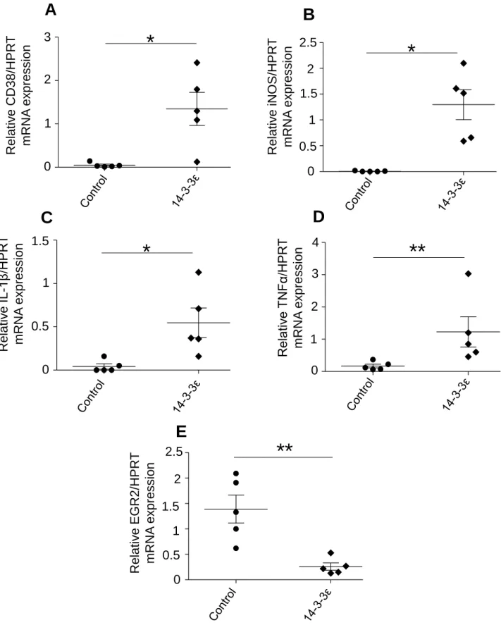
Fig. S1: Stimulation of M1 markers (iNOS, CD38, IL-1β, TNFα) and decrease of M2 3
marker (EGR2) in response to 14-3-3ε treatment murine bone marrow macrophages. 4
Murine BMM were stimulated with 14-3-3ε (1 µg/ml). Total RNA was extracted and 5
mRNA levels of CD38 (A) (n=5), iNOS (B) (n=5), IL-1β (C) (n=5), TNFα (D) (n=5) and 6
EGR2 (E) (n=5) were determined by qRT-PCR. Bars show the mean ± s.e.m. (Mann 7
Whitney test) *P<0.05; **P<0.01; ***P<0.001; ns: not significant. mRNA expression 8
levels of CD38, iNOS, IL-1β and TNFα showed significant fold increase of 28; 216; 9
13 and 7.3 respectively compared with the controls which show the property of 14-3-10
3ε to induce the expression of BMM type M1 genes markers. Moreover, mRNA 11
expression levels of EGR2, a M2 phenotype marker, was significantly reduced (5.3-12
fold decrease) after a 14-3-3ε stimulation compared to the BMM controls. 13
14
Fig. S2: Assessment of 14-3-3ε recombinant protein proper effect by inhibiting 15
endotoxins with polymyxin B on murine articular chondrocytes. 16
Murine articular chondrocytes cultures were stimulated with 14-3-3ε (1 µg/ml) or LPS 17
(10 ng/ml) with or without PMB (30 µg/ml). Total RNA was extracted and mRNA 18
levels of MMP13 was determined by qRT-PCR (A) (n=3). Protein level of MCP1 in 19
cell supernatants was measured by ELISA (B) (n=3). Bars show the mean ± s.e.m. 20
(One-way ANOVA with a Bonferroni post test) *P<0.05; **P<0.01; ***P<0.001; ns: not 21
significant. Treatment with PMB inhibited MMP13 mRNA expression (15% compared 22
to 14-3-3ε stimulation), MCP1 release (1% compared to 14-3-3ε stimulation) whereas 23
MMP13 mRNA expression and MCP1 protein release in response to LPS stimulation 24
33
were totally inhibited by PMB. This result show that recombinant 14-3-3ε has its self-1
effect independent from endotoxins. 2
0 1 2 3 4 5 0 20 40 60 80 100
Figure 1
*
IL 6 rele a se ( ng /mL ) Control 14-3-3ε 1 µg/ml*
M CP1 rele a se ( ng /mL ) Control 14-3-3ε 1 µg/ml 100 80 60 40 20 0 5 4 3 2 1 0B
A
Figure 2
0 2 4 6 Control 14-3-3ε 1 µg/ml*
Rela tiv e IL 6 /1 8 S m R N A ex pressionA
6 4 2 0 0 10 20 30 40 50 IL 6 rele a se ( ng /mL )C
Control 14-3-3ε 1 µg/ml*
10 0 50 30 20 40 0 20 40 60 80 M CP1 rele a se ( ng /mL )D
20 0 60 40 80*
Control 14-3-3ε 1 µg/ml 0 2 4 6 Rela tiv e M CP1 /1 8 S m RN A e x p ression 6 4 2 0B
*
Control 14-3-3ε 1 µg/ml0.0 0.2 0.4 0.6 0.8 1.0
Figure 3
**
Rela tiv e IL 6 /1 8 S m RN A e x p ression Control 14-3-3ε 1 µg/mlA
0 5 10 15 IL 6 rele ase ( ng /mL ) Control 14-3-3ε 1 µg/ml 5 0 15 10**
D
F
1 0 0.6 0.2 0.4 0.8 0.0 0.2 0.4 0.6 0.8 1.0**
Control 14-3-3ε 1 µg/ml Rela tiv e M CP1 /1 8 S m RN A e x p ressionB
1 0 0.6 0.2 0.4 0.8 0.0 0.5 1.0 1.5 Re la tiv e CD3 8 /1 8 S m RNA e x p ression**
Control 14-3-3ε 1 µg/mlC
0 0.5 1 1.5 0 200 400 600 M CP1 rele a se ( ng /mL ) 200 0 400 600 Control 14-3-3ε 1 µg/ml**
E
0.0 0.5 1.0 1.5 T NF α rele a se ( ng /mL ) 0.5 0 1 1.5 Control 14-3-3ε 1 µg/ml**
0 1000 2000 3000 4000 5000
W T
TLR2 KO
TLR4 KO
0 1000 2000 3000 4000W T
TLR2 KO
TLR4 KO
Figure 4
A
IL 6 rele a se ( ng /mL ) 1 0 2 4 3 Control 14-3-3ε 1 µg/ml WT TLR2 KO TLR4 KO*** ***
M CP1 rele a se ( ng /mL ) 2 0 3 5 4B
Control 14-3-3ε 1 µg/ml 1 P>0.05 WT TLR2 KO TLR4 KO-0.5 0.0 0.5 1.0 1.5 -0.5 0.0 0.5 1.0 1.5 -0.5 0.0 0.5 1.0 1.5 -0.5 0.0 0.5 1.0 1.5
Figure 5a
***
**
***
M M P3 rel eas e (i nduc ti on) 0.5 0 1 1.5D
***
***
***
R el ativ e M M P -3/HP R T m R N A ex pressi on ( ind uctio n)A
0.5 0 1 1.5***
***
*** ***
*
Rel at iv e M M P -3/HP R T m R N A ex pressi on ( ind uctio n)C
0.5 0 1 1.5*** *** ***
***
**
***
M M P3 rel eas e (i nduc ti on)F
0.5 0 1 1.5 -0.5 -0.5 -0.5 0.0 0.5 1.0 1.5***
***
***
B
0.5 0 1 1.5 R el ativ e M M P -3/HP R T m R N A ex pressi on ( ind uctio n) -0.5 -0.5 0.0 0.5 1.0 1.5 0.5 0 1 1.5 M M P 3 rel ea se (i nd uctio n)***
***
***
E
-0.5 -0.5 -0.5 14-3-3ε 1µg/ml α -T LR 4 1 µg /mL none none α -T LR 4 5 µg /mL 14-3-3ε 1µg/ml α -T LR4 1 µg /mL none none α -T LR 4 5 µg /mL 14-3-3ε 1µg/ml α -T LR 2 1 µg /mL none none α -T LR 2 5 µg /mL 14-3-3ε 1µg/ml α -T LR 2 1 µg /mL none none α -T LR 2 5 µg /mL 14-3-3ε 1µg/ml O xP A P C 0, 3 µ g/ mL none none O xP A P C 3 µ g/ mL O xPAP C 30 µ g/ mL 14-3-3ε 1µg/ml O xP A P C 0, 3 µg/ mL none none OxP A P C 3 µ g /mL OxP A P C 3 0 µ g /mLFigure 5b
-0.5 0.0 0.5 1.0 1.5***
***
**
R el ativ e IL6 /H P R T m R N A ex pressi on ( ind uctio n)G
0.5 0 1 1.5 -0.5 14-3-3ε 1µg/ml α -T LR 4 1 µg /mL none none α -T LR 4 5 µg /mL -0.5 0.0 0.5 1.0 1.5***
***
***
IL6 r el ea se (i nd uctio n)J
0.5 0 1 1.5 -0.5 14-3-3ε 1µg/ml α -T LR 4 1 µg /mL none none α -T LR 4 5 µg /mL -0.5 0.0 0.5 1.0 1.5***
*
R el ativ e IL6 /H P R T m R N A ex pr ess ion (i nduc ti on) 0.5 0 1 1.5H
-0.5 14-3-3ε 1µg/ml α -T LR 2 1 µg /mL none none α -T LR 2 5 µg /mL -0.5 0.0 0.5 1.0 1.5***
***
***
IL6 r el ea se (i nd uctio n)K
0.5 0 1 1.5 -0.5 14-3-3ε 1µg/ml α -T LR 2 1 µg /mL none none α -T LR 2 5 µg /mL -0.5 0.0 0.5 1.0 1.5***
***
***
R el ativ e IL6 /H P R T m R N A ex pressi on ( ind uctio n)I
0.5 0 1 1.5***
-0.5 14-3-3ε 1µg/ml O xPAP C 0, 3 µ g/ mL none none O xPAP C 3 µ g/ mL O xP A P C 30 µg/ mL -0.5 0.0 0.5 1.0 1.5***
***
***
*
***
**
IL6 r el ea se ( ind uc ti on )L
0.5 0 1 1.5 -0.5 14-3-3ε 1µg/ml O xP A P C 0, 3 µ g/ mL none none O xP A P C 3 µ g/ mL O xPAP C 30 µ g/ mLFigure 6
R el ati v e IL6 /H P R T m R N A ex pressi on** ***
Control 14-3-3ε 1 µg/ml 20 0 60 40 80 100 WT TLR2 KO TLR4 KO 500 0 1000 1500** ***
Control 14-3-3ε 1 µg/ml R el ativ e M M P 3/HP R T m R N A ex pressi on WT TLR2 KO TLR4 KOC
D
R el ativ e M M P 13 /H P R T m R N A ex pressi on** ***
20 0 60 40 80 100 Control 14-3-3ε 1 µg/ml WT TLR2 KO TLR4 KO WT TLR2 KO TLR4 KO IL 6 rele a se ( ng /mL ) 10 0 30 20 40 Control 14-3-3ε 1 µg/mlA
B
0 1 2 3 Rela tiv e CD 3 8 /HP R T m RN A e x p ression 0
A
1 2 3*
0.0 0.5 1.0 1.5 2.0 2.5*
0 2 2.5 1 0.5 1.5 Rela tiv e iN OS /HP R T m RN A e x p ressionB
Rela tiv e IL -1 β /HP R T m R N A e x p re ssio n 1.5 1 0.5 0C
0.0 0.5 1.0 1.5*
0 1 2 3 4 Rela tiv e T NF α/ HPR T m R N A ex pression 4 2 1 0D
3**
0.0 0.5 1.0 1.5 2.0 2.5 Rela tiv e E GR2 /HP R T m RN A e x p ression**
2.5 1.5 1 0.5 0E
2Fig. S1: Stimulation of M1 markers (iNOS, CD38, IL-1β, TNFα) and decrease of M2 marker (EGR2) in response to 14-3-3ε treatment murine bone marrow macrophages. Murine BMM were stimulated with 14-3-3ε (1 µg/ml). Total RNA was extracted and mRNA levels of CD38 (A) n=5, iNOS (B) n=5, IL-1β (C) n=5, TNFα (D) n=5 and EGR2 (E) n=5 were determined by qRT-PCR. Bars show the mean ± s.e.m. (Mann Whitney test) *P<0.05; **P<0.01; ***P<0.001; ns : not significant.