HAL Id: hal-03246636
https://hal.sorbonne-universite.fr/hal-03246636
Submitted on 2 Jun 2021
HAL is a multi-disciplinary open access
archive for the deposit and dissemination of
sci-entific research documents, whether they are
pub-lished or not. The documents may come from
teaching and research institutions in France or
abroad, or from public or private research centers.
L’archive ouverte pluridisciplinaire HAL, est
destinée au dépôt et à la diffusion de documents
scientifiques de niveau recherche, publiés ou non,
émanant des établissements d’enseignement et de
recherche français ou étrangers, des laboratoires
publics ou privés.
registry
Josefine Baekgaard, Paer-Selim Abback, Marouane Boubaya, Jean-Denis
Moyer, Delphine Garrigue, Mathieu Raux, Benoit Champigneulle, Guillaume
Dubreuil, Julien Pottecher, Philippe Laitselart, et al.
To cite this version:
Josefine Baekgaard, Paer-Selim Abback, Marouane Boubaya, Jean-Denis Moyer, Delphine Garrigue,
et al.. Early hyperoxemia is associated with lower adjusted mortality after severe trauma: results from
a French registry. Critical Care, BioMed Central, 2020, 24 (1), pp.604. �10.1186/s13054-020-03274-x�.
�hal-03246636�
R E S E A R C H
Open Access
Early hyperoxemia is associated with lower
adjusted mortality after severe trauma:
results from a French registry
Josefine S. Baekgaard
1,2*, Paer-Selim Abback
3, Marouane Boubaya
4, Jean-Denis Moyer
3, Delphine Garrigue
5,
Mathieu Raux
6, Benoit Champigneulle
7, Guillaume Dubreuil
8, Julien Pottecher
9, Philippe Laitselart
10,
Fleur Laloum
11, Coralie Bloch-Queyrat
4, Frédéric Adnet
1, Catherine Paugam-Burtz
3and Traumabase® Study Group
Abstract
Background: Hyperoxemia has been associated with increased mortality in critically ill patients, but little is known
about its effect in trauma patients. The objective of this study was to assess the association between early
hyperoxemia and in-hospital mortality after severe trauma. We hypothesized that a PaO
2≥ 150 mmHg on admission
was associated with increased in-hospital mortality.
Methods: Using data issued from a multicenter prospective trauma registry in France, we included trauma patients
managed by the emergency medical services between May 2016 and March 2019 and admitted to a level I trauma
center. Early hyperoxemia was defined as an arterial oxygen tension (PaO
2) above 150 mmHg measured on hospital
admission. In-hospital mortality was compared between normoxemic (150 > PaO
2≥ 60 mmHg) and hyperoxemic
patients using a propensity-score model with predetermined variables (gender, age, prehospital heart rate and
systolic blood pressure, temperature, hemoglobin and arterial lactate, use of mechanical ventilation, presence of
traumatic brain injury (TBI), initial Glasgow Coma Scale score, Injury Severity Score (ISS), American Society of
Anesthesiologists physical health class > I, and presence of hemorrhagic shock).
Results: A total of 5912 patients were analyzed. The median age was 39 [26
–55] years and 78% were male. More
than half (53%) of the patients had an ISS above 15, and 32% had traumatic brain injury. On univariate analysis, the
in-hospital mortality was higher in hyperoxemic patients compared to normoxemic patients (12% versus 9%, p <
0.0001). However, after propensity score matching, we found a significantly lower in-hospital mortality in
hyperoxemic patients compared to normoxemic patients (OR 0.59 [0.50
–0.70], p < 0.0001).
Conclusion: In this large observational study, early hyperoxemia in trauma patients was associated with reduced
adjusted in-hospital mortality. This result contrasts the unadjusted in-hospital mortality as well as numerous other
findings reported in acutely and critically ill patients. The study calls for a randomized clinical trial to further
investigate this association.
Keywords: Hyperoxemia, Hyperoxia, Trauma, Critical care, Oxygen
© The Author(s). 2020 Open Access This article is licensed under a Creative Commons Attribution 4.0 International License, which permits use, sharing, adaptation, distribution and reproduction in any medium or format, as long as you give appropriate credit to the original author(s) and the source, provide a link to the Creative Commons licence, and indicate if changes were made. The images or other third party material in this article are included in the article's Creative Commons licence, unless indicated otherwise in a credit line to the material. If material is not included in the article's Creative Commons licence and your intended use is not permitted by statutory regulation or exceeds the permitted use, you will need to obtain permission directly from the copyright holder. To view a copy of this licence, visithttp://creativecommons.org/licenses/by/4.0/. The Creative Commons Public Domain Dedication waiver (http://creativecommons.org/publicdomain/zero/1.0/) applies to the data made available in this article, unless otherwise stated in a credit line to the data.
* Correspondence:josefinebeakgaard@me.com
1Urgences et Samu 93, AP-HP, Avicenne Hospital, Inserm U942, 93000
Bobigny, France
2Department of Anesthesia, Section 4231, Centre of Head and Orthopedics,
Rigshospitalet, University of Copenhagen, Juliane Maries Vej 10, DK-2100 Copenhagen, Denmark
Introduction
Each year, 5.8 million people die as result of trauma
making it the leading cause of death for individuals
below 45 years of age [
1
]. Furthermore, trauma
consti-tutes a major economic burden, as trauma-related costs
were estimated to $671 billion in 2013 in the USA alone
[
2
]. Efforts to lower the mortality and morbidity
follow-ing trauma are therefore of highest importance. The
pre-hospital management of severe trauma patients requires
a rapid approach during which it is recommended to
provide supplemental oxygen to both treat and prevent
hypoxemia [
3
,
4
]. As a result, high fractions of inspired
oxygen (FiO
2) are commonly administered during this
initial phase and may result in hyperoxemia on hospital
admission. However, exposure to high oxygen levels,
even during a short period of time, has been associated
with cerebral and coronary vasoconstriction, deleterious
effects on lung function, and increased production of
re-active oxygen species [
5
–
10
].
In a large meta-analysis on randomized controlled
trials (RCT), which compared liberal and
conserva-tive oxygenation administration in acutely ill
pa-tients, the relative risk of in-hospital mortality was
increased amongst patients treated with a liberal
oxygen approach compared to a conservative oxygen
approach [
11
]. A recent systematic review also
inves-tigated the relationship between hyperoxemia and
mortality in critically ill patients and found a similar
association [
12
].
Despite an increasing awareness of the potentially
deleterious effects of elevated arterial oxygen partial
pressure (PaO
2) in acutely ill patients [
13
], the
preva-lence of hyperoxemia in the emergency department (ED)
and the intensive care unit (ICU) remains high [
14
–
16
].
Furthermore, a recent cohort study found a link between
early hyperoxemia in the ED and mortality [
14
].
However, the association between hyperoxemia and
mortality in the trauma population remains
controver-sial. In one RCT, authors found no effect of exposure to
different levels of FiO
2on mortality amongst patients
suffering from traumatic brain injury (TBI) [
17
]. A
re-cent observational study on 24,148 mechanically
venti-lated patients with TBI found no effects of hyperoxemia
on mortality either [
18
].
Taken as a whole, knowledge on the effects of
hyperoxemia in trauma patients is sparse and the
evidence for systematic oxygen therapy in these
pa-tients is thus inadequate, especially in the pre-hospital
setting [
19
].
The primary objective of this study was to assess the
association between elevated PaO
2on hospital admission
and in-hospital mortality in level I trauma centers. We
hypothesized that a PaO
2≥ 150 mmHg on admission was
associated with increased in-hospital mortality.
Methods
Study design
This was an observational study using a multicenter,
prospective trauma registry in France, the TraumaBase©.
The TraumaBase consecutively collects data on trauma
patients from 15 trauma centers in France. A central
ad-ministrator monitors the data and the TraumaBase is
approved by the Institutional Review Board as well as
the National Commission on Informatics and Liberties.
The study is reported in accordance with the STROBE
guidelines [
20
].
Setting
Between May 2016 and March 2019, data collected from
the 14 level 1 trauma centers was reviewed (one center
had not yet included patients). As previously described
[
21
], the French EMS system consists of two levels of
tri-age that will trigger a paramedic-staffed ambulance or a
physician-staffed mobile ICU (Service Mobile d’Urgence
et de Réanimation
(SMUR)). In case of major trauma,
the SMUR will always be activated and accompany the
patient to a specialized trauma center.
Participants
Trauma patients above 17 years of age with a PaO
2measured and registered in the TraumaBase® registry
were included. Hypoxemic patients (PaO
2< 60 mmHg
on arrival) and patients withdrawn from life-sustaining
therapy were excluded. Baseline characteristics on
hyp-oxemic patients can be found in the Additional file
1
.
Variables
The following variables were extracted from the
data-base: age (years), gender, American Society of
Anesthesi-ologists (ASA) score, initial Glasgow Coma Scale (GCS)
score, pre-hospital systolic blood pressure and heart rate,
mechanism and site of injury, volume fluid replacement
(mL of colloids and/or crystalloids), catecholamine
administration, use of mechanical ventilation, body
temperature, arterial blood gas analysis on admission,
lactate level, hemoglobin level, creatinine level, presence
of hemorrhagic shock (defined as at the transfusion of at
least four units of packed red blood cells within 6 h),
TBI (at least one visible lesion on computed
tomog-raphy), Injury Severity Score (ISS), in-hospital length of
stay, and in-hospital mortality.
Statistical methods
Patients were divided into two groups of exposure a
priori according to their initial PaO
2on hospital
ad-mission:
normoxemia
(PaO
260–150 mmHg) and
hyperoxemia
(PaO
2≥ 150 mmHg). The 150 mmHg
an RCT on ICU patients [
22
], as well as in several
observational studies [
23
–
25
].
Our primary aim was to assess the correlation between
hyperoxemia on hospital admission and in-hospital
mor-tality. Two pre-planned subgroup analyses on patients
with an initial GCS < 8 and mechanically ventilated
pa-tients were also planned.
Categorical variables are expressed as numbers with
percentages (%) and continuous variables as means with
standard deviations (SD), or medians with interquartile
ranges [IQR]. Characteristics were compared using a
chi
2test for categorical data and t test or
Mann-Whitney U test for continuous data.
Since hyperoxemia is caused by exposure to high
oxy-gen levels, the association between hyperoxemia and
in-hospital mortality was assessed using propensity score to
reduce potential selection bias due to measured baseline
covaries. The variables included in the model were
chosen a priori by comparing pre-hospital variables and
baseline characteristics between patients that died and
survived to hospital discharge. Significant determinants
of mortality were included.
The score was estimated using logistic regression, and
the primary analyses were made using inverse
probabil-ity of treatment weighting (IPTW).
To verify the robustness of the results, two sensitivity
analyses were performed using a propensity score analysis
with a matching method with a 1:1 ratio within a caliper
of 0.05 standard deviation of the logit propensity score
and a stratification on the quintiles of the propensity
score. To account for missing data, analyses were
con-ducted using multiple imputations by chained equations
with 10 imputations obtained after 10 iterations [
26
]. A
complete-case analysis was also performed to verify the
results. The propensity scores came from 10 independent
complete data sets and were averaged according to
the
“across approach” [
27
]. Balance in potentials
con-founders were assessed by standardized mean differences
which came from a complete imputed data set [
28
]. A
multivariate full model including factors used in the
pro-pensity score was also performed to verify the results of
the propensity score.
Finally, several sensitivity analyses were performed.
An analysis removing patients who died within 24 h of
hospital admission was carried out to allow sufficient time
for deleterious effects such as lung complications of
oxy-gen to develop, and an analysis on patients with a GCS < 8
as well as an analysis on intubated patients was done.
Fur-thermore, other cutoffs for hyperoxemia were examined
(PaO
2≥ 100 mmHg and PaO
2≥ 200 mmHg), and the
PaO
2/FiO
2was explored using the Berlin definition [
29
].
All tests were two-tailed, and the results were
consid-ered statistically significant when p < 0.05. Analyses were
performed using R statistical software [
30
].
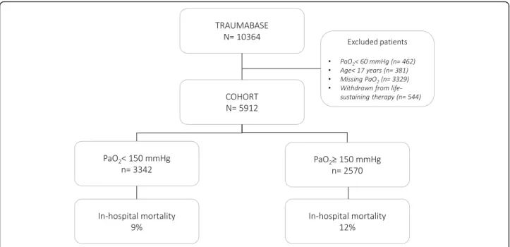
Results
Of 6654 adult trauma patients with PaO
2values available
in the database, 544 were excluded as they were
with-drawn from life-sustaining therapy and 462 were
ex-cluded as they were hypoxemic on arrival, leaving 5912
patients for analysis (Fig.
1
).
The median age was 39 years and the majority were
males (Table
1
). More than half of all patients had an
ISS score above 15, and one third presented with TBI.
The overall in-hospital mortality was 10%.
On hospital admission, the median PaO
2of the
en-tire
cohort
was
133 mmHg:
3342
(57%)
were
normoxemic, and 2570 (43%) were hyperoxemic.
Numerous baseline characteristics were significantly
different
between
normoxemic
and
hyperoxemic
patients: a higher proportion of hyperoxemic patients
were
mechanically
ventilated
(a
comparison
of
baseline characteristics between intubated and
spon-taneously
breathing
patients
can
be
found
in
Additional file
2
), they had lower prehospital GCS
scores and more suffered from a TBI. On univariate
analysis, the in-hospital mortality was higher for
hyperoxemic patients (12% versus 9%, p < 0.0001)
(Table
1
).
In a propensity score model, patients were matched
based upon significant determinants of mortality
amongst the baseline characteristics (Table
2
). The
model revealed an inverse relationship between
hyper-oxemia
and
in-hospital
mortality:
mortality
was
significantly decreased in hyperoxemic patients
com-pared to normoxemic patients (OR 0.59 [0.50–0.70],
p
< 0.0001) and hyperoxemia thus appeared as a
pro-tective factor. The accuracy of the model is presented
in Fig.
2
. Here, the balances in potentials confounders
were also checked, and the absolute mean differences
were all less than 5% after using propensity score
(IPTW and matching methods). The multivariate full
model including factors used in propensity score
veri-fied
the
results
of
the
propensity
score
(Add-itional file
3
). A complete-case analysis presented very
similar results (OR 0.60 [0.46–0.78], p < 0.0001).
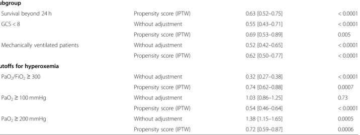
In a sensitivity analysis, where patients deceased within
24 h were excluded, the results remained statistically
sig-nificant (OR 0.63 [0.52–0.76], p < 0.0001) (Table
3
).
Likewise, in our subgroup analysis on patients with a
GCS < 8, mortality was also decreased in hyperoxemic
patients (OR 0.69 [0.53–0.89], p = 0.005). The same was
true in a subgroup analysis on intubated patients (OR
0.62 [0.50
–0.77], p < 0.0001) (Table
3
).
Furthermore, our sensitivity analyses also showed a
beneficial effect on mortality using PaO
2/FiO
2≥ 300, and
different cut-off levels for hyperoxemia (PaO
2≥ 100
mmHg and PaO
2≥ 200 mmHg) left our results largely
Discussion
In this large observational study of nearly 6000 trauma
patients, we found hyperoxemia above 150 mmHg on
hospital admission to be independently associated with a
significantly decreased in-hospital mortality compared to
normoxemia. This result challenges our initial
hypoth-esis. Our results were unaltered by a sensitivity analysis,
where patients deceased within 24 h were excluded.
The beneficial effects of supplemental oxygen for the
critically ill patient have remained undisputed for
decades and have resulted in international guidelines on
initial
trauma
management
recommending
high
fractions of inspired oxygen. However, although it is
suspected that hyperoxemia may be deleterious (due to
increased oxidative stress, vasoconstriction, and potential
hyperoxemic lung injury) [
31
], the evidence both in
favor and against supplemental oxygen, and thus the risk
of hyperoxemia, is almost non-existent in trauma
pa-tients [
19
].
Supplemental oxygen seems to possess a potential to
rescue threatened neurons after brain injury or in the
is-chemic penumbra [
32
,
33
], and it is known to prolong
the safe apnea time [
34
]. Nevertheless, numerous
physio-logic arguments exist against liberal administration of
oxygen in critically ill patients. For example, excess
oxy-gen has been associated with the formation of reactive
oxygen species which are detoxified in the mitochondria
by a variety of antioxidants. Furthermore, acute states
such as shock induce an increased production of reactive
oxygen species worsening the imbalance between
pro-oxidants and antipro-oxidants [
6
].
In recent years, the optimal targets of both SpO
2and
PaO
2have therefore been challenged in acutely ill
pa-tients. A large meta-analysis showed increased rates of
mortality for patients with oxygen saturation (SpO
2)
above 96% compared to 94–96% [
11
]. However, as the
trial sequential analysis was driven primarily by a single
large randomized trial [
35
], the authors were unable to
exclude a small beneficial effect of liberal oxygen. Only
one RCT on trauma patients was included, and here no
effect of liberal oxygen was observed. Another
meta-analysis on patients with cardiac arrest showed beneficial
effects of oxygen intra-arrest while post-arrest arrest
hyperoxemia was associated with increased mortality
[
36
]. A recent systematic review found a higher all-cause
mortality in ICU patient with hyperoxemia [
25
];
however, in subgroup analyses on patients with TBI and
patients
on
mechanical
ventilation,
results
were
inconclusive.
In trauma patients, studies on liberal versus
conserva-tive oxygen approaches are sparse. To date, only two
small RCTs have been done on patients with TBI, and
here, one found difference between a liberal and
restrict-ive oxygen approach on mortality [
17
], and the other
found no differences in terms of neurological outcome
[
37
]. Furthermore, the few retrospective studies available
have shown inconsistent results: one recent large study
showed no difference in in-hospital mortality between
hyperoxemic and normoxemic trauma patients [
18
],
others have shown a deleterious effect of hyperoxemia
[
38
,
39
], and yet two studies have found a strong
rela-tionship between hyperoxemia and better long-term,
Table 1 Baseline characteristics of all included trauma patients including a comparison of norm- and hyperoxemic patients. Results
are presented as medians with [interquartile ranges], numbers with (percentages), or as otherwise indicated
All patients Normoxemic 60 < PaO2< 150 mmHg Hyperoxemic PaO2≥ 150 mmHg p value N = 5912 n = 3342 n = 2570 Age 39 [26–55] 41 [17–96] 36 [17–96] < 0.0001 Sex (female) 1273 (21.6) 703 (21.1) 570 (22.3) 0.3 ASA-score > 1 1903 (34.5) 1168 (37.0) 735 (31.1) < 0.0001 Mechanism of injury
Falls from height 1368 (21.5) 747 (22.4) 521 (20.3) 0.089
Falls from standing 240 (4.1) 132 (4.0) 108 (4.2)
Vehicle incident/collision 3339 (56.5) 1895 (56.7) 1444 (56.2)
Shootings 590 (10.0) 211 (9.3) 279 (10.9)
Fight 204 (3.5) 117 (3.5) 87 (3.4)
Other 270 (4.6) 139 (4.2) 131 (5.1)
Site of injury
Head and neck 2823 (51.1) 1461 (46.9) 1362 (64.4) < 0.0001
Face 1389 (25.1) 707 (22.7) 682 (28.3) < 0.0001
Abdomen 1833 (33.2) 1022 (32.8) 811 (33.6) 0.56
Chest 2865 (51.8) 1647 (52.9) 1218 (50.5) 0.079
External 924 (16.7) 533 (17.1) 391 (16.2) 0.39
Extremities 3080 (55.7) 1684 (54.1) 1396 (57.9) 0.0055
Duration of prehospital care (minutes), median [IQR] 70 [48–100] 79 [49–97] 70 [45–105] 0.58 Prehospital systolic blood pressure (mmHg) 127 [110–141] 130 [0–256] 124 [0–230] < 0.0001 Prehospital heart rate (bpm) 89 [75–105] 88 [0–170] 76 [0–155] < 0.0001 Prehospital intubation 1840 (31.7) 651 (19.9) 1189 (47.0) < 0.0001 Prehospital GCS score 15 [11–15] 15 [3–15] 14 [3–15] < 0.0001 Values on hospital arrival
pH 7.4 [7.3–7.4] 7.4 [7.3–7.4] 7.3 [7.3–7.4] < 0.0001 PaO2 133 [93–216] 97 [81–117] 230 [186–308] – PCO2 40 [35–44] 39 [35–43] 40 [26–45] < 0.0001 Temperature (°C) 36.5 [35.9–37.0] 36.6 [26.4–41.0] 36.4 [26.0–40] < 0.0001 Lactate (mmol/L) 1.9 [1.2–3.0] 1.8 [0.2–23.4] 2 [0.2–25] < 0.0001 Creatinine (μmol/L) 77 [65–92] 77 [8–1004] 77 [7–926] 0.64 Hemoglobin (mmol/L) 13 [11.5–14.2] 13.3 [3.9–21.6] 12.6 [1.1–20.0] < 0.0001 Catecholamine administration 815 (14.3) 322 (10.0) 493 (19.9) < 0.0001 Fluid replacement 500 [250–1000] 500 [0–7000] 750 [0–5500] < 0.0001 ISS score 16 [9–25] 13 [8–24] 18 [10–27] < 0.0001 ISS score > 15 2935 (52.9) 1433 (45.9) 1502 (62.0) < 0.0001 Traumatic brain injury 1836 (31.6) 824 (25.1) 1012 (40.1) < 0.0001
Hemorrhagic shock 545 (9.4) 202 (6.2) 343 (13.6) < 0.0001
In-hospital mortalitya 481 (10.0) 239 (8.7) 242 (11.6) < 0.0001
Cause of death (available for 426 patients) < 0.01
Hemorrhagic shock 46 (10.8) 21 (10.0) 25 (11.5)
Septic chock 6 (1.4) 3 (1.4) 3 (1.4)
Multi organ failure 98 (23.0) 59 (28.2) 39 (18.0)
functional, and cognitive outcomes [
24
,
40
]. As such the
physiologic consequences of hyperoxemia on outcomes
after TBI remain uncovered. In several studies, a
de-crease in cerebral perfusion of up to 30% has been
ob-served in individuals exposed to hyperoxia [
41
–
43
],
while other studies have suggested that hyperoxia aids in
one of the cornerstones in treatment of traumatic brain
injury: decreasing intracranial pressure [
44
–
46
].
Supple-mental oxygen could also be beneficial in TBI by simply
increasing the level of oxygen in the brain. In stroke
pa-tients, supplemental oxygen has been proposed to rescue
threatened neurons, and thus the brain, from further
de-terioration [
47
]. Nonetheless, studies so far have failed
to show an association between supplemental oxygen
and improved physical function [
35
,
48
]. Further
re-search in larger cohorts should look into this to help
un-cover the induced pathways.
In accordance with several of the above studies, we
found a clinical benefit of early hyperoxemia in the
current study. Of note, however, all the latter studies
focus solely on trauma patients with TBI, whereas we
chose to include all trauma patients to present a broader
and more pragmatic perspective, as isolated TBI may
not always be evident in the acute phase. Nonetheless, in
our subgroup analysis of patients with GCS < 8, our
re-sults were unchanged.
Table 1 Baseline characteristics of all included trauma patients including a comparison of norm- and hyperoxemic patients. Results
are presented as medians with [interquartile ranges], numbers with (percentages), or as otherwise indicated (Continued)
All patients Normoxemic 60 < PaO2< 150 mmHg
Hyperoxemic PaO2≥ 150 mmHg
p value
N = 5912 n = 3342 n = 2570
Traumatic brian injury 58 (13.6) 26 (12.4) 32 (14.7)
Other 21 (4.9) 15 (7.1) 6 (2.8)
The provided pre-hospital vital signs are the first vital signs recorded on-scene
Abbreviations: ASA, American Society of Anesthesiologists; GCS, Glasgow Coma Scale score; ISS, Injury Severity Score; Hemorrhagic shock, defined as administration of at least four units of packed red blood cells within 6 h; Fluid replacement, mL of colloids and/or crystalloids
a
Missing in 18%. Imputated in the propensity score analysis
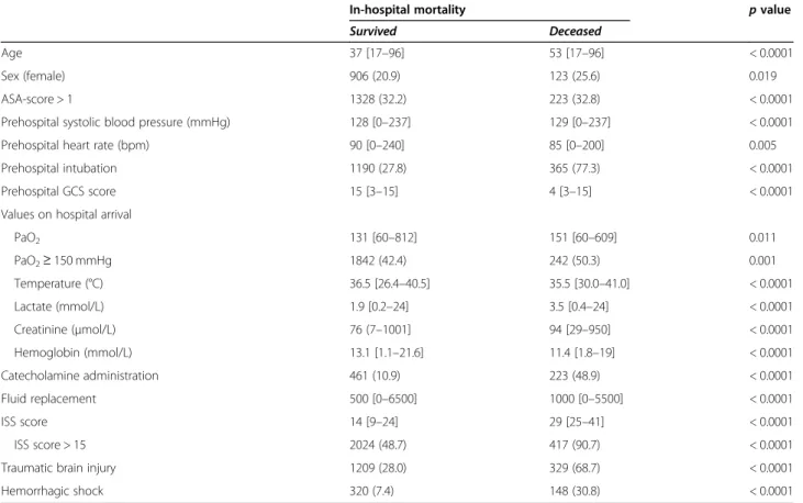
Table 2 Baseline differences amongst trauma patients that survived to hospital-discharge or died in-hospital. Results are presented
as medians with [interquartile ranges], numbers with (percentages), or as otherwise indicated
In-hospital mortality p value
Survived Deceased
Age 37 [17–96] 53 [17–96] < 0.0001
Sex (female) 906 (20.9) 123 (25.6) 0.019
ASA-score > 1 1328 (32.2) 223 (32.8) < 0.0001
Prehospital systolic blood pressure (mmHg) 128 [0–237] 129 [0–237] < 0.0001
Prehospital heart rate (bpm) 90 [0–240] 85 [0–200] 0.005
Prehospital intubation 1190 (27.8) 365 (77.3) < 0.0001
Prehospital GCS score 15 [3–15] 4 [3–15] < 0.0001
Values on hospital arrival
PaO2 131 [60–812] 151 [60–609] 0.011 PaO2≥ 150 mmHg 1842 (42.4) 242 (50.3) 0.001 Temperature (°C) 36.5 [26.4–40.5] 35.5 [30.0–41.0] < 0.0001 Lactate (mmol/L) 1.9 [0.2–24] 3.5 [0.4–24] < 0.0001 Creatinine (μmol/L) 76 (7–1001] 94 [29–950] < 0.0001 Hemoglobin (mmol/L) 13.1 [1.1–21.6] 11.4 [1.8–19] < 0.0001 Catecholamine administration 461 (10.9) 223 (48.9) < 0.0001 Fluid replacement 500 [0–6500] 1000 [0–5500] < 0.0001 ISS score 14 [9–24] 29 [25–41] < 0.0001 ISS score > 15 2024 (48.7) 417 (90.7) < 0.0001
Traumatic brain injury 1209 (28.0) 329 (68.7) < 0.0001
Hemorrhagic shock 320 (7.4) 148 (30.8) < 0.0001
Abbreviations: ASA, American Society of Anesthesiologists; GCS, Glasgow Coma Scale score; Hemorrhagic shock, defined as administration of at least four units of packed red blood cells within 6 h; Fluid replacement, mL of colloids and/or crystalloids
Fig. 2 In-hospital mortality in normoxemic (60 mmHg <PaO2< 150 mmHg) and hyperoxemic (PaO2> 150 mmHg) trauma patients using a
propensity score model
Table 3 Sensitivity analyses. In-hospital mortality amongst subgroups of trauma patients (reference: Normoxemia)
Subgroup
Survival beyond 24 h Propensity score (IPTW) 0.63 [0.52–0.75] < 0.0001
GCS < 8 Without adjustment 0.55 [0.43–0.71] < 0.0001
Propensity score (IPTW) 0.69 [0.53–0.89] 0.005 Mechanically ventilated patients Without adjustment 0.52 [0.42–0.65] < 0.0001
Propensity score (IPTW) 0.62 [0.50–0.77] < 0.0001 Cutoffs for hyperoxemia
PaO2/FiO2≥ 300 Without adjustment 0.32 [0.27–0.38] < 0.0001
Propensity score (IPTW) 0.74 [0.62–0.88] 0.0007
PaO2≥ 100 mmHg Without adjustment 1.03 [0.86–1.25] 0.73
Propensity score (IPTW) 0.54 [0.46–0.64] < 0.0001
PaO2≥ 200 mmHg Without adjustment 1.38 [1.15–1.65] 0.0005
Propensity score (IPTW) 0.72 [0.59–0.87] 0.0006
The comparison of studies on hyperoxemia is difficult
as some studies compare SpO
2values, others FiO
2values, and others PaO
2values. Besides, when utilizing
the PaO
2,there is no consensus on the arbitrarily
prede-termined PaO
2cut-off [
25
]. In the current study, we
chose to use 150 mmHg as the threshold for
hyperoxe-mia as it presented a large percentage of our population
(43%), and in addition, this approach has been used
pre-viously [
22
–
25
]. Numerous other studies have chosen
values
above
300 mmHg
to
present
hyperoxemia,
thereby considering values below 300 mmHg as
nor-moxemic, which appears problematic. Furthermore,
many studies have used the worst PaO
2(the highest
PaO
2) as their exposure variable [
49
,
50
]. We chose to
use the first PaO
2recorded at hospital admission to
re-flect the pre-hospital treatment. This has previously been
done [
38
,
51
]. Finally, the exposure duration should also
be taken into account. As such, the attempt to answer
whether or not hyperoxemia is harmful—in any patient
population—should always aim to consider the variable
measured (SpO
2, PaO
2or FiO
2), the concentration of
the given variable, and the exposure duration.
Our results reflect the liberal use of pre-hospital
oxy-gen administration of severe trauma patients, and we
found a high percentage (43%) of patients with
hyperox-emia at hospital admission. Although the duration of
hyperoxemia in our study must be assumed to be
rela-tively short (mean prehospital time from trauma until
admission of 70 min), several studies have found that
deleterious effects of hyperoxemia may occur already
during the first hours of administration. For example,
both human and animal data have shown development
of lung injury after just a few hours of exposure to
hyperoxemia [
10
,
22
,
52
]. Furthermore, prehospital
sup-plemental oxygen administration for patients with
myo-cardial infarction has been associated with increased
myocardial injury and infarct size at 6 months [
53
], and
in a recent study, an association between an even shorter
exposure time to hyperoxia and mortality was found in
mechanically ventilated patients in the emergency
de-partment [
14
].
Nevertheless, in a recent small single center
observa-tional study, authors found no impact on 30-day
mortal-ity in trauma patients with early hyperoxemia [
54
], and
in our current study on trauma patients, we even found
a significant association between hyperoxemia on
admis-sion and decreased mortality compared to normoxemia
on admission. The threshold for potentially toxic
con-centrations and duration of administration of oxygen are
poorly defined, and the mechanisms behind a favorable
effect may, at least partly, be explained by hemodynamic
stabilization during shock, improvement in tissue bed
oxygenation in both peri-contusional and remote
neur-onal tissue, and more aerobic neural metabolic profiles
[
55
]. These could be some of the explanation behind a
positive effect of short-term hyperoxemia in the current
study along with the actual ability of the affected
individ-ual to increase their PaO
2as demonstrated in the PaO
2/
FiO
2sensitivity analysis. Regarding the exact threshold,
the current study also shows that this may point towards
mild hyperoxemia being the most beneficial, as the
bene-ficial effect seemed to decrease when a higher PaO
2was
used to define hyperoxemia.
Limitations
The primary limitation of the current study lies within
its retrospective design, where, for instance, missing data
often is seen. In our study, in-hospital mortality was
un-fortunately missing in 18%. Furthermore, the PaO
2value
was also missing in a substantial proportion of patients,
leaving these patients for exclusion. It is impossible to
know whether these were missing completely at random
or not. However, for a large proportion, they seem to be
missing completely at random, as other results of an
ar-terial blood gas were available. Nonetheless, the large
number of included patients allowed not only the
pro-pensity score analysis to include all the necessary
vari-ables for corrections but also important subgroup
analyses. One must, however, keep in mind that the risk
of hidden confounders still exists. Furthermore, although
the first PaO
2recorded at hospital admission partly
represents the prehospital management, the median of
several consecutive PaO
2’s may have provided a more
accurate picture. Moreover, in contrast to some other
retrospective studies, we chose not to include a
compari-son group of hypoxemic patients, as the deleterious
ef-fect of hypoxemia is well established. This allows a
cleaner comparison to the randomized trials available,
where randomization is aimed at normoxemia versus
hyperoxemia, thus not including a hypoxemic group.
We chose in-hospital mortality as our primary
out-come as this seemed to be the most patient centered
outcome available in the database. However, in future
studies, other outcomes such as lactate levels and
cat-echolamine administration could be interesting to look
at, to gain a deeper understanding of the resulting
physiological changes with different PaO
2levels.
Finally, the results of this study are based upon the
French pre-hospital system which is characterized by the
presence of emergency physicians in the field. The
char-acteristics of the patients and the nature of their initial
management can therefore not easily be extrapolated to
EMS systems in other countries. Our results must
there-fore be compared with other systems of prehospital care.
Conclusion
In the current study, we found early hyperoxemia in
se-vere trauma patients to be associated with a reduced
in-hospital mortality. This result may support systematic
administration of oxygen in trauma patients during the
initial management in the prehospital setting, but the
retrospective nature of the study warrants its careful
in-terpretation. The study calls for a randomized clinical
trial to further investigate this association.
Supplementary information
Supplementary information accompanies this paper athttps://doi.org/10. 1186/s13054-020-03274-x.
Additional file 1. Supplementary table on baseline characteristics for hypoxemic patients (PaO2< 60 mmHg).
Additional file 2. Supplementary table on baseline characteristics for intubated vs spontaneously breathing patients.
Additional file 3. Multivariate full model including factors used in propensity score.
Abbreviations
ASA score:American Society of Anesthesiologists score; ED: Emergency department; FiO2: High inspired concentrations of oxygen; GCS: Glasgow
Coma Scale; ICU: Intensive care unit; IPTW: Inverse probability of treatment weighting; IQR: Interquartile ranges; PaO2: Arterial oxygen partial pressure;
RCT: Randomized controlled trials; SD: Standard deviations; SMUR: Service Mobile d’Urgence et de Réanimation; SpO2: Oxygen saturation; TBI: Traumatic
brain injury Acknowledgements
Collaborating author names of the Traumabase® group: Romain Pirracchio
Service d’Anesthésie-réanimation, Hôpital Européen Georges Pompidou, Université Paris Descartes, Paris, France
Anne Godier
Service d’Anesthésie-réanimation, Hôpital Européen Georges Pompidou, Université Paris Descartes, Paris, France
Anatole Harrois
Université Paris Sud, Université Paris Saclay, Department of Anesthesiology and Critical Care, Assistance Publique-Hôpitaux de Paris (AP-HP), Bicêtre Hôpitaux Universitaires Paris Sud, 78 rue du Général Leclerc, 94275 Le Kremlin Bicêtre, F-94275, Le Kremlin Bicêtre, France
Thomas Geeraerts
Anesthesiology and Critical Care Department, University Hospital of Toulouse, University Toulouse 3-Paul Sabatier, Toulouse, France Eric Meaudre
Department of Anesthesiology and Intensive Care, Military Hospital, Hôpital d’Instruction des Armées Sainte-Anne, France and Emergency department, Military Hospital, Hôpital d’Instruction des Armées Sainte-Anne, France Sylvain Ausse
Department of Anesthesiology and Critical Care, Percy military hospital, Clamart, France
Tobias Gauss
Department of Anesthesia and Critical Care, Beaujon Hospital, AP-HP, University of Paris, Paris, France
Alain Meyer
Unités de Réanimation Chirurgicale et de Surveillance Continue, Hôpitaux Universitaires de Strasbourg, Strasbourg, France
Sophie Hamada
Université paris Sud, Université Paris Saclay, Department of Anesthesiology and Critical Care, Assistance Publique-Hôpitaux de Paris (AP-HP), Bicêtre Hôpitaux Universitaires Paris Sud, 78 rue du Général Leclerc, 94275 Le Kremlin Bicêtre, F-94275, Le Kremlin Bicêtre, France
Arthur Neuschwander
Service d’Anesthésie-réanimation, Hôpital Européen Georges Pompidou, Université Paris Descartes, Paris, France
Fabrice Cook
Department of Anaesthesia and Intensive Care Medicine, Henri Mondor University Hospital, Creteil, France
Helene Vinour
Anaesthesiology and Critical Care Department, University Hospital of Toulouse, University Toulouse 3-Paul Sabatier, Toulouse, France Jean Luc Hanouz
Department of Anesthesiology and Critical Care Medicine, Pôle Réanimations Anesthésie SAMU, Caen University Hospital, Caen, France
Arnaud Foucrier
Department of Anesthesia and Critical Care, Beaujon Hospital, AP-HP, University of Paris, Paris, France
Mathieu Boutonnet
Department of Anesthesiology and Critical Care, Percy military hospital, Clamart, France
Pascal Raclot
Reims University Hospital, Robert Debré Hospital, Intensive Care Unit, Reims, France
James Arthur
Sorbonne Université and Department of Anesthesiology and Critical Care, AP-HP, Hôpitaux Universitaires Pitié-Salpêtrière
Nathalie Bruneau
Pôle d’Anesthésie-Réanimation, CHU de Lille, Lille, France Jean Cotte
Department of Anesthesiology and intensive care, military hospital, Hôpital d’Instruction des Armées Sainte-Anne, France
Marc Leone
Department of Anesthesiology and Intensive Care Medicine, Aix-Marseille University, Assistance Publique Hôpitaux de Marseille, Hôpital Nord, Marseille, France
Gerard Audibert
Department of Anesthesiology and surgical Intensive Care, University Hospital of Nancy, Nancy, France
Others:
For ideas, input and constructive criticism Jacob Steinmetz
Department of Anesthesia, Centre of Head and Orthopedics, Rigshospitalet, University of Copenhagen, Denmark
Lars S. Rasmussen
Department of Anesthesia, Centre of Head and Orthopedics, Rigshospitalet, University of Copenhagen, Denmark
Authors’ contributions
All authors have contributed to the analysis design, manuscript conception and drafting, the statistical analysis, and/or editorial review. Individual contributions were as follows:
JSB:• Substantial contributions to the conception and design of the work and the acquisition, analysis, and interpretation of data for the work.• Revising it critically for important intellectual content.• Final approval of the version to be published.• Agreement to be accountable for all aspects of the work in ensuring that questions related to the accuracy or integrity of any part of the work are appropriately investigated and resolved. PA:• Substantial contributions to the conception and design of the work and the acquisition, analysis, and interpretation of data for the work.• Revising it critically for important intellectual content.• Final approval of the version to be published.• Agreement to be accountable for all aspects of the work in ensuring that questions related to the accuracy or integrity of any part of the work are appropriately investigated and resolved. MB:• Substantial contributions to the analysis and interpretation of data for the work.• Revising it critically for important intellectual content. • Final approval of the version to be published.• Agreement to be accountable for all aspects of the work in ensuring that questions related to the accuracy or integrity of any part of the work are appropriately investigated and resolved. JM:• Substantial contributions to the conception and design of the work and the acquisition, analysis, and interpretation of data for the work.• Revising it critically for important intellectual content.• Final approval of the version to be published.• Agreement to be accountable for all aspects of the work in ensuring that questions related to the accuracy or integrity of any part of the work are appropriately investigated and resolved. DG:• Substantial contributions to the acquisition and interpretation of data for the work.• Revising it critically for important intellectual content. • Final approval of the version to be published.• Agreement to be accountable for all aspects of the work in ensuring that questions related to the accuracy or integrity of any part of the work are appropriately investigated and resolved.
MR:• Substantial contributions to the acquisition and interpretation of data for the work.• Revising it critically for important intellectual content. • Final approval of the version to be published.• Agreement to be accountable for all aspects of the work in ensuring that questions related to the accuracy or integrity of any part of the work are appropriately investigated and resolved. BC:• Substantial contributions to the acquisition and interpretation of data for the work.• Revising it critically for important intellectual content. • Final approval of the version to be published.• Agreement to be accountable for all aspects of the work in ensuring that questions related to the accuracy or integrity of any part of the work are appropriately investigated and resolved. GD:• Substantial contributions to the acquisition and interpretation of data for the work.• Revising it critically for important intellectual content. • Final approval of the version to be published.• Agreement to be accountable for all aspects of the work in ensuring that questions related to the accuracy or integrity of any part of the work are appropriately investigated and resolved. JP:• Substantial contributions to the acquisition and interpretation of data for the work.• Revising it critically for important intellectual content. • Final approval of the version to be published.• Agreement to be accountable for all aspects of the work in ensuring that questions related to the accuracy or integrity of any part of the work are appropriately investigated and resolved. PL:• Substantial contributions to the acquisition and interpretation of data for the work.• Revising it critically for important intellectual content. • Final approval of the version to be published.• Agreement to be accountable for all aspects of the work in ensuring that questions related to the accuracy or integrity of any part of the work are appropriately investigated and resolved. FL:• Substantial contributions to the acquisition and interpretation of data for the work.• Revising it critically for important intellectual content. • Final approval of the version to be published.• Agreement to be accountable for all aspects of the work in ensuring that questions related to the accuracy or integrity of any part of the work are appropriately investigated and resolved. CBQ:• Substantial contributions to the acquisition and interpretation of data for the work.• Revising it critically for important intellectual content. • Final approval of the version to be published.• Agreement to be accountable for all aspects of the work in ensuring that questions related to the accuracy or integrity of any part of the work are appropriately investigated and resolved. FA:• Substantial contributions to the conception and design of the work and the acquisition, analysis, and interpretation of data for the work.• Drafting the work.• Final approval of the version to be published. • Agreement to be accountable for all aspects of the work in ensuring that questions related to the accuracy or integrity of any part of the work are appropriately investigated and resolved.
CPB:• Substantial contributions to the acquisition and interpretation of data for the work.• Revising it critically for important intellectual content. • Final approval of the version to be published.• Agreement to be accountable for all aspects of the work in ensuring that questions related to the accuracy or integrity of any part of the work are appropriately investigated and resolved. Funding
The Traumabase® has been sponsored by the Regional Health Agency of Ile de France for 2014–2017.
Availability of data and materials
The datasets generated during and/or analyzed during the current study may be available from the corresponding author on reasonable request. Ethics approval and consent to participate
The data come from the Traumabase®. This registry obtained approval from the Advisory Committee for Information Processing in Health Research (CCTI RS, 11.305bis) and from the National Commission on Informatics and Liberties (CNIL, 911461), and the analysis meets the requirement of the local and national ethics committee (Comité de Protection des Personnes, Paris VI).
Consent for publication
All authors have reviewed and approved the manuscript and are willing to attest to their qualifications as authors, disclose potential conflicts of interest, and release copyright should the manuscript be accepted for publication. Competing interests
None.
Author details
1Urgences et Samu 93, AP-HP, Avicenne Hospital, Inserm U942, 93000
Bobigny, France.2Department of Anesthesia, Section 4231, Centre of Head
and Orthopedics, Rigshospitalet, University of Copenhagen, Juliane Maries Vej 10, DK-2100 Copenhagen, Denmark.3Department of Anesthesia and
Critical Care, Beaujon Hospital, AP-HP, University of Paris, Paris, France.4URC
CRC, Avicenne Hospital, Bobigny, France.5Department of Anesthesia and
Critical Care, CHU de Lille, Lille, France.6Sorbonne Université, INSERM, UMRS1158 Neurophysiologie Respiratoire Expérimentale et Clinique; AP-HP Groupe Hospitalier Universitaire APHP-Sorbonne Université, site
Pitié-Salpêtrière, Département d’Anesthésie Réanimation, F-75013 Paris, France.7Surgical Intensive Care Unit, Georges Pompidou European Hospital, AP-HP, Paris, France.8Department of Anesthesia and Critical Care, AP-HP,
Bicêtre Hospital, Paris, France.9Department of Anesthesia and Surgical
Critical Care, Strasbourg University Hospital, Strasbourg, France.10Department
of Anesthesia, Percy Army Training Hospital, Paris, France.11Department of Anesthesia and Critical Care, University Hospital of Reims, Reims, France.
12Service d’Anesthésie-réanimation, Hôpital Européen Georges Pompidou,
Université Paris Descartes, Paris, France.13Université Paris Sud, Université Paris
Saclay, Department of Anesthesiology and Critical Care, Assistance Publique-Hôpitaux de Paris (AP-HP), Bicêtre Hôpitaux Universitaires Paris Sud, 78 rue du Général Leclerc, 94275 Le Kremlin Bicêtre, F-94275 Le Kremlin Bicêtre, France.14Anesthesiology and Critical Care Department, University
Hospital of Toulouse, University Toulouse 3-Paul Sabatier, Toulouse, France.
15Department of Anesthesiology and Intensive Care, Military Hospital, Hôpital
d’Instruction des Armées Sainte-Anne, Toulon, France.16Emergency
department, Military Hospital, Hôpital d’Instruction des Armées Sainte-Anne, Toulon, France.17Department of Anesthesiology and Critical Care, Percy military hospital, Clamart, France.18Unités de Réanimation Chirurgicale et de
Surveillance Continue, Hôpitaux Universitaires de Strasbourg, Strasbourg, France.19Department of Anaesthesia and Intensive Care Medicine, Henri
Mondor University Hospital, Creteil, France.20Anaesthesiology and Critical Care Department, University Hospital of Toulouse, University Toulouse 3-Paul Sabatier, Toulouse, France.21Department of Anesthesiology and Critical Care
Medicine, Pôle Réanimations Anesthésie SAMU, Caen University Hospital, Caen, France.22Reims University Hospital, Robert Debré Hospital, Intensive Care Unit, Reims, France.23Sorbonne Université and Department of
Anesthesiology and Critical Care, AP-HP, Hôpitaux Universitaires
Pitié-Salpêtrière, Paris, France.24Pôle d’Anesthésie-Réanimation, CHU de Lille,
Lille, France.25Department of Anesthesiology and Intensive Care Medicine, Aix-Marseille University, Assistance Publique Hôpitaux de Marseille, Hôpital Nord, Marseille, France.26Department of Anesthesiology and surgical
Intensive Care, University Hospital of Nancy, Nancy, France.
Received: 21 November 2019 Accepted: 4 September 2020
References
1. WHO | Injuries and violence: the facts. Available from:http://www.who.int/ violence_injury_prevention/key_facts/en/. [cited 2017 Aug 11].
2. Cost of Injury & Calculators | WISQARS | Injury Center | CDC. 2018. Available from:https://www.cdc.gov/injury/wisqars/cost/index.html. [cited 2019 May 10]. 3. American College of Surgeons. ATLS: Advanced Trauma Life Support for
Doctors (Student Course Manual), 9th edition. 2012.
4. Mosby. PHTLS: Basic and Advanced Prehospital Trauma Life Support. 5 edn. 2003.
5. Cornet AD, Kooter AJ, Peters MJ, Smulders YM. The potential harm of oxygen therapy in medical emergencies. Crit Care. 2013;17:313. 6. Damiani E, Donati A, Girardis M. Oxygen in the critically ill: friend or foe?
Curr Opin Anaesthesiol. 2018;31:129–35.
7. Nagato AC, Bezerra FS, Lanzetti M, Lopes AA, Silva MAS, Porto LC, et al. Time course of inflammation, oxidative stress and tissue damage induced by hyperoxia in mouse lungs. Int J Exp Pathol. 2012;93:269–78. 8. Schwingshackl A, Lopez B, Teng B, Luellen C, Lesage F, Belperio J, et al.
Hyperoxia treatment of TREK-1/TREK-2/TRAAK-deficient mice is associated with a reduction in surfactant proteins. Am J Physiol Lung Cell Mol Physiol. 2017;313:L1030–46.
9. Aboab J, Jonson B, Kouatchet A, Taille S, Niklason L, Brochard L. Effect of inspired oxygen fraction on alveolar derecruitment in acute respiratory distress syndrome. Intensive Care Med. 2006;32:1979–86.
10. Staehr-Rye AK, Meyhoff CS, Scheffenbichler FT, Vidal Melo MF, Gätke MR, Walsh JL, et al. High intraoperative inspiratory oxygen fraction and risk of major respiratory complications. BJA Br J Anaesth. 2017;119:140–9. 11. Chu DK, Kim LH-Y, Young PJ, Zamiri N, Almenawer SA, Jaeschke R, et al.
Mortality and morbidity in acutely ill adults treated with liberal versus conservative oxygen therapy (IOTA): a systematic review and meta-analysis. Lancet Lond Engl. 2018;391:1693–705.
12. You J, Fan X, Bi X, Xian Y, Xie D, Fan M, et al. Association between arterial hyperoxia and mortality in critically ill patients: a systematic review and meta-analysis. J Crit Care. 2018;47:260–8.
13. Helmerhorst HJ, Schultz MJ, van der Voort PH, Bosman RJ, Juffermans NP, de Jonge E, et al. Self-reported attitudes versus actual practice of oxygen therapy by ICU physicians and nurses. Ann Intensive Care. 2014;4:23. 14. Page D, Ablordeppey E, Wessman BT, Mohr NM, Trzeciak S, Kollef MH, et al.
Emergency department hyperoxia is associated with increased mortality in mechanically ventilated patients: a cohort study. Crit Care Lond Engl. 2018;22:9. 15. Suzuki S, Eastwood GM, Peck L, Glassford NJ, Bellomo R. Current oxygen
management in mechanically ventilated patients: a prospective observational cohort study. J Crit Care. 2013;28:647–54.
16. Helmerhorst HJF, Schultz MJ, van der Voort PHJ, Bosman RJ, Juffermans NP, de Wilde RBP, et al. Effectiveness and clinical outcomes of a two-step implementation of conservative oxygenation targets in critically ill patients: a before and after trial. Crit Care Med. 2016;44:554–63.
17. Taher A, Pilehvari Z, Poorolajal J, Aghajanloo M. Effects of normobaric hyperoxia in traumatic brain injury: a randomized controlled clinical trial. Trauma Mon. 2016;21:e26772.
18. Ó Briain D, Nickson C, Pilcher DV, Udy AA. Early hyperoxia in patients with traumatic brain injury admitted to intensive care in Australia and New Zealand: a retrospective multicenter cohort study. Neurocrit Care. 2018;29:443–51. 19. Eskesen TG, Baekgaard JS, Steinmetz J, Rasmussen LS. Initial use of supplementary
oxygen for trauma patients: a systematic review. BMJ Open. 2018;8:e020880. 20. von Elm E, Altman DG, Egger M, Pocock SJ, Gøtzsche PC, Vandenbroucke
JP, et al. The Strengthening the Reporting of Observational Studies in Epidemiology (STROBE) statement: guidelines for reporting observational studies. Int J Surg Lond Engl. 2014;12:1495–9.
21. Hamada SR, Gauss T, Duchateau F-X, Truchot J, Harrois A, Raux M, et al. Evaluation of the performance of French physician-staffed emergency medical service in the triage of major trauma patients. J Trauma Acute Care Surg. 2014;76:1476–83.
22. Girardis M, Busani S, Damiani E, Donati A, Rinaldi L, Marudi A, et al. Effect of conservative vs conventional oxygen therapy on mortality among patients in an intensive care unit: the oxygen-ICU randomized clinical trial. JAMA. 2016;316:1583–9.
23. Jouffroy R, Saade A, Saint Martin LC, Philippe P, Carli P, Vivien B. Prognosis value of partial arterial oxygen pressure in patients with septic shock subjected to pre-hospital invasive ventilation. Am J Emerg Med. 2019;37:56–60. 24. Alali AS, Temkin N, Vavilala MS, et al. Matching early arterial oxygenation to
long-term outcome in severe traumatic brain injury: target values. J Neurosurg. 2019;132(2):537–44.https://doi.org/10.3171/2018.10.JNS18964. 25. Ni Y-N, Wang Y-M, Liang B-M, Liang Z-A. The effect of hyperoxia on
mortality in critically ill patients: a systematic review and meta analysis. BMC Pulm Med. 2019;19:53.
26. White IR, Royston P, Wood AM. Multiple imputation using chained equations: issues and guidance for practice. Stat Med. 2011;30:377–99. 27. Mitra R, Reiter JP. A comparison of two methods of estimating propensity
scores after multiple imputation. Stat Methods Med Res. 2016;25:188–204. 28. Austin PC. An introduction to propensity score methods for reducing the effects
of confounding in observational studies. Multivar Behav Res. 2011;46:399–424. 29. Definition Task Force ARDS, Ranieri VM, Rubenfeld GD, Thompson BT,
Ferguson ND, Caldwell E, et al. Acute respiratory distress syndrome: the Berlin Definition. JAMA. 2012;307:2526–33.
30. R Foundation for Statistical Computing, Vienna, Austria. R Core Team. R: A language and environment for statistical computing. Vienna: R Foundation for Statistical Computing; 2016. Available from:https://www.R-project.org/. 31. Asfar P, Singer M, Radermacher P. Understanding the benefits and harms of
oxygen therapy. Intensive Care Med. 2015;41:1118–21.
32. Pountain SJ, Roffe C. Does routine oxygen supplementation in patients with acute stroke improve outcome? BMJ. 2012;345:e6976.
33. Michalski D, Härtig W, Schneider D, Hobohm C. Use of normobaric and hyperbaric oxygen in acute focal cerebral ischemia - a preclinical and clinical review. Acta Neurol Scand. 2011;123:85–97.
34. Kelly C. Oxygen therapy: time to move on? Ther Adv Respir Dis. 2014;8:191–9. 35. Roffe C, Nevatte T, Sim J, Bishop J, Ives N, Ferdinand P, et al. Effect of
routine low-dose oxygen supplementation on death and disability in adults with acute stroke: the stroke oxygen study randomized clinical trial. JAMA. 2017;318:1125–35.
36. Patel JK, Kataya A, Parikh PB. Association between intra- and post-arrest hyperoxia on mortality in adults with cardiac arrest: a systematic review and meta-analysis. Resuscitation. 2018;127:83–8.
37. Lång M, Skrifvars MB, Siironen J, Tanskanen P, Ala-Peijari M, Koivisto T, et al. A pilot study of hyperoxemia on neurological injury, inflammation and oxidative stress. Acta Anaesthesiol Scand. 2018;62:801–10.
38. Davis DP, Meade W, Sise MJ, Kennedy F, Simon F, Tominaga G, et al. Both hypoxemia and extreme hyperoxemia may be detrimental in patients with severe traumatic brain injury. J Neurotrauma. 2009;26:2217–23.
39. Rincon F, Kang J, Vibbert M, Urtecho J, Athar MK, Jallo J. Significance of arterial hyperoxia and relationship with case fatality in traumatic brain injury: a multicentre cohort study. J Neurol Neurosurg Psychiatry. 2014;85:799–805.
40. Asher SR, Curry P, Sharma D, Wang J, O’Keefe GE, Daniel-Johnson J, et al. Survival advantage and PaO2 threshold in severe traumatic brain injury. J Neurosurg Anesthesiol. 2013;25:168–73.
41. Watson NA, Beards SC, Altaf N, Kassner A, Jackson A. The effect of hyperoxia on cerebral blood flow: a study in healthy volunteers using magnetic resonance phase-contrast angiography. Eur J Anaesthesiol. 2000;17:152–9. 42. Bulte DP, Chiarelli PA, Wise RG, Jezzard P. Cerebral perfusion response to
hyperoxia. J Cereb Blood Flow Metab. 2007;27:69–75.
43. Borzage MT, Bush AM, Choi S, Nederveen AJ, Václavů L, Coates TD, et al. Predictors of cerebral blood flow in patients with and without anemia. J Appl Physiol (1985). 2016;120:976–81.
44. Tolias CM, Reinert M, Seiler R, Gilman C, Scharf A, Bullock MR. Normobaric hyperoxia--induced improvement in cerebral metabolism and reduction in intracranial pressure in patients with severe head injury: a prospective historical cohort-matched study. J Neurosurg. 2004;101:435–44. 45. Reinert M, Barth A, Rothen HU, Schaller B, Takala J, Seiler RW. Effects of
cerebral perfusion pressure and increased fraction of inspired oxygen on brain tissue oxygen, lactate and glucose in patients with severe head injury. Acta Neurochir (Wien). 2003;145:341–9 discussion 349-350.
46. Rockswold SB, Rockswold GL, Zaun DA, Liu J. A prospective, randomized phase II clinical trial to evaluate the effect of combined hyperbaric and normobaric hyperoxia on cerebral metabolism, intracranial pressure, oxygen toxicity, and clinical outcome in severe traumatic brain injury. J Neurosurg. 2013;118:1317–28.
47. Singhal AB. Oxygen therapy in stroke: past, present, and future. Int J Stroke. 2006;1:191–200.
48. Rønning OM, Guldvog B. Should stroke victims routinely receive supplemental oxygen? A quasi-randomized controlled trial. Stroke. 1999;30:2033–7. 49. Helmerhorst HJF, Roos-Blom M-J, van Westerloo DJ, de Jonge E. Association
between arterial hyperoxia and outcome in subsets of critical illness: a systematic review, meta-analysis, and meta-regression of cohort studies. Crit Care Med. 2015;43:1508–19.
50. Helmerhorst HJF, Arts DL, Schultz MJ, van der Voort PHJ, Abu-Hanna A, de Jonge E, et al. Metrics of arterial hyperoxia and associated outcomes in critical care. Crit Care Med. 2017;45:187–95.
51. Kilgannon JH, Jones AE, Parrillo JE, Dellinger RP, Milcarek B, Hunter K, et al. Relationship between supranormal oxygen tension and outcome after resuscitation from cardiac arrest. Circulation. 2011;123:2717–22. 52. Andrade PV, dos Santos JM, Silva HCA, Wilbert DD, Cavassani SS,
Oliveira-Júnior IS. Influence of hyperoxia and mechanical ventilation in lung inflammation and diaphragm function in aged versus adult rats. Inflammation. 2014;37:486–94.
53. Dion S, Karen S, Stephen B, Ziad N, Michael S, Bray Janet E, et al. Air versus oxygen in ST-segment–elevation myocardial infarction. Circulation. 2015;131: 2143–50.
54. Harpsø M, Granfeldt A, Løfgren B, Deakin CD. No effect of hyperoxia on outcome following major trauma. Open Access Emerg Med. 2019;11:57–63. 55. Bitterman H. Bench-to-bedside review: oxygen as a drug. Crit Care Lond
Engl. 2009;13:205.
Publisher
’s Note
Springer Nature remains neutral with regard to jurisdictional claims in published maps and institutional affiliations.