HAL Id: hal-01677544
https://hal.sorbonne-universite.fr/hal-01677544
Submitted on 8 Jan 2018
HAL is a multi-disciplinary open access archive for the deposit and dissemination of sci-entific research documents, whether they are pub-lished or not. The documents may come from teaching and research institutions in France or abroad, or from public or private research centers.
L’archive ouverte pluridisciplinaire HAL, est destinée au dépôt et à la diffusion de documents scientifiques de niveau recherche, publiés ou non, émanant des établissements d’enseignement et de recherche français ou étrangers, des laboratoires publics ou privés.
Trichosporon : another yeast-like organism responsible
for immune reconstitution inflammatory syndrome in
patients with hematological malignancy
Fanny Alby-Laurent, Catherine Dollfus, Hafid Ait-Oufella, Jérôme Rambaud,
Ollivier Legrand, Marie-Dominique Tabone, Christophe Hennequin
To cite this version:
Fanny Alby-Laurent, Catherine Dollfus, Hafid Ait-Oufella, Jérôme Rambaud, Ollivier Legrand, et al.. Trichosporon : another yeast-like organism responsible for immune reconstitution inflammatory syndrome in patients with hematological malignancy. Hematological Oncology, Wiley, 2017, 35 (4), pp.900 - 904. �10.1002/hon.2309�. �hal-01677544�
Case Report
Trichosporon: another yeast-like organism responsible for
immune reconstitution in
flammatory syndrome in
patients with hematological malignancy
Fanny Alby-Laurent1,2, Catherine Dollfus1, Hafid Ait-Oufella3, Jerome Rambaud2, Ollivier Legrand4, Marie-Dominique Tabone1and Christophe Hennequin5,6,7,8*
1
Service d’Hémato-Oncologie Pédiatrique, AP-HP, Hôpital A. Trousseau, Paris, France
2
Service de Réanimation Pédiatrique, AP-HP, Hôpital A. Trousseau, Paris, France
3
Service de Réanimation Médicale, AP-HP, Hôpital St Antoine, Paris, France
4
Service d’hématologie clinique et de thérapie cellulaire, AP-HP, Hôpital St Antoine, Paris, France
5
Assistance Publique–Hôpitaux de Paris, Hôpital St Antoine, Service de Parasitologie-Mycologie, Paris, France
6
Inserm, U1135, CIMI-Paris, Paris, France
7
CNRS, ERL 8255, CIMI-Paris, Paris, France
8
Sorbonne Universités, UPMC Univ Paris 06, CR7, Centre d’Immunologie et des Maladies Infectieuses (CIMI-Paris), Paris, France
*Correspondence to: C. Hennequin, Service de
Parasitologie-Mycologie, Hôpital St Antoine, 34 Rue Crozatier, 75012 Paris, France. E-mail: christophe. hennequin@upmc.fr Received 13 December 2015 Revised 8 April 2016 Accepted 27 April 2016
Abstract
Trichosporon has recently emerged as a life-threatening opportunistic fungal pathogen, no-tably in patients with hematological malignancy. Fungemia, sometimes associated with cu-taneous lesions and/or pneumonitis, is the major clinical form. Here, we report two cases of patients suffering from acute leukaemia who developed hepatic and/or splenic lesions apart fromTrichosporon positive blood cultures. The appearance of hepatic and splenic lesions following the recovery from neutropenia is highly suggestive of a chronic disseminated in-fection, now considered as an immune reconstitution inflammatory syndrome. Treatment with corticosteroid therapy led to clinical improvement in both cases. Copyright © 2016 John Wiley & Sons, Ltd.
Keywords: Trichosporon; IRIS; neutropenia; chronic disseminated infection
Introduction
Fungal infections are frequent and severe complications in patients with hematologic malignancy. Recently, non-Aspergillus and non-Candida fungal pathogens have emerged as life-threatening opportunist microor-ganisms [1]. Trichosporon, one of these emerging path-ogens, is mostly responsible for fungemia, sometimes associated with cutaneous lesions and/or pneumonitis, while involvement of other organs is rare [2]. Trichosporon infections are particularly difficult to man-age due to a natural resistance to echinocandins and, to a lesser extent, to polyenes [3].
Here, we report two cases of Trichosporon
fungemia, complicated with the occurrence, in the fol-lowing weeks, of hepatic and/or splenic abscesses, highly suggestive of chronic disseminated infections. Such clinical forms due to Candida are well known and are considered as immune reconstitution in flamma-tory syndromes and as such can benefit from cortico-steroid therapy [4].
Case 1 (Figure 1)
A 65-year-old man was referred to our institution for peripheral blood blastosis. Bone marrow biopsy confirmed the diagnosis of acute myeloid leukaemia. Cytotoxic chemo-therapy with cytarabin plus idarubicin (day 0) was initiated, combined with posaconazole prophylaxis (200 mg/day). On day 21, while still neutropenic and treated with cefepime Bristol-Myers Squibb, Reuil-Malmaison, France, the patient became febrile without any other clinical symptoms. Liposo-mal amphotericin B was introduced at 3 mg/kg/day, but the patient remained febrile. On day 24, twoflasks of blood cul-tures returned positive for a yeast-like organism, further identified as Trichosporon asahii, using a home-made data-base for MALDI-TOF identification [5]. A ß-D-glucan assay returned positive at 115 pg/ml (N< 80 pg/ml; Fungitell, CapeCod East Falmouth, MA, USA) while a galactomanan antigen assay was negative. At the same time, there was a dramatic increase of the serum level of C-reactive protein and a significant increase of hepatic enzymes. After fungal identification, liposomal amphotericin B was replaced with (wileyonlinelibrary.com) DOI: 10.1002/hon.2309
voriconazole Pfizer, Paris, France (400 mg i.v. bid for 24 h then 200 mg bid). While clinical breakpoints have not yet been determined for Trichosporon, minimal inhibitory con-centrations (MIC) determined using the E-test method (BioMérieux, Marcy l’Etoile, France) were indicative of sus-ceptibility to amphotericin B,fluconazole and voriconazole. After a couple of days of improvement and despite an in-creasing number of neutrophils, fever reappeared and the pa-tient’s clinical condition worsened. A very large panel of microbiological and serological tests only revealed few colo-nies of C. glabrata and coagulase-negative Staphylococcus
in a BAL specimen. On day 30, ß-D-glucan remained posi-tive at 212 pg/ml. The abdominal CT-scan was considered normal. Because of difficulties in drug monitoring, voriconazole was replaced by i.v.fluconazole Pfizer, Paris, France (400 mg/day) after a 6-day course. At the same time, bone marrow examination revealed a hemophagocytic syn-drome treated with corticosteroids (1 mg/kg/d) for a 6-day course. This allowed control of the fever and a decrease in the C-reactive protein. Nineteen days after the fungemia epi-sode, positron emission tomography (PET)/CT revealed multiple splenic nodules, further confirmed by a CT-scan,
Figure 1. Case 1: (A) Patient’s clinical course; (B) Abdominal CT-scan showing the evolution of splenic nodules surrounded with pe-ripheral hyposignal lesions
901 Chronic disseminatedTrichosporon infection
Copyright © 2016 John Wiley & Sons, Ltd. Hematol Oncol 2017; 35: 900–904 DOI: 10.1002/hon
suggestive of a fungal dissemination (Figure 1). Blood cul-tures were negative, but ß-D-glucan remained high at 251 pg/ml. Fluconazole was replaced by voriconazole again (280 mg every 12 h), and the fever stopped after a few days. The general status of the patient improved progressively, and, 1 month later, the patient was afebrile, the biological markers of inflammation and hepatic enzymes had returned to normal and the ß-D-glucan level decreased to 149 pg/ml. Three months later, abdominal CT-scan and positron emis-sion tomography/CT showed stability of the splenic leemis-sions. Voriconazole was maintained for several months because of future predictable neutropenia episodes. Unfortunately,
allogenic bone marrow transplantation was excluded leading to the prescription of palliative monthly cytarabine courses. Relapse of the hematological disease occurred 9 months later, and the patient eventually died.
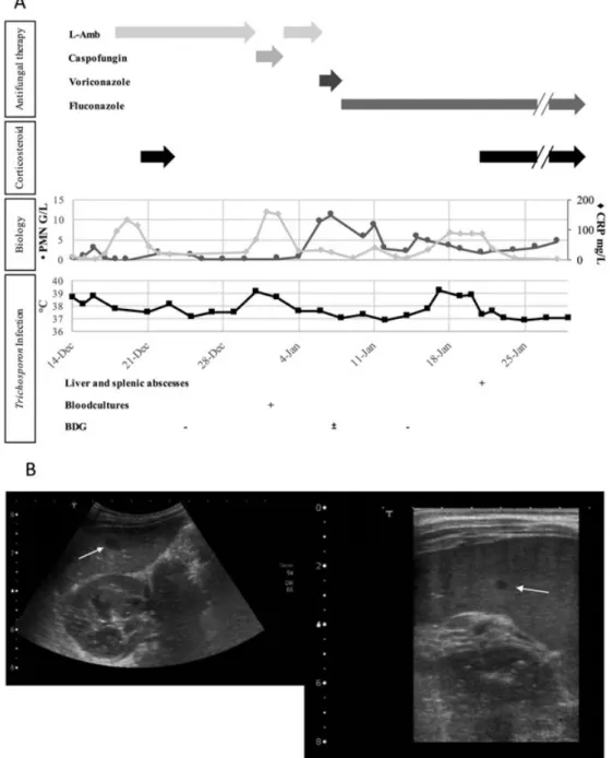
Case 2 (Figure 2)
A 17-month-old boy was initially referred to our emer-gency department with a 10-day history of fever with dete-rioration of his general status (day 0). Clinical examination showed peripheral lymphadenopathies associated with
Figure 2. Case 2: (A) Patient’s clinical course; (B) Abdominal ultrasound showing hepatic (left) (diameter around 15 mm) and splenic (right) (diameter<10 mm) abscesses
liver and spleen enlargement. Temperature was at 38.7 °C. The child was neutropenic (neutrophil counts at 0.3 × 10⁹/l) with circulating blasts at 14 × 109/l. Respiratory failure led to admission in intensive care unit, where empirical antibi-otic therapy combining piperacilline-tazobactam and
van-comycin was initiated. On day 2, bone marrow
examination confirmed the diagnosis of acute lymphoblas-tic leukaemia. On day 3, while the child was still febrile, clinical exam revealed an ano-rectal abscess. Piperacillin-tazobactam was switched for imipenem, ciprofloxacin, ornidazole and liposomal amphotericin B was started at 3 mg/kg/day. On day 10, the ano-rectal abscess turned into a cellulitis, requiring a colostomy in order to allow the ab-scess to heal. Five days later, the patient became afebrile and induction chemotherapy with prednisone, vincristine andL-Asparaginase was then started. At this time, because of digestive colonization by Candida lusitaniae, liposomal amphotericin B was replaced by caspofungin (70 mg/m2 thefirst day and then 50 mg/m2/day). Three days later, a white necrotic lesion appeared on the colostomy mucosa. Caspofungin was then switched back to liposomal amphotericin B, which resulted in rapid apyrexia. How-ever, a blood culture and the colostomy lesion grew posi-tive for a yeast-like organism further identified as T. asahii [5]. Liposomal amphotericin B was then changed for voriconazole (14 mg/kg/day i.v.) for 2 days, then flu-conazole (10 mg/kg/day i.v.). This treatment led to rapid improvement of the clinical condition. MIC of antifungals showed a decrease in susceptibility to amphotericin B, but susceptibility to fluconazole, voriconazole and posaconazole. However, 20 days after the positive Trichosporon bloodculture, and while still treated with flu-conazole, the child became febrile again. Bacterial and fun-gal bloodcultures remained negative. An abdominal ultrasound revealed two hepatic and multiple splenic nod-ules, suggestive of fungal abscesses (Figure 2). Dexameth-asone treatment (1 mg/12 h) resulted in an immediate apyrexia and was maintained for 15 days. An attempt to stop steroids led to the reappearance of fever. Therefore, dexamethasone (0.5 mg/12 h) was maintained for another 2 weeks, followed by a prolonged treatment with hydrocor-tisone. Afterwards, a chemotherapy protocol was pursued leading to complete cytological and molecular remission. Fluconazole treatment was stopped after 4 months, based on the recovery of a normal abdominal ultrasound. Six months after fluconazole discontinuation, no sign of re-lapse of the fungal infection has appeared.
Discussion
Trichosporon belongs to the long list of emerging fungal pathogens that complicate the course of hematological ma-lignancies [6,7]. One peculiarity of Trichosporon is that it belongs to the basidiomycete phylum [7], and as such, it
is naturally resistant to echinocandins. In addition, and to a variable extent according to species, susceptibility to amphotericin B may be decreased [3], making thera-peutic management challenging. Thereby, as seen in pa-tient 2, Trichosporon infections commonly occur as a breakthrough infection in patients receiving either amphotericin B or echinocandin therapy [8]. Most of those infections emerge during an episode of neutropenia in patients with hematological malignancy [6]. Dissemi-nated infection with positive blood culture(s), sometimes associated with cutaneous and/or pulmonary involve-ment, is the predominant clinical picture of invasive Trichosporon infection [2].
Immune reconstitution inflammatory syndrome (IRIS), initially reported in the context of AIDS-related opportu-nistic infections [9], has been described in other settings, notably in neutropenic patients [10]. The rapid relief of an effective immune response, combined with immuno-modulatory effects linked to some pathogens’ antigens, and to antimicrobials by themselves may lead to severe clinical pictures [9]. While mimicking treatment failure, the chronology of the symptoms, the persistently nega-tive cultures and the potential benefit of limiting the im-mune response, either by delaying the ARV therapy or even adding corticosteroid therapy, support this difficult-to-prove diagnosis [4]. While the effect of corti-costeroids is doubtful in patient 1, it seems truly efficient in case 2, where the discontinuation led to the recurrence of the fever.
Fungal pathogens seem particularly prone to induce such phenomena. Indeed, Cryptococcus neoformans, Pneumocystis jiroveci, Aspergillus fumigatus and Histoplasma capsulatum have been associated with IRIS too [11–13]. Recently, some reports suggest that chronic disseminated candidiasis, so-called hepato-splenic candi-diasis, because of the organs usually involved, should be considered as an IRIS [4]. Indeed, concomitantly with the recovery of neutrophils, some patients, previously neutropenic and having undergone a Candida blood-stream disseminated infection, documented or not with positive blood cultures, develop abscesses in the liver, the spleen or more rarely the kidneys [4,14]. Our obser-vations are highly suggestive of such a phenomenon in-duced by Trichosporon infection. In both cases, haematogenous dissemination of Trichosporon was docu-mented during the neutropenic phase. Hepatic and/or splenic lesions were diagnosed 34 and 19 days later, re-spectively, while neutrophil count had returned to nor-mal. Although there was no confirmation of the fungal nature of these lesions, imaging features were highly suggestive of fungal abscesses. The role of amphotericin B, received by our two patients, in the development of such lesions can be postulated. Indeed, this drug exhibits only limited activity against Trichosporon and has a well-known pro-inflammatory activity notably through 903 Chronic disseminatedTrichosporon infection
Copyright © 2016 John Wiley & Sons, Ltd. Hematol Oncol 2017; 35: 900–904 DOI: 10.1002/hon
the release of inflammatory cytokines [15]. A similar case of a chronic disseminated T. asahii infection in a leukemic child has been previously reported [16]. The patient was treated with liposomal amphotericin B, then with an amphotericin B lipid complex when an abdomi-nal ultrasound and CT-scan revealed multiple hepatic abscesses and splenomegaly. A favourable outcome was obtained when itraconazole therapy was initiated. No corticosteroid therapy was associated.
In vitro susceptibility testing and case reports suggest that voriconazole could be the best therapeutic alternative for such infections ([3] and personal data). Indeed, MICs of voriconazole against Trichosporon spp, including T. asahii, are usually the lowest as compared with the other antifungal drugs [3]. Nevertheless, in the context of cytotoxic chemo-therapy, caution must be taken in the case of concomitant treatment with vincristine because of possible drug–drug interaction [17].
In conclusion, Trichosporon, a recently emerged fungal pathogen, may be responsible for chronic disseminated in-fection following fungemia. As shown for chronic dissem-inated candidiasis, corticosteroid therapy may be a valuable adjunctive therapy in such cases.
Conflict of interest
On behalf of all authors, the corresponding author states that there is no conflict of interest
Ethics statement
According to the French legislation, patients or their repre-sentatives were informed that the clinical details of their history could be published, and none expressed opposition.
References
1. Chitasombat MN, Kofteridis DP, Jiang Y, Tarrand J, Lewis RE, Kontoyiannis DP. Rare opportunistic (non-Candida, non-Cryptococcus) yeast bloodstream infections in patients with cancer. J Infect 2012; 64(1): 68–75.
2. Colombo AL, Padovan ACB, Chaves GM. Current knowledge of Trichosporon spp. and Trichosporonosis. Clin Microbiol Rev 2011; 24(4): 682–700.
3. Arikan S, Hascelik G. Comparison of NCCLS microdilution method and E-test in antifungal susceptibility testing of clinical
Trichosporon asahii isolates. Diagn Microbiol Infect Dis 2002; 43(2): 107–11.
4. Legrand F, Lecuit M, Dupont B, et al. Adjuvant corticosteroid therapy for chronic disseminated candidiasis. Clin Infect Dis 2008; 46(5): 696–702.
5. de Almeida Junior JN, Figueiredo DS, Toubas D, et al. Useful-ness of matrix-assisted laser desorption ionisation-time-of-flight mass spectrometry for identifying clinical Trichosporon isolates. Clin Microbiol Infect 2014; 20(8): 784–90.
6. Girmenia C, Pagano L, Martino B, et al. Invasive infections caused by Trichosporon species and Geotrichum capitatum in patients with hematological malignancies: a retrospective multi-center study from Italy and review of the literature. J Clin Microbiol 2005; 43(4): 1818–28.
7. Chagas-Neto TC, Chaves GM, Colombo AL. Update on the ge-nus Trichosporon. Mycopathologia 2008; 166(3): 121–32. 8. Bayramoglu G, Sonmez M, Tosun I, Aydin K, Aydin F.
Break-through Trichosporon asahii fungemia in neutropenic patient with acute leukemia while receiving caspofungin. Infection 2008; 36(1): 68–70.
9. Manzardo C, Guardo AC, Letang E, Plana M, Gatell JM, Miro JM. Opportunistic infections and immune reconstitution in flam-matory syndrome in HIV-1-infected adults in the combined anti-retroviral therapy era: a comprehensive review. Expert Rev Anti Infect Ther 2015; 13(6): 751–67.
10. Sun HY, Singh N. Immune reconstitution inflammatory syn-drome in non-HIV immunocompromised patients. Curr Opin In-fect Dis 2009; 22(4): 394–402.
11. Mok HP, Hart E, Venkatesan P. Early development of immune reconstitution inflammatory syndrome related to Pneumocystis pneumonia after antiretroviral therapy. Int J STD AIDS 2014; 25(5): 373–7.
12. Jung J, Hong HL, Lee SO, et al. Immune reconstitution in flam-matory syndrome in neutropenic patients with invasive pulmo-nary aspergillosis. J Infect 2015; 70(6): 659–67.
13. Passos L, Talhari C, Santos M, Ribeiro-Rodrigues R, Ferreira LC, Talhari S. Histoplasmosis-associated immune reconstitution inflammatory syndrome. An Bras Dermatol 2011; 86(4 Suppl 1): S168–72.
14. Chaussade H, Bastides F, Lissandre S, et al. Usefulness of corti-costeroid therapy during chronic disseminated candidiasis: case reports and literature review. J Antimicrob Chemother 2012; 67(6): 1493–5.
15. Sau K, Mambula SS, Latz E, Henneke P, Golenbock DT, Levitz SM. The antifungal drug amphotericin B promotes inflammatory cytokine release by a Toll-like receptor- and CD14-dependent mechanism. J Biol Chem 2003; 278(39): 37561–8.
16. Meyer MH, Letscher-Bru V, Waller J, Lutz P, Marcellin L, Herbrecht R. Chronic disseminated Trichosporon asahii infec-tion in a leukemic child. Clin Infect Dis 2002; 35(2): e22–5. 17. Moriyama B, Henning SA, Leung J, et al. Adverse interactions
between antifungal azoles and vincristine: review and analysis of cases. Mycoses 2012; 55(4): 290–7.